HAL Id: hal-02160924
https://hal-amu.archives-ouvertes.fr/hal-02160924
Submitted on 20 Jun 2019
HAL is a multi-disciplinary open access
archive for the deposit and dissemination of
sci-entific research documents, whether they are
pub-lished or not. The documents may come from
teaching and research institutions in France or
abroad, or from public or private research centers.
L’archive ouverte pluridisciplinaire HAL, est
destinée au dépôt et à la diffusion de documents
scientifiques de niveau recherche, publiés ou non,
émanant des établissements d’enseignement et de
recherche français ou étrangers, des laboratoires
publics ou privés.
Bacterial respiratory chain diversity reveals a
cytochrome c oxidase reducing O2 at low overpotentials
Xie Wang, Romain Clément, Magali Roger, Marielle Bauzan, Ievgen
Mazurenko, Anne de Poulpiquet, Marianne Ilbert, Elisabeth Lojou
To cite this version:
Xie Wang, Romain Clément, Magali Roger, Marielle Bauzan, Ievgen Mazurenko, et al.. Bacterial
respiratory chain diversity reveals a cytochrome c oxidase reducing O2 at low overpotentials. Journal
of the American Chemical Society, American Chemical Society, 2019, �10.1021/jacs.9b03268�.
�hal-02160924�
Bacterial respiratory chain diversity reveals a cytochrome c
oxidase reducing O
2
at low overpotentials
Xie Wang,
†Romain Clément,
†Magali Roger,
‡Marielle Bauzan,
§Ievgen Mazurenko,
†Anne de
Poulpiquet,
†Marianne Ilbert,
†Elisabeth Lojou*
†.
†
Aix-Marseille Univ, CNRS, BIP, UMR 7281, 31 Chemin Aiguier, 13009 Marseille, France.
‡School of Natural and Environmental Sciences; Newcastle University, Devonshire building, NE1 7RX, Newcastle
upon Tyne, England.
§Aix-Marseille Univ, CNRS, IMM, FR 3479, 31 Chemin Aiguier, 13009 Marseille, France.
KEYWORDS: Catalysis, enzymes, electrochemistry, oxygen reduction, cytochrome c oxidase, acidophilic bacterium
ABSTRACT: Cytochrome c oxidases (CcO) are the terminal enzymes in energy converting chains of microorganisms where they reduce oxygen into water. Their affinity for O2 makes them attractive biocatalysts for technological devices in which O2 concentration is limited, but the high overpotentials they display on electrodes, severely limit their applicative use. Here, the CcO of the acidophilic bacterium Acidithiobacillus ferrooxidans, is studied on various carbon materials by direct protein electrochemistry and mediated one with redox mediators either diffusing or co-immobilized at the elec-trode surface. The entrapment of the CcO in a network of hydrophobic carbon nanofibers permits a direct electrochemi-cal communication between the enzyme and the electrode. We demonstrate that the CcO displays a µM affinity for O2, and reduces O2 at exceptionally high electrode potentials in the range of +700 – +540 mV vs NHE over a pH range of 4-6. The kinetics of interactions between the enzyme and its physiological partners are fully quantified. Based on these results, an electron transfer pathway allowing O2 reduction in the acidic metabolic chain is proposed.
INTRODUCTION
Oxygen reduction reaction (ORR) efficiency is one parameter that currently limits the large-scale devel-opment of fuel cells. Intensive research is carried out to discover catalysts able to enhance the ORR kinet-ics.1 Among them, enzymes are biocatalysts which present the advantage of large availability and high specificity.2-3 In terms of catalytic current output for O2 reduction and low overvoltage, multicopper oxi-dases (MCO), displaying typical onset O2 reduction in the +500-+800 mV vs NHE range, are among the most used. Their efficiency is nevertheless limited by low affinity towards oxygen in the range of 100 µM.2
Cytochrome c oxidases (CcOs) play a key role in mi-tochondrial and bacterial energy converting chains.4 They are the last electron acceptors in the chain, and have the function of O2 reduction to water, thus par-ticipating in the build-up of the proton gradient across the cell membrane required for ATP synthesis. 5-7
The aa3 CcOs belong to the heme-copper-containing terminal oxidases. These enzymes are widely distrib-uted over and crucial to many prokaryotes and eukar-yotes. Several crystallographic structures have been resolved such as those of the widely studied aa3 CcOs from Paracoccus denitrificans 5 and bovine heart
mi-tochondria.8 The catalytic cycle involves O2 binding to the fully reduced enzyme at the active site composed of the heme a3 and CuB center. The electron transfer pathway towards this binuclear center proceeds from a soluble cytochrome through the exposed surface of the CcOs to electron relays comprising the dicopper center CuA and heme a. However, fundamental ques-tions are still open such as the initial step of O2 diffu-sion to the active site, the effect of redox states of the enzyme on the proton-pumping pathway, or the type of interactions between soluble c-type cytochromes and the CuA domain according to the microorganism.9-12
Due to their physiological function, CcO affinity for O2 is two or three orders of magnitude higher than that of MCOs, making them attractive alternative for biotechnological applications.13-14 A further require-ment for the valuable use of this type of enzyme as bioelectrocatalysts is a low overvoltage for the reduc-tion of O2. Actually, redox titrareduc-tions either by UV or EPR spectroscopies were carried out on a few aa3 CcOs providing equilibrium potentials of their metal centers. Redox potentials of CuA and CuB were found to range between 150-240 mV and 225-340 mV at pH 7, respectively.15-19 Equilibrium measurements showed
two distinct transitions at pH 7 attributed to the two hemes, in the range of 140-390 mV.15-18, 20 Very few data are available for O2 catalytic reduction when CcOs are immobilized on electrochemical interfaces. Early studies followed catalytic O2 reduction in the presence of cytochrome c as a redox mediator with CcO either attached to gold electrodes by His-tag or embedded in reconstituted lipid bilayer.21-25 O2 reduc-tion thus proceeded at the redox potential of the cy-tochrome c, close to +250 mV vs NHE. More recently, immobilization of the ba3 CcO from Thermus thermophilus 26 and of the aa3 CcO from P. denitrificans on gold nanoparticles allowed catalytic reduction of O2 in the absence of any redox mediator. However, especially in the case of the aa3 CcO, O2 reduction occurred with a high overpotential, display-ing an onset potential for O2 reduction close to 0 mV vs NHE at pH 7.26 The marked differences observed for aa3 CcO between redox potentials obtained at equilibrium in solution or operating potentials under turn-over conditions in the immobilized state remains unclear. One explanation may arise from the for-mation of non native heme a species recently demon-strated by surface enhanced resonance Raman spec-troscopy when Rhodobacter sphaeroides CcO was immobilized on a thiol-modified gold electrode.27
Acidithiobacillus ferrooxidans is an acidophilic bac-terium living in pH environment as low as pH 2. One of its metabolic chains couples ferrous iron oxidation to O2 reduction by an aa3 CcO (Supporting Infor-mation Figure S1).28 Despite a similar function, A. ferrooxidans CcO differs from the usual CcOs in at least three respects: i) the CuA domain faces acidic periplasm with a pH value close to pH 2, ii) the aci-dophilic environment most likely suppresses the con-tribution of electrostatic interactions in the inter-protein recognition, and iii) an additional subunit with a cupredoxin fold, AcoP for Acidophile Cyto-chrome c Oxidase Partner, was identified in tight interaction with CcO.29 Several other redox proteins have been identified and purified in the A. ferrooxidans ET chain, which all share a high redox potential in the range of +300/+600 mV vs NHE at pH 5.30-31 As a direct consequence of the high redox poten-tial of the entire energy converting chain, it may be expected that A. ferrooxidans CcO also operates at a higher redox potential than CcOs from neutrophiles. Actually, almost 40 years ago, redox titrations per-formed on whole membranes of A. ferrooxidans showed two redox components attributed to low and high spin hemes. Redox transitions were found at +725 and +610 mV vs NHE at pH 3.2 and +500 mV and +420 mV vs NHE at pH 7.32 Although they were not attributed to heme a and heme a3 at that time, this is the most reasonable attribution indicating that CcO in A. ferrooxidans would indeed operate at high po-tentials. However, although A. ferrooxidans CcO was purified for the first time more than twenty years ago, very few biochemical or biophysical studies were
car-ried out on this enzyme, likely because of the harsh growth conditions for this bacterium, and the ensuing difficulty to accumulate enough biomass for subse-quent purification.33 Using spectroscopic methods, it was shown that the diheme cytochrome c4 (Cyt c4) interacted with the CcO, displaying an intermolecular ET rate constant of 11 s-1.34 Since a mutant in the envi-ronment of the low potential heme showed a tenfold lower rate constant, it was proposed that the ET oc-curred from this low potential heme to CcO. In a recent work, we quantified the ET rate between AcoP and Cyt c4.30 We showed that AcoP could be reduced at the electrochemical interface through the reduced high potential heme of Cyt c4. We also underlined the versatility of Cyt c4 which can be electroactive while in interactions with either hydrophobic or positive or negative interfaces. As AcoP copurifies with the CcO, this would suggest that an ET pathway differing from the classical one from the cytochrome to the CuA domain of the CcO may be involved.
Here, using mainly electrochemistry as the analyti-cal method, we demonstrate the unique capability of A. ferrooxidans CcO to reduce O2 at electrochemical interfaces. We first examine the O2 reduction in the presence of artificial redox mediators with increasing redox potentials. We then look for suitable electrode modification enabling a direct wiring of the CcO. Using hydrophobic carbon nanofibers as a host net-work, we demonstrate that the acidophilic aa3-type CcO of A. ferrooxidans displays the lowest overpotential for O2 reduction reported to date among CcOs. This CcO is stable in vitro, and active over a wide range of pH values and a high oxygen affinity at the same time. We finally evaluate the cata-lytic properties of CcO towards O2 reduction in more physiological conditions. We demonstrate that the O2 reduction by the CcO can be mediated by both hemes of Cyt c4, its physiological partner, although with a higher rate constant using the low potential heme. Having measured the redox potentials of all the indi-vidual proteins, including CuA, this allows us to pro-pose a new ET pathway in the metabolic chain from Fe2+ oxidation to O2 reduction. Considering its low overpotential for O2 reduction associated to a high affinity for O2, A. ferrooxidans CcO should be consid-ered in the future as an attractive biocatalyst for the cathode of biofuel cells, where noble metals at the anode are replaced by enzymes, microorganisms or bioinspired catalysts.
EXPERIMENTAL METHODS
Biomass accumulation of A. ferrooxidans. A. ferrooxidans ATCC 23270 was routinely grown on sterile iron-medium at 28°C. The medium, as previ-ously described 35, was slightly modified in this study, and contained: 14% (v/v) FeSO4, 7 H2O stock at 25% (w/v) diluted in H2O and adjusted to pH 1.6 with H2SO4 (3.5% (w/v) FeSO4·7H2O); 25% (v/v) basal salts diluted in H2O [0.4 g·L-1 (NH4)2SO4, 0.4 g·L-1 KH2PO4,
0.4 g·L-1 MgSO4·7 H2O, and 0.3 g·L-1 trisodium citrate (C6H5Na3O7·2H2O), adjusted to pH 1.6 with H2SO4]; and 61% (v/v) distilled water sterilized by autoclave sterilization for 15 min. Trisodium citrate (C6H5Na3O7·2H2O) was used to decrease oxidized iron species precipitation.
One liter pre-culture was inoculated with 100 mL stock culture to a final cell concentration of 107 bacte-ria per mL of culture. The pre-culture was cultivated until exponential phase to reach a cell concentration of about 109 bacteria per mL of culture. The pre-culture was then used to inoculate several flasks con-taining 20 L of medium. In order to increase the bio-mass yield, additional ferrous iron was added on the third day of cultivation. The cells were counted by Petroff-Hausser counting chamber (Electronic Mi-croscopy Sciences) and the concentration of ferrous iron was quantified by ferrozine assay,36 at different time points during growth. Growth curve is provided in Supporting Information Figure S2. Cells were har-vested on the fourth day. First, several low velocity centrifugations (2,000 × g, 4 min) were used to elimi-nate most of precipitated iron species. Then, the su-pernatant was centrifuged at 9,000 × g for 15 min to harvest the cells. Cells were stored at -80 °C for future use.
Purification of A. ferrooxidans CuA-soluble
domain (CoxB). Gene encoding CuA-soluble domain (CoxB) was amplified using A. ferrooxidans ATCC 23270 genomic DNA as a template. The PCR fragment was inserted into a pDEST14 vector using Gateway technology. E. coli BL21(DE3) strain was transformed with the resulting plasmid for overexpression. Among the conditions and vectors tested, A. ferrooxidans CoxB appears to be always produced as inclusion body in E. coli. Nevertheless, insoluble CoxB can be solubil-ized and re-folded to a native state according to the reported procedure19: cells were grown in LB medium containing 200 µg.mL-1 ampicillin at 37°C to A600 nm 0.7-0.8. The expression of the recombinant CoxB pro-tein was induced by the addition of 1 mM IPTG. After 4 h of induction, cells were harvested by centrifuga-tion at 9000 × g for 10 min and the cell pellet was re-suspended with 25 mM Tris-HCl buffer at pH 7 sup-plemented with anti-protease (1 tablet/ 50 mL) and DNase I (10 µg.mL-1). Cells were broken following two passages through a French Press at 1000 bar and the insoluble material were isolated by centrifugation at 10,000 × g for 20 min. The pellet containing the inclu-sion bodies was washed 3-times with 25 mM Tris-HCl buffer at pH 7 containing 1% Triton X100. The inclu-sion bodies were solubilized with 8 M urea in NaAc 50 mM buffer pH 5 supplemented with 50 mM DTT and non-solubilized material was eliminated by centrifu-gation at 11,000 × g for 30 min. The proteins were re-folded by eliminating the urea using step-wise dialy-sis. The amount of urea in the dialysis buffer was decreased by 1 M for each step and 1 mM CuCl2 was added during the last step allowing the in vitro
recon-stitution of the CuA cofactor. The re-folded proteins were loaded onto a MonoS column pre-equilibrated with NaAc 50 mM buffer pH 5 and proteins were elut-ed with a linear gradient of NaCl. Fractions containing CoxB were pooled, washed and concentrated (Vivaspin 10,000 MWCO PES, Sartorius). Protein concentration was determined by BCA assay. Support-ing Information Figure S3 gives the UV-visible spec-trum of CoxB, typical of the CuA-domain.19
Purification of A. ferrooxidans CcO. CcO was pu-rified from A. ferrooxidans ATCC 23270 using ~ 40 g of cells. Cells were re-suspended with potassium phos-phate buffer (50 mM potassium phosphos-phate, 10 mM EDTA and 20% glucose) at pH 7.4 (25 mL/ g cells), then flash-frozen in liquid nitrogen and subsequently thawed. Lysozyme (1 g·L-1) was added and the cell suspension was incubated at 30°C for 2 h. The mixture was centrifuged at 15,000 × g for 15 min and the cell pellet was then re-suspended with potassium phos-phate buffer at pH 7.4 (100 mM potassium phosphos-phate, 20% glucose, 4 tablets of anti-protease and 2 mg DNase). Cells were broken following 3 passages through a French Press at 2500 bar and then centri-fuged at 9,000 × g for 15 min. The membrane proteins were isolated by ultracentrifugation at 45,000 rpm for 45 min. The pellet was re-suspended with sodium acetate (NaAc) buffer at pH 4.8 (50 mM NaAc, 5% glycerol and 1 tablet of anti-protease) and membrane proteins were solubilized with n-dodecyl β-d-maltoside (DDM) at a concentration of 1.5 mg deter-gent/ mg total proteins for 1.5 h at 4 °C, then centri-fuged at 45,000 rpm for 45 min. The supernatant con-taining solubilized membrane proteins was concen-trated (Vivaspin 50,000 MWCO PES, Sartorius), then loaded onto a 10% - 30% saccharose gradient and centrifuged overnight at 43,000 rpm. Fractions were collected and tested for the presence of the CcO by UV-Visible absorption spectroscopy. Fractions con-taining the CcO were pooled, dialyzed against buffer A (50 mM NaAc buffer at pH 4.8, 0.01% DDM, 0.05% 6-Aminocaproic acid (ACA) and 5% glycerol), and loaded onto a DEAE column pre-equilibrated with the same buffer. CcO does not bind to this column, and it was directly found in the flow through. Some contam-inants tightly bound to the column were eliminated. CcO containing sample was then loaded onto HTP (hydroxyapatite) column that was pre-equilibrated with buffer A. Proteins were eluted using a linear gradient of 1 M potassium phosphate buffer at pH 4.8 supplemented with 0.01% DDM, 0.05% 6-Aminocaproic acid (ACA) and 5% glycerol. Fractions containing the CcO were finally pooled, washed, concentrated, and dialyzed against storage buffer A. The final protein con-centration was quantified by BCA assay. The purity of the CcO was estimated by loading 34 µg of proteins onto a 15% SDS-polyacrylamide gel. Protein samples were frozen in liquid nitrogen and stored at -80°C. Freshly thawed proteins were used for the electro-chemical experiments. As depicted in Supporting
Information Figure S4, outer membrane contaminants were found in the CcO final fraction and could not be separated from the enzyme. Their potential contribu-tion will be discussed later in the result seccontribu-tion.
Electrochemistry measurements. Cyclic voltam-metry (CV) was performed using an Autolab PGSTAT30 potentiostat controlled by Nova software (Eco Chemie). The electrochemical cell was equipped with three electrodes, a pyrolytic graphite (PG), a platinum wire as an auxiliary electrode and an Ag/AgCl electrode as the reference electrode. All po-tentials are converted to the normal hydrogen elec-trode (NHE) by adding 0.2 V. NH4Ac 20 mM buffer at pH 4.8 was used as the electrolyte, unless otherwise specified. All the electrochemical measurements were made at least in triplicate at a controlled temperature of 28°C. The PG electrode surface (S = 0.07 cm2) was renewed by polishing with fine sand paper (P1200), and then briefly sonicated to remove free carbon par-ticles. The membrane electrode configuration was used to entrap 2 µl of protein sample in a thin layer between the electrode and a dialysis membrane of suitable cutoff 37.
Carbon nanofiber (CNF) and carbon nanotube (CNT) preparation. CNFs were synthesized as previ-ously described by chemical vapor deposition.38 Multi-walled CNTs functionalized by carboxylic functions (CNT-COOH) were purchased from NanoLab Inc. (U.S.A.). Their surface chemistry was previously re-ported.39-40 The material suspensions were prepared as previously described.38-39 Briefly, all solutions were ultra-sonicated at least 4 h to suspend the nanotubes. CNFs were prepared at 4 mg·mL-1 in 50% N,N-dimethylformamide (DMF) solution in H2O. CNT-COOH were solubilized in H2O at 1 mg·mL-1. In order to modify the PG electrode, 4 µL of nanomaterial solution was dried on the PG surface at 60°C to form a layer of nanomaterials. Finally, 5 µL of protein was added on the nanomaterial layer and dried at 4°C for further study.
Protein and enzyme preparation on electrode. Regarding intermolecular electron-transfer experi-ments between CcO and cytochromes, a mixture of both proteins was first incubated at 4°C for 3 h. The pre-incubated protein mixture was further incubated 1 h to 2h on the PG electrode within the membrane configuration in the working buffer at room tempera-ture. Otherwise, individual protein or enzyme sam-ples were used freshly at the electrode. No DDM was added in the electrolyte. Michaelis-Menten constant (KM) of CcO with redox mediators and with its sub-strate, O2, were calculated from Michaelis-Menten fitted curves describing the relationship between the current and O2 concentration. The second-order electron transfer rates between CcO and its physiolog-ical partner or artificial redox mediators were calcu-lated as previously reported.37, 41-44 The equations re-lated either to diffusing redox mediators or to the
cytochrome immobilized in the thin layer at the elec-trode are detailed in Supporting Information.
Oxygen concentration calibration in aqueous solution. The whole experiment was operated in anaerobic chamber. The calibration method was adapted from the Clark cell measurement, except that a platinum electrode was used in the same electro-chemical cell and working conditions as described above. Chronoamperometry was used to investigate the reduction of oxygen at the platinum electrode at different O2 concentrations. Standard dissolved oxy-gen concentration in air saturated water was found in the literature at 243.75 µM at 28°C and atmospheric pressure.45 The concentration of oxygen in aqueous electrolyte was changed by mixing a nitrogen saturat-ed buffer and a pure oxygen saturatsaturat-ed buffer. The measurements were made in the absence of stirring to avoid O2 diffusion into the air phase. A standard curve was obtained linking oxygen reduction current to oxygen concentration (Supporting information Figure S5). Enzymes were studied within the same range of oxygen concentrations as calibrated.
Homology modelling of CcO from A.
ferrooxi-dans. Structure modelling was performed on the
Expasy server using Swiss-Model.46 The 3D-structure of the 4-subunit aa3-type oxidase from P. denitrificans (pdb-entry: 1QLE) was used as target file for coordi-nates.
RESULTS AND DISCUSSION
Artificially mediated O2 electroreduction by A. ferrooxidans CcO reveals a high affinity for O2
and a high redox potential. The catalytic properties of the CcO was first examined by entrapping the en-zyme in a thin layer between a dialysis membrane and the surface of the pyrolytic graphite (PG) electrode. No electrocatalytic signal could be observed on the bare PG electrode when only CcO and O2 were pre-sent (Figure 1A, (a)). To obtain a first indirect evalua-tion of the operating potential of A. ferrooxidans CcO, redox species with increasing redox potentials were used to mediate O2 reduction by the enzyme. In me-diated electrochemistry (MET), a redox molecule behaving as a fast and reversible electrochemical sys-tem, as well as showing suitable redox potential and affinity for the enzyme, serves as the electron shuttle between the enzyme and the electrode surface
.
47-48In the MET mode, there is no need for electrical wiring of the enzyme. Catalysis occurs at the potential of the redox mediator. The extent of catalysis will be a func-tion of the driving force, controlled by the potential of the mediator, and of the affinity between the redox mediator and its binding site on the enzyme. Figure 1A (b-d) shows the typical CVs obtained with three redox mediators, (i.e. potassium ferricyanide (FeCN), ferrocenemethanol (FcMeOH), and ferrocenecarboxylic acid (FC) covering a broad range of potentials, in the presence of CcO at the membrane
PG electrode. A catalytic wave at the redox potential of each mediator is observed, i.e. at +0.37, +0.42 and +0.51 vs NHE, respectively. The second order rate constants are reported in Table 1, showing, as ex-pected, that the catalytic ET transfer rate increases as the driving force increases (Details on the calculation are provided in Supporting information S3 and Fig-ures S56, S7, and S8). The second order rate constant reaches 7.3 106 M-1·s-1 in the case of FeCN. The addition of KCN in the electrolyte induces a complete disap-pearance of the catalytic signal as a result of the well-
known inhibitory effect of cyanide binding to the CuB-heme a3 catalytic site.49 From mediated electrocatalysis, it can be concluded that CcO may operate at least at potentials higher than +500 mV at pH 4.8. The affinity for O2 was quantified based on the FcMeOH mediated catalysis (Figure 1B). A KM value of 8.7 ± 1 µM is found, which is, as expected, higher than reported for bd-type oxidases (in the nM range),50 but much lower than MCOs (in the 50 µM - 1 mM range), the enzymes mostly used for O2 reduction in biotechnological devices 51 (Table 2).
Figure 1. Enzymatic reduction of O2 by A. ferrooxidans CcO in the presence of artificial redox mediators with increasing redox
potentials. (A) CVs under O2 without CcO (black curves) or with 14 µM CcO (green curves) at the PG membrane electrode (a)
CcO alone, (b-d) in the presence in solution of FeCN, FcMeOH, and FC at the concentration of 9 µM. The grey curve in (b) is obtained after 6 mM KCN addition in the O2-saturated electrolyte. Scan rate = 5 mV·s-1. 20 mM NH4Ac buffer at pH 4.8. (B)
Determination of the affinity of CcO for O2. KM value is obtained from the fitting according to Michaelis-Menten equation of the
relationship between catalytic current and O2 concentration. The catalytic current was measured in a glove box by CV in the presence of 14 µM CcO at the PG membrane electrode with 40 µM FcMeOH and increasing O2 concentration in the electrolyte.
Scan rate = 5 mV·s-1. 20 mM NH4Ac buffer at pH 4.8.
Table 1.Kinetic constants of the mediated reduction of O2 by A. ferrooxidans CcO. Redox potentials of
physio-logical or artificial mediators, Michaelis constants for the redox mediators (KM(app)) and second order rate
con-stants (k) are measured at pH 4.8. Details of the calcula-tion are given in Supporting Informacalcula-tion. ND means non determined values
.
Direct electroreduction of O2 by A. ferrooxidans
CcO incorporated in carbon nanomaterials. In direct electrochemistry, the active site of the enzyme or an electronic relay, as a cofactor, must be wired to the electrode. Catalysis thus occurs at the potential of the active site or the cofactor being the entry-leaving site of electrons in the protein. Such a wiring imposes
a tunnel distance between active sites or cofactors in the enzyme and the electrode. A distance less than 20 Å was shown to be required to allow relevant interfa-cial electron transfer52. Control of the orientation of the enzyme maintaining the tunnel distance can be obtained by appropriate surface chemistry, yielding to a film of wired protein on the surface of the electrode. Direct electrochemistry gives access to kinetic infor-mation relative to the enzyme itself.53 The absence of any direct electrochemical response on bare PG elec-trode was noted in Figure 1A (a). This situation is common for large membrane proteins, and can be due either to low quantity of enzyme available at the elec-trode surface, or to damage of the enzyme once im-mobilized, or to unfavorable wiring that prevents any direct electrical communication. In an attempt to increase the surface area, and the amount of immobi-lized enzyme, a film of carboxylic acid-functionaimmobi-lized carbon nanotubes (CNT-COOH) was deposited at the PG electrode. No electrocatalytic reduction of O2 can be observed either, although the enzyme is present and active at the surface as demonstrated by the me-diated catalytic reduction obtained after addition of
FeCN (Figure 2A). Since CcO is an integral membrane protein, a first explanation may come from the hydro-philic nature of both the PG electrode and the CNT-COOH deposit. One more explanation comes from the chemical functions present on the two electrode surfaces that can repel the protein. Actually, the PG electrode is known to present many oxygenated
spe-cies on the surface, including carboxylic ones, yielding a surface pK close to 4.8. The CNTs used in the pre-sent work display as well a high content of oxygen, coming in large proportion from carboxylic groups 39. Their zeta potential was measured to be negative from -40 to -60 mV in the pH range from 3 to 8.
Figure 2. Direct enzymatic reduction of O2 by A. ferrooxidans CcO entrapped in carbon nanomaterials. (A) CVs under O2 before
(grey curve) and after (black curve) addition of 14 µM CcO at the CNT-modified PG electrode. Orange curve has been obtained after addition of 100 µM FeCN. Scan rate = 5 mV·s-1. 20 mM NH4Ac buffer at pH 4.8. (B) CVs at the CNF-modified PG electrode
under O2 before (grey curve) and after (green curve) addition of 14 µM CcO. Black dashed curve has been obtained after 400 µM
KCN addition in the electrolyte. Scan rate = 5 mV·s-1. 20 mM NH4Ac buffer at pH 4.8. (C) CVs at the CNF-modified PG elec-trode under O2 before (grey curve) and after addition of 14 µM CcO in the presence of 0 (black line), 20 (green line) and 40 (yellow line) mM Zn2+ in solution. Scan rate = 5 mV·s-1. 20 mM NH4Ac buffer at pH 4.8. (D) CVs at the CNF-modified PG electrode for O2 reduction by 14 µM CcO at pH 4 (green), 4.8 (light blue), 6 (dark blue) and 7 (black). The CVs are plotted after
subtraction of the CVs for the CNF-based electrode alone at the different pHs. Scan rate = 5 mV·s-1.
(E) Evolution of the onset-potentials for O2 reduction obtained from Figure 2D at the function of pH. Dotted grey line corresponds to the values of the
O2/H2O potentials. (F) Evolution of the catalytic current measured at + 350 mV at a unique CNF-modified electrode with
ad-sorbed CcO, when the pH of the electrolyte is decreased sequentially from pH 4 to pH 3, then increased from pH 3 to pH 7, and decreased again to pH 4.8.
When looking at the structure of A. ferrooxidans CcO, two main features can be underlined explaining the electrochemical behavior above (Figure 3). At first, as expected for an integral membrane protein, it is mainly hydrophobic, and charged amino acid residues are only present at the bottom and top sides of the protein. Second, the bottom side of the protein which faces the cytoplasm is more positive than the top side facing the periplasm (+14 net charge against +5). Electrostatic interactions will then favor the anchoring of the CcO through the bottom side on negative surfaces such as
the PG electrode or the CNT-based film. In this orienta-tion, the distances between the electrode and CuA, and heme a or heme a3 – CuB active site are around 50 and 40 Å respectively, hampering any direct ET.
It should be more efficient to provide a hydrophobic matrix to entrap the CcO in view of direct wiring. This strategy can be reached by reconstitution of lipid layers at the electrode, or more simply by modification of the electrode by hydrophobic materials. One way is to modify gold electrodes by hydrophobic thiols 26. Alt-hough this strategy allowed Meyer et al. to wire P. denitrificans aa3 CcO, O2 reduction occurred with a
large overpotential. Search for materials other than thiol-modified gold should be preferred for enhanced stability. We previously used 100-200 nm diameter carbon nanofibers (CNF), which are composed of coni-cal shape sp2 carbon layers. These materials were proved to be very efficient to wire membrane-bound proteins such as hydrogenases, which was explained by the hydrophobic character of the carbon nanofibers, with no carboxylic groups even detected on the surface 38
. O2 reduction on such CNFs deposited as a film on the PG electrode occurs at an onset potential around +250 mV at pH 4.8 (Figure 2B). When the A. ferrooxidans CcO is immobilized on the CNF film, a cathodic signal develops with a high onset potential close to +650 mV at pH 4.8. This signal is stable at least over 20 CV cycles at 5 mV·s-1. It is absent under N2 atmosphere, and com-pletely vanishes after KCN addition, demonstrating that this signal is related to catalytic O2 reduction by the immobilized CcO.
Zn2+ cations, known to block proton pathways in CcO54, hence to inhibit O2 reduction, did not affect the catalyt-ic signal for Zn2+ concentrations up to 40 mM (Figure 2C). This result may origin from the immobilization of the enzyme in the CNF network, preventing access of Zn2+ to the channel. It is however more probable that the low pH at which catalysis occurs, decreases the inhibition rate, as it is accepted that protons competes with Zn2+ site.
Figure 3. Homology model of A. ferrooxidans CcO. (A)
Schematic representation of the distances in between redox centers and between redox centers and the surface of the protein (blue sphere CuA, heme a in red, heme a3 –
CuB center in red and grey spheres, respectively). (B)
Dis-tribution of charges on the surface of the protein. The side view represents the surface seen parallel to the membrane while the top and bottom views are from the periplasmic and cytoplasmic side, respectively (perpendicular to the membrane plane). The displayed structure is a homology model for the A. ferrooxidans enzyme from the structure of the 4-subunit oxidase of Paracoccus denitrificans (pdb-entry: 1QLE). Surface coloring is based on calculation for pH 4.8 (Swiss-Pdb Viewer) with positive charges in blue and negative charges in red.
The pH of the electrolyte was varied showing that the onset for O2 reduction follows a linear dependence of 70 mV/UpH, being respectively + 700 mV at pH 4 and + 490 mV at pH 7 (Figures 2D and 2E). The second obser-vation to be made is the decrease of the catalytic cur-rent when the pH is increased (Figure 2F). This is a reversible process, since transfer of the electrode back to pH 4 restores the activity. The dependence of the catalytic current with pH may suggest a mechanism similar to that proposed for ba3 CcO from T. thermophilus. It was postulated that the decrease of the catalytic current with pH may be linked to the inversion in the difference of potentials between heme b and heme a3, so that the ET between heme b and heme a3 is impeded when the pH increases 26.
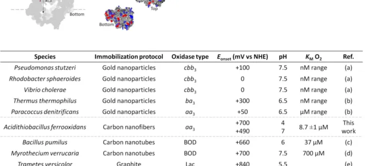
Table 2. Onset potentials for electroreduction of O2 by CcOs compared to some MCOs, and KM for O2. The onset potentials for
O2 electroreduction are given as a function of the species and type of CcO. (a) 4; (b) 26 (c),40, 55 (d),40, 56-57 (e)58-59 The onset potentials for O2 electroreduction
ob-tained using A. ferrooxidans CcO entrapped in CNF are exceptionally high compared to the other reported values (Table 2). This finding shows not only that the enzyme maintains its activity once purified, but also that immobilization of the enzyme in CNF does not induce high overpotentials. They confirm the interest of using the enzyme from the high potential respira-tory chain of A. ferrooxidans.
The electron transfer pathway in the direct wiring of the CcO for O2 reduction needs however to be determined. The accepted electron pathway in neutrophile CcOs is from CuA centre to heme a and CuB-heme a3 active site. We consequently determined the redox potential of the CuA-domain of A. ferrooxidans CcO (Figure 4A). A value of +340 mV vs NHE at pH 4, at least 100 mV higher than in neutrophile,18, 60 is found. However, the onset for di-rect electrocatalysis of O2 with the CcO embedded in the carbon nanofibers is +700 mV vs NHE at pH 4, it is thus unlikely that electron could enter CcO through CuA. during the direct electron transfer. In addition, CuA-domain was not electroactive when immobilized in CNFs (Figure 4B), most probably because of the hydrophobic character of CNFs, ruling out that CuA center could be the entry point for ET.
Figure 4. (A) CV under N2 of the CuA-domain (CoxB)
at the concentration of 80 µM at the PG electrode; Insert: relationship between the midpoint potential and the pH of the electrolyte. (B) CVs under N2 at the CNF modified
PG electrode of 80 µM CuA-domain. 20 mM NH4Ac
buffer, pH 4.8. Scan rate = 20 mV·s-1.
Outer membrane proteins are present in the CcO sample (Supporting Information Figure S4), especially the outer-membrane cytochrome Cyc2 30. Cyc2 has a redox potential of +560 mV 28 at pH 4.8. However, Cyc2 is not a physiological redox partner of CcO, and it was previously shown that it could not transfer electrons to CcO in solution34. Furthermore, in the presence of CcO sample on the CNF, no non-catalytic signals could be detected which may have been as-cribed to the outer-membrane cytochrome. Also, the shape of the catalytic CV on the CNF features a typical slope in a large range of potentials characteristic for the distribution of adsorbed enzyme orientations during the direct electron transfer, while it would
have had more Nernstian shape with rapid saturation in the case of mediated electron transfer.61 All togeth-er, these observations allow us to rule out the mediat-ed O2 rmediat-eduction by Cyc2 or another soluble mmediat-ediator. Besides, AcoP always copurifies with the CcO (Sup-porting Information Figure S4).28 Given its redox po-tential, the AcoP copper site might be the entry point for electrons towards the CcO active site. However, we showed in this work that purified AcoP is not electroactive on CNF, as expected from our previous results where no electroactivity could be found with AcoP adsorbed on hydrophobic SAM layer 30 (Sup-porting Information Figure S9). This suggests that AcoP might not be involved in the ET between CcO and CNF.
The hydrophobic character of the CNFs, able to dis-place DDM detergent, provides a favorable environ-ment for CcO which can adopt the orientation where both the heme a and the CuB-heme a3 active sites are at distances close to 10 Å to the CNF-based electrode, thus compatible for ET (Figure 3). Noteworthily, the onset potential measured under turn-over condition is in good accordance with the value of one heme we have measured in this work by redox titration on membrane fragments from A. ferrooxidans (Support-ing Information Figure S10). Two transitions are ob-served, at +405 and +540 mV vs NHE at pH 7. From a literature survey of equilibrium redox potentials of heme a and heme a3 in aa3 CcO, the highest potential transition is most probably linked to the heme a17, 62 . It is thus reasonable to propose a direct ET process between CNF and CcO through the heme a site to the CuB-heme a3 active site. This direct ET pathway, al-lows O2 to be reduced at a much higher potential than the physiological pathway through the CuA.
O2 reduction by A. ferrooxidans CcO in a
physi-ological context. Cyt c4 is the physiphysi-ological partner of CcO in the metabolic chain. The ET rate between Cyt c4 and CcO was determined by incubation of both proteins, and entrapment of the mixture at the mem-brane PG electrode. In the presence of O2, the reversi-ble redox waves at +310 mV and +430 mV, characteris-tic of the low potential (HemeL) and high potential (HemeH) hemes of Cyt c4 respectively, turn into sig-moidal waves. These processes are linked to the cata-lytic reduction of O2 mediated by Cyt c4, since it does not occur under N2, or under O2 in the absence of enzyme, or under O2 in the absence of Cyt c4 (Figure 5A and Supporting Information Figure S11). Both HemeL and HemeH are able to mediate the electroreduction of O2 at the electrode. Note that we exclude the possibility of an intramolecular ET be-tween two hemes fast enough to impact the catalysis on the experiment timescale. It is based on our previ-ous results with Cyt c4 which demonstrated a constant ratio of two peaks whatever the experiment condi-tions, and literature data of similar cytochromes.30,
63-64
Modeling of the electrochemical signal by analysis of the kinetics of the inter-protein ET in the particular case of the redox mediator entrapped in a thin layer (Figure 5B, details are provided in Supporting Infor-mation) allows to calculate a second order rate con-stant of 9.3 105 M-1.s-1 for the ET between HemeL and CcO, five times higher than between HemeH and CcO (1.8 105 M-1.s-1) (Table 1, Figure 5B and Supporting In-formation Figure S12). For comparison, aa3 CcO and mono-hemic Cyt c of bovine heart were also en-trapped at the membrane PG electrode. A second order rate constant of 4.1 105 M-1.s-1 was found at pH 7, thus in the same order as the constant obtained for the reaction between CcO and Cyt c4 (Supporting Information Figures S12 and S13 and Supporting In-formation Table S1). In accordance with a higher affin-ity of CcO to HemeL as compared to HemeH, the vari-ation of the catalytic current as a function of consecu-tive CV cycles shows a continuous decrease of the ratio between the catalytic contributions of HemeL versus HemeH (Figures 5C and 5D). At the beginning of the CV experiments, catalysis mostly occurs on HemeL. After around 30 min of cycling, the catalytic current stabilizes, and both hemes contribute to the catalysis. This variation strongly suggests the re-quirement of a slow rearrangement of the proteins at the electrochemical interface, with a much faster recognition of HemeL for CcO. The ET processes ob-served between CcO and Cyt c4 at the electrode pro-vide an explanation to previous kinetic data obtained by spectrophotometry in solution.34 It was shown that a key residue in the hydrophobic area near HemeL affected the ET rate, indicating interaction between CcO and Cyt c4 via this low potential heme. However, ET was still observed, suggesting that HemeH would also be involved in the whole process. This ET path-way was disregarded, and it was concluded that Cyt c4 would act as a wire between Rusticyanin and CcO. However, the presence of AcoP in the CcO sample was also unknown at that time.
Figure 5. Diheme Cyt c4 mediates the reduction of O2 by
A. ferrooxidans CcO. (A) CVs of Cyt c4 and CcO under O2
(green curve) or under N2 (black curve), and CV of CcO
alone under O2 (grey line) at the membrane PG
elec-trode. 50 µM Cyt c4 and 2.5 µM CcO were incubated
together and entrapped at the membrane PG electrode in 20 mM NH4Ac buffer, pH 4.8. Scan rate: 5 mV s-1. (B)
Determination of the catalytic rate constants of the first and second O2 reduction waves: experimental CV of O2
reduction by CcO mediated by Cyt c4 (grey line) and
COMSOL model (dashed line). (C) Evolution with con-secutive cycles of the mediated catalytic process: 1st cycle (black line), 11th cycle (green line). Grey line is obtained under N2; (D) Evolution with time of the mediated
cata-lytic current ratio Ic(HemeL) / Ic(HemeH), shown in solid
line; Stability of the total catalytic current for O2
reduc-tion by CcO mediated by Cyt c4 in dashed line. 2.5 µM
CcO and 50 µM Cyt c4 were incubated and studied at the
PG membrane electrode. 20 mM NH4Ac buffer, pH 4.8.
Scan rate 5 mV s-1.
Based on the kinetic data obtained in this work, we can now propose a new ET pathway between Cyt c4 and CcO. In neutrophiles, ET proceeds from monohemic Cyt c to CuA-domain of CcO. The redox potential of the CuA-domain of A. ferrooxidans CcO (+340 mV vs NHE at pH 4) is compatible with an intermolecular ET with HemeL of Cyt c4 (+310 mV), like the “classic” neutrophile ET pathway. Besides, we previously demonstrated the formation of a complex between AcoP and Cyt c4, allowing AcoP to be re-duced by HemeH of Cyt c4, although with a slow in-termolecular ET rate in the order of a few s-1.30 No interaction between AcoP and HemeL was found. As AcoP copurifies with CcO, behaving as an additional subunit of the CcO (Supporting Information Figure
S4), an additional electron pathway between HemeH and CcO active center through AcoP can be proposed for O2 reduction (Figures 6A and 6B).
Figure 6. (A) Schematic representation of the three
partners based on Cyt c4 structure (pdb 1H1O), model of
CcO as in Figure 3, and model of AcoP.30 (B) Proposed ET pathways from Cyt c4 to CcO in A. ferrooxidans, showing
ET between HemeL of Cyt c4 and CuA-domain, and
HemeH of Cyt c4 and AcoP.
CONCLUSION
Protein film electrochemistry has been applied to characterize the electrocatalytic properties of the CcO from the acidophile bacterium A. ferrooxidans. We have demonstrated that both hemes of Cyt c4 can mediate ET for O2 reduction by CcO, although with a higher rate constant for electron transfer from the low potential heme. We propose a new electron transfer pathway from Cyt c4 to CcO, where Cyt c4 transfers two electrons from its two hemes to CcO in parallel, one through the CuA domain and one through the additional subunit AcoP. Whether this type of ET pathway is specific to acidophiles, and what would be its physiological relevance, is an open question. We succeeded in generating direct ET between CcO and the CNF-modified electrode, and propose a molecular basis for such direct wiring. The attractive electrocatalytic properties of the CcO from A. ferrooxidans are especially highlighted: CcO is an acidophilic oxidase, with optimal pH lower than 4, it displays µM O2 affinity, and reduces O2 at a high re-dox potential, at least 300 mV higher than the cur-rently studied oxidases from neutrophiles. Not only does the direct wiring of the CcO open new avenues in the understanding of the catalytic mechanism of O2 reduction at low pH - in particular proton channeling can now be studied - but from a more applied point of view it offers the possibility of new developments in the domain of fuel cells. The coupling of the CcO from A. ferrooxidans with novel bioinspired catalysts for H2 oxidation, only active at low pHs, 65 might be a very promising option to explore in the future.
ACKNOWLEDGEMENT
This work was supported by ANR (ENZYMOR-ANR-16-CE05- 0024), “Région PACA” (Optolen pro-ject) and Aix-Marseille University for X. Wang’s fund-ing. The authors acknowledge Prof R. Gadiou (ISM2, Mulhouse, France) for the synthesis of CNF materials in the framework of BIOPAC ANR project. The
au-thors want to thank Dr M.T. Giudici-Orticoni, and Dr W. Nitschke for fruitful discussions, P. Infossi (BIP, CNRS Marseille), P. Pai (IBDM, CNRS Marseille), D. Byrne-Kodjabachian (IMM, CNRS Marseille) and R. Puppo (Proteomic Analysis Center, IMM, CNRS Mar-seille, France) for technical support.
ASSOCIATED CONTENT
Supporting Information. Chemicals, materials biomass
production and protein purifications, O2 calibration
curve, determination of CcO midpoint potential by redox titration, methods for calculation of the kinetic stants, modeling of the mediated catalytic signals, con-trol CV experiments. “This material is available free of charge via the Internet at http://pubs.acs.org.”
REFERENCES
1. Pegis, M. L.; Wise, C. F.; Martin, D. J.; Mayer, J. M., Oxygen Reduction by Homogeneous Molecular Cata-lysts and ElectrocataCata-lysts. Chem. Rev. 2018, 118, 2340-2391.
2. Mano, N.; de Poulpiquet, A., O2 Reduction in Enzymatic Biofuel Cells. Chem. Rev. 2018, 118, 2392-2468. 3. Mazurenko, I.; Wang, X.; de Poulpiquet, A.; Lojou, E., O2 Reduction in Enzymatic Biofuel Cells.
Chem. Rev. 2018, 118, 2392-2468.
4. Melin, F.; Xie, H.; Meyer, T.; Ahn, Y. O.; Gennis, R. B.; Michel, H.; Hellwig, P., The unusual redox proper-ties of C-type oxidases. Biochim. Biophys.
Acta-Bioenergetics 2016, 1857, 1892-1899.
5. Iwata, S.; Ostermeier, C.; Ludwig, B.; Michel, H., Structure at 2.8 angström resolution of cytochrome c oxidase from Paracoccus denitrificans. Nature 1995, 376, 660-669.
6. von Ballmoos, C.; Gennis, R. B.; Adelroth, P.; Brzezinski, P., Kinetic design of the respiratory oxidases.
Proc. Natl Acad. Sci. USA 2011, 108, 11057-11062.
7. Wikstrom, M.; Sharma, V., Proton pumping by cytochrome c oxidase - A 40 year anniversary. Biochim.
Biophys. Acta 2018, 1859, 692-698.
8. Tsukihara, T.; Aoyama, H.; Yamashita, E.; Tomizaki, T.; Yamaguchi, H.; Shinzawaitoh, K.; Nakashima, R.; Yaono, R.; Yoshikawa, S., Structure of metal sites of oxidized bovine heart cytochrome c oxi-dase at 2.8 angström. Science 1995, 269, 1069-1074. 9. Wikstrom, M.; Krab, K.; Sharma, V., Oxygen Activation and Energy Conservation by Cytochrome c Oxidase. Chem. Rev. 2018, 118, 2469-2490.
10. Rocha, M.; Springett, R., Electron transfer be-tween cytochrome c and the binuclear center of cyto-chrome oxidase. J. Theor. Biol. 2019, 460, 134-141.
11. Luo, F. J.; Shinzawa-Itoh, K.; Hagimoto, K.; Shimada, A.; Shimada, S.; Yamashita, E.; Yoshikawa, S.; Tsukihara, T., Structure of bovine cytochrome c oxidase in the ligand-free reduced state at neutral pH. J. Theor.
Biol. 2019, 460, 134-141.
12. Mahinthichaichan, P.; Gennis, R. B.; Tajkhorshid, E., Cytochrome aa(3) Oxygen Reductase Utilizes the Tunnel Observed in the Crystal Structures To Deliver O-2 for Catalysis. Biochem. 2018, 57, 2150-2161.
13. Morris, R. L.; Schmidt, T. M., Shallow breathing: bacterial life at low O-2. Nat. Rev. Microbiol. 2013, 11, 205-212.
14. Mano, N., Features and applications of bilirubin oxidases. Appl. Microbiol. Biotechnol. 2012, 96, 301-307. 15. Carithers, R. P.; Palmer, G., Characterization of the potentiometric behavior of soluble cytochrome-oxidase by magnetic circular dichroism - Evidence in support of heme-heme interaction. J. Biol. Chem. 1981, 256, 7967-7976.
16. Junemann, S.; Meunier, B.; Gennis, R. B.; Rich, P. R., Effects of mutation of the conserved lysine-362 in cytochrome c oxidase from Rhodobacter sphaeroides.
Biochem. 1997, 36, 14456-14464.
17. Kao, W. C.; Kleinschroth, T.; Nitschke, W.; Baymann, F.; Neehaul, Y.; Hellwig, P.; Richers, S.; Vonck, J.; Bott, M.; Hunte, C., The obligate respiratory supercomplex from Actinobacteria. Biochim. Biophys.
Acta-Bioenergetics 2016, 1857, 1705-1714.
18. Maneg, O.; Ludwig, B.; Malatesta, F., Different interaction modes of two cytochrome-c oxidase soluble Cu-A fragments with their substrates. J. Biol. Chem. 2003, 278, 46734-46740.
19. Lappalainen, P.; Aasa, R.; Malmstrom, B. G.; Saraste, M., Soluble Cu(A)-binding domain from the Paracoccus cytochrome-c-oxidase. J. Biol. Chem. 1993, 268, 26416-26421.
20. Sidhu, G. S.; Hendler, R. W., Characterization of 2 low EM forms of cytochrome a3 and their carbon-monoxide complexes in mammalian cytochrome-c-oxidase. Biophys. J. 1990, 57, 1125-1140.
21. Haas, A. S.; Pilloud, D. L.; Reddy, K. S.; Babcock, G. T.; Moser, C. C.; Blasie, J. K.; Dutton, P. L., Cyto-chrome c and cytoCyto-chrome c oxidase: Monolayer assem-blies and catalysis. J. Phys. Chem. B 2001, 105, 11351-11362. 22. Ataka, K.; Giess, F.; Knoll, W.; Naumann, R.; Haber-Pohlmeier, S.; Richter, B.; Heberle, J., Oriented attachment and membrane reconstitution of his-tagged cytochrome c oxidase to a gold electrode: In situ moni-toring by surface-enhanced infrared absorption spectros-copy. J. Am. Chem. Soc. 2004, 126, 16199-16206.
23. Friedrich, M. G.; Plum, M. A.; Santonicola, M. G.; Kirste, V. U.; Knoll, W.; Ludwig, B.; Naumann, R. L. C., In situ monitoring of the catalytic activity of cyto-chrome c oxidase in a biomimetic architecture. Biophys.
J. 2008, 95, 1500-15100.
24. Schadauer, F.; Geiss, A. F.; Srajer, J.; Siebenhofer, B.; Frank, P.; Reiner-Rozman, C.; Ludwig, B.; Richter, O. M. H.; Nowak, C.; Naumann, R. L. C., Silica Nanoparticles for the Oriented Encapsulation of Membrane Proteins into Artificial Bilayer Lipid Mem-branes. Langmuir 2015, 31, 2511-2516.
25. Hill, H. A. O.; Walton, N. J.; Higgins, I. J., Elec-trochemical reduction of dioxygen using a terminal oxi-dase. FEBS Lett. 1981, 126, 282-284.
26. Meyer, T.; Melin, F.; Xie, H.; von der Hocht, I.; Choi, S. K.; Noor, M. R.; Michel, H.; Gennis, R. B.; Soulimane, T.; Hellwig, P., Evidence for Distinct Electron Transfer Processes in Terminal Oxidases from Different Origin by Means of Protein Film Voltammetry. J. Am.
Chem. Soc. 2014, 136, 10854-10857.
27. Sezer, M.; Kielb, P.; Kuhlmann, U.; Mohrmann, H.; Schulz, C.; Heinrich, D.; Schlesinger, R.; Heberle, J.;
Weidinger, I. M., Surface Enhanced Resonance Raman Spectroscopy Reveals Potential Induced Redox and Con-formational Changes of Cytochrome c Oxidase on Elec-trodes. J. Phys. Chem. B 2015, 119, 9586-9591.
28. Castelle, C.; Guiral, M.; Malarte, G.; Ledgham, F.; Leroy, G.; Brugna, M.; Giudici-Orticoni, M. T., A new iron-oxidizing/O2-reducing supercomplex spanning both inner and outer membranes, isolated from the extreme acidophile Acidithiobacillus ferrooxidans. J. Biol. Chem.
2008, 283, 25803-25811.
29. Roger, M.; Biaso, F.; Castelle, C. J.; Bauzan, M.; Chaspoul, F.; Lojou, E.; Sciara, G.; Caffarri, S.; Giudici-Orticoni, M. T.; Ilbert, M., Spectroscopic Characteriza-tion of a Green Copper Site in a Single-Domain Cupredoxin. Plos One 2014, 9.
30. Wang, X.; Roger, M.; Clement, R.; Lecomte, S.; Biaso, F.; Abriata, L. A.; Mansuelle, P.; Mazurenko, I.; Giudici-Orticoni, M. T.; Lojou, E.; Ilbert, M., Electron transfer in an acidophilic bacterium: interaction between a diheme cytochrome and a cupredoxin. Chem. Sci. 2018, 9, 4879-4891.
31. Roger, M.; de Poulpiquet, A.; Ciaccafava, A.; Ilbert, M.; Guiral, M.; Giudici-Orticoni, M. T.; Lojou, E., Reconstitution of supramolecular organization involved in energy metabolism at electrochemical interfaces for biosensing and bioenergy production. Anal. Bioanal.
Chem. 2014, 406, 1011-1027.
32. Ingledew, W. J.; Cobley, J. G., A potentiometric and kinetic study on the respiratory chain of ferrous iron grown Thiobacillus ferrooxidans. Biochim. Biophys. Acta
1980, 590, 141-158.
33. Kai, M.; Yano, T.; Tamegai, H.; Fukumori, Y.; Yamanaka, T., Thiobacillus ferrooxidans cytochrome c oxidase - purification and molecular and enzymatic fea-tures. J. Biochem. 1992, 112, 816-821.
34. Malarte, G.; Leroy, G.; Lojou, E.; Abergel, C.; Bruschi, M.; Giudici-Orticoni, M. T., Insight into molecu-lar stability and physiological properties of the diheme cytochrome CYC41 from the acidophilic bacterium
Acidithiobacillus ferrooxidans. Biochem. 2005, 44,
6471-6481..
35. Yarzabal, A.; Duquesne, K.; Bonnefoy, V., Rusticyanin gene expression of Acidithiobacillus ferrooxidans ATCC 33020 in sulfur- and in ferrous iron
media. Hydrometal. 2003, 71, 107-114.
36. Lovley, D. R.; Phillips, E. J. P., Rapid assay for microbially reducible ferric iron in aquatic sediments.
Appl. Environ. Microbiol. 1987, 53, 1536-1540.
37. dos Santos, M. M. C.; de Sousa, P. M. P.; Goncalves, M. L. S.; Krippahl, L.; Moura, J. J. G.; Lojou, E.; Bianco, P., Electrochemical studies on small electron transfer proteins using membrane electrodes. J.
Electroanal. Chem. 2003, 541, 153-162.
38. de Poulpiquet, A.; Marques-Knopf, H.; Wernert, V.; Giudici-Orticoni, M. T.; Gadiou, R.; Lojou, E., Carbon nanofiber mesoporous films: efficient platforms for bio-hydrogen oxidation in biofuel cells. Phys. Chem. Chem.
Phys. 2014, 16, 1366-1378.
39. Mazurenko, I.; Monsalve, K.; Rouhana, J.; Par-ent, P.; Laffon, C.; Goff, A. L.; Szunerits, S.; Boukherroub, R.; Giudici-Orticoni, M.-T.; Mano, N., How the intricate interactions between carbon nanotubes and two
biliru-bin oxidases control direct and mediated O2 reduction.
ACS Appl. Mater. Inter. 2016, 8, 23074-23085.
40. Monsalve, K.; Mazurenko, I.; Gutierrez-Sanchez, C.; Ilbert, M.; Infossi, P.; Frielingsdorf, S.; Giudici-Orticoni, M. T.; Lenz, O.; Lojou, E., Impact of Carbon Nanotube Surface Chemistry on Hydrogen Oxi-dation by Membrane-Bound Oxygen-Tolerant Hydrogenases. Chem. Electro. Chem 2016, 3, 2179-2188. 41. Lojou, E.; Bianco, P.; Bruschi, M., Kinetic stud-ies on the electron transfer between various c-type cyto-chromes and iron (III) using a voltammetric approach.
Electrochim. Acta 1998, 43, 2005-2013.
42. Ferapontova, E. E.; Ruzgas, T.; Gorton, L., Di-rect electron transfer of heme- and molybdopterin cofac-tor-containing chicken liver sulfite oxidase on alkanethiol-modified gold electrodes. Anal. Chem. 2003, 75, 4841-4850.
43. Nicholson, R. S.; Shain, I., Theory of stationary electrode polarography for a chemical reaction coupled between 2 charge transfers. Anal. Chem. 1965, 37, 178. 44. de Sousa, P. M. P.; Pauleta, S. R.; Rodrigues, D.; Goncalves, M. L. S.; Pettigrew, G. W.; Moura, I.; Moura, J. J. G.; dos Santos, M. M. C., Benefits of membrane elec-trodes in the electrochemistry of metalloproteins: medi-ated catalysis of Paracoccus pantotrophus cytochrome c peroxidase by horse cytochrome c: a case study. J. Biol.
Inorg. Chem. 2008, 13, 779-787.
45. Weiss, R. F., Solubility of nitrogen, oxygen and argon in water and sea water. Deep-Sea Res. 1970, 17, 721. 46. Waterhouse, A.; Bertoni, M.; Bienert, S.; Studer, G.; Tauriello, G.; Gumienny, R.; Heer, F. T.; de Beer, T. A. P.; Rempfer, C.; Bordoli, L.; Lepore, R.; Schwede, T., SWISS-MODEL: homology modelling of protein struc-tures and complexes. Nucleic Acids Research 2018, 46, W296-W303.
47. Gallaway, J. W.; Barton, S. A. C., Kinetics of redox polymer-mediated enzyme electrodes. J. Am.
Chem. Soc. 2008, 130, 8527-8536.
48. Milton, R. D.; Minteer, S. D., Direct enzymatic bioelectrocatalysis: differentiating between myth and reality. J. Royal Soc. Inter. 2017, 14.
49. Kalinovich, A. V.; Azarkina, N. V.; Vygodina, T. V.; Soulimane, T.; Konstantinov, A. A., Peculiarities of cyanide binding to the ba (3)-type cytochrome oxidase from the thermophilic bacterium Thermus thermophilus.
Biochem.-Moscow 2010, 75, 342-352.
50. Dmello, R.; Hill, S.; Poole, R. K., The cyto-chrome bd quinol oxidase in Escherichia coli has an extremely high oxygen affinity and two oxygen-binding haems: Implications for regulation of activity in vivo by oxygen inhibition. Microbiol. 1996, 142, 755-763.
51. de Poulpiquet, A.; Ranava, D.; Monsalve, K.; Giudici-Orticoni, M. T.; Lojou, E., Biohydrogen for a New Generation of H2/O2 Biofuel Cells: A Sustainable Energy Perspective. Chem. Electro. Chem. 2014, 1, 1724-1750. 52. Page, C. C.; Moser, C. C.; Chen, X. X.; Dutton, P. L., Natural engineering principles of electron tunnelling in biological oxidation-reduction. Nature 1999, 402, 47-52.
53. Sensi, M.; del Barrio, M.; Baffert, C.; Fourmond, V.; Leger, C., New perspectives in hydrogenase direct electrochemistry. Curr. Op. Electrochem. 2017, 5, 135-145. 54. Mills, D. A.; Schmidt, B.; Hiser, C.; Westley, E.; Ferguson-Miller, S., Membrane potential-controlled inhibition of cytochrome c oxidase by zinc. J. Biol.Chem.
2002, 277, 14894-14901.
55. Mazurenko, I.; Monsalve, K.; Infossi, P.; Giudici-Orticoni, M. T.; Topin, F.; Mano, N.; Lojou, E., Impact of substrate diffusion and enzyme distribution in 3D-porous electrodes: a combined electrochemical and modelling study of a thermostable H2/O2 enzymatic fuel cell. Energ. Environ. Sci. 2017, 10, 1966-1982.
56. Brocato, S.; Lau, C.; Atanassov, P., Mechanistic study of direct electron transfer in bilirubin oxidase.
Electrochim. Acta 2012, 61, 44-49.
57. dos Santos, L.; Climent, V.; Blanford, C. F.; Armstrong, F. A., Mechanistic studies of the 'blue' Cu enzyme, bilirubin oxidase, as a highly efficient electrocatalyst for the oxygen reduction reaction. Phys.
Chem. Chem. Phys. 2010, 12, 13962-13974.
58. Lee, C. W.; Gray, H. B.; Anson, F. C.; Malmstrom, B. G., Catalysis of the reduction of dioygen at graphite electrodes coated with fungal laccase-A. J.
Electroan. Chem. 1984, 172, 289-300.
59. Thuesen, M. H.; Farver, O.; Reinhammar, B.; Ulstrup, J., Cyclic voltammetry and electrocatalysis of the blue copper oxidase Polyporus versicolor laccase. Acta
Chem. Scandin. 1998, 52, 555-562.
60. Immoos, C.; Hill, M. G.; Sanders, D.; Fee, J. A.; Slutter, C. E.; Richards, J. H.; Gray, H. B., Electrochemis-try of the Cu-A domain of Thermus thermophilus cyto-chrome ba(3). J. Biol. Inorg. Chem. 1996, 1 (6), 529-531. 61. Fourmond, V.; Leger, C., Modelling the volt-ammetry of adsorbed enzymes and molecular catalysts.
Curr. Op. Electrochem. 2017, 1, 110-120.
62. Pardhasaradhi, K.; Ludwig, B.; Hendler, R. W., potentiometric and spectral studies with the 2-subunit cytochrome aa3 from Paracoccus denitrificans - compari-son with the 13-subunit beef-heart enzyme. Biophys. J.
1991, 60, 408-414.
63. Monari, S.; Battistuzzi, G.; Borsari, M.; Di Roc-co, G.; Martini, L.; Ranieri, A.; Sola, M., Heterogeneous Electron Transfer of a Two-Centered Heme Protein: Redox and Electrocatalytic Properties of Surface-Immobilized Cytochrome c(4). J. Phys. Chem. B 2009, 113, 13645-13653.
64. Melin, F.; Schoepp-Cothenet, B.; Abdulkarim, S.; Noor, M. R.; Soulimane, T.; Hellwig, P., Electrochemi-cal study of an electron shuttle diheme protein: The cytochrome c(550) from T. thermophilus. Inorg. Chim. Acta 2017, 468, 252-259.
65. Gentil, S.; Lalaoui, N.; Dutta, A.; Nedellec, Y.; Cosnier, S.; Shaw, W. J.; Artero, V.; Le Goff, A., Carbon-Nanotube-Supported Bio-Inspired Nickel Catalyst and Its Integration in Hybrid Hydrogen/Air Fuel Cells. Angew.