HAL Id: inserm-02965704
https://www.hal.inserm.fr/inserm-02965704
Submitted on 13 Oct 2020
HAL is a multi-disciplinary open access
archive for the deposit and dissemination of
sci-entific research documents, whether they are
pub-lished or not. The documents may come from
teaching and research institutions in France or
abroad, or from public or private research centers.
L’archive ouverte pluridisciplinaire HAL, est
destinée au dépôt et à la diffusion de documents
scientifiques de niveau recherche, publiés ou non,
émanant des établissements d’enseignement et de
recherche français ou étrangers, des laboratoires
publics ou privés.
interferon response
Margarida Sa Ribero, Nolwenn Jouvenet, Marlène Dreux, Sébastien Nisole
To cite this version:
Margarida Sa Ribero, Nolwenn Jouvenet, Marlène Dreux, Sébastien Nisole. Interplay between
SARS-CoV-2 and the type I interferon response. PLoS Pathogens, Public Library of Science, 2020, 16 (7),
pp.e1008737. �10.1371/journal.ppat.1008737�. �inserm-02965704�
REVIEW
Interplay between SARS-CoV-2 and the type I
interferon response
Margarida Sa Ribero
ID1, Nolwenn Jouvenet
2*, Marlène Dreux
ID1*, Se´bastien Nisole
ID3*
1 CIRI, Inserm, U1111, Universite´ Claude Bernard Lyon 1, CNRS, E´ cole Normale Supe´rieure de Lyon, Univ Lyon, Lyon, France, 2 Institut Pasteur, CNRS UMR3569, Paris, France, 3 IRIM, CNRS UMR9004, Universite´ de Montpellier, Montpellier, France
*nolwenn.jouvenet@pasteur.fr(NJ);marlene.dreux@ens-lyon.fr(MD);sebastien.nisole@inserm.fr(SN)
Abstract
The severe acute respiratory syndrome coronavirus-2 (SARS-CoV-2) is responsible for the
current COVID-19 pandemic. An unbalanced immune response, characterized by a weak
production of type I interferons (IFN-Is) and an exacerbated release of proinflammatory
cytokines, contributes to the severe forms of the disease. SARS-CoV-2 is genetically related
to SARS-CoV and Middle East respiratory syndrome-related coronavirus (MERS-CoV),
which caused outbreaks in 2003 and 2013, respectively. Although IFN treatment gave some
encouraging results against SARS-CoV and MERS-CoV in animal models, its potential as a
therapeutic against COVID-19 awaits validation. Here, we describe our current knowledge
of the complex interplay between SARS-CoV-2 infection and the IFN system, highlighting
some of the gaps that need to be filled for a better understanding of the underlying molecular
mechanisms. In addition to the conserved IFN evasion strategies that are likely shared with
SARS-CoV and MERS-CoV, novel counteraction mechanisms are being discovered in
SARS-CoV-2–infected cells. Since the last coronavirus epidemic, we have made
consider-able progress in understanding the IFN-I response, including its spatiotemporal regulation
and the prominent role of plasmacytoid dendritic cells (pDCs), which are the main
IFN-I–pro-ducing cells. While awaiting the results of the many clinical trials that are evaluating the
effi-cacy of IFN-I alone or in combination with antiviral molecules, we discuss the potential
benefits of a well-timed IFN-I treatment and propose strategies to boost pDC-mediated IFN
responses during the early stages of viral infection.
Introduction
The severe acute respiratory syndrome coronavirus 2 (SARS-CoV-2) is a beta-coronavirus that
emerged at the end of 2019 in China and rapidly spread around the world, causing a pandemic
[
1
,
2
]. SARS-CoV-2 infection is responsible for COVID-19, a disease associated with mild
symptoms in the majority of cases but that can progress to an acute respiratory distress
syn-drome [
1
,
3
]. So far (July 16th, 2020), the virus has infected more than 13 million people and
caused more than 500,000 deaths worldwide. SARS-CoV-2 is genetically related to other
beta-coronaviruses that have caused epidemics: SARS-CoV and MERS-CoV (for Middle East
respi-ratory syndrome-related coronavirus), in 2003 and 2013, respectively. Beta-coronaviruses are
enveloped positive-sense single-stranded RNA viruses. The 30-kb genome of SARS-CoV-2 has
a1111111111
a1111111111
a1111111111
a1111111111
a1111111111
OPEN ACCESSCitation: Sa Ribero M, Jouvenet N, Dreux M,
Nisole S (2020) Interplay between SARS-CoV-2 and the type I interferon response. PLoS Pathog 16 (7): e1008737.https://doi.org/10.1371/journal. ppat.1008737
Editor: Kenneth Stapleford, NYU Langone Health,
UNITED STATES
Published: July 29, 2020
Copyright:© 2020 Sa Ribero et al. This is an open access article distributed under the terms of the
Creative Commons Attribution License, which permits unrestricted use, distribution, and reproduction in any medium, provided the original author and source are credited.
Funding: Research in the laboratory of NJ is
funded by the CNRS, the Institut Pasteur, the European Molecular Biology Organisation (EMBO) Young Investigator Program, and the Agence Nationale de la Recherche Scientifique (ANR 16 CE15 0025 01 VIRO-STORM). Research related to the topic of this review in the team led by MD is supported by the Agence Nationale de la Recherche (ANR Flash COVID-19 and ANR 19-CE15-0025-01 JCJC iSYN). Research related to the topic of this review in the team led by SN is supported by the Labex EpiGenMed, an Investissements d’avenir program (ANR-10-LABX-12-01), the Re´gion Occitanie and the Agence Nationale de la Recherche (ANR Flash COVID-19). The funders had no role in study design, data
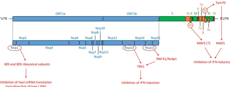
82% nucleotide identity with SARS-CoV and contains at least 14 open reading frames (ORFs)
[
4
,
5
] (
Fig 1
). It comprises a 5
0-untranslated region (5
0-UTR); ORF1a/b, encoding a
polypro-tein proteolytically processed into 16 nonstructural propolypro-teins (Nsp1–16); structural propolypro-teins
including spike (S), envelope (E), membrane (M), and nucleocapsid (N); 9 accessory proteins
(ORF3a, 3b, 6, 7a, 7b, 8, 9b, 9c, and 10); and a 3
0-UTR [
4
,
5
] (
Fig 1
).
Type I interferon (IFN-I) response is critical for providing an efficient protection against
viral infections. IFN-I production is rapidly triggered by the recognition by host sensors of
path-ogen-associated molecular patterns (PAMPs), such as viral nucleic acids [
6
]. IFN-I–induced
sig-naling converges on transcription factors, which rapidly induces the expression of hundreds of
genes called interferon-stimulated genes (ISGs) (reviewed in [
7
,
8
]). This antiviral signaling
cas-cade occurs in virtually all cell types exposed to IFN-I. ISGs, along with other downstream
mol-ecules controlled by IFN-I (including proinflammatory cytokines), have diverse functions,
ranging from direct inhibition of viral replication to the recruitment and activation of various
immune cells [
9
,
10
]. A robust, well-timed, and localized IFN-I response is thus required as a
first line of defense against viral infection because it promotes virus clearance, induces tissue
repair, and triggers a prolonged adaptive immune response against viruses.
Like most, if not all, RNA viruses, coronavirus RNA is detected by cytosolic sensors
includ-ing retinoic acid-inducible gene 1 (RIG-I/DExD/H-box helicase 58 [DDX58]) and melanoma
differentiation-associated gene 5 (MDA5/IFN induced with helicase C domain 1 [IFIH1]) [
11
,
12
]. Upon activation, RIG-I and MDA5 interact with the downstream adaptor, the
mitochon-drial antiviral signaling protein (MAVS, also known as IFN-B promoter stimulator 1 [IPS-1],
CARD adaptor inducing IFN-beta [CARDIF], or virus-induced signaling adaptor [VISA]) on
mitochondria. MAVS activation leads, via the recruitment of tumor necrosis factor
receptor-associated factor 3 (TRAF3), TRAF family member-receptor-associated NF-
κB activator
(TANK)-bind-ing kinase 1 (TBK1) and inhibitor of nuclear factor
κB (IκB) kinase-ε (IKKε), to the
phosphor-ylation of the IFN gene “master regulators” IFN regulatory factor (IRF)3 and IRF7. Upon
Fig 1. SARS-CoV-2 genomic organization and encoded proteins. ORF1a/1b encode a polyprotein, which is proteolytically processed into Nsp1–16, represented
in blue. Structural proteins, including S, E, M, and N proteins are in green. Accessory proteins encoded at the 30end of the viral genome comprise ORF3a, 3b, 6, 7a,
7b, 8, 9b, 9c, and 10 and are colored in orange. Untranslated extremities of the genome (50-UTR and 30-UTR) are also represented. In red are depicted SARS-CoV-2
proteins that interfere with IFN induction pathway as well as their known or hypothetic target [5,37,147]. E, envelope; IFN, interferon; M, membrane; MAVS, mitochondrial antiviral-signaling protein; N, nucleocapsid; Nrdp1, neuregulin receptor degradation protein-1; Nsp, nonstructural protein; ORF, open reading frame; RNF41, ring finger protein 41; S, spike; SARS-CoV-2, severe acute respiratory syndrome coronavirus-2; TANK, TRAF family member-associated NF-κB activator; TBK1, TANK-binding kinase 1; Tom70, translocase of outer mitochondrial membrane 70; UTR, untranslated region.
https://doi.org/10.1371/journal.ppat.1008737.g001
collection and analysis, decision to publish, or preparation of the manuscript.
Competing interests: The authors have declared
phosphorylation, IRF3 and/or IRF7 dimerize and translocate into the nucleus, where they
induce the expression of IFN-I and a subset of ISGs referred to as early ISGs (reviewed in
[
13
]). Secreted IFN-I then bind to the interferon alpha and beta receptor (IFNAR, composed
of the IFNAR1 and IFNAR2 subunits), leading to the activation of the Jak tyrosine kinases
tyrosine kinase 2 (Tyk2) and Janus kinase 1 (JAK1), which in turn phosphorylate the signal
transducer and activator of transcription (STAT)1 and STAT2 [
14
,
15
]. Phosphorylated
STATs heterodimerize and associate with the DNA binding protein IRF9 to form a complex
known as IFN-stimulated growth factor 3 (ISGF3). The ISGF3 complex translocates into the
nucleus and binds to interferon-stimulated response elements (ISREs) in ISG promoters, thus
inducing the expression of hundreds of ISG products that establish the antiviral state at the site
of viral infection [
15
]. The antiviral response is intensified by various signaling factors,
includ-ing sensors and transcriptional regulators, which are themselves ISGs induced by ISGF3 and/
or directly by the IRF3/IRF7 transcriptional activators. Aside from the IFN-I response, the
rec-ognition of double-stranded viral RNA elements by the protein kinase receptor (PKR) triggers
a translational arrest in infected cells (reviewed in [
8
,
16
,
17
]). This host response is highly
con-nected to the IFN-I response because PKR is also an ISG (reviewed in [
16
,
18
]).
IFN-I response requires fine-tuning because its overactivation is deleterious to the host.
Notably, some ISGs are involved in the regulation of cell metabolism, intracellular RNA
degra-dation, translation arrest, and cell death, for which changes can be potentially detrimental to
the host. IFN-I also potentiates the recruitment and activation of various immune cells. Thus,
although a robust IFN-I response is required as a first line of defense against viral infections,
systemic/uncontrolled or prolonged IFN-I production can lead to inflammatory diseases. For
example, an exacerbated IFN-I response contributes to the development of autoimmune
dis-eases [
19
]. COVID-19 is no exception to the rule, and it is therefore critical to understand the
regulation of the IFN-I response upon infection.
SARS-CoV-2 and IFN-I response
SARS-Cov-2 is a poor inducer of IFN-I response in vitro and in animal models as compared
with other respiratory RNA viruses [
20
,
21
]. IFN-I levels in the serum of infected patients are
below the detection levels of commonly used assays, yet ISG expression is detected [
4
,
22
], thus
suggesting that a limited IFN-I production could be sufficient to induce ISGs. Alternatively,
IFN-I production could be restricted to specific immune cells, such as plasmacytoid dendritic
cells (pDCs). Despite a more efficient replication in human lung tissues, SARS-CoV-2 induced
even less IFN-I than SARS-CoV [
4
], which is itself a weak inducer in human cells [
23
–
25
]. An
ineffective IFN-I response seems to be a hallmark of other coronavirus infections, as observed
with MERS-CoV in ex vivo respiratory tissue cultures [
26
] and with animal coronaviruses
such as the porcine epidemic diarrhea virus (PEDV) or the mouse hepatitis virus (MHV),
which are alpha- and beta-coronaviruses, respectively [
27
,
28
]. Indeed, coronaviruses have
developed multiple strategies to escape and counteract innate sensing and IFN-I production.
Inhibition of IFN-I induction and signaling by SARS-CoV-2
SARS-CoV encodes at least 10 proteins that allow the virus to either escape or counteract the
induction and antiviral action of IFN (
Fig 2
and
Table 1
) [
29
–
48
]. Initial observations already
suggest that the SARS-CoV-2 anti-IFN arsenal is at least as efficient as that of SARS-CoV [
4
,
20
,
22
], although detailed mechanistic studies are required to determine whether the IFN
antagonists identified in other coronaviruses have equivalently competent counterparts in
SARS-CoV-2. A virus–cell protein interaction map performed with 26 of the 29 SARS-CoV-2
proteins expressed in human embryonic kidney (HEK)293T identified several innate immune
signaling proteins as partners of viral proteins cells (
Fig 1
) [
5
]. SARS-CoV-2 ORF9b, like
SARS-CoV ORF9b, interacts with MAVS through its association with Tom70, thus suggesting
a conserved mechanism of IFN-I evasion [
5
,
40
] (
Fig 1
). Furthermore, SARS-CoV-2 Nsp13
and Nsp15 were found to interact with TBK1 and the TBK1 activator ring finger protein 41
(RNF41)/Nrdp1, respectively [
5
] (
Fig 1
). Nsp15, which is a highly conserved endoribonuclease
encoded by various coronaviruses, including SARS-CoV [
30
,
49
,
50
], antagonizes the
induc-tion of IFN-I by cleaving the 5
0-polyuridines of the negative-sense viral RNA, as demonstrated
for MHV and PEDV in various cellular models [
31
,
32
,
50
] (
Table 1
and
Fig 2
). If further
vali-dated, the interaction between SARS-CoV-2 nsp15 and TBK1 may reveal that the viral
endori-bonuclease antagonizes IFN induction via at least 2 mechanisms. SARS-CoV ORF3b was
reported to inhibit IFN induction and to act either on IRF3 or possibly on MAVS because it
translocates to mitochondria when overexpressed in Vero cells [
36
,
42
]. Despite the fact it
encodes a shorter protein than SARS-CoV, SARS-CoV-2 ORF3b was recently found to
sup-press IFN induction even more efficiently [
51
]. By screening 15,000 SARS-CoV-2 sequences,
the authors identified a natural variant encoding a longer ORF3b and displaying an even
greater inhibitory activity [
51
]. Finally, SARS-CoV-2 Nsp1 was also recently found to bind 40S
ribosomal subunits (
Fig 1
), thus inhibiting host mRNA translation, including that of IFN-I
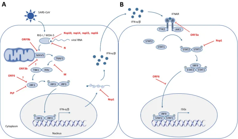
Fig 2. SARS-CoV interfering with IFN induction and signaling. On this cartoon are schematically represented the signaling pathways triggered by SARS-CoV RNA
recognition by the cytoplasmic RNA sensors RIG-I and MDA5, which leads to IFN induction (A) and subsequent IFN signaling in surrounding cells, resulting in the expression of ISGs (B). SARS-CoV proteins that have been reported to interfere with these pathways are indicated. IFN, interferon; IFNAR, interferon alpha and beta receptor; IκB, inhibitor of nuclear factor κB; IKKε, IκB kinase-ε; IRF, IFN regulatory factor; ISG, IFN-stimulated gene; JAK, Janus kinase; M, membrane; MAVS, mitochondrial antiviral signaling protein; MDA5, melanoma differentiation-associated gene 5; N, nucleocapsid; Nsp, nonstructural protein; ORF, open reading frame; P, phosphate; PLP, papain-like protease; RIG-I, retinoic acid-inducible gene 1; SARS-CoV, severe acute respiratory syndrome coronavirus; STAT, signal transducer and activator of transcription; TANK, TRAF family member associated NF-κB activator; TBK1, TANK-binding kinase 1; TRAF3, tumor necrosis factor receptor-associated factor 3; TYK2, tyrosine kinase 2.
[
52
], a feature that was previously demonstrated for other coronavirus-encoded Nsp1,
includ-ing SARS-CoV [
43
,
45
] (
Fig 2
and
Table 1
).
Another viral strategy to inhibit IFN-I signaling is to enhance the host retrocontrol of this
pathway. Several ISGs are themselves repressors of the IFN-I response, and their regulatory
functions operate at the viral and host mRNA transcription and translation steps, acting via a
wide-range of mechanisms (reviewed in [
7
,
53
]). For example, the inducible negative
regula-tors such as the suppressor of cytokine signaling (SOCS1 and SOCS3) act at various levels of
the Jak–STAT pathway or by targeting IRF7 for degradation [
54
]. In the context of SARS-CoV,
the S protein induces the expression of SOCS3 expression in B cells [
55
]. Induction of SOCS1/
3 expression is also detected in SARS-CoV–infected cells, albeit to a lower extent as compared
with other respiratory viruses [
56
]. Recent genomic screen approaches identified a set of
Table 1. SARS-CoV proteins interfering with IFN-I induction and signaling.
Protein Mechanism Affected Step Experimental Approach Cellular Model References Inhibition of IFN-I Induction
Nsp14 Guanine-N7-methyltransferase activity— methylates capped RNA transcripts
Sensing Yeast genetic system—validated using a SARS-CoV replicon
Yeast [29] Nsp15
(EndoU)
Cleaves the 50-polyuridines from negative-sense
viral RNA
Sensing Protein overexpression (mutants for other coronaviruses)
MA104 cells [30–32] Nsp16 20-O-methyltransferase activity involved in viral
RNA capping
Sensing SARS-CoV mutants Vero, Calu-3, and HAE cells; mice
[33]
Nsp10 Cofactor of Nsp16, required for RNA cap methylation
Sensing In vitro reconstitution of SARS-CoV mRNA cap methylation
In vitro [34] N Inhibits TRIM25-mediated RIG-I
ubiquitination
Sensing Protein overexpression HEK293T and A549 cells
[35–37] Nsp3 (PLP) Deubiquitinates cellular substrates, possibly
including RIG-I
Sensing Protein overexpression HEK293 cells [38] Inhibits IRF3 phosphorylation IRF3 activation Protein overexpression HEK293, HeLa, and
MA104 cells
[30,39] ORF9b Targets MAVS, TRAF3, and TRAF6 to
degradation
Downstream signaling
Protein overexpression HEK293 and A549 cells
[40] M Impedes the formation of TRAF3/TBK1/IKKε
complex
Downstream signaling
Protein overexpression; also observed in infected cells
HEK293 and HeLa cells
[41]
ORF6 Unknown Downstream
signaling
Protein overexpression HEK293T and A549 cells
[36] ORF3b Mechanism unclear; may target MAVS Downstream
signaling
Protein overexpression HEK293T and A549 cells
[36,42] Nsp1 Promotes cellular mRNA degradation and
blocks host mRNA translation
Expression of IFN-I Protein overexpression—validated using a SARS-CoV mutant virus
HEK293 and Vero cells
[43–45]
Inhibition of IFN-I Signaling
ORF3a Promotes IFNAR1 degradation IFN binding to its receptor
Protein overexpression Huh7 cells [46] Nsp1 Decreases STAT1 phosphorylation STAT1 activation Protein overexpression—validated using
a SARS-CoV mutant virus
HEK293T and Vero cells
[47] ORF6 Sequesters karyopherin alpha 2 and beta 1 Nuclear translocation
of STAT1
Protein overexpression—Validated using a SARS-CoV mutant virus
HEK293(T), A549, and Vero cells
[36,48]
Abbreviations: EndoU, endoribonuclease; HAE, human airway epithelial; HEK, human embryonic kidney; IFN, interferon; IFNAR, interferon alpha and beta receptor;
IFN-I, type I IFN; IκB, inhibitor of nuclear factor κB; IKKε, IκB kinase-ε; IRF, IFN regulatory factor; M, membrane; MAVS, mitochondrial antiviral signaling protein; Nsp, nonstructural protein; ORF, open reading frame; PLP, papain-like protease; RIG-I, retinoic acid-inducible gene 1; SARS-CoV, severe acute respiratory syndrome coronavirus; STAT, signal transducer and activator of transcription; TANK, TRAF family member-associated NF-κB activator; TBK1, TANK-binding kinase 1; TRAF, tumor necrosis factor receptor-associated factor; TRIM25, tripartite motif containing 25.
repressors of the IFN-I response depending on the cell type and activation pathway involved
[
57
–
59
]. Hence, one might anticipate that distinct repressors of the IFN-I response are induced
depending on the cell type targeted by SARS-CoV-2, the level of replication, and the
microen-vironment. For example, in the context of coronaviruses, an inefficient detection of MHV
infection likely results from an inhibition of the basal levels of sensors mRNA expression in
several cell types [
60
]. It is conceivable that this inhibition might involve negative regulators
such as the IFN-inducible RNF125, which targets signaling components such as RIG-I,
MDA5, and MAVS for degradation [
61
].
Interplay between host translation and the IFN-I response
Inhibition of protein synthesis is a conserved host response to prevent viral infections. The
host translation is dynamically regulated by PKR, activated via recognition of viral RNA
(reviewed by [
62
]). Activated PKR inhibits the eukaryotic initiation factor 2 (eIF2α), a major
regulator of the initiation phase of mRNA translation, by phosphorylating its
α subunit. The
PKR-induced translational arrest shuts down the negative feedback on the IFN-I response,
which can thus result in a prolonged and/or amplified IFN-I response [
63
]. Because PKR is an
ISG, the translational arrest is, in turn, potentiated by the IFN-I response (reviewed in [
64
]).
This highlights a paradoxical situation in which translation arrest prevents viral replication but
also set a threshold of viral detection to commensurate the host transcriptional antiviral
response to the level of infection [
63
].
Whether the PKR pathway is modulated by SARS-CoV2 is unknown, yet different
corona-viruses regulate PKR-eIF2α axis and host translation. For example, the MERS-CoV protein 4a
(p4a) accessory protein impedes PKR activation [
65
,
66
]. Future studies are needed to further
uncover the relationship between IFN-I response and host translation and their dynamics in
the context of SARS-CoV2 infection.
Dynamics of the IFN-I response define severity of infection
Dynamics of the IFN-I response at the levels of individual cells and cell
population
The IFN-I response varies among different cell types and within different microenvironments.
Studies at the single-cell level suggest that the amplitude and kinetic of the response is also
hetero-geneous for a given cell type. Mathematic modeling revealed that IFN-I response is, at least in
part, stochastic because only a fraction of cells are able to produce IFN-I upon activation by
ago-nists of the sensors and are sensitive to the paracrine stimulation by IFN-I [
67
–
71
]. The
heteroge-neity of the IFN-I response can be imprinted by the state of the cell at the activation time,
including its global translation activity, metabolism, expression levels of signaling molecules
(sen-sors, adaptors, and receptors) [
67
–
70
]. Additionally, the distinct onsets of the IFN-I induction
depend on the rapidity and amplitude of viral replication. This heterogeneous responsiveness at
the individual cell level consequently shapes the dynamics of the host antiviral response at the
whole population level [
69
–
71
]. This model of the IFN-I response dynamics yielded in the context
of various RNA viruses provides a framework likely at play for coronavirus infections. A delayed
induction of the ISG expression via virus-induced modulation of the basal activity of
transcrip-tional activity of STAT1 and PKR pathways leads to a peak of coronavirus replication preceding
the ISG response [
72
]. Additionally, in vivo study of the dynamic of MHV infection showed that
a fast and robust IFN-I production by pDCs down-regulate the IFN-I response by other cells [
73
].
This suggests that the IFN-I response at play in different cell types might drive the control of
coro-navirus infection and potentially contribute to the progression of the disease.
Impaired or delayed IFN-I response contributes to the disease
As mentioned above, coronaviruses possess various mechanisms to defeat the IFN-I response
within infected cells, and this inhibition ability is associated with clinical severity (reviewed in
[
74
]). Clinical studies showed that coronaviruses evade innate immunity during the first 10
days of infection, which corresponds to a period of widespread inflammation and steadily
increasing viral load [
75
,
76
]. Elevated virus replication eventually leads to inflammation and
hypercytokinemia, referred to as a “cytokine storm” [
77
–
80
] (
Fig 3
). The delayed IFN-I
response indeed promotes the accumulation of pathogenic monocyte–macrophages [
77
,
81
].
This cell infiltrate results in lung immunopathology, vascular leakage, and suboptimal T cell
response [
77
,
81
]. Immune phenotypic profiling in peripheral blood mononuclear cells
(PBMCs) of COVID-19 patients similarly revealed that high viremia is associated with an
exac-erbated IFN-I response, an aggravated cytokine secretion, and inflammation, driving clinical
Fig 3. Working model of the failure of IFN-I to control of SARS-CoV-2 infection, leading to COVID-19. While
IFN-I can control viral infection (upper panel), IFN-I deficiency is believed to play a key role in SARS-CoV-2 pathogenesis (lower panel). As shown for related coronaviruses, a delayed IFN-I signaling is associated with robust virus replication and severe complications, i.e., inflammation and “cytokine storm,” notably via the accumulation of monocytes resulting in lung immunopathology, vascular leakage, and suboptimal T cell response. ACE2, angiotensin I converting enzyme 2; IFNAR, interferon alpha and beta receptor; IFN-I, type I interferon; ISG, IFN-stimulated gene; pDC, plasmacytoid dendritic cell; SARS-CoV-2, severe acute respiratory syndrome coronavirus-2.
severity [
22
]. Although the IFNAR signaling pathway was up-regulated at an earlier disease
stage, down-regulation of ISGs, together with exacerbated NF-κB activation, promotes a
cyto-kine storm and hyperinflammation, found in critically ill patients [
22
].
Collectively, these findings highlight the negative impact of a delayed IFN-I response on
viral control and disease severity. However, the underlining mechanisms that drive the
tempo-ral control of the IFN-I response in patients are still elusive. In particular, the host and vitempo-ral
determinants driving the on/off switch of the IFN-I response in infected cells, noninfected
cells, and/or stimulated immune cells need to be investigated. Such studies will certainly
bene-fit from longitudinal studies of immune profiling in SARS-CoV2 infected patients at the
sin-gle-cell level and in combination with the clinical data.
Robust and localized IFN-I response by pDCs
By producing 1,000-fold more IFN-I than any other cell types, pDCs are at the heart of the
antiviral IFN-I response [
82
,
83
]. They also produce other proinflammatory cytokines, which
contribute to modulate the function of several immune cells, such as the mobilization of
natu-ral killer (NK) cells or the licensing of virus-specific T cell responses [
82
–
85
]. Because pDCs
are refractory to most viral replication, their antiviral response cannot be inhibited by viral
proteins [
82
,
86
]. This unopposed response likely contributes to the exceptional magnitude of
pDC IFN-I production [
82
,
87
–
89
]. pDCs are circulating immune cells; nonetheless, their
response is mostly localized at the infection site because their activation requires physical
con-tact with infected cells [
82
,
83
,
90
]. The contact site between pDCs and infected cells, which we
named the interferogenic synapse, is a specialized platform for PAMP transfer from infected
cell to the Toll-like receptor 7 (TLR7) sensor in pDC, leading to an antiviral response [
91
].
Previous studies on SARS- and MERS-CoV demonstrated that the rapid production of
IFN-I by pDCs is essential for the control of potentially lethal coronavirus infections in mouse
models [
77
,
92
]. pDCs migrate into the lungs at the early phase of infection (i.e., pDC number
peaks at day 2), temporally coinciding with the peak of IFNα production [
92
,
93
]. The pDC
number was found to be reduced in blood of COVID-19 patients as compared with control
patients [
22
], potentially resulting from a prior response followed by a vanished number of
cir-culating pDCs and/or their mobilization to the infected site. Future studies are needed to
address how pDC responsiveness evolves in the course of SARS-CoV-2 infection and how
pDCs respond to contact with coronavirus-infected cells.
Are IFNs protective of or detrimental to SARS-CoV-2?
Despite the abovementioned viral inhibitory mechanisms of IFN-I response (
Table 1
),
exoge-nous IFN-I in cell cultures efficiently inhibit SARS-CoV, SARS-CoV-2, and MERS-CoV
spread [
5
,
20
,
26
,
51
,
94
–
101
]. Consistently, IFN-I was shown to have a protective effect against
SARS-CoV and MERS-CoV, alone or in combination with other antivirals, in various animal
models including mice, marmosets, and macaques [
97
,
102
,
103
]. IFN-I and III interfere with
virus infection by inducing the expression of several hundred ISGs [
7
]. Numerous
well-described ISGs exhibit direct antiviral activities by targeting specific stages of the viral life
cycle, including entry into host cells, replication, protein translation, and assembly of new
virus particles. As mentioned above, many signaling regulators are themselves ISGs, thus
lead-ing to amplification of the antiviral IFN-I pathway.
Detrimental effects of IFNs on SARS-CoV-2 replication
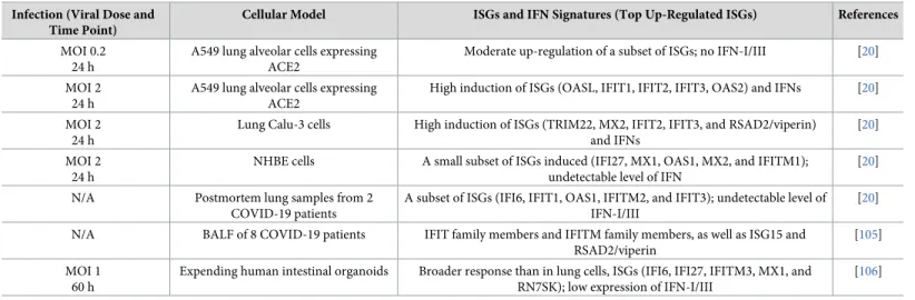
As a first step towards identifying ISGs able to restrict SARS-CoV-2 replication, transcriptomic
responses to infection have been analyzed in different cellular models, including primary cells,
organoids, and clinical samples [
20
,
104
–
106
], as summarized in
Table 2
. These studies
dem-onstrate that, despite triggering very little to no IFN expression (
Table 2
), SARS-CoV-2
repli-cation induces moderate levels of a limited number of ISGs. A small subset of infected cells
may be refractory to the antagonistic mechanisms of SARS-CoV-2, producing minute but
suf-ficient amounts of IFNs to trigger ISG induction in larger population of cells. Alternatively,
ISGs may be up-regulated in noninfected cells, which were analyzed together with infected
ones. Indeed, interpretation of genome-wide investigations of virus–pathogen interactions are
often obscured by analyses of mixed populations of infected and uninfected cells [
107
].
Of note, by contrast to low-multiplicity of infection (MOI) infection of A549 cells
express-ing angiotensin I convertexpress-ing enzyme 2 (ACE2), normal human bronchial epithelial (NHBE)
cells, and patient samples, high-MOI infections of A549-ACE2 and Calu-3 cells led to the high
induction of IFNs and ISGs, including ISGs with broad antiviral activities [
20
] (
Table 2
). This
discrepancy of IFN production/signaling between the levels of viral replication and/or
propor-tion of infected cells might reflect that the counteracpropor-tion measures employed by SARS-CoV-2
are less potent at high MOI. Alternatively, as suggested by Blanco-Melo and colleagues,
high-MOI infections in cell culture may generate more PAMPs, such as defective noninfectious
viral particles, than low-MOI infections [
108
].
Despite being expressed at moderate levels in vitro and in vivo, several up-regulated ISGs
identified by these transcriptomic studies (
Table 2
) exhibit well-characterized broad-spectrum
antiviral activities and could thus have additive restrictive effects on SARS-CoV-2 replication.
For instance, the 3 members of the interferon-induced protein with tetratricopeptide repeats
(IFITM) family, known to inhibit entry of numerous enveloped RNA viruses [
109
], similarly
restrict entry of SARS-CoV, MERS-CoV, and the globally circulating human coronaviruses
229E and NL63 in 293T and A549 cell lines [
110
,
111
]. OAS1 and mycovirus resistance protein
(Mx)A could also contribute to the IFN-I–mediated inhibitory effect on SARS-CoV-2 because
a clinical study revealed that single nucleic polymorphisms in the OAS1 3
0-UTR and MxA
pro-moter region appear associated with host susceptibility to SARS-CoV in the Chinese Han
pop-ulation [
112
]. Moreover, the fact that MERS-CoV nonstructural protein 4B (NS4b) is a 2
0-5
0-Table 2. ISGs and IFN signature of SARS-CoV-2–infected samples. Infection (Viral Dose and
Time Point)
Cellular Model ISGs and IFN Signatures (Top Up-Regulated ISGs) References
MOI 0.2 24 h
A549 lung alveolar cells expressing ACE2
Moderate up-regulation of a subset of ISGs; no IFN-I/III [20] MOI 2
24 h
A549 lung alveolar cells expressing ACE2
High induction of ISGs (OASL, IFIT1, IFIT2, IFIT3, OAS2) and IFNs [20] MOI 2
24 h
Lung Calu-3 cells High induction of ISGs (TRIM22, MX2, IFIT2, IFIT3, and RSAD2/viperin) and IFNs
[20] MOI 2
24 h
NHBE cells A small subset of ISGs induced (IFI27, MX1, OAS1, MX2, and IFITM1); undetectable level of IFN
[20] N/A Postmortem lung samples from 2
COVID-19 patients
A subset of ISGs (IFI6, IFIT1, OAS1, IFITM2, and IFIT3); undetectable level of IFN-I/III
[20] N/A BALF of 8 COVID-19 patients IFIT family members and IFITM family members, as well as ISG15 and
RSAD2/viperin
[105] MOI 1
60 h
Expending human intestinal organoids Broader response than in lung cells, ISGs (IFI6, IFI27, IFITM3, MX1, and RN7SK); low expression of IFN-I/III
[106]
Abbreviations: ACE2, angiotensin I converting enzyme 2; BALF, bronchoalveolar lavage fluid; IFI, interferon-inducible protein; IFIT, interferon-induced protein with
tetratricopeptide repeats; IFN, interferon; IFN-I/III, type I/III IFN; ISG, IFN-stimulated gene; MOI, multiplicity of infection; MX, myxovirus resistance protein; N/A, not applicable; NHBE, normal human bronchial epithelial; OAS, 20-50-oligoadenylate synthetase; RSAD2, Radical S-Adenosyl Methionine Domain Containing 2;
RN7SK, RNA component of 7SK nuclear ribonucleoprotein; SARS-CoV-2, severe acute respiratory syndrome coronavirus-2; TRIM22, tripartite motif containing 22.
oligoadenylate synthetase (OAS)-RNase L antagonist [
113
] suggests that the OAS pathway
contributes to the antiviral effects of IFNs on coronavirus replication. ISGs positively
potenti-ating IFN signaling, such as IFIH1/MDA5, TANK, IRF7, and STAT1, were also increased in
the bronchoalveolar lavage fluid (BALF) of COVID-19 patients as compared with healthy
con-trols [
105
] and could potentially contribute to the amplification of IFN-I response against
SARS-CoV-2 replication.
Zinc finger antiviral protein (ZAP), which is encoded by an ISG, contributes to the
anti-SARS-CoV-2 effect of IFNs in human lung Calu-3 cells [
114
]. ZAP is known for restricting the
replication of numerous viruses such as retroviruses and filoviruses [
115
]. The protein recruits
the cellular mRNA degradation machinery to viral RNA via 5
0-C-phosphate-G-3
0(CpG)
dinu-cleotide recognition [
115
].
To further determine which individual ISG or combination of ISGs mainly restricts
SARS--CoV-2 replication in vitro, several previously established approaches could be used, such as,
for example, screening for single or combined ISG activity using a lentiviral vector-based
library, as successfully performed by Schoggins and colleagues for other viral infections [
116
–
119
]. Indeed, this library of around 380 human ISGs was recently screened in human
hepa-toma cells for antiviral activity against HCoV-229E [
120
]. The screen identified IFN-inducible
lymphocyte antigen 6 complex, locus E (LY6E) as a potent inhibitor of the replication of
multi-ple coronaviruses, including SARS-CoV, SARS-CoV-2, and MERS-CoV, by blocking fusion of
viral and cellular membranes [
120
]. Mice studies revealed that LY6E directly protects primary
B cells and dendritic cells from murine coronavirus infection [
120
]. Pursuing the identification
and characterization of IFN effectors with potent anti-SARS-CoV-2 activities will reveal
weak-ness points in the life cycle of SARS-CoV-2 and may lead to the design of drugs that activate
antiviral ISGs or either mimic or amplify their action.
Beneficial effects of IFN on SARS-CoV-2 replication
Recent advances in systematic screening strategies have revealed the existence of a small subset
of ISGs exhibiting proviral activities [
116
,
119
]. These proviral ISGs act either by exhibiting
direct proviral activities such as facilitating viral entry [
119
] or via their abilities to negatively
regulate IFN signaling and facilitate the return to cellular homeostasis. The receptor tyrosine
kinase AXL is a well-characterized example of an ISG that is used by enveloped virus for
cellu-lar internalization [
121
–
123
]. Alternatively, ISGs that possess antiviral activities against a viral
family can be hijacked by unrelated viruses to favor infection. This is the case for IFITM2 and
IFITM3, which potently block entry of a broad range of enveloped viruses [
109
] while
promot-ing entry step of human coronavirus OC43 (HCoV-OC43) in human cells [
124
].
SARS-CoV-2 uses ACE2 and transmembrane serine protease 2 (TMPRSS2) to enter cells
[
125
]. Viral tropism is thus largely dictated by ACE2 and TMPRSS2 coexpression. Analysis of
human, nonhuman primate, and mouse single-cell RNA-sequencing (scRNA-seq) data sets
generated from healthy or diseased individuals revealed that expression of
ACE2 is primarily
restricted to type II pneumocytes in the lung, absorptive enterocytes within the gut, and goblet
secretory cells of the nasal mucosa [
126
]. Interestingly, this meta-analysis identified an
associa-tion between ACE2 expression and canonical ISGs or components of the IFN-signaling
path-way in different tissues. Independent analyzes of publicly available data sets concluded that
ACE2 expression pattern is similar to ISGs [
127
]. In vitro validations were performed by
treat-ing primary human upper airway cells with numerous inflammatory cytokines. IFNα2, and to
some extent IFNy, led to greater and more significant ACE2 up-regulation compared with all
other tested cytokines [
126
]. Substantial up-regulation of
ACE2 was also observed in primary
expression was also up-regulated upon ex vivo influenza A infection in human lung explants
isolated following surgical resection [
126
]. Because the majority of cells robustly up-regulating
ACE2 were epithelial, this observation potentially explains why previous analyses to define
canonical ISGs within immune populations did not identify
ACE2 as an induced gene [
116
].
Finally, STAT1, STAT3, IRF8, and IRF1 binding sites were identified within
−1,500 to +500 bp
of the transcription start site of ACE2 [
126
]. Despite need for direct evidence that IFNs
up-reg-ulate ACE2 in target cells in vivo, altogether these studies suggest that ACE2 could be an ISG
that enhances SARS-CoV-2 internalization in human epithelial cells [
125
,
126
].
Elucidating tissue and cell type specificity of ISGs, as well as their mechanisms of action, is
essential for understanding the potential dual role of IFNs during human SARS-CoV-2
infec-tion. It may also guide the use of IFNs in clinical trials.
Clinical implications of the dual role of IFN on SARS-CoV-2 replication
Although IFN-I treatment gave some encouraging results against SARS-CoV and MERS-CoV
in vitro and in animal models, including mice, marmosets, and macaques [
97
,
102
,
103
,
128
,
129
], additional knowledge to optimize its therapeutic efficiency in humans is required [
130
–
134
]. Previous information yielded from these animal studies provided guidance for treating
the current pandemic virus. First, it became clear from these former studies that IFNβ is a more
potent inhibitor than IFNα as shown both in vitro and in patients [
129
,
132
]. Second, the timing
of IFN-I treatment seems determinant for infection outcomes. Indeed, as shown in mice and in
macaques, IFN-I is protective when administered prior to SARS-CoV or MERS-CoV infection
or early in the course of infection, whereas late administration could be either ineffective or
det-rimental [
77
,
135
]. In humans as well, IFN-I–based therapies were not beneficial to critically ill
patients with multiple comorbidities and who were diagnosed late with MERS-CoV, thus
point-ing out that IFN-I has to be administered early after infection [
134
,
136
].
The first clinical trials using IFN-I alone or in combination with other antivirals are
cur-rently carried out in COVID-19 patients in several countries. For instance, the multicenter,
adaptive, randomized, open clinical trial DisCoVeRy evaluates, among other treatment, the
efficacy of IFNβ as a treatment for COVID-19 in hospitalized adults in Europe. A recent
open-label, randomized, phase 2 trial performed in adults with COVID-19 in Hong Kong showed
that the triple combination of IFNβ-1b, lopinavir–ritonavir, and ribavirin was safe and
supe-rior to lopinavir–ritonavir alone in alleviating symptoms and shortening the duration of viral
shedding and hospital stay in patients with mild to moderate COVID-19 [
137
]. It has to be
noted that the patients were treated in the early stages of the disease because the median
num-ber of days from symptom onset to start of study treatment was 5 days, further reinforcing the
fact that the timing of IFN-I treatment is key [
137
].
Other therapeutic approaches are under investigation to avoid the adverse effects of IFN-I
therapy and/or its potential inefficacity when administrated too late postinfection. One
strat-egy is to use aerosol formulations of recombinant IFN to deliver the cytokine directly inside
the lung [
138
,
139
]. This approach has several benefits because it is a noninvasive route of
administration, and the local concentration reached in the tissue can be higher than through
systemic injection and is thus expected to minimize the adverse effects of IFN. Nebulized
IFNα-2b was used on COVID-19 patients in Wuhan, alone or in combination with arbidol
[
140
]. The study, performed on 77 adults, showed a significant reduction of the duration of
detectable virus in the upper respiratory tract in IFNα-2b–treated patients, with or without
arbidol [
140
]. Another study currently ongoing in Beijing aims at evaluating the efficacy and
safety of recombinant human IFNα spray in preventing SARS-CoV-2 infection in highly
exposed medical staffs (ChiCTR2000030013).
Type III IFNs (IFNλs or IFN-III) are gaining an increased interest in antiviral therapies
[
141
–
143
]. Like IFN-I, they activate the JAK–STAT signaling pathway. They do so via a
recep-tor that is largely restricted to cells of epithelial origin, including respirarecep-tory epithelial cells
(reviewed in [
144
]). IFN-IIIs are induced upon viral infections, and they are growing evidence
that they provide important first-line defense against viral infections of the respiratory and
gas-trointestinal tracts [
145
–
147
]. In mice, IFN-III was shown to protect epithelial cells of the
respiratory and tract from infections with several respiratory viruses, including MERS-CoV
[
147
]. A study investigating SARS-CoV-2 infection of intestinal epithelial cells, using both
colon-derived cell lines and primary colon organoids, showed that IFN-III response was more
efficient than IFN-I at controlling viral replication [
148
]. However, IFN-IIIs produced by
den-dritic cells in the lung were recently shown to cause barrier damage and to compromise host
tissue tolerance and predispose to lethal bacterial superinfections [
149
]. Therefore, although
the antiviral properties are promising, the benefit of IFN-III to treat COVID-19 patients awaits
careful evaluation. The first clinical trials using IFN-III are ongoing, including one launched at
the Massachusetts General Hospital to evaluate the safety and efficacy of pegylated IFNλ on a
small number of COVID-19 patients (NCT04343976).
Besides the use of recombinant IFN as a therapeutic treatment, one interesting alternative
strategy would be to boost the natural innate immune defenses of COVD-19 patients at early
stages of the disease. Because pDCs are seemingly crucial to control coronavirus infections [
77
,
92
], a possibility would be to either amplify or prolong their activation to make them produce
more IFN-I and IFN-III. A number of negative feedback loops prevent an exacerbated
activa-tion of pDCs, which can be deleterious for the organism in the long term. Thus, transitorily
inhibiting these negative retrocontrols may increase the antiviral activity of pDCs. For
instance, the bone marrow stromal cell antigen 2 (BST2) is an ISG that activates the
immuno-globulin-like transcript 7 (ILT7) inhibitory receptor expressed by pDCs to interrupt the IFN-I
response [
150
]. The blockade of this interaction using either antibodies or inhibitory
mole-cules should thus increase the duration of pDC activation. One could also envisage to take
advantage of viral proteins that counteract the antiviral activity of BST2, such as HIV-1 viral
protein U (vpu) [
151
]. Other pDC inhibitory molecules include natural monamines such as
histamine, dopamine, or serotonin, which bind to the C-X-C motif chemokine receptor 4
(CXCR4) at the surface of pDCs [
152
]. Because the CXCR4 antagonist AMD3100 (also known
as plerixafor) blocks the binding of monoamines to pDCs, it can prevent the amine-dependent
inhibition of pDC activation [
152
]. AMD3100 is already used in clinics as an
immunostimula-tory molecule able to mobilize hematopoietic stem cells in cancer patients [
153
]. Finally, we
recently reported that the peptidyl-prolyl isomerase peptidyl-prolyl
cis-trans isomerase
NIMA-interacting 1 (Pin1) switches off the IFN-I expression by pDCs by inducing IRF7 degradation
[
154
]. A number of Pin1 inhibitors have been developed and could be tested for their potential
activity on human pDCs [
155
] and could represent another possible therapeutic strategy to
boost pDC-mediated IFN-I production.
Conclusions and perspectives
SARS-CoV-2 emerged in the human population around 7 months ago, yet it seems well
adapted to avoid and inhibit the IFN-I response in its new host. Such efficient strategies allow
the virus to replicate and disseminate in infected individuals without encountering the initial
host defense. This modest IFN response could explain why viremia peaks at early stages of the
disease, at the time of symptoms appearance, and not around 7 to 10 days following symptoms,
like during SARS-CoV and MERS-CoV infections. IFNβ treatment would be expected to
improve the antiviral response of patients at the early stage of COVID-19 and, if possible, at
the site of infection. Indeed, IFNβ appeared to be pivotal to improve patient states in a
com-bined therapy regiment of IFNβ, lopinavir–ritonavir, and ribavirin [
137
]. Nonetheless,
IFN-resistant viral mutants may arise and be able to control IFN even more efficiently than parental
viruses.
The exacerbated production of proinflammatory cytokines observed at later stage of
COVID-19 might challenge the efficiency of an IFNβ treatment administrated after
appear-ance of symptoms. There is indeed an increasing appreciation of the detrimental effects of
inappropriate, excessive, or mistimed IFN-I responses in viral infections [
156
]. The underlying
mechanisms by which IFN-I promote disease severity likely include immunopathology due to
excessive inflammation and direct tissue damage. At the late stages of COVID-19,
immuno-modulatory drugs that diminish inflammation may benefit patients. The therapeutic benefits
of such treatments have been demonstrated in the context of influenza infection in clinical
tri-als [
157
] and in mice models [
158
]. Discovery of host markers associated with disease
progres-sion will be instrumental to defining the appropriate treatment and time of administration.
Luckily, most COVID-19 patients develop no or mild symptoms. In these patients, the
virus ends up being cleared by the immune system, possibly through a partial protection
con-ferred by cross-reactive CD4+ T cells that have been found in between 40% and 60% of
unex-posed individuals [
159
]. It is therefore probable that a viral replication that is under control
thanks to an efficient adaptive immunity prevents a systemic viral spread and the subsequent
cytokine storm. Prior and coinfections along with age, gender, immunological state, and
comorbidities also likely play a key role in the ability of the patients to efficiently respond to
SARS-CoV-2 infection.
The current studies that aim to better understand the mechanisms that render some
patients particularly sensitive to SARS-CoV-2 infections raise hope for the possibility of
treat-ing patients with drugs that either enhance the IFN response at the early stage of the disease or
dampen it at later stages.
Acknowledgments
We thank Nathalie J. Arhel for helpful discussion.
References
1. Wang C, Horby PW, Hayden FG, Gao GF. A novel coronavirus outbreak of global health concern. Lan-cet. 2020; 395(10223):470–3.https://doi.org/10.1016/S0140-6736(20)30185-9PMID:31986257; PubMed Central PMCID: PMC7135038.
2. Zhu N, Zhang D, Wang W, Li X, Yang B, Song J, et al. A Novel Coronavirus from Patients with Pneu-monia in China, 2019. N Engl J Med. 2020; 382(8):727–33.https://doi.org/10.1056/NEJMoa2001017
PMID:31978945; PubMed Central PMCID: PMC7092803.
3. Wu F, Zhao S, Yu B, Chen YM, Wang W, Song ZG, et al. A new coronavirus associated with human respiratory disease in China. Nature. 2020; 579(7798):265–9. https://doi.org/10.1038/s41586-020-2008-3PMID:32015508; PubMed Central PMCID: PMC7094943.
4. Chan JF, Kok KH, Zhu Z, Chu H, To KK, Yuan S, et al. Genomic characterization of the 2019 novel human-pathogenic coronavirus isolated from a patient with atypical pneumonia after visiting Wuhan. Emerg Microbes Infect. 2020; 9(1):221–36.https://doi.org/10.1080/22221751.2020.1719902PMID:
31987001; PubMed Central PMCID: PMC7067204.
5. Gordon DE, Jang GM, Bouhaddou M, Xu J, Obernier K, White KM, et al. A SARS-CoV-2 protein inter-action map reveals targets for drug repurposing. Nature. 2020; 583: 459–468.https://doi.org/10.1038/ s41586-020-2286-9PMID:32353859.
6. Streicher F, Jouvenet N. Stimulation of Innate Immunity by Host and Viral RNAs. Trends Immunol. 2019; 40(12):1134–48.https://doi.org/10.1016/j.it.2019.10.009PMID:31735513.
7. Schoggins JW. Interferon-Stimulated Genes: What Do They All Do? Annu Rev Virol. 2019; 6(1):567– 84.https://doi.org/10.1146/annurev-virology-092818-015756PMID:31283436.
8. Schneider WM, Chevillotte MD, Rice CM. Interferon-stimulated genes: a complex web of host defenses. Annu Rev Immunol. 2014; 32:513–45. https://doi.org/10.1146/annurev-immunol-032713-120231PMID:24555472; PubMed Central PMCID: PMC4313732.
9. Crouse J, Kalinke U, Oxenius A. Regulation of antiviral T cell responses by type I interferons. Nat Rev Immunol. 2015; 15(4):231–42.https://doi.org/10.1038/nri3806PMID:25790790.
10. Makris S, Paulsen M, Johansson C. Type I Interferons as Regulators of Lung Inflammation. Front Immunol. 2017; 8: 259.https://doi.org/10.3389/fimmu.2017.00259PMID:28344581; PubMed Central PMCID: PMC5344902.
11. Li J, Liu Y, Zhang X. Murine coronavirus induces type I interferon in oligodendrocytes through recogni-tion by RIG-I and MDA5. J Virol. 2010; 84(13): 6472–82.https://doi.org/10.1128/JVI.00016-10PMID:
20427526; PubMed Central PMCID: PMC2903279.
12. Zalinger ZB, Elliott R, Rose KM, Weiss SR. MDA5 Is Critical to Host Defense during Infection with Murine Coronavirus. J Virol. 2015; 89(24):12330–40.https://doi.org/10.1128/JVI.01470-15PMID:
26423942; PubMed Central PMCID: PMC4665247.
13. Loo YM, Gale M Jr. Immune signaling by RIG-I-like receptors. Immunity. 2011; 34(5):680–92.https:// doi.org/10.1016/j.immuni.2011.05.003PMID:21616437; PubMed Central PMCID: PMC3177755.
14. Darnell JE Jr., Kerr IM, Stark GR. Jak-STAT pathways and transcriptional activation in response to IFNs and other extracellular signaling proteins. Science. 1994; 264(5164):1415–21.https://doi.org/10. 1126/science.8197455PMID:8197455.
15. Schindler C, Levy DE, Decker T. JAK-STAT signaling: from interferons to cytokines. J Biol Chem. 2007; 282(28):20059–63.https://doi.org/10.1074/jbc.R700016200PMID:17502367.
16. Dabo S, Meurs EF. dsRNA-dependent protein kinase PKR and its role in stress, signaling and HCV infection. Viruses. 2012; 4(11):2598–635.https://doi.org/10.3390/v4112598PMID:23202496; PubMed Central PMCID: PMC3509664.
17. Ivashkiv LB, Donlin LT. Regulation of type I interferon responses. Nat Rev Immunol. 2014; 14(1):36– 49.https://doi.org/10.1038/nri3581PMID:24362405; PubMed Central PMCID: PMC4084561.
18. McCormick C, Khaperskyy DA. Translation inhibition and stress granules in the antiviral immune response. Nat Rev Immunol. 2017; 17(10):647–60.https://doi.org/10.1038/nri.2017.63PMID:
28669985.
19. Uggenti C, Lepelley A, Crow YJ. Self-Awareness: Nucleic Acid-Driven Inflammation and the Type I Interferonopathies. Annu Rev Immunol. 2019; 37:247–67. https://doi.org/10.1146/annurev-immunol-042718-041257PMID:30633609.
20. Blanco-Melo D, Nilsson-Payant BE, Liu WC, Uhl S, Hoagland D, Moller R, et al. Imbalanced Host Response to SARS-CoV-2 Drives Development of COVID-19. Cell. 2020; 181(5):1036–45 e9.https:// doi.org/10.1016/j.cell.2020.04.026PMID:32416070; PubMed Central PMCID: PMC7227586.
21. Chu H, Chan JF, Wang Y, Yuen TT, Chai Y, Hou Y, et al. Comparative replication and immune activa-tion profiles of SARS-CoV-2 and SARS-CoV in human lungs: an ex vivo study with implicaactiva-tions for the pathogenesis of COVID-19. Clin Infect Dis. 2020: ciaa410.https://doi.org/10.1093/cid/ciaa410PMID:
32270184; PubMed Central PMCID: PMC7184390.
22. Hadjadj J, Yatim N, Barnabei L, Corneau A, Boussier J, Smith N, et al. Impaired type I interferon activ-ity and inflammatory responses in severe COVID-19 patients. Science. 2020: eabc6027.https://doi. org/10.1126/science.abc6027PMID:32661059.
23. Spiegel M, Pichlmair A, Martinez-Sobrido L, Cros J, Garcia-Sastre A, Haller O, et al. Inhibition of Beta interferon induction by severe acute respiratory syndrome coronavirus suggests a two-step model for activation of interferon regulatory factor 3. J Virol. 2005; 79(4):2079–86.https://doi.org/10.1128/JVI. 79.4.2079-2086.2005PMID:15681410; PubMed Central PMCID: PMC546554.
24. Cheung CY, Poon LL, Ng IH, Luk W, Sia SF, Wu MH, et al. Cytokine responses in severe acute respi-ratory syndrome coronavirus-infected macrophages in vitro: possible relevance to pathogenesis. J Virol. 2005; 79(12):7819–26.https://doi.org/10.1128/JVI.79.12.7819-7826.2005PMID:15919935; PubMed Central PMCID: PMC1143636.
25. Ziegler T, Matikainen S, Ronkko E, Osterlund P, Sillanpaa M, Siren J, et al. Severe acute respiratory syndrome coronavirus fails to activate cytokine-mediated innate immune responses in cultured human monocyte-derived dendritic cells. J Virol. 2005; 79(21):13800–5.https://doi.org/10.1128/JVI.79.21. 13800-13805.2005PMID:16227300; PubMed Central PMCID: PMC1262618.
26. Chan RW, Chan MC, Agnihothram S, Chan LL, Kuok DI, Fong JH, et al. Tropism of and innate immune responses to the novel human betacoronavirus lineage C virus in human ex vivo respiratory organ cul-tures. J Virol. 2013; 87(12):6604–14.https://doi.org/10.1128/JVI.00009-13PMID:23552422; PubMed Central PMCID: PMC3676115.
27. Zhang Q, Shi K, Yoo D. Suppression of type I interferon production by porcine epidemic diarrhea virus and degradation of CREB-binding protein by nsp1. Virology. 2016; 489:252–68.https://doi.org/10. 1016/j.virol.2015.12.010PMID:26773386; PubMed Central PMCID: PMC7111358.
28. Roth-Cross JK, Martinez-Sobrido L, Scott EP, Garcia-Sastre A, Weiss SR. Inhibition of the alpha/beta interferon response by mouse hepatitis virus at multiple levels. J Virol. 2007; 81(13):7189–99.https:// doi.org/10.1128/JVI.00013-07PMID:17459917; PubMed Central PMCID: PMC1933268.
29. Chen Y, Cai H, Pan J, Xiang N, Tien P, Ahola T, et al. Functional screen reveals SARS coronavirus nonstructural protein nsp14 as a novel cap N7 methyltransferase. Proc Natl Acad Sci U S A. 2009; 106 (9):3484–9.https://doi.org/10.1073/pnas.0808790106PMID:19208801; PubMed Central PMCID: PMC2651275.
30. Frieman M, Ratia K, Johnston RE, Mesecar AD, Baric RS. Severe acute respiratory syndrome corona-virus papain-like protease ubiquitin-like domain and catalytic domain regulate antagonism of IRF3 and NF-kappaB signaling. J Virol. 2009; 83(13):6689–705.https://doi.org/10.1128/JVI.02220-08PMID:
19369340; PubMed Central PMCID: PMC2698564.
31. Deng X, Hackbart M, Mettelman RC, O’Brien A, Mielech AM, Yi G, et al. Coronavirus nonstructural protein 15 mediates evasion of dsRNA sensors and limits apoptosis in macrophages. Proc Natl Acad Sci U S A. 2017; 114(21):E4251–E60.https://doi.org/10.1073/pnas.1618310114PMID:28484023; PubMed Central PMCID: PMC5448190.
32. Hackbart M, Deng X, Baker SC. Coronavirus endoribonuclease targets viral polyuridine sequences to evade activating host sensors. Proc Natl Acad Sci U S A. 2020; 117(14):8094–103.https://doi.org/10. 1073/pnas.1921485117PMID:32198201; PubMed Central PMCID: PMC7149396.
33. Menachery VD, Yount BL Jr., Josset L, Gralinski LE, Scobey T, Agnihothram S, et al. Attenuation and restoration of severe acute respiratory syndrome coronavirus mutant lacking 2’-o-methyltransferase activity. J Virol. 2014; 88(8):4251–64.https://doi.org/10.1128/JVI.03571-13PMID:24478444; PubMed Central PMCID: PMC3993736.
34. Bouvet M, Debarnot C, Imbert I, Selisko B, Snijder EJ, Canard B, et al. In vitro reconstitution of SARS-coronavirus mRNA cap methylation. PLoS Pathog. 2010; 6(4):e1000863.https://doi.org/10.1371/ journal.ppat.1000863PMID:20421945; PubMed Central PMCID: PMC2858705.
35. Lu X, Pan J, Tao J, Guo D. SARS-CoV nucleocapsid protein antagonizes IFN-beta response by target-ing initial step of IFN-beta induction pathway, and its C-terminal region is critical for the antagonism. Virus Genes. 2011; 42(1):37–45.https://doi.org/10.1007/s11262-010-0544-xPMID:20976535; PubMed Central PMCID: PMC7088804.
36. Kopecky-Bromberg SA, Martinez-Sobrido L, Frieman M, Baric RA, Palese P. Severe acute respiratory syndrome coronavirus open reading frame (ORF) 3b, ORF 6, and nucleocapsid proteins function as interferon antagonists. J Virol. 2007; 81(2):548–57.https://doi.org/10.1128/JVI.01782-06PMID:
17108024; PubMed Central PMCID: PMC1797484.
37. Hu Y, Li W, Gao T, Cui Y, Jin Y, Li P, et al. The Severe Acute Respiratory Syndrome Coronavirus Nucleocapsid Inhibits Type I Interferon Production by Interfering with TRIM25-Mediated RIG-I Ubiquiti-nation. J Virol. 2017; 91(8): e02143–16.https://doi.org/10.1128/JVI.02143-16PMID:28148787; PubMed Central PMCID: PMC5375661.
38. Clementz MA, Chen Z, Banach BS, Wang Y, Sun L, Ratia K, et al. Deubiquitinating and interferon antagonism activities of coronavirus papain-like proteases. J Virol. 2010; 84(9):4619–29.https://doi. org/10.1128/JVI.02406-09PMID:20181693; PubMed Central PMCID: PMC2863753.
39. Devaraj SG, Wang N, Chen Z, Chen Z, Tseng M, Barretto N, et al. Regulation of IRF-3-dependent innate immunity by the papain-like protease domain of the severe acute respiratory syndrome corona-virus. J Biol Chem. 2007; 282(44):32208–21.https://doi.org/10.1074/jbc.M704870200PMID:
17761676; PubMed Central PMCID: PMC2756044.
40. Shi CS, Qi HY, Boularan C, Huang NN, Abu-Asab M, Shelhamer JH, et al. SARS-coronavirus open reading frame-9b suppresses innate immunity by targeting mitochondria and the MAVS/TRAF3/ TRAF6 signalosome. J Immunol. 2014; 193(6):3080–9.https://doi.org/10.4049/jimmunol.1303196
PMID:25135833; PubMed Central PMCID: PMC4179872.
41. Siu KL, Kok KH, Ng MH, Poon VK, Yuen KY, Zheng BJ, et al. Severe acute respiratory syndrome coro-navirus M protein inhibits type I interferon production by impeding the formation of TRAF3.TANK. TBK1/IKKepsilon complex. J Biol Chem. 2009; 284(24):16202–9.https://doi.org/10.1074/jbc.M109. 008227PMID:19380580; PubMed Central PMCID: PMC2713514.
42. Freundt EC, Yu L, Park E, Lenardo MJ, Xu XN. Molecular determinants for subcellular localization of the severe acute respiratory syndrome coronavirus open reading frame 3b protein. J Virol. 2009; 83 (13):6631–40.https://doi.org/10.1128/JVI.00367-09PMID:19403678; PubMed Central PMCID: PMC2698541.
43. Narayanan K, Huang C, Lokugamage K, Kamitani W, Ikegami T, Tseng CT, et al. Severe acute respi-ratory syndrome coronavirus nsp1 suppresses host gene expression, including that of type I interferon, in infected cells. J Virol. 2008; 82(9):4471–9.https://doi.org/10.1128/JVI.02472-07PMID:18305050; PubMed Central PMCID: PMC2293030.
44. Kamitani W, Narayanan K, Huang C, Lokugamage K, Ikegami T, Ito N, et al. Severe acute respiratory syndrome coronavirus nsp1 protein suppresses host gene expression by promoting host mRNA deg-radation. Proc Natl Acad Sci U S A. 2006; 103(34):12885–90.https://doi.org/10.1073/pnas. 0603144103PMID:16912115; PubMed Central PMCID: PMC1568942.
45. Lokugamage KG, Narayanan K, Huang C, Makino S. Severe acute respiratory syndrome coronavirus protein nsp1 is a novel eukaryotic translation inhibitor that represses multiple steps of translation initia-tion. J Virol. 2012; 86(24):13598–608.https://doi.org/10.1128/JVI.01958-12PMID:23035226; PubMed Central PMCID: PMC3503042.
46. Minakshi R, Padhan K, Rani M, Khan N, Ahmad F, Jameel S. The SARS Coronavirus 3a protein causes endoplasmic reticulum stress and induces ligand-independent downregulation of the type 1 interferon receptor. PLoS ONE. 2009; 4(12):e8342.https://doi.org/10.1371/journal.pone.0008342
PMID:20020050; PubMed Central PMCID: PMC2791231.
47. Wathelet MG, Orr M, Frieman MB, Baric RS. Severe acute respiratory syndrome coronavirus evades antiviral signaling: role of nsp1 and rational design of an attenuated strain. J Virol. 2007; 81
(21):11620–33.https://doi.org/10.1128/JVI.00702-07PMID:17715225; PubMed Central PMCID: PMC2168762.
48. Frieman M, Yount B, Heise M, Kopecky-Bromberg SA, Palese P, Baric RS. Severe acute respiratory syndrome coronavirus ORF6 antagonizes STAT1 function by sequestering nuclear import factors on the rough endoplasmic reticulum/Golgi membrane. J Virol. 2007; 81(18):9812–24.https://doi.org/10. 1128/JVI.01012-07PMID:17596301; PubMed Central PMCID: PMC2045396.
49. Ivanov KA, Hertzig T, Rozanov M, Bayer S, Thiel V, Gorbalenya AE, et al. Major genetic marker of nidoviruses encodes a replicative endoribonuclease. Proc Natl Acad Sci U S A. 2004; 101(34):12694– 9.https://doi.org/10.1073/pnas.0403127101PMID:15304651; PubMed Central PMCID: PMC514660.
50. Deng X, Baker SC. An "Old" protein with a new story: Coronavirus endoribonuclease is important for evading host antiviral defenses. Virology. 2018; 517:157–63.https://doi.org/10.1016/j.virol.2017.12. 024PMID:29307596; PubMed Central PMCID: PMC5869138.
51. Konno Y, Kimura I, Uriu K, Fukushi M, Irie T, Koyanagi Y, et al. SARS-CoV-2 ORF3b is a potent inter-feron antagonist whose activity is further increased by a naturally occurring elongation variant. bioRxiv 088179 [Preprint]. 2020 [cited 2020 May 15]. Available from:https://www.biorxiv.org/content/10.1101/ 2020.05.11.088179v1
52. Thoms M, Buschauer R, Ameismeier M, Koepke L, Denk T, Hirschenberger M, et al. Structural basis for translational shutdown and immune evasion by the Nsp1 protein of SARS-CoV-2. bioRxiv 102467 [Preprint]. 2020 [cited 2020 May 15]. Available from:https://www.biorxiv.org/content/10.1101/2020. 05.18.102467v1
53. Li MM, MacDonald MR, Rice CM. To translate, or not to translate: viral and host mRNA regulation by interferon-stimulated genes. Trends Cell Biol. 2015; 25(6):320–9.https://doi.org/10.1016/j.tcb.2015. 02.001PMID:25748385; PubMed Central PMCID: PMC4441850.
54. Yu CF, Peng WM, Schlee M, Barchet W, Eis-Hubinger AM, Kolanus W, et al. SOCS1 and SOCS3 Tar-get IRF7 Degradation To Suppress TLR7-Mediated Type I IFN Production of Human Plasmacytoid Dendritic Cells. J Immunol. 2018; 200(12):4024–35.https://doi.org/10.4049/jimmunol.1700510[pii]. PMID:29712772.
55. Chiang SF, Lin TY, Chow KC, Chiou SH. SARS spike protein induces phenotypic conversion of human B cells to macrophage-like cells. Mol Immunol. 2010; 47(16):2575–86.https://doi.org/10.1016/ j.molimm.2010.06.014PMID:20667598; PubMed Central PMCID: PMC7112600.
56. Okabayashi T, Kariwa H, Yokota S, Iki S, Indoh T, Yokosawa N, et al. Cytokine regulation in SARS coronavirus infection compared to other respiratory virus infections. J Med Virol. 2006; 78(4):417–24.
https://doi.org/10.1002/jmv.20556PMID:16482545; PubMed Central PMCID: PMC7166776.
57. Lumb JH, Li Q, Popov LM, Ding S, Keith MT, Merrill BD, et al. DDX6 Represses Aberrant Activation of Interferon-Stimulated Genes. Cell Rep. 2017; 20(4):819–31.https://doi.org/10.1016/j.celrep.2017.06. 085PMID:28746868; PubMed Central PMCID: PMC5551412.
58. Li J, Ding SC, Cho H, Chung BC, Gale M, Chanda SK, et al. A short hairpin RNA screen of interferon-stimulated genes identifies a novel negative regulator of the cellular antiviral response. mBio. 2013; 4 (3):e00385–13.https://doi.org/10.1128/mBio.00385-13PMID:23781071; PubMed Central PMCID: PMC3684836.