HAL Id: tel-00856589
https://tel.archives-ouvertes.fr/tel-00856589
Submitted on 2 Sep 2013
HAL is a multi-disciplinary open access archive for the deposit and dissemination of sci-entific research documents, whether they are pub-lished or not. The documents may come from teaching and research institutions in France or abroad, or from public or private research centers.
L’archive ouverte pluridisciplinaire HAL, est destinée au dépôt et à la diffusion de documents scientifiques de niveau recherche, publiés ou non, émanant des établissements d’enseignement et de recherche français ou étrangers, des laboratoires publics ou privés.
and conventional MRI high field
Emilie Gerardin
To cite this version:
Emilie Gerardin. Morphometry of the human hippocampus from MRI and conventional MRI high field. Other [cond-mat.other]. Université Paris Sud - Paris XI, 2012. English. �NNT : 2012PA112375�. �tel-00856589�
THÈSE DE DOCTORAT DE
L’UNIVERSITE PARIS-SUD
ÉCOLE DOCTORALE : "Sciences et Technologies de l'Information desTélécommunications et
des Systèmes"
LABORATOIRE : Centre de Recherche de l’Institut du Cerveau et de la Moelle Epinière, Paris
DISCIPLINE : Physique
soutenue le 13/12/1985
présentée par
Emilie GERARDIN
M
ORPHOMETRIE DE L
'
HIPPOCAMPE HUMAIN A PARTIR D
'
IRM CONVENTIONNELLES
ET D
'
IRM A TRES HAUT CHAMP
Directeur de thèse : Olivier COLLIOT
Rapporteurs : Christian BARILLOT Arnaud CACHIA
Examinateurs : Nicholas AYACHE Emmanuelle CORRUBLE Jean-François MANGIN
Membres invités : Marie CHUPIN Joan GLAUNES
Contents iii
List of Figures ix
List of Tables xiii
1 Introduction 1
1 Background
5
2 The hippocampus 7
2.1 Localization, or How to find the hippocampus without GPS . . . 8
2.2 General Shape, or The hippocampus to the naked eye . . . 8
2.3 Topography, or The hippocampus through a magnifying glass . . . 11
2.4 Subdivisions, Histology : The hippocampus under the microscope . . . 14
2.4.1 Different types of cortex in the temporal lobe . . . 14
2.4.2 The cytoarchitecture of the hippocampus . . . 15
2.5 Around the hippocampus . . . 17
2.6 Cognition . . . 18
2.6.1 Hippocampus and memories . . . 18
2.6.2 Spatial navigation . . . 23
2.7 Implication in pathologies . . . 23
2.7.1 Alzheimer’s disease . . . 23
2.7.2 Epilepsy . . . 24
2.8 In vivo visualization using MRI . . . 25
2.9 Conclusion . . . 27
3 Shape Analysis 29 3.1 What is a shape ? . . . 30
3.2 Shape Descriptors . . . 30 3.2.1 Contour-based descriptors . . . 32 3.2.2 Interior-based descriptors . . . 38 3.2.3 Deformation-based descriptors . . . 42 3.2.4 Thickness measurements . . . 44 3.3 Conclusion . . . 47
2 Standard MRI
49
4 Automatic classification of patients with Alzheimer’s disease based on hippocam-pal shape features 51 4.1 Abstract . . . 514.2 Introduction . . . 52
4.3 Material and methods . . . 53
4.3.1 Participants . . . 53
4.3.2 MRI acquisition . . . 54
4.3.3 Automatic hippocampal segmentation . . . 54
4.3.4 SPHARM decomposition . . . 55
4.3.5 Feature extraction and selection . . . 56
4.3.6 Classification using SVM . . . 56
4.3.7 Validation . . . 57
4.3.8 Comparison with a voxel-based SVM approach . . . 57
4.3.9 Comparison with classification approaches based on SPHARM-PDM 58 4.3.10 Statistical group analysis using SPHARM-PDM . . . 59
4.4 Results . . . 59
4.4.1 Classification results . . . 59
4.4.2 Comparison with a voxel-based SVM approach . . . 62
4.4.3 Comparison with classification approaches based on SPHARM-PDM 63 4.4.4 Statistical group analysis using SPHARM-PDM . . . 64
4.5 Discussion . . . 65
4.6 Acknowledgments . . . 68
5 Evaluation on a large database 71 5.1 Material . . . 72
5.1.1 The ADNI database . . . 72
5.1.2 Participants . . . 72
5.2 Methods . . . 74
5.2.1 Classification experiments . . . 74
5.2.2 Method based on the hippocampus . . . 74
5.2.3 Other methods included in the comparison study . . . 75
5.2.4 Classification methods . . . 76
5.3 Results . . . 78
5.4 Discussion . . . 78
6 Morphometry of a neuro-psychiatric disease: Gilles De La Tourette Syndrome 81 6.1 Abstract . . . 82
6.2 Introduction . . . 82
6.3 Materials and methods . . . 84
6.3.1 Subjects . . . 84
6.3.2 Magnetic resonance imaging acquisition . . . 85
6.3.3 Cortical thickness measurements . . . 85
6.3.4 Hippocampal morphometry . . . 86
6.3.5 Statistical analyses . . . 86
6.4 Results . . . 87
6.4.1 Clinical subgroups of patients with Gilles de la Tourette syndrome . 87 6.4.2 Cortical thickness in all patients with Gilles de la Tourette syndrome 87 6.4.3 Cortical thickness in clinical subgroups of patients with Gilles de la Tourette syndrome . . . 89
6.4.4 Hippocampal volumes and morphology in clinical subgroups of patients with Gilles de la Tourette syndrome . . . 90
6.4.5 Correlations between YGTSS and Y-BOCS scores and cortical thick-ness in patients with Gilles de la Tourette syndrome . . . 90
6.5 Discussion . . . 93
6.5.1 Cortical thickness in patients with Gilles de la Tourette syndrome compared with controls . . . 93
6.5.2 Different patterns of cortical thinning in clinical subgroups of pa-tients with Gilles de la Tourette syndrome . . . 94
6.5.3 Cortical structural changes correlated with the YGTSS and the Y-BOCS 95 6.5.4 Brain correlates of cortical thinning . . . 96
6.6 Limitations . . . 96
3 Ultra high field MRI
97
7 Building an atlas of hippocampal internal structure from post-mortem MRI 99
7.1 Background . . . 99
7.2 Segmentation of hippocampal subregions . . . 101
7.3 Basic principles of segmentation . . . 103
7.3.1 Identification of the head, the body and the tail . . . 103
7.3.2 Segmentation of subfields . . . 106
7.4 Application to create a 3D atlas . . . 108
8 Reproducing Kernel Hilbert Spaces 111 8.1 Elements of Hilbert Space Theory . . . 111
8.1.1 Definitions . . . 111
8.1.2 Reproducing Kernel Hilbert Space . . . 112
9 Shape modeling of the hippocampal ribbon and thickness estimation 113 9.1 Requirements for thickness measures . . . 113
9.2 Shape modeling of the hippocampal ribbon and thickness estimation . . . . 114
9.2.1 Vector field estimation using a variational approach . . . 115
9.2.2 Maximization of the functional . . . 117
9.2.3 Streamlines computation and estimation of thickness . . . 117
9.2.4 Extension to anisotropic images . . . 118
9.2.5 Implementation details . . . 119
9.3 Experiments on synthetic datasets . . . 122
9.3.1 Hairpin . . . 122
9.3.2 Sinusoid . . . 123
9.3.3 Sphere . . . 124
9.3.4 Ribbon . . . 126
9.4 Conclusion . . . 128
10 Validation using the postmortem atlas 131 10.1 Materials and methods . . . 131
10.2 Results . . . 132
10.2.1 Estimation on the original atlas segmentation . . . 132
10.2.2 2D/3D comparison . . . 132
10.2.3 Robustness to changes in slice thickness . . . 134
10.3 Conclusion . . . 136
11.1 Flows of diffeomorphisms for registration . . . 142
11.1.1 Shapes modeled as currents . . . 142
11.1.2 Shape registration based on currents . . . 143
11.1.3 Diffeomorphism-based registration . . . 143
11.2 Measure-based diffeomorphic matching of hippocampal skeletons . . . 143
11.2.1 Multi-subject registration . . . 144
11.2.2 Dissimilarity measure between surfaces . . . 144
11.2.3 Optimization technique . . . 144
11.2.4 Choosing the right parameters . . . 145
11.3 Conclusion . . . 145
12 Application to in-vivo data 147 12.1 7T MRI acquisition and segmentation . . . 147
12.2 Shape modeling and template estimation . . . 148
12.3 Results . . . 148
12.4 Discussion . . . 151
Conclusion 157
A Automatic classification of patients with Alzheimer’s disease from structural
MRI: A comparison of ten methods using the ADNI database 159
2.1 Inferomedial aspect of the right hemisphere. . . 8
2.2 Dorsolateral view of the human hippocampus after removal of the overlying structures and opening of the lateral ventricle. . . 9
2.3 Coronal view of the hippocampus. . . 9
2.4 Intraventricular aspect of the hippocampus. . . 10
2.5 The Egyptian god Ammon, who inspired the name “cornu Ammonis ”. . . 10
2.6 General view of the internal structure of the hippocampus. . . 11
2.7 Diagram in the coronal plane illustrates stages of infolding of the components of the left hippocampus. . . 12
2.8 Aspect of the hippocampus after opening of the temporal horn of the lateral ventricle. . . 13
2.9 Coronal sections of hippocampus, and 3D diagrams, showing planes of section. . 14
2.10 Laminar organization of the neocortex. . . 15
2.11 Diagram and 9.4T MRI view of the structure of the hippocampus (coronal section). 17 2.12 Layers of the hippocampus. . . 17
2.13 Polysynaptic intrahippocampal pathway. . . 19
2.14 Direct intrahippocampal pathway. . . 20
2.15 Bilateral resection of the anterior temporal lobe in patient HM. . . 21
2.16 Anterograde amnesia in Hollywood. . . 21
2.17 Coronal slices of T1 weighted MRI, showing the atrophy of the hippocampus . . 24
2.18 Visualization of the hippocampal structure in MRI. . . 25
2.19 SACHA: Automatic segmentation of hippocampus and amygdala. . . 26
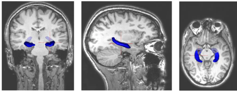
2.20 Coronal, sagittal and axial MRI views of the hippocampus. A 3D structure is superposed to the scans for a better representation. . . 27
2.21 Coronal slice of 7T MRI, at the level of the hippocampal head. . . 27
3.1 Three objects that have the same shape, with different positions, orientations, and scales. . . 31
3.2 Spharm-PDM representation of an hippocampal surface . . . 35
3.4 The attribute vector in two dimensions. . . 37
3.5 Representation of a 3D medial atom . . . 41
3.6 Theory of transformations according to D’Arcy Thompson . . . 42
3.7 Illustration of currents for curves and surfaces . . . 44
3.8 Steps of the unfolding of the medial temporal lobe. . . 46
4.1 AD vs controls classification . . . 60
4.2 MCI vs controls classification. . . 60
4.3 Classification rates for AD vs controls, as a function of the parameters C andγ. . 61
4.4 Classification rates for MCI vs controls, as a function of the parameters C andγ. 62 4.5 Illustration of the influence of the two most discriminative SPHARM coefficients on hippocampal shape for AD vs controls classification. . . 63
4.6 Group differences between AD and controls. . . 64
4.7 Group differences between MCI and controls. . . 64
5.1 CN vs AD: classification results for the differents methods. . . 79
5.2 CN vs MCIc: classification results for the differents methods. . . 79
6.1 Regions of cortical thinning in all patients with Gilles de la Tourette syndrome (GTS) compared with controls. . . 89
6.2 Region of cortical thinning in clinical subgroups with Gilles de la Tourette syn-drome compared with controls. . . 90
6.3 Structural changes in patients with Gilles de la Tourette syndrome with associated obsessive-compulsive disorder (GTS-OCD) compared with controls. . . 91
6.4 Cortical regions negatively correlated with the YGTSS score in all patients with Gilles de la Tourette syndrome. . . 92
6.5 Y-BOCS score correlations in the Gilles de la Tourette patients with associated obsessive-compulsive disorders (GTS-OCD). . . 92
7.1 Typical example of hippocampal subfield markings . . . 100
7.2 Cross-sectional slices of an ultra-high resolution MRI scan, manual delineation of the hippocampal subfields and corresponding automated segmentation. . . . 101
7.3 Coronal slices of the MR images of the hippocampus, and segmentation pre-sented in [Yushkevich et al., 2009] . . . 102
7.4 High-resolution scans of the hippocampus. . . 104
7.5 Identification of the most anterior slice of the body. . . 105
7.6 Identification of the most posterior part of the body. . . 105
7.7 Anterior limit of the hippocampus. . . 106
7.9 Steps of the manual delineation of the subparts of the hippocampal formation,
in a coronal slice of the body. . . 107
7.10 3D atlas of hippocampal subregions. . . 109
7.11 3D views of the atlas of hippocampal subregions. . . 110
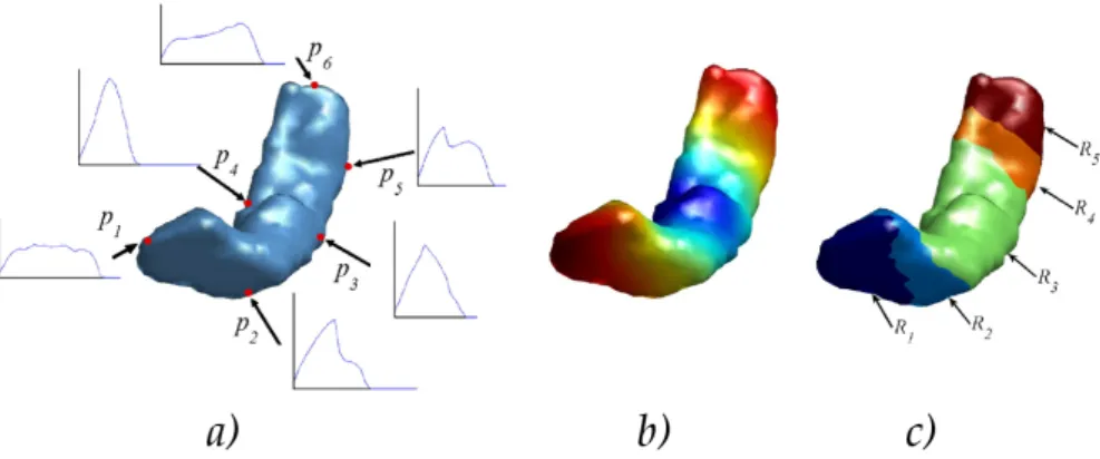
9.1 Exemple of problem which can occur when using a definition of thickness based on straight lines . . . 114
9.2 Initial estimation of² . . . 116
9.3 Computation of the diffusion coefficients in the simplest case. . . 122
9.4 Computation of diffusion coefficients for the kernel restricted toΩ . . . 123
9.5 Anisotropic kernels are built by introducing different weights for loops and hori-zontal or vertical links. . . 124
9.6 Left: Isotropic diffusion kernel (l=1,t=15). Right: Anisotropic diffusion kernel (l=2,t=15). . . 124
9.7 Folded ribbon. T: thickness of the ribbon; d: distance between the two branches. 125 9.8 Comparison of two types of kernel in a folded ribbon . . . 125
9.9 Mean thickness error for the hairpin with distance d = 2 between the two branches. Circles are for the kernel adapted to the geometry of the shape, triangles are for the kernel in the entire image. . . 126
9.10 Sinusoid shape . . . 126
9.11 Sinusoid shape: position of the computed skeleton. The graph displays the mean value of the function f on the skeleton points, depending on the size parameter t .127 9.12 Section and volumetric representation of the ribbon-shaped phantom. . . 128
9.13 Superimposition of the estimated skeleton with the “ground truth ” . . . 129
10.1 Top and bottom views of the hippocampal ribbon skeleton . . . 132
10.2 Skeleton inside the original volume. The surfaces are cut at the level of the hippocampal body . . . 133
10.3 Thickness estimation plotted on the computed skeleton, based on the original segmentation of the postmortem atlas. . . 133
10.4 Computed streamlines in a cut at the level of the hippocampal body. . . 134
10.5 Computed thickness measures in a cut at the level of the hippocampal body. . . 135
10.6 Superimposition of the skeleton and thickness maps obtained when the method was applied in 2D or 3D, in the body. . . 136
10.7 Comparison between the thickness computed with the 2D and the 3D method, in the hippocampal body. . . 137
10.8 Superimposition of the skeleton obtained from the original volume (in yellow) and of the skeleton obtained from the subsampled volume (in blue). . . 138 10.9 Comparison of the estimated thickness on the original and sub-sampled volumes.139
12.1 Template estimation for the hippocampal ribbons of controls and TLE patients. . 149
12.2 Mean thickness of the left hippocampus (averaged across the hippocampal body) in controls and TLE patients . . . 150
12.3 Mean thickness of right hippocampus (averaged across the hippocampal body) in controls and TLE patients . . . 150
12.4 Thickness maps for Controls-L vs TLE-IL (Top and bottom views). . . 151
12.5 Thickness maps for Controls-R vs TLE-IR (Top and bottom views) . . . 152
12.6 Thickness maps for Controls-L vs TLE-CL (Top and bottom views) . . . 153
4.1 Comparison between our approach and the method proposed in [Kloppel et al.,
2008] on the ADNI group. . . 63
5.1 Clinical and demographic characteristics of the studied population. . . 73
5.2 Summary of the approaches in this study. . . 77
6.1 Clinical characteristics and medication of Gilles de la Tourette syndrome . . . 88
9.1 Comparison of thickness for the synthetic spherical shell with different resolu-tions. Expected thickness is 6. . . 127
9.2 Comparison of the thickness for the synthetic 3D phantom with different resolu-tions. . . 128
10.1 Correlation between thickness values and mean distance between skeletons computed from the original volume and those computed from the subsampled volume. . . 135
C
H A P1
I
N T R O D U C T I O N
The hippocampus is one of the most fascinating structures of the brain. Phylogenetically, it is one of the oldest parts of the mamallian brain, having an archaic three-layer structure. In small mammals such as rodents, the hippocampus occupies an important portion of the brain. Somehow paradoxically, this archaic structure is crucial for cognitive functions that seem at the core of our humaneness: memory and temporal consciousness, i.e. the ability to remember our past and imagine our future [Dalla Barba, 2002]. Anatomically, the hippocampus is no less intriguing [Duvernoy & Cattin, 2005]. It is made of two convoluted sheets of gray matter that are folded one onto another: the cornu Ammonis and the dentate gyrus. The cytoarchitecture and connectivity of the hippocampus are also particularly rich with many distinct fields exhibiting distinct cell types, density and organization. Last but not least, the dentate gyrus of the hippocampus is one of the few brain areas where adult neurogenesis has been demonstrated, proving false the long held dogma that neurogenesis only occurs during development [Altman et al., 1962; Eriksson et al., 1998].
∗ ∗ ∗
Magnetic resonance imaging (MRI) allows studying the anatomy of the hippocampus in vivo. Using segmentation procedures, which can be manual [Hasboun et al., 1996] or automatic [Chupin et al., 2009a], one can delineate the contour of this structure and quantify its volume. Volumetric studies have been performed in various pathologies in which the hippocampus plays a major role. For instance, in Alzheimer’s disease (AD), hippocampal vol-umetry can distinguish AD patients and elderly controls with high sensitivity and specificity. In temporal lobe epilepsy (TLE), hippocampal volumetry allows detecting atrophy, which is suggestive of hippocampal sclerosis, and, when found, is associated with good postsurgical prognosis for patients. However, volumetry is a very crude and limited way to assess the structure of the hippocampus and cannot capture the full spectrum of abnormalities. This results both in limited insight on the nature of the alterations and in limited sensitivity to detect them. Indeed, in Alzheimer’s disease, the sensitivity of hippocampal volumetry is
much lower at the prodromal stage of mild cognitive impairment (MCI) than at the dementia stage. In temporal lobe epilepsy, hippocampal volume is “normal”in about 20% of patients and more sensitive methods are thus needed.
Therefore, it is important to propose models of hippocampal shape that can assess the full spectrum of its complexity. In the past years, a large number of approaches for analyzing the anatomical shapes have been proposed in the medical imaging and computer vision communities, with successful applications to different pathologies and brain structure, including the hippocampus. However, these approaches suffer from two limitations. First, many of them were designed for group analysis and not for the classification of individual patients which is necessary to assist diagnosis. Second, these approaches usually analyze the external shape of the hippocampus. This may limit their sensitivity and makes the interpretation of the detected changes difficult. In which subparts of the hippocampus do the changes occur: cornu Ammonis, subiculum, dentate gyrus? What is the nature of these changes: local gray matter loss, changes in convolution, in organization? This is due to the fact that only the external border of the hippocampus is visible on conventional anatomical MRI using T1-weighted sequences at 1.5T or 3T. On the contrary, ultra high-field MRI (7T and higher) provides new contrasts and increased spatial resolution, opening a new window on the internal organization of the hippocampus. These new images offer a completely different view of hippocampal anatomy and new shape models are needed to exploit them.
∗ ∗ ∗
This thesis is devoted to the development of shape models of the hippocampus and their application to different brain pathologies. These developments are made in two distinct contexts: whole-hippocampus morphometry using conventional MRI and shape models of hippocampal substructure using ultra-high field MRI.
We were first interested in the context of conventional MRI at 1.5T or 3T which is easily accessible and can be applied to large cohorts of subjects. Using these images, only the external border of the hippocampus can be segmented. As mentioned above, a substantial number of hippocampal shape analysis methods have been proposed. However, most of these approaches were designed for group analysis and not for the individual classification of patients. This limits their application to assist the diagnosis of pathologies such as Alzheimer’s disease for instance. We thus proposed a method to automatically classify between patients with Alzheimer’s disease or MCI and elderly controls, based on hippocampal shape features. In this approach, we modeled hippocampal shape with previously proposed descriptors, the spherical harmonics, which provide a flexible multiscale representation. These descriptors were combined with a support vector machine for automatic classification. We first evaluated the approach in a group of 23 AD patients, 23 MCI patients and 25 elderly controls (recruited at Caen University Hospital). We further evaluated the approach using a larger population of 509 patients from the ADNI database. This work was done in collaboration with Rémi Cuingnet who compared 10 methods for the automatic classification of AD patients. Finally, we present another application of morphometry to a neuropsychiatric disorder: Gilles de la Tourette syndrome. This work was done as part of the work of Yulia Worbe. Our contribution concerns hippocampal and cortical morphometry.
proposed a new approach for modeling the shape of the gray matter ribbon formed by cornu Ammonis and the subiculum (which we refer to as the hippocampal ribbon in the following). The hippocampal ribbon has a laminar organization: it presents as a convoluted sheet of gray matter, which is horizontally organized as a superposition of layers. This two-dimensional organization within the sheet suggests that, like the cortex, its thickness is a fundamental measurement to study its anatomy. It seemed thus natural to model its shape using a thick-ness measure and a central skeleton. To that purpose, we proposed a method to compute a skeleton for thin surfaces as well as a robust estimation of the thickness which is based on an original variational formulation. This was done by estimating a smooth vector field which goes through the ribbon. This approach relies on representations of such vector fields using the theory of Reproducing Kernel Hilbert Spaces (RKHS). It provides a proper regularization which prevents numerical instabilities usually present in skeletonization approaches. This point of view leads to a well-posed problem as well as to an effective maximization procedure. Thickness is then computed as the length of the streamlines from one boundary to the other, following the vector field. The methodology can be applied either to the full volumic seg-mentation (3D case) or separately to each coronal slice (2D case). An attractive feature of the approach is that, thanks to the use of RKHS norms, one obtains a diffeomorphic flow from the internal to the external surface. To validate this approach, we first created a very high resolution atlas (with nearly isotropic 300 mm resolution) of the hippocampal substructure. To that purpose, we manually segmented a postmortem hippocampal specimen that had been previously acquired at 9.4T at the University of Pennsylvania [Yushkevich et al., 2009] (images available for download on the internet). We then applied the approach to in vivo 7T acquisitions acquired as part of our collaboration with University of Minnesota.
∗ ∗ ∗
This thesis contains three main parts. Part I provides background information on the hippocampus and reviews existing shape analysis approaches. Part II is devoted to the study of whole-hippocampus morphometry from conventional MRI at 1.5T or 3T. Part III is focused on the morphometry of hippocampal substructure using ultra-high field MRI (in vivo 7T and post-mortem 9.4T MRI). These three parts are further organized as follows.
In part I, chapter 2 presents the anatomy of the hippocampus, its histological and cytoar-chitectonic features, its connectivity and briefly describes its role in cognition and pathologies. Chapter 3 reviews existing approaches for shape modeling of brain structures.
In part II, chapter 4 describes the method for the automatic classification of patients with AD or MCI, using hippocampal shape features, along with a first evaluation on 71 subjects1. Chapter 5 then presents the evaluation of the method on a larger population of 509 patients from the ADNI database2. Chapter 6 then presents another application of morphometry to a
1This chapter was published as a journal paper in NeuroImage [Gerardin et al., 2009]
2This was done in collaboration with Rémi cuingnet and was included in a journal paper in
neuropsychiatric disorder: Gilles de la Tourette syndrome3.
In part III, chapter 7 presents the construction of a very high resolution atlas of hip-pocampal subtructure. Chapter 8 gives a few elements on the theory of reproducing kernel hilbert spaces (RKHS). Chapter 9 presents the method for shape modeling of the hippocam-pal ribbon and thickness estimation. In chapter 10, we validate this approach using the postmortem atlas. Chapter 11 decribes the method used for template estimation at the group level, based on large deformation diffeomorphic metric mapping and currents. In chapter 12, we apply the approach to in vivo 7T MRI data.
3This chapter was published as a journal paper in Brain [Worbe et al., 2010]. This was part of the
C
H A P2
T
H E H I P P O C A M P U S
The hippocampus is a brain structure which plays a crucial role in fundamental cognitive processes like memory or emotions. It is also involved in different neurological and psy-chiatric diseases, such as Alzheimer’s disease, epilepsy or depression. This chapter gives a comprehensive description of the anatomical structure of the hippocampal formation. It also provides a brief introduction to the role this structure plays in normal human brain function. Finally, it presents some pathologies in which the hippocampus can be damaged.
In the following, we will address aspects of the anatomy of the hippocampal formation. This is a difficult task, due to two major problems:
• the complexity of the hippocampal structure, both in terms of geometry and of histol-ogy.
• the large number of existing terminologies to describe the parts of the hippocampal formation.
The hippocampus, also referred to the hippocampal formation, is a region that includes the hippocampus proper, or cornu Ammonis, the gyrus dentatus and the subiculum. Hip-pocampal anatomy is complex, and it is hard to describe it only with words. Thus, we incite the reader to refer to the illustrations given in this chapter for a better understanding of its three-dimensional configuration.
The rest of this chapter is organized as follows. Section 2.1 presents the localization of the hippocampus within the medial temporal lobe of the brain. We then describe its overall shape (Section 2.2) and the anatomy of its macroscopic subparts (Section 2.3). Section 2.4 is devoted to the histological features of its inner structure. Section 2.5 presents its connectivity and its relationships with neighbouring structures. We then briefly summarize its role in cognition (Section 2.6) and pathologies (Section 2.7). Finally, we present the role of MRI in studying the hippocampus in vivo (Section 2.8).
2.1 Localization, or How to find the hippocampus
without GPS
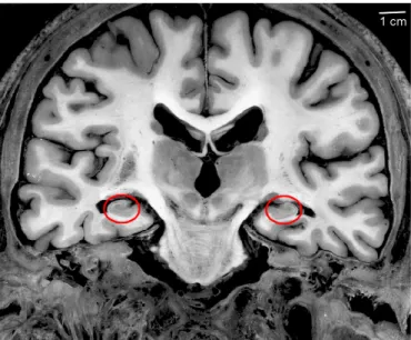
The hippocampus is a bilateral structure which belongs to the limbic system, a group of structures that works together to produce and regulate emotions and to form new memories. The hippocampus is phylogenetically one of the oldest structures in the mammallian brain, the hippocampi are thus situated deeply in both of the cerebral hemispheres. More precisely, each hippocampus belongs to the fifth circonvolution of the temporal lobe, and forms the medial wall and floor of the lateral ventricle. Thus, to locate the hippocampus, we have to look at the internal (medial) aspect of the hemisphere (figure 2.1).
Figure 2.1:
Inferomedial aspect of the right hemisphere. The red arrow indicates the tem-poral pole, while the green arrow shows the emplacement of the hippocampal region. The hippocampus being a deep structure, only a small part of it is visible superficially. From [Du-vernoy & Cattin, 2005]To see the hidden part of the hippocampus and its whole shape, some overlying struc-tures have to be removed; the result can be seen in figure 2.2.
An alternative solution is to observe sections of the brain, where the hippocampus and the surrounding structures can be more easily located. Coronal sections at the middle part of the hippocampus reveal its typical internal structure, as shown in figure 2.3.
2.2 General Shape, or The hippocampus to the
naked eye
The hippocampus is a small elongated structure, and has a total length of between 4 and 4.5 cm. The classical gross anatomical image of the human hippocampal formation is a bow which anterior extremity is enlarged and which posterior extremity narrows like a comma. The general shape of the hippocampus is shown on figure 2.4.
Figure 2.2:
Dorsolateral view of the human hippocampus after removal of the overlying structures and opening of the lateral ventricle. From [Insausti & Amaral, 2004].Figure 2.3:
Coronal view of the hippocampus. The hippocampal region is circled in red. From [Duvernoy & Cattin, 2005].• A head, or anterior segment,which is transversely oriented. It is the largest part of the structure. It shows elevations, the digitationes hippocampi.
• A body, or middle segment, which is sagittally oriented. The gyrus dentatus forms the axis of this elongated part.
• A tail, or posterior segment, whose organization is similar to that of the body, but with a transverse orientation.
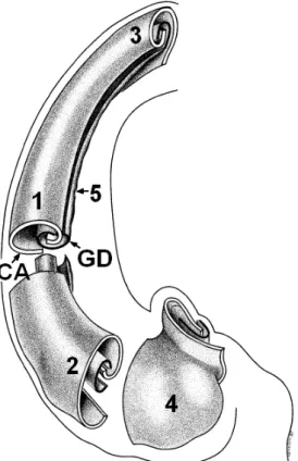
Figure 2.4:
Intraventricular aspect of the hippocampus. H: head, B: body, T: tail, f: fimbria, s: subiculum. From [Duvernoy & Cattin, 2005].easily made, from the enlargement of the structure in the uncal part, the transition between body and tail is more difficult to identify.
The term “hippocampus”(latin for sea horse) was given by the italian anatomist Arantius in 1587, and inspired by the three-dimensional form of the human hippocampus which reminds of this sea creature; the head and tail of the brain structure corresponding to those of the animal. The hippocampal shape also inspired other comparisons: the cornu Ammonis, or Ammon’s Horn is named after the mythological Egyptian god (figure 2.5)
Figure 2.5:
The Egyptian god Ammon, who inspired the name “cornu Ammonis”. Within this gross structure, the hippocampus is bilaminar, the two layers consisting of the cornu Ammonis (or hippocampus proper) and the gyrus dentatus. These two interlocking layers are visible in figure 2.6. They are separated from each other by the hippocampal sulcus.Figure 2.6:
General view of the internal structure of the hippocampus. The cornu Ammonis (CA) and gyrus dentatus (GD) form two interlocking, U-shaped laminae. 1: hippocampal body, 2: hippocampal head, 3 hippocampal tail, 4: digitationes hippocampi, 5: margo denticulatus. From [Duvernoy & Cattin, 2005].[Kier et al., 1997] examined hippocampal development in normal fetal specimens using MR imaging, dissection, and histology, in order to explain the progressive infolding of the fetal dentate gyrus, cornu Ammonis and subiculum around the hippocampal sulcus. However, the explanation of this particular configuration remains uncertain: in early development, the two laminae are continuous, then as a result of the marked expansion of the neocortex and unequal growth of the various components of the hippocampus, the cornu Ammonis would fold into the ventricular cavity, forming the hippocampal sulcus. The gyrus dentatus becomes concave and seems to slip beneath the medial end of the cornu Ammonis. At the end, the two layers fit into each other, as shown in figure 2.7.
2.3 Topography, or The hippocampus through a
magnifying glass
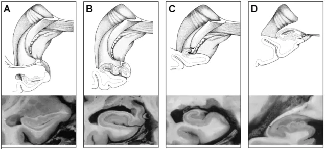
The next paragraphs describe the aspect of the hippocampus and the respective position of the two layers, in the three segments of the hippocampus, namely the body, head and tail. Figure 2.9 summarizes this complex anatomical construction.
Figure 2.7:
Diagram in the coronal plane illustrates stages of infolding of the components of the left hippocampus. A : Early in fetal development, the dentate gyrus (D), cornu ammonis (C), and subiculum (S) are arranged serially along the temporal horn( T). B and C : gradual infolding of the components around the hippocampal sulcus that first forms between the dentate gyrus and cornu Ammonis (large arrow in A). The hippocampal sulcus (small arrow in Figure B and C) shifts later to a location between the dentate gyrus and subiculum, and eventually becomes obliterated. From [Kier et al., 1997].Hippocampal body
The hippocampal formation has its simplest shape in its body: the fields of the cornu Ammonis and the gyrus dentatus fold over each other and form the characteristic C-shape of the hippocampus. In coronal sections of the hippocampal body, we can see that the gyrus dentatus is a narrow, dorsally concave lamina. Its concavity envelopes the last segment of the cornu Ammonis. The hippocampal sulcus becomes vestigial, and the two layers are fused together (a few residual cavities may persist): it becomes impossible to distinguish between them. Only a small segment of the gyrus dentatus is visible, and bordered by the superficial hippocampal sulcus along its entire length. It exhibits various folds on the surface, these folds form the “teeth”or dentes of the gyrus (Figure 2.8). These rounded protrusions diminish in size caudally. The dentes are surface manifestations of general folding in the gyrus dentatus.The hippocampal body is limited in its lateral part by the horn of the lateral ventricule. The white matter of the parahippocampal gyrus forms the inferior boundary, while the hippocampus is prolonged by the subicular region ( the “bed”of the hippocampus ) in its infero-medial limit. The hippocampal body is bordered medially by the fimbria. The fimbria is a narrow, white strip which more or less hides the superficial part of the gyrus dentatus. Note that the intraventricular hippocampal surface is almost entirely hidden by voluminous choroid plexuses; only the hippocampal head is devoid of these plexuses.
Hippocampal head
As we approach the anterior limit of the hippocampal formation, its configuration becomes more and more complex. Prominent bulges, the digitationes hippocampi, or digitations, become visible in the hippocampal head. The term “pes (foot) hippocampi”is sometimes used for the digitationes hippocampi. There are usually three or four digitations, sagittally oriented and separated by small but definite sulci. In coronal sections, the digitations are seen to be transverse foldings of the cornu Ammonis. Each digitation is surrounded by a digital extension of the gyrus dentatus. These folds vary in thickness, as is frequent in cortical gyri in general. When the hippocampal digitations appear at the junction of the body and head, the fimbria gives way to a thick alveus which covers them.Figure 2.8:
Aspect of the hippocampus after opening of the temporal horn of the lateral ventricle. A small segment of the gyrus dentatus is visible. The arrow shows the superficial hippocampal sulcus and the dentes of the gyrus. From [Duvernoy & Cattin, 2005].The hippocampal head is free of choroid plexuses. The anterior part of the hippocampus is adjacent to the amydgala. It is determined by a thin layer of white matter, namely the alveus. The temporal horn of the lateral ventricle may be visible between the hippocampus and the amygdala. The presence of white matter of the parahippocampal gyrus or the entorhinal area determines the inferior limit.
Hippocampal tail
The tail is the most posterior part of the human hippocampal forma-tion. At this level, the structure again loses its simple C-shaped organizaforma-tion. This segment is smaller than the hippocampal head, but its orientation and internal structure are relatively similar: although digitations do not appear at the surface of the tail, it is composed of a vast layer of the cornu Ammonis centered by the digital extensions of the gyrus dentatus. The distinction between the different fields becomes very complex at the most posterior segments. The fimbria, which in the initial segment hides the margo denticulatus, separates from it, ascending to join the crus of fornix. The extraventricular, superficial part of the hippocampal tail has relations similar to those of the body.Because of the curvature of the hippocampus, the gyrus dentatus and the cornu Ammonis have the same reciprocal position in a coronal section of the body as in a sagittal section of the head or of the tail.
Figure 2.9:
Coronal sections of hippocampus, and 3D diagrams, showing planes of section. A,B: Head; C: Body; D: Tail. The modifications of the respective positions of the gyrus dentatus and the cornu Ammonis can be observed from anterior to posterior levels. From [Duvernoy & Cattin, 2005].2.4 Subdivisions, Histology : The hippocampus
under the microscope
The different fields of the hippocampus have different histological characteristics. A thorough knowledge of hippocampal histology is necessary for a better understanding of its global shape and internal configuration, as well as its connections with the surrounding structures. In chapter 7, we will establish a coherent protocol of segmentation of the subfields of the hippocampus, and try to relate anatomical features to the underlying cytoarchitectonic structures.
2.4.1 Different types of cortex in the temporal lobe
We will start with a brief overview of the cytoarchitecture of the human brain.
The cerebral cortex is a highly folded sheet of grey matter encasing the brain, home to most higher cognitive functions. We can easily distinguish at least two types of cortices: the major component of the human brain is made of a six-layered cortex, the neocortex, while the olfactory cortex and the hippocampal formation are elements of the paleocortex, or allocortex, which consists of only three layers. Evolutionarily speaking, the three-layered organization is considered to be “older”, so this type of cortex is also known as archicortex whereas the “newer”six-layered cerebral cortex is “neocortex”). The cortex adjacent to the hippocampus changes from three layers to six layers, this transitional zone is classified as mesocortex, or periallocortex, and includes the parahippocampal gyrus and the entorhinal region. These cytologically different types of cortex exhibit distinct patterns of architecture.
Figure 2.10:
Laminar organization of the neocortex. The different layers contain different cell types. They are numbered from I to VI, from superficial to deep. From [de Economo & Koskinas, 1925].Horizontal lamination of the neocortex
The neocortex consists of various types of neurons, horizontally structured in layers, which differ by cell composition and density [Brodmann, 1909]. There are six layers, numbered from I to VI, from superficial to deep. Each layer is characterized by the neuronal cell types it contains and its connections with other cortical and subcortical regions. The cortical layers are illustrated in figure 2.10.2.4.2 The cytoarchitecture of the hippocampus
The gray matter parts of the hippocampal formation (the cornu Ammonis and the gyrus dentatus) are covered by two white matter structures: the alveus and the fimbria. The hippocampus is composed of allocortex, a three-layered structure. Distinct organizations are identifiable in the cornu Ammonis and the gyrus dentatus.
Cornu Ammonis
In the cornu Ammonis, the three layers (starting from hippocampal sulcus to alveus) are as follows:• A molecular cell layer which contains apical dendrites of the pyramidal cells and a number of interneurons. This layer is usually divided into three distinct sub-layers: the stratum moleculare contains a few interneurons and dendritic connections to the pyramidal layer and is adjacent with the molecular layer of the dentate gyrus; the stratum lacunosum contains axons of the perforating fibers and the Schaffer collaterals, which run parallel to the surface of the cornu Ammonis, and the stratum radiatum contains mostly apical dendrites of the pyramidal cells.
• A pyramidal cell layer (stratum pyramidale), which contains the main cells of the cornu Ammonis: the pyramidal cells. Their dendrites extends into the molecular layer.
• A polymorphic layer (stratum oriens) which contains basilar dendrites of the pyrami-dal cells, and scattered neurons (basket cells). In humans, this layer merges with the stratum pyramidale.
Alternatively, a six-layered nomenclature of the hippocampus can be used, formed successively by the stratum moleculare, the stratum radiatum, the stratum lacunosum, the stratum pyramidale, the straum oriens, and the alveus.
The repartition of these layers varies along the cornu Ammonis. Thus, the cornu Ammo-nis can be divided into different sectors, using cytoarchitectonic criteria . A varying number of nomenclatures have been proposed, for example Rose [Rose, 1927] divided the cornu ammonis into five zones labeled H1 to H5. In the following, we will use the nomenclature introduced by Lorente de No [Lorente de No, 1934]. He described four fields of pyramidal cells, dividing the hippocampus into four fields and labeled them CA1-CA4. However, it should be noted that some imprecisions persist in the definition of the limit of these fields. Borders between the various fields of the hippocampal formation are especially difficult to establish in humans compared to other species. This is due, in part, to the overlapping of neuronal layers at the interfaces of fields.
CA1 is the largest sector of the cornu Ammonis and is continuous with the subiculum. It contains small scattered pyramidal cells, with triangular somata. CA2 consists of a narrow band of densely packed pyramidal cells, with ovoid somata. The curve, or genu, of the cornu Ammonis (where it enters the concavity of the gyrus dentatus) is a densely packed stratum of pyramidal cells, and is designated as CA3. Its pyramidal somata are similar to those in CA2, but density in CA2 is greater. A specificity of CA3 is the presence of the stratum lucidum, a supplementary layer which contains fine, non myelinated fibers, the mossy fibers, that originate in the dentate gyrus. CA3 continues within the concavity of the gyrus dentatus; this field is designated as CA4. CA4 contains scattered, large ovoid pyramidal cells and intertwined large and myelinated fibers.
Gyrus dentatus
The dentate gyrus fits inside the hippocampus. It is a trilaminate cortical structure. The three layers (starting from the stratum moleculare of CA to CA4) are as follows:• A molecular layer (stratum moleculare) which is relatively cell free. It contains den-drites of granule cells.
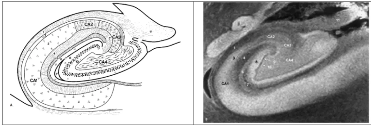
Figure 2.11:
Diagram and 9.4T MRI view of the structure of the hippocampus (coronal section). Cornu Ammonis: 1 = alveus, 2 = stratum oriens, 3 = stratum pyramidale, 3’ = stratum lucidum, 4 = stratum radiatum, 5 = stratum lacunosum, 6 = stratum moleculare, 7 and 7’ = vestigial hippocampal sulcus. Gyrus dentatus: 8 = stratum moleculare, 9 = stratum granulosum, 10 = polymorphic layer, 11 = fimbria. From [Duvernoy & Cattin, 2005].• A granule cell layer (stratum granulosum), the main layer of the gyrus dentatus. It is a thin layer, comprised of densely packed granular neurons, which have small round bodies. Efferent neurons from the granule cells are mossy fibers that synapse only with cells of hippocampal areas CA2 and CA3.
• A polymorphic cell layer, sometimes referred as to the hilus. It is the thinnest of the three dentate layers and contains the axons that cross from the granular layer to the cornu Ammonis.
Figure 2.12:
Layers of the hippocampus. From [Duvernoy & Cattin, 2005].2.5 Around the hippocampus
We give here a brief description of the surrounding anatomy of the hippocampus.
The nearest neighbour to the hippocampus is the amygdala. The amygdala is a structure mainly involved in emotional processes. The amygdala lies anteriorly and superiorly to the
hippocampus. The temporal horn of the lateral ventricle can sometimes be seen between the hippocampal head and the amygdala.
Anteriorly and inferiorly to the hippocampal head lies the parahippocampal gyrus. It includes the entorhinal cortex and also the perirhinal cortex. The parahippocampal region plays an important role in visual recognition, but there is also evidence that it makes a contri-bution to memory which can be distinguished from the contricontri-bution of the hippocampus [Eichenbaum et al., 2000]. Medially to the hippocampal head, the hippocampus borders the transverse fissure, in close proximity to the brainstem.
The lateral ventricles surround the hippocampal body by its lateral aspects. The parahip-pocampal gyrus runs parallel to the hippocampus along the base of its body.
Where the hippocampal tail becomes thinner, it is bordered laterally by the posterior lateral ventricle. The tail is adjacent to the posterior aspects of the thalamus, these two structures being separated by the lateral ventricle. The hippocampal tail finally joins the white matter bundle known as the fornix.
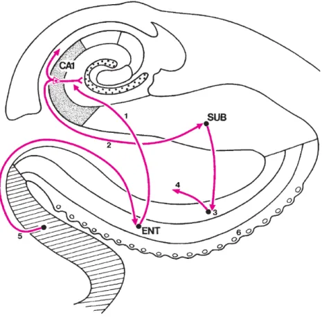
We now present a summary of the major intrahippocampal connections: the polysynap-tic pathway and the direct pathway [Duvernoy & Cattin, 2005].
The first system, the so-called polysynaptic pathway, links all parts of the hippocampus by a long neuronal chain. The superficial layers of the entorhinal cortex provide the most prominent input to the hippocampus. Within the hippocampus, the flow of information forms a loop. The first step in the flow loop, known as the perforant path, is a set of fibers originating from the entorhinal cortex and projecting to the gyrus dentatus. Axons called mossy fibers, emerge from the dentate gyrus before entering CA3, and then CA2 and CA1 regions through the Schaeffer collaterals. Finally, the information is sent from the CA1 subfield to the subiculum (Figure 2.13).
We can identify another circuitry: the direct pathway, which directly reaches out the neurons of the hippocampus, without following the usual polysynaptic chain. The direct pathway finds its origin in layer III of the entorhinal cortex. From this layer, fibers directly reach the pyramidal neurons of CA1. CA1 neurons project onto the subiculum, the axons of which return to the deep layers of the entorhinal area (Figure 2.14).
2.6 Cognition
The hippocampus plays an important role in various cognitive processes. Their description is beyond the scope of this thesis. Here, we briefly review some of its key roles.
2.6.1 Hippocampus and memories
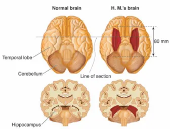
The patient HM
Advances in research can sometimes occur in a surprising way. The importance of the hippocampus in the encoding and retrieval of memory processes has been recognized following the works of Scoville and Milner [Scoville & Milner, 1957]. Their primal goal was to study the effects of the surgical resection of medial temporal lobe for treatment of epilepsy. They describe for the first time in 1957 the case of a 29 year old man, the patient “H.M”. This patient had one of the most severe cases of amnesia ever observed, his amnesia being the result of neurosurgery performed on him to treat the symptoms of his epilepsy.Figure 2.13:
Polysynaptic intrahippocampal pathway. A-E are parts of the neural chain forming this pathway. 1: alveus, 2: stratum pyramidale, 3: Schaffer collaterals, 4: axons of pyramidal neurons, 5: strata lacunosum and radiatum, 6: stratum moleculare, 7: vestigial hippocampal sulcus, 8: stratum moleculare, 9: stratum granulosum. From [Duvernoy & Cattin, 2005].He has been followed for over 40 years by more than 100 researchers, and is the subject of dozens of research papers and book chapters. This man was a high school graduate, and had a IQ of 104 before his operation. After a bicycle accident in the age of nine, he began at the age of ten to suffer from frequent and moderate seizures. At sixteen, his seizures increased in power and intensity, despite the use of anticonvulsants. Surgery was decided upon and carried out on 1 September 1953: a removal of both medial temporal lobes was carried out, extending posteriorly for a distance of 8 cm (see figure 2.15). Two thirds of both hippocampi were removed. After the intervention, the authors mention that HM continues to suffer from seizures, but that “they were less disabling than before”.
A psychological examination was conducted on 26 April 1955. The examinators noted that this patient was suffering from retrograde amnesia limited to a period of 11 years prior to his surgery at age 27. His memories formed before age 16 are still intact. In addition, it suffering from anterograde amnesia1: although he can store new information temporarily in his short-term memory, he can no longer form any new long-term memories, and did not remember, for example, seeing the doctor a few minutes before. Psychological testing showed that his IQ was 112, (slightly better than before the operation), he presented no
1Two other examples of anterograde amnesia : Dory from Nemo and the hero of the movie
Figure 2.14:
Direct intrahippocampal pathway. The entorhinal area projects directly onto CA1 pyramidal neurons (1), which innervate the subiculum (2). Subicular axons project back to the deep layers of the entorhinal cortex (3). The neurons of these layers send axons to the association cortex (4). From [Duvernoy & Cattin, 2005].disturbances of perception, abstract thinking, of reasoning skills or motivation. In summary, patient HM suffered only anterograde and retrograde amnesia but showed no disturbances of intelligence or personality. This amnesia apparently came from the absence of the temporal lobe. For the first time, an obvious link between memory and brain structure was revealed.
What tells HM about memory
The experimental approach of the modern study of memory began with patient HM. Since 1953, he has been the involved in over one hundred experiments (for review see [Corkin, 2002]), using different paradigms. However, a study conducted by Milner in 1962 [Milner, 1962] remains indispensable for understanding the current view of the neurobiology of memory. In this study, the patient had to draw a complex figure without seeing directly his hand but with a mirror. This task requires to correct all his habits of visuomotor coordination. During testing, this patient has improved its performance in a manner similar to controls but has no memory of his previous trials, claiming to never have done this test. From this original experience, a conclusion can be made: there is a memory accessible to consciousness (the declarative memory) and a second type, a memory independent of the first, which deal with motor skills (the non-declarative memory). For the patient HM, only the first was impaired after the operation while the second was intact. TheFigure 2.15:
Bilateral resection of the anterior temporal lobe in patient HM.Figure 2.16:
Anterograde amnesia in Hollywood : In Memento, Guy Pearce plays Leonard, who suffers severe anterograde amnesia after sustaining a head injury. Leonard retains his identity and the memories of events that occurred before the accident, but loses all ability to form new memories. In Finding Nemo, a reef fish called Dory, has a profound memory deficit which prevents her from learning or retaining any new information, remembering names, or knowing where she is going. As a result, she gets lost when left alone and is often found in a state of confusion.modern notion that there are different types of memory was born. Four points have to be retained about HM’s case :
• Memory has a distinct neurobiological support: it is a function separable from other cognitive abilities, that one can quantify and experimentally study.
• The medial temporal lobe is not needed for short-term memory, since HM retained a number or a visual image for a short time. However these memories are not stored: it exibhits the difference between encoding and storage.
• Memory is not “stored”into the medial temporal lobe, since childhood memories of HM are intact.
• There is a dissociation between declarative and non-declarative memory (see below for more details).
Different types of memory
Memory is the capacity of the nervous system to benefit from experience. It takes many forms, from simple to complex, from highly specific to most general. Any claim about “memory”or”memory impairment”immediately requires clarification: which kind of memory are we talking about?As we have seen in the previous paragraph, we can identify at least two types of memories: an explicit (declarative) and an implicit (non-declarative) memory. The distinction between declarative and nondeclarative memory is fundamental, because it has turned out that different kinds of memory are supported by different brain systems.
Other memory system organizations have been proposed, they are briefly presented in the following.
■ Short-term memory
Our memory is structured in several sub-systems, each comprising different categories of memories. A classic kind of categorisation is based on the amount of time the memory is stored: thus we can distinguish short-term memory, including working memory, from long-term memory.Working memory refers to the capacity to maintain temporarily a limited amount of information in mind, which can then be used to support various cognitive tasks, including language comprehension, learning, reasoning, and preparation for action [Baddeley, 1992].
■ Long term memory
Long-term memory keeps information lasting for several days or even years. It is divided into four different types of memory: episodic, semantic, procedural, and perceptual.Episodic memory concerns events experienced personnally by the individual and their context (date, place, emotions). It gives the subject the impression of reliving the moment. This the autobiographical part of the episodic memory. It is also the registration of individual information in the specific context of occurrence.
Semantic memory is the general knowledge about the world and ourselves (our profes-sion or our age). It stores the concepts of words and their meaning. Semantic memory is considered a network of associations between the word and concept, such as canary and bird. This memory is usually the most sustainable. It does not assume remembering specific events.
Procedural memory records the actions whose use becomes automatic over time (tying shoelaces, drive a car), elaborate mental procedures (protocol to solve a math problem, game strategies).
Perceptual memory retains the information provided by the senses of the shape of ob-jects, texture, or smell. It is requested without the knowledge of the subject automatically. Perceptual memory comes into action before the percept has one meaning: in the context of visual perception, we perceive a form prior to identifying it.
In conclusion, we can note that the separation between these different types of memory is hard to do: autobiographical memory includes episodic elements, as well as semantic elements. In addition, these memories are complementary and interact.
2.6.2 Spatial navigation
The hippocampus also has a main role in spatial navigation functions, particularly in finding shortcuts and new routes between familiar places. A study at University College London focused on the hippocampus in taxi drivers [Maguire et al., 1998, 2000]. London’s taxi drivers must learn a large number of places and the most direct routes between them (they have to pass a strict test before being licensed to drive). The study showed that the posterior hippocampus was larger in taxi drivers than in a control group. The anterior hippocampus, however, was larger in controls. In that study, the authors examined the correlation between these volume changes and time spent as a taxi driver. They found that the amount of time an individual worked as a taxi driver correlated positively and negatively respectively, with the volume of posterior and anterior portion of the hippocampus.
2.7 Implication in pathologies
Various clinical conditions are associated with alterations of the hippocampal formation. They may selectively affect different parts of hippocampus, each of the different hippocampal cytoarchitectonic fields being more or less vulnerable to damage. Conditions associated with hippocampal changes include, among others, dementia (Alzheimer’s disease, fronto-temporal dementia . . . ), epilepsy and psychiatric disorders (depression, schizophrenia . . . ). Here, we briefly present its implication in Alzheimer’s disease and temporal lobe epilepsy.
2.7.1 Alzheimer’s disease
In 1907, Alois Alzheimer described for the first time the main neuropathologic characteristics of a disease which mainly caused impairment of memory, and disorientation followed by depression. Pathological examination revealed atrophy and specific brain lesions, localized in the cortical grey matter. The lesions of Alzheimer’s disease (AD) are of two types: the amyloid plaques and the neurofibrirally tangles (NFT).
The progression of AD pathology is stereotyped. In [Braak & Braak, 1991], the progression is divided into six stages, based on the distribution of NFT: the lesions are first located in the trans-entorhinal cortex area (stade I), then spread into the entorhinal cortex (stade II), extend to the hippocampus and the limbic lobe(stade III and IV ), involve the association neocortex (stade V ) and finally the primary cortex (stade VI). These stages are divided into entorhinal (I and II), limbic (III and IV) and neocortical phases (V and VI), and are closely associated with clinical and cognitive deterioration, reflecting the degeneration of the cortical areas associated with these functions. The medial temporal lobe and the hippocampus are thus affected by NFT and neuronal loss at the earliest stages of the disease.
At present, the only way to obtain a definite diagnosis for AD is to perform an autopsy of the brain of probable AD sufferer, revealing the density and distribution of amyloid plaques and NFT. The diagnosis of probable AD relies on clinical criteria, based on neuropsychological examination. An important question for research is to make progress towards earlier and more accurate diagnosis of AD, by discovering markers of early AD. In particular, magnetic
resonance imaging (MRI) allows visualizing brain atrophy which reflects neuronal loss. A vast amount of research has thus been carried out to extract diagnostic markers of AD from MRI.
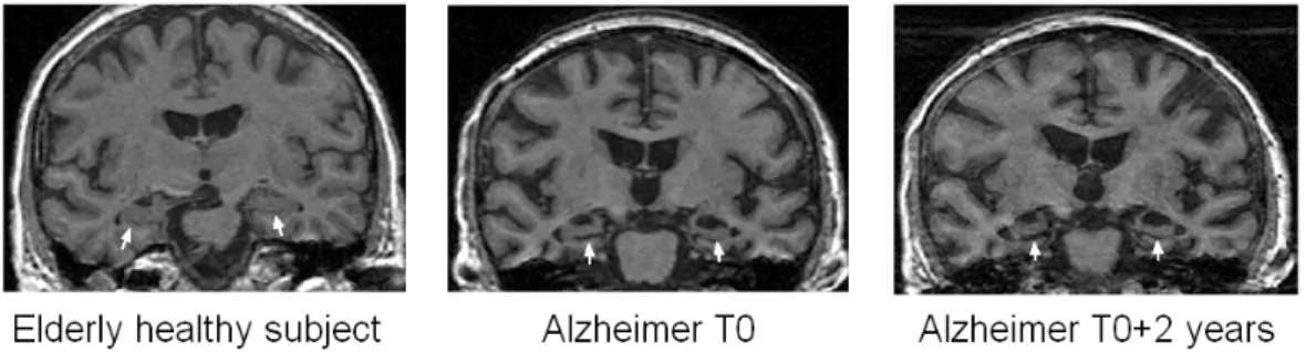
Figure 2.17:
Coronal slices of T1 weighted MRI, showing the atrophy of the hippocampus. The first panel comes from an elderly healthy subject. The two next panels show the hip-pocampal atrophy in an AD patient at T0and T0+ 2 years. The hippocampi are marked withthe white arrows.
Several volumetric MRI studies have highlighted increased atrophy of the hippocampus in the earliest stages of the disease [Laakso et al., 1995; Bobinski et al., 1999]. They showed that the volumetry of the hippocampal region is a reliable marker of the moderate to severe stages of the disease, with a sensitivity and specificity above 80% [Frisoni et al., 1999; Du et al., 2001]. However, at the stage of mild cognitive impairment (MCI), the sensitivity of hippocampal volumetry is much lower [Convit et al., 1997; Pennanen et al., 2004]. This may be because early pathology selectively affects some specific parts of the hippocampus, NFT and neuronal loss being dominant in CA1 and the subiculum [Hyman et al., 1984; Van Hoesen & B.T., 1990]. New imaging techniques and mathematical shape models that could measure local atrophy have thus the potential to provide more efficient diagnostic tools.
2.7.2 Epilepsy
Epilepsy is a brain disorder characterized by generalized or focal epileptic seizures. Among the different forms of focal epilepsies, temporal lobe epilepsy (TLE) is of special interest since this is the most common variant in adults accounting for about 40% of cases [Engel, 1996]. The association between hippocampal abnormalities and temporal lobe epilepsy is well documented in the literature. In particular, hippocampal sclerosis is a frequent finding in patients with TLE [Eriksson et al., 2008]. However, it is not yet clear whether the epilepsy is caused by hippocampal abnormalities, or whether the hippocampus is damaged by cumulative effects of seizures.
In about 70% of TLE patients, seizures cannot be controlled with medication. In carefully selected patients, epilepsy surgery can effectively control seizures [Wiebe et al., 2001]. MRI plays an important role in the pre-surgical evaluation by allowing the identification of atrophy or altered signal intensity in the hippocampus suggesting hippocampal sclerosis. When atrophy is found, more than 70% of surgically treated TLE patients achieve seizure freedom after surgery [Wiebe et al., 2001; Wiebe, 2003]. On the other hand, hippocampal volumes
are normal in about 20% of patients with electro-clinical signs of mesial TLE [Jackson et al., 1994]. In these “MRI-negative”TLE cases, the MRI is therefore unable to show a potential surgical target. In the absence of identifiable structural pathology on MRI, these patients currently undergo invasive EEG monitoring with intracranially implanted electrodes and have a lower chance of success after surgery than those in whom a lesion is found. It is thus important to design new MR image analysis approaches that could unveil subtle hippocampal abnormalities that cannot be detected by standard means.
2.8 In vivo visualization using MRI
Magnetic Resonance Imaging (MRI) is the imaging technique of first choice for visualization of the hippocampal structure due to the high contrast between different tissues (figure 2.18).
Figure 2.18:
Vizualization of the hippocampal structure in conventional T1-weighted sequence. The hippocampus, marked with the arrows, can be seen in coronal (a), sagittal (b) and axial (c) planes.It provides anatomical images of three-dimensional nature, composed of small volume elements (voxels). Creating images requires no anatomical tracer injection and no radiation exposure. MR signal is essentially produced by protons. Each proton rotates around its own axis. When a strong magnetic field and a radio frequency (RF) pulse are applied at angles orthogonal to each other, the protons as a group begin to precess about their own axis in synchrony with each other. When the RF pulse is turned off, the system returns to normal and in doing so create small local magnetic fields which in turn gives rise to small electric currents in receiving coils. It is this current that is ultimately measured in MRI.
Classically, at 1.5T or 3T, the hippocampus is visualized using 3D T1-weighted sequences with about 1mm isotropic resolution (figure 2.20). Using T1-weighted sequences, manual segmentation of the hippocampus can be performed using standardized protocols [Hasboun et al., 1996; Pruessner et al., 2000]. However, manual segmentation is very time consuming (over 1 hour per structure) and suffers from considerable intra and inter-rater variability. Automatic segmentation approaches have thus been proposed [Fischl et al., 2002; Coupé et al., 2011; Lötjönen et al., 2011]. In our laboratory, Marie Chupin has developed the automatic approach SACHA [Chupin et al., 2007, 2009b] that segments the hippocampus and the amygdala from MR images 2.19. The segmentation uses simultaneous region deformation constrained by anatomical landmarks and knowledge derived from probabilistic atlases.
This method was validated by comparison with manual segmentations in healthy subjects, patients with Alzheimer’s disease, and patients with epilepsy. The validation showed a high accuracy of the segmentation, with a relative error in volume of approximately 8% compared to manual segmentation.
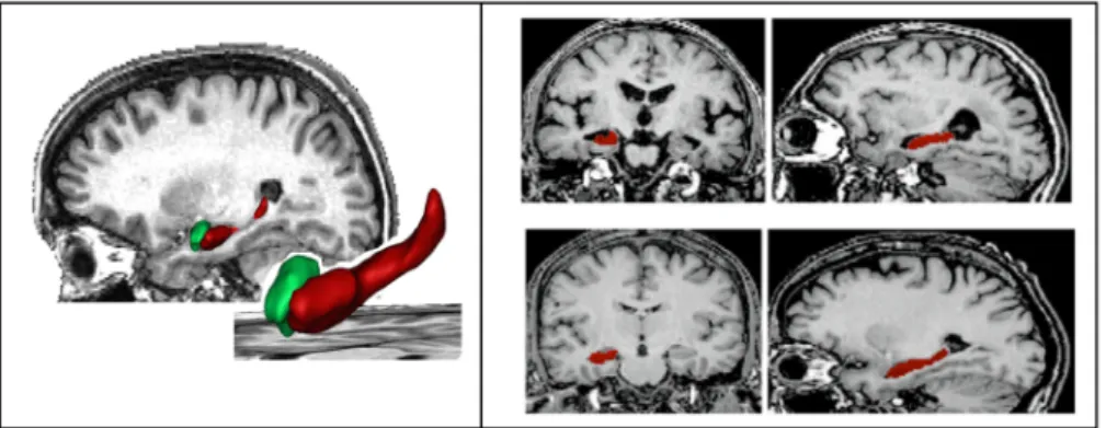
Figure 2.19:
SACHA: Automatic segmentation of hippocampus and amygdala. Left panel: Illustration of the final result on a sagittal reconstruction and on 3D surface renderings corresponding to the automated segmentations of the hippocampus (red) and the amyg-dala(green). Right panel, top: patient with Alzheimer’s disease (coronal and sagittal recon-structions, from left to right). Right panel, bottom: healthy elderly control (coronal and sagittal reconstructions, from left to right). From [Chupin et al., 2009b; Colliot et al., 2008].However, conventional T1-weighted sequences allow only visualizing the external bound-aries of the hippocampus and not its internal subparts such as the cornu Ammonis or the dentate gyrus. This is because:
• most of these substructures are too small to be correctly imaged with 1mm resolution; • T1-weighted images display almost no contrast between them.
On the contrary, T2-weighted spin echo sequences provide contrast between some of these subfields. [Mueller et al., 2007] used T2-weighted sequences with 0.4x0.5x2mm resolution at 4T to measure the subiculum, CA1, CA2 and CA3/4 and the dentate gyrus with applications in epilepsy [Mueller et al., 2009] and Alzheimer’s disease [Mueller & Weiner, 2009]. However, their approach involves the definition of many arbitrary landmarks. Moreover, using 2mm thick slices prevents from building 3-dimensional models of the subparts.
7T MRI provides new contrast and increased spatial resolution. At 7T, Chupin et al. [2009c] used T2-weighted spin echo sequences with 250µmx250µmx1mm resolution to segment CA, the dentate gyrus (together with CA4), the subiculum, the alveus and the fimbria. They further applied their approach to patients with temporal lobe epilepsy [Henry et al., 2011]. Wisse et al. [2012] also proposed manual segmentation of the hippocampal subfields at 7T. Kerchner et al. [2010] used 7T MRI to detect subregional atrophy of CA in Alzheimer’s disease. However, they did not perform volumetry of the different subfields. In our laboratory, we are currently imaging patients with Alzheimer’s disease, MCI and healthy controls at 7T. On a restricted subset of patients, Claire Boutet performed subfield volumetry and found marked atrophy in the left subiculum [Boutet, 2012].
Figure 2.20:
Coronal, sagittal and axial MRI views of the hippocampus. A 3D structure is superposed to the scans for a better representation.Figure 2.21:
Coronal slice of 7T MRI, at the level of the hippocampal head. Contrary to conventional T1-MRI, we can clearly distinguish the Cornu ammonis and the gyrus dentatus. From [Chupin et al., 2009c]2.9 Conclusion
In this chapter, we described the anatomy of the hippocampal formation, its different sub-parts and connections. We also briefly reviewed its role in cognition and in two disorders: Alzheimer’s disease and epilepsy. Volumetric measurements of the hippocampus can prove useful to assist the diagnosis of Alzheimer’s disease or for the presurgical evaluation of pa-tients with temporal lobe epilepsy. However, these global measures cannot detect local alterations of the hippocampus and their diagnostic value is therefore limited. On the con-trary, mathematical shape models have the potential to detect subtle abnormalities. In the next chapter, we will review existing approaches for shape analysis of brain structures.
C
H A P3
S
H A P E
A
N A L Y S I S
Advances in medical imaging technology have provided the ability to acquire high resolution 3D images of the human brain. In particular, magnetic resonance imaging (MRI) is a fantastic non-invasive mean for investigating human anatomy in-vivo and provides high resolution images whose utility is beyond the simple visual inspection. Quantitative study of anatomical shape and its variation is of high importance to understand the mechanisms and the impacts of diseases such as neurodegenerative or neurodevelopmental disorders. Many studies rely on the statistical analysis of volume measurements [Aylward et al., 1999; Colliot et al., 2008]. While they can detect volumetric changes between patients and healthy controls, they do not capture the complexity of a shape. Medical images can provide highly detailed shape information for analysis of morphological variability within a single population or among different groups of subjects.
Indeed, a quantitative shape analysis would be of special interest to understand mor-phological changes caused by neurological or psychiatric diseases like schizophrenia [Styner et al., 2004], autism [Dager et al., 2007], epilepsy [Hogan et al., 2004] or Alzheimer’s dis-ease [Apostolova et al., 2006a]. Brain anatomy is thought to change with the progression of these diseases. One motivation for shape analysis is its potential ability to provide relevant information which helps doctors providing a diagnosis. Beyond assisting the diagnosis, shape analysis can also offer a way to quantify the development of a disease or the effect of a treatment [Gerig et al., 2003], or investigate anatomical differences associated with age or gender [Bouix et al., 2005]. It is also important for a shape analysis method to not only demonstrate that there are shape differences, but also to identify where and how these dif-ferences occur. For example, recent evidence suggests that Alzheimer’s disease affects the shape of some parts of the hippocampus more than the others [Apostolova et al., 2006a; Mueller & Weiner, 2009]. These results could allow to focus future research on these specific substructures.
Shape characterization can also aid understanding anatomical variability by providing statistical anatomical atlases. Currently, most anatomical atlases show a single instance of the normal anatomy of brain structures [Duvernoy & Cattin, 2005; Paxinos & Mai, 2004].