HAL Id: tel-03157478
https://tel.archives-ouvertes.fr/tel-03157478
Submitted on 3 Mar 2021HAL is a multi-disciplinary open access archive for the deposit and dissemination of sci-entific research documents, whether they are pub-lished or not. The documents may come from teaching and research institutions in France or abroad, or from public or private research centers.
L’archive ouverte pluridisciplinaire HAL, est destinée au dépôt et à la diffusion de documents scientifiques de niveau recherche, publiés ou non, émanant des établissements d’enseignement et de recherche français ou étrangers, des laboratoires publics ou privés.
transport in tumors
Cristina Vaghi
To cite this version:
Cristina Vaghi. Mathematical modeling of antibody nanoconjugates transport in tumors. Numerical Analysis [math.NA]. Université de Bordeaux, 2020. English. �NNT : 2020BORD0326�. �tel-03157478�
Thèse présentée pour obtenir le grade de
DOCTEUR DE L’UNIVERSITÉ DE BORDEAUX
École doctorale de Mathématiques et InformatiqueSpecialité: MATHÉMATIQUES APPLIQUÉES ET CALCUL SCIENTIFIQUE
C
RISTINA
V
AGHI
sous la direction de Sébastien Benzekry, Raphaëlle Fanciullino et Clair Poignard
Mathematical modeling of antibody
nanoconjugates transport in tumors
Soutenue le 16 décembre 2020 devant le jury composé de :
Sébastien Benzekry CR, HDR, Inria Bordeaux-Sud-Ouest, MONC Directeur de thèse
Helen Byrne Pr, University of Oxford Rapportrice
Raphaëlle Fanciullino MCU, HDR, Université d’Aix-Marseille Directrice de thèse
Florence Gattacceca MCU, HDR, Université d’Aix-Marseille Examinatrice
Hans Peter Grimm PhD, Roche Innovation Center Basel Examinateur
Jonathan P. Mochel Associate Professor, Iowa State University Invité
Roberto Natalini DR, CNR Roma Rapporteur
Clair Poignard DR, Inria Bordeaux-Sud-Ouest, MONC Directeur de thèse
A
BSTRACT
Title: Mathematical modeling of antibody nanoconjugates transport in tumors
Abstract: Nanomedicine offers promising and innovative tools to treat cancer. Recently, liposomes
conjugated with an antibody were developed to target breast cancer cells while sparing healthy tis-sues from the toxicity of the chemotherapy. These nanoparticles are called antibody-nanoconjugates (ANCs) and are currently tested in a preclinical trial. However, the pharmacokinetics, biodistribu-tion, and efficacy of these nanoparticles are not well known and could be improved. Mathematical modeling can help in understanding the intratumor penetration of the nanoparticles and in quanti-fying the treatment efficacy.
Pharmacokinetic-pharmacodynamic modeling evaluates the dose-response relationship in vivo and can be used to optimize the therapy schedule. Here, we described several biological processes us-ing ordinary differential equations: (i) the untreated tumor growth with a novel reduced Gompertz model, (ii) the nanoparticle biodistribution using a two-compartment pharmacokinetic model, and (iii) the therapeutic response with a resistance model. All the models were validated against ex-perimental data in the statistical framework of nonlinear mixed-effects modeling, which models simultaneously the dynamic of the population and the inter-individual variability.
Furthermore, we derived a spatial mathematical model with the two-scale asymptotic expansion method to describe the fluid and nanoparticle transport within the tumor tissue. This approach al-lowed us to evaluate the barriers that impair a homogeneous distribution of nanoparticles at the tumor site. Moreover, we propose a computational framework to predict tumor accumulation of nanoparticles using individual imaging data.
Keywords: antibody-nanoconjugates, nonlinear mixed-effects modeling, two-scale asymptotic
ex-pansion
Résumé: La nanomédecine offre des perspectives ambitieuses pour le traitement du cancer.
Récem-ment, des liposomes conjugués à des anticorps spécifiques ont été développés pour cibler les cellules tumorales du cancer au sein, en réduisant la toxicité de la chimiothérapie dans les tissus sains. Ces nanoparticules, appelées ANC (pour antibody nano-conjugate), sont actuellement testées dans une phase préclinique. Cependant, la pharmacocinétique, la biodistribution et l’efficacité de ces nanopar-ticules ne sont pas bien caracterisées quantitativement et pourrait être ameliorées. La modélisation mathématique peut aider à mieux comprendre la dynamique de la pénétration des ANC dans la tumeur et à améliorer l’efficacité du traitement.
La modélisation pharmacocinétique-pharmacodynamique permet d’évaluer la réponse du traitement
in vivoen fonction de la dose injectée. Dans ce travail, nous avons décrit plusieurs phénomènes bi-ologiques avec des équations differentielles ordinaires : (i) la croissance tumorale avec un nouveau modèle réduit de Gompertz, (ii) la biodistribution des nanoparticules avec un modèle pharmacociné-tique à deux compartiments, et (iii) la réponse au traitement avec un modèle de résistance. Tous les modèles ont été calibrés dans le cadre des modèles non linéaires à effets mixtes, qui décrivent la dynamique globale de la population ainsi que la variabilité individuelle.
De plus, nous avons dérivé un modèle mathématique spatial avec la technique de développement asymptotique double-échelle pour décrire le transport des fluides et des nanoparticules dans le tissu tumoral. Cette méthodologie nous permet d’évaluer les barrières microscopiques qui empêchent une distribution homogène des ANC dans la tumeur. Finalement, nous proposons un schéma computa-tionnel pour prédire l’accumulation des nanoparticules à partir des données individuels d’imagerie.
Keywords: nanoparticules, modèles non linéaires à effets mixtes, pharmacocinétique,
pharmaco-dynamique, développement asymptotique double-échelle
R
ÉSUMÉ
Le cancer du sein est le plus diagnostiqué chez les femmes au monde. Les progrès faits par la science dans le cadre des traitements médicaux et chirurgicaux ont permis d’améliorer les perspec-tives de survie ainsi que la qualité de vie des personnes affectées par cette maladie. Néanmoins, beaucoup reste à faire pour améliorer les médicaments. La toxicité des chimiothérapies est no-tamment un grand problème. Pour limiter ces effets secondaires, une approche innovante consiste à appliquer des nanotechnologies pour encapsuler l’agent cytotoxique dans des nanoparticules, afin de cibler les cellules tumorales et de préserver les tissus sains. Cependant, la dynamique de pénétration de ces nanoparticules dans le tissu tumoral n’est pas bien caractérisée quantitativement. L’objectif de ce projet de recherche est d’utiliser la modélisation mathématique pour décrire le transport des nanoparticules dans la tumeur, afin de mieux comprendre leurs propriétés et en optimiser l’efficacité. Cette thèse se base sur une collaboration interdisciplinaire avec des pharmaciens de l’équipe SMARTc de l’université d’Aix-Marseille, qui ont développé des immunoliposomes, aussi appelés ANC (pour antibody nano-conjugate). Ces nanoparticules à base lipidique contiennent un agent cytotox-ique (docétaxel) et présentent sur leur surface un anticorps (trastuzumab) qui se lie aux récepteurs Her2 des cellules du cancer au sein. Elles sont actuellement évaluées dans une phase préclinique. Les questions auxquelles nous nous sommes intéressés sont les suivantes: Comment est-il possible de prévoir la taille de la tumeur et l’efficacité des nanoparticules ? Quelle est la programmation optimale du traitement ? Comment le microenvironnement de la tumeur (comme la pression du fluide interstitiel, la densité des vaisseaux, la taille de la tumeur ou l’expression de Her2) affecte-t-il le transport des nanoparticules ? Comment est-il possible de personnaliser la dose et le protocole d’administration des médicaments à partir de données histologiques sur le microenvironnement de la tumeur ?
Les résultats de ce travail apportent une contribution directe au développement des anticorps-nanoconjugués dans le traitement du cancer du sein.
La modélisation mathématique peut permettre de mieux comprendre la dynamique sous-jacente au processus de transport des nanoparticules dans la tumeur, ce qui est la première étape pour évaluer l’efficacité et adapter la posologie. Quand les ANC sont injectées par voie intraveineuse, elles doivent traverser différentes barrières: le transport vasculaire jusqu’au site de la tumeur, le transport trans-épithélial pour passer des capillaires au tissu tumoral, la pénétration dans le
des nanoparticules jusqu’au tissu tumoral rend difficile de quantifier la concentration de nanopar-ticules qui va réellement agir sur les cellules cancéreuses à partir de la dose injectée. Des modèles biophysiques peuvent décrire ce processus et déterminer la portion de médicament qui arrive aux cellules tumorales ainsi que leur efficacité.
Ce manuscrit se divise en deux parties: dans la première partie, nous étudions un modèle pharmacocinétique-pharmacodynamique afin de caractériser la biodistribution des nanoparticules ainsi que l’efficacité in vivo ; dans la deuxième partie, un modèle spatial de transport de fluide et de nanoparticules dans la tumeur est dérivé avec la méthode du développement asymptotique double-échelle.
***
La modélisation pharmacocinétique-pharmacodynamique permet d’évaluer la réponse du traite-ment in vivo en fonction de la dose injectée. Dans ce travail, nous avons décrit plusieurs phénomènes biologiques avec des équations différentielles ordinaires: (i) la croissance tumorale avec un nouveau modèle réduit de Gompertz, (ii) la biodistribution des nanoparticules avec un modèle pharmacociné-tique à deux compartiments, et (iii) la réponse au traitement avec un modèle de résistance. Tous les modèles ont été calibrés dans le cadre des modèles non linéaires à effets mixtes, qui décrivent la dynamique globale de la population ainsi que la variabilité inter-individuelle.
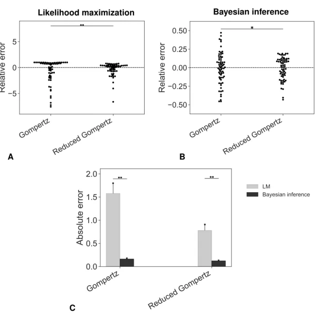
Nous avons dérivé un modèle de Gompertz réduit dans le cadre statistique des modèles non linéaires à effets mixtes pour décrire la croissance des tumeurs non traitées. Ce modèle, qui com-porte un paramètre au niveau de la population et un paramètre spécifique à l’individu, a montré un bon pouvoir descriptif (similaire au modèle de Gompertz) et a amélioré les prédictions du temps d’initiation de la tumeur. En outre, grâce à l’interprétation biologique des paramètres du modèle, nous avons pu valider le modèle sur un nouvel ensemble de données en estimant les paramètres à partir d’expériences indépendantes. Notre modèle devrait être testé afin de prédire la taille des tumeurs individuelles (c’est-à-dire pour les prévisions dans le futur). En effet, une bonne caractérisa-tion de la croissance de la tumeur est d’une importance fondamentale pour faire des prédiccaractérisa-tions sur la réponse individuelle aux traitements. Bien que notre méthode reste à étendre à des données clin-iques, ces résultats sont prometteurs pour l’estimation personnalisée de l’âge d’une tumeur à partir de mesures limitées au moment de la détection. L’estimation de l’âge de la tumeur d’un patient pour-rait en effet être instructive pour la pratique clinique, par exemple pour contribuer à l’élaboration de modèles informatiques personnalisés de métastases.
La biodistribution des ANC et leur efficacité ont été évaluées à l’aide d’un modèle pharmacocinétique-pharmacodynamique. Un modèle pharmacocinétique (PK) à deux comparti-ments basé sur un système d’équations différentielles ordinaires a été utilisé pour décrire l’échange de nanoparticules entre les compartiments systémique et tumoral. Le modèle PK a été calibré sur qua-tre ensembles différents de données, ce qui nous a permis de comparer la distribution des anticorps-nanoconjugués et des liposomes (sans trastuzumab greffé en surface) dans les compartiments central
et tumoral. Un modèle pharmacodynamique (PD) de résistance a été utilisé pour évaluer l’efficacité des nanoparticules. La comparaison entre les liposomes et les ANC n’a pas montré de différences significatives en termes d’activité cytotoxique, bien que la résistance acquise aux ANC soit légère-ment inférieure à la résistance aux liposomes. Les mécanismes d’action des liposomes et des im-munoliposomes devraient être davantage mis en évidence, même si la plus grande efficacité globale des ANC pourrait être due à une internalisation plus efficace grâce à l’anticorps greffé à la surface. De plus, l’efficacité des deux nanoparticules était supérieure à celle des médicaments libres grâce à l’amélioration de la pharmacocinétique des deux formulations liposomales. Toutefois, la PK des ANC et des liposomes pourrait être évaluée plus en détail. La mesure de la concentration plasmatique du médicament nous permettrait d’estimer le volume de distribution et la clairance systémique des deux formulations liposomales. En effet, les immunoliposomes pourraient être reconnus et éliminés plus rapidement par le système des macrophages ou ils pourraient être absorbés par d’autres organes en raison du trastuzumab greffé. Dans une perspective d’avenir, un modèle pharmacocinétique physi-ologique (PBPK) pourrait permettre de mieux comprendre les phénomènes impliqués dans les pro-cessus d’absorption, de distribution, de métabolisme et d’excrétion. En outre, l’efficacité in vivo des nanoparticules pourrait être modélisée en tenant compte du docétaxel et du trastuzumab dans le processus de la croissance tumorale.
La modélisation pharmacocinétique-pharmacodynamique est fondamentale dans le processus de développement des médicaments: la pharmacocinétique permet d’évaluer l’exposition au site d’action en fonction de la dose injectée, tandis que la pharmacodynamique décrit la réponse au traitement. De nouveaux schémas de traitement pourraient être testés expérimentalement en fonc-tion des prédicfonc-tions de notre modèle. Il est important de noter que la modélisafonc-tion de la PKPD permet de traduire la PK en applications cliniques en quantifiant la dose initiale requise chez l’homme.
***
La grande hétérogénéité des tissus tumoraux ainsi que les propriétés des nanomédicaments ont un impact sur l’administration des médicaments au niveau du site de la tumeur. Pour comprendre les principaux facteurs qui affectent la distribution des nanoparticules, nous avons dérivé un modèle spatial de transport de médicaments qui prend en compte les caractéristiques microscopiques de la tumeur sur la dynamique globale. L’étude de la distribution des ANC dans la tumeur se base sur trois points : (i) l’élaboration d’un modèle théorique avec des équations mathématiques pour la de-scription des phénomènes biologiques, (ii) la simulation numérique des modèles dérivés et (iii) leur validation avec des données expérimentales. On propose un modèle théorique de concentration des ANC dans les vaisseaux et dans le compartiment interstitiel couplé avec l’écoulement des fluides dans le tissu tumoral. La pénétration des nanoparticules dans la tumeur se produit par diffusion et convec-tion. Il faut donc d’abord caractériser l’environnement tumoral, c’est-à-dire le transport des fluides interstitiel et sanguin. Le tissu tumoral peut être vu comme un milieu poreux hétérogène irrigué par des capillaires très irréguliers et perméables. Avec la méthode d’homogénéisation nous avons construit un modèle asymptotique qui décrit l’écoulement des fluides au niveau macroscopique, en
un couplage d’équations de Darcy et présente des comportements différents selon la perméabilité et la structure capillaire paramétrée. Ainsi, le modèle prend en considération l’hétérogénéité et la porosité du tissu tumoral. Enfin, nous avons développé un modèle asymptotique de diffusion, convection et réaction des nanoparticules avec la méthode d’homogénéisation pour décrire la con-centration des ANC dans les capillaires et dans le compartiment interstitiel. Le terme de réaction prend en compte l’interaction des nanoparticules avec les cellules tumorales. Après avoir fait une analyse mathématique du modèle dérivé, nous avons obtenu des résultats numériques en utilisant la méthode des différences finies. Les simulations numériques ont montré que l’environnement tu-moral (i.e., la structure des capillaires et leur perméabilité) a un fort impact sur la pénétration des ANC, en accord avec les observations biologiques. Ce résultat qualitatif est une étape essentielle pour garantir la validité du modèle. La calibration du modèle a été effectuée empiriquement ou à l’aide des différentes données à disposition: des données in vitro qui permettent d’analyser le mécanisme d’absorption par les cellules (pour déterminer le terme de réaction) ; les données in vivo donnent des informations sur la perméabilité des vaisseaux et sur la clairance des nanoparticules dans le plasma. Enfin, nous avons proposé une méthodologie pour intégrer les données d’imagerie individuelles dans le modèle mathématique spatial. En particulier, nous avons utilisé des données ex vivo pour récupérer les tenseurs de perméabilité et avons effectué des simulations individuelles de la péné-tration des nanoparticules dans le compartiment interstitiel de la tumeur. Ces prédictions ont été comparées aux résultats du modèle pharmacocinétique calibré à partir de mesures macroscopiques. Ces résultats sont prometteurs pour la personnalisation des traitements. Les technologies émergentes d’acquisition d’images permettent de quantifier les propriétés microscopiques de la tumeur in vivo. Ces données peuvent être intégrées dans le modèle afin de prédire l’accumulation de nanoparticules et de programmer la dose optimale pour améliorer l’efficacité thérapeutique.
Plusieurs stratégies pourraient être employées pour améliorer la délivrance des médicaments par les nanoparticules au niveau du site de la tumeur. Selon notre analyse, la pression du fluide inter-stitiel est la principale barrière d’une pénétration inefficace dans le tissu tumoral. Pour diminuer la pression du fluide interstitiel, la normalisation vasculaire pourrait être une solution possible. La diminution de la surface vasculaire et de la conductivité hydraulique des parois des vaisseaux ré-duirait la filtration du fluide et la pression du fluide interstitiel. Cependant, elle entraînerait égale-ment une perte de filtration des nanoparticules en raison de la perte de perméabilité des parois des vaisseaux. Une deuxième stratégie possible pourrait être la normalisation de la matrice interstitielle. L’augmentation de la conductivité hydraulique interstitielle permettrait de réduire la pression du flu-ide interstitiel. De plus, la normalisation de la matrice interstitielle pourrait améliorer la diffusion des nanoparticules, qui est affectée par le collagène. En outre, les propriétés des immunoliposomes pourraient être optimisées. Dans ce travail, nous avons observé que la taille des nanoparticules joue un rôle important dans leur transport. En effet, les grosses particules sont moins susceptibles d’être extravasées dans l’interstitium de la tumeur que les petites particules et leur diffusion est entravée. Par conséquent, la réduction de la taille pourrait favoriser une pénétration homogène dans le tissu
tumoral. En outre, le taux de greffe du trastuzumab pourrait affecter l’affinité de liaison des nanopar-ticules avec les cellules et les taux d’internalisation. Les expériences in vitro pourraient fournir des informations sur cette propriété des immunoliposomes. De plus, le médicament encapsulé dans les nanoparticules pourrait être modulé pour optimiser la quantité de médicament qui atteint le site de la tumeur. Une étude récente a montré qu’il pourrait y avoir une dose seuil du nombre de NP qui pourrait améliorer la délivrance du médicament (à savoir, 1 trillion de nanoparticules chez la souris). En outre, nous avons observé que les ANC pourraient améliorer la vascularisation. L’impact des immunoliposomes sur la vascularisation de la tumeur pourrait être étudié afin d’évaluer les effets sur la perméabilité des parois des vaisseaux. En particulier, la couronne de protéines qui se forme à la surface des nanoparticules pourrait apporter de l’oxygène à la tumeur, améliorant ainsi sa per-méabilité. En outre, la couronne protéique pourrait être différente selon la composition chimique de la nanoparticule, ce qui pourrait mettre en évidence les différences d’accumulation de la tumeur entre les liposomes et les immunoliposomes.
A
CKNOWLEDGEMENTS
Je tiens d’abord à remercier mes directeurs de thèse Sébastien Benzekry, Clair Poignard et Raphaëlle Fanciullino. Merci à Sébastien Benzekry pour avoir transmis sa passion pour les mathéma-tiques appliquées à l’oncologie, et à Clair Poignard pour les enseignements en analyse mathématique et numérique. J’ai apprécié de travailler avec vous, tant d’un point de vue scientifique qu’humain. Je vous remercie pour l’estime et la confiance réciproques. Merci à Raphaëlle Fanciullino pour la connaissance en biologie et pharmacie qui ont apporté une grande contribution à mon travail de recherche.
Thanks to Helen Byrne and Roberto Natalini for having read this thesis and for the nice and encouraging comments. Thanks to Florence Gattacceca, Hans Peter Grimm, Jonathan P. Mochel and Rebecca Shipley for being part of the jury.
Merci à toute l’équipe SMARTc pour les discussions scientifiques et pour votre accueil à Marseille. Un grand merci en particulier à Anne Rodallec pour les données expérimentales et pour sa disponi-bilité infinie à répondre à toutes mes questions, et à Joseph Ciccolini pour les conseils scientifiques. Je suis très fière d’avoir fait partie de l’équipe MONC. Merci à toutes et tous pour vos enseigne-ments et pour la bonne atmosphère qui règne dans l’équipe. En particulier, merci à Olivier Saut pour toute la disponibilité et le soutien, Annabelle Collin pour les précieux conseils sur les simula-tions numériques, Christèle Etchegaray pour les inestimables conseils sur mon travail. Merci aussi à Sylvie Embolla pour sa gentillesse et disponibilité. Merci à tous les doctorants, postdocs et mem-bres des équipe MONC et MEMPHIS pour les beaux moments passés ensemble, qui ont rendu ces trois années très agréables : Sergio, Pedro, Cédrick, Arturo, Antoine, Guillaume, Giulia, Gwladys, Sébastien, Tommaso, Costanza et tous ceux qui sont partis au fil des ans. Merci en particulier à mes « co-bureau » Thibaut, Remi, Cécile, Floriane, Océane et Anne-Sophie pour votre disponibilité et soutien.
Ces trois années ont été très intenses et pleines des belles surprises. D’abord, merci à Bordeaux pour l’accueil, le bon vin et la bonne cuisine. Un très grand merci au Chœur Voyageur pour m’avoir fait découvrir la beauté du chant, pour m’avoir portée autour de la Nouvelle Aquitaine (mais pas seulement), pour son esprit et son bonheur qui m’ont accompagnée pendant ces dernières années. Merci à tous mes amis de Bordeaux pour les beaux moments passés ensemble.
a Silvia, con cui ho condiviso ogni avventura - e anche disavventura - dal primo giorno in cui sono arrivata a Bordeaux: grazie per avermi fatto sentire a casa e in famiglia, per essere sempre stata presente e per il sostegno reciproco. Grazie a Giuliano, che ho avuto la fortuna di rincontrare a Bordeaux dopo esser stati compagni di università: grazie per essere stato un punto di riferimento per ogni mio dubbio matematico (e culturale) e per l’amicizia che ci lega da così tanti anni.
Grazie a tutti i miei amici in Italia per la loro costante presenza. Grazie in particolare a Mary, Marta, Lucrezia, Eleonora e Arianna per essere fonte di ispirazione dai tempi del liceo. Grazie a Elisa, Manuela, Annagiulia, Maria Laura, Claudia e Roberta per il forte legame che abbiamo dall’università. Grazie alla mia famiglia che mi ha sempre sostenuto anche a distanza. Grazie a tutti i miei zii e cugini, a cui sono molto affezionata. Grazie ai miei fratelli, Stefano ed Emanuela, per essere sempre disponibili ad aiutarmi e consigliarmi. Un immenso grazie ai miei genitori, Bruna e Massimo, per aver sempre alimentato la mia curiosità e per il supporto incondizionato.
Infine, grazie a te, Giorgio, per tutto quello che abbiamo vissuto e che vivremo insieme. Grazie per essere sempre presente ad ascoltarmi, sostenermi, consigliarmi e per darmi ogni giorno una marcia in più.
A
CRONYMS
NP Nanoparticle
ANC Antibody-nanoconjugate
NLMEM Nonlinear mixed effects modeling
IFP Interstitial fluid pressure
IFV Interstitial fluid velocity
MVP Microvascular pressure
EPR Enhanced permeability retention
PK Pharmacokinetics
PD Pharmacodynamics
C
ONTENTS
Abstract iv Resumé v Acknowledgements xi Acronyms xiii 1 Introduction 1 1.1 Biological background . . . 31.2 State of the art. . . 11
1.3 Experimental data. . . 16
1.4 Main contributions and outline of the thesis. . . 18
I
P
HARMACOKINETIC-
PHARMACODYNAMIC MODELING25
2 Untreated tumor growth analysis 27 2.1 Overview on tumor growth . . . 292.2 Introduction . . . 32
2.3 Material and methods . . . 33
2.4 Results. . . 38
2.5 Discussion . . . 46
2.6 Validation of the reduced Gomperz model . . . 52
2.7 Further applications of the reduced Gompertz model: assessing the differences in tumor growth kinetics . . . 54
2.8 Conclusion . . . 56
3 Evaluation of the nanoparticle dose-response relationship: in vitro studies 57 3.1 Introduction . . . 59
3.2 Material and methods . . . 60
3.3 Results. . . 63
3.4 Discussion . . . 63
4 In vivo pharmacokinetic-pharmacodynamic modeling of liposomes and
immunolipo-somes 67
4.1 Introduction . . . 69
4.2 Biodistribution and tumor growth data . . . 70
4.3 Mathematical modeling and calibration . . . 72
4.4 Results. . . 75
4.5 Discussion . . . 81
II
S
PATIAL MATHEMATICAL MODELING OF NANOPARTICLE TRANSPORT IN TUMOR TIS-SUES
87
5 Macro-scale models for fluid flow in tumor tissues: impact of the microstructure prop-erties 89 5.1 Introduction . . . 915.2 Microscale model of fluid transport in tumors. . . 94
5.3 Continuum macroscale models using two scale asymptotic analysis . . . 98
5.4 Derivation of the macro-scale models . . . 104
5.5 Numerical convergence of the multiscale model to the homogenized models (2D) . . 109
5.6 Numerical simulations . . . 112
5.7 Discussion . . . 118
6 Macro-scale model of nanoparticle and drug transport in the tumor interstitium 121 6.1 Introduction . . . 123
6.2 A microscopic model of nanoparticle transport in the tumor interstitium . . . 124
6.3 Derivation of a macroscale model of nanoparticle concentration in the tumor tissue . 125 6.4 Numerical simulations . . . 128
6.5 Discussion . . . 137
7 A computational framework to predict individual tumor nanoparticle accumulation 141 7.1 Introduction . . . 143
7.2 Ex vivo data: microscopy fluorescence imaging . . . 144
7.3 Methods. . . 144
7.4 Results. . . 150
7.5 Discussion . . . 154
Conclusion and perspectives 157
xvii
A Cell doubling time . . . 161
B Supplementary figures and tables to Chapter 2 . . . 169
C Supplementary material to Chapter 3 . . . 180
CHAPTER
1
I
NTRODUCTION
Breast cancer is the most common malignant tumor diagnosed in women worldwide[1]. In the
last decades, several innovative treatments have improved life expectancies and the quality of life of people who are affected by this disease. Indeed, the majority of early-stage breast malignancies is curable with currently available therapeutical options. However, advanced metastatic breast cancer is still an incurable disease and the goals of therapies are to control symptoms and prolong survival with low-toxic treatments[2].
Toxicity associated with cytotoxic drugs is a challenging problem in breast cancer therapy. To overcome this issue, nanoparticles (NPs) can be developed to target the tumor site and to spare healthy tissues. In this thesis, we focus on liposomes conjugated with cancer cell-specific antibodies [3], which are called antibody-nanoconjugates (ANCs). Mathematical modeling can help in better
understanding the properties of these nanoparticles, enabling us to quantify the intra-tumor pene-tration and their efficacy. Importantly, it provides a link between different kinds of data (such as in
vitro, in vivo and ex vivo data) that can be used to understand the nanoparticle contribution in the tumor growth kinetics. This can be applied to make predictions of the individual NPs accumulation and response to the therapy and potentially schedule personalized treatments.
This thesis is the result of a three year research project in the INRIA team MONC at the Institut de Mathematiques de Bordeaux (University of Bordeaux). The study has been carried out in collab-oration with the SMARTc team1in Marseille, who realized the experiments and provided the data to analyze.
The document is divided into two parts: in the first part, the pharmacokinetic-pharmacodynamic of the nanoparticles is investigated to assess the nanoparticles dose-response relationship; in the sec-ond part, a spatio-temporal model of ANC transport in the tumor tissue is introduced to quantify the intra-tumor penetration of the nanoparticles according to the characteristics of the tumor architec-ture. This chapter introduces the biological background and relevant questions that were addressed, as well as the methodology that has been applied.
1SMARTc, CRCM Inserm UMR1068, CNRS UMR7258, Aix Marseille University, France
Contents
1.1 Biological background. . . 3
1.1.1 Breast cancer overview . . . 3
1.1.2 Nanomedicine . . . 4
1.1.2.1 Liposomes: mechanism of drug delivery in cancer treatment . . 5
1.1.3 Development of antibody-nanoconjugates against breast cancer . . . 8
1.1.4 Open questions . . . 10
1.2 State of the art. . . 11
1.2.1 Overview of mathematical models in cancer nanomedicine . . . 11
1.2.2 The journey of nanoparticles to solid tumors: determinants and barriers . 12 1.2.3 Techniques to improve nanoparticle delivery. . . 15
1.3 Experimental data . . . 16
1.3.1 In vitrodata . . . 16
1.3.2 In vivodata . . . 17
1.3.3 Ex vivo data . . . 17
1.1. Biological background 3
1.1
Biological background
1.1.1
Breast cancer overview
Breast cancer is a disease that develops primarily in the breast tissue, typically in the lobules or the ducts of the breast. It occurs when mutations of genes that regulate cell growth let cells proliferate in an uncontrolled way. Therapies and prognosis strongly depend on the histological and molecular characteristics and several classifications of breast cancer have been made (see Figure1.1). Here, we shortly explain the major features of breast cancer and its treatment and we invite the reader to read[2] and [4] for further details.
Histology. Invasive ductal carcinoma is the most common breast cancer (50%-70% of patients)
followed by invasive lobular carcinoma (5%-15% of cases)[4].
in the Ashkenazi group (10.2%) in a US nationwide study28. Germline BRCA testing will now be performed
as a companion diagnostic in patients with metastatic breast cancer29 given the availability of poly(ADP-
ribose) polymerase (PARP) inhibitors, which prolong progression- free survival (PFS) and improve quality of life30,31, as a targeted therapy for BRCA mutation carriers
in HER2-negative metastatic breast cancer32,33.
Several syndromes related to germline mutations of genes involved in DNA repair and maintaining genomic integrity have been shown to be linked to, to a lesser degree, the inherited breast cancer risk (TABLE 1). Next-
generation sequencing has enabled panels of genes to be
screened — beyond BRCA1 and BRCA2 — to determine the inherited breast cancer risk34–36, and include ATM,
CHEK2, PALB2, PTEN, STK11 and TP53 (REF.37). Lifestyle and other environmental factors
Breast cancer epidemiology pattern differences across countries are further compounded by cultural factors, lifestyle factors and national awareness campaigns. The increase in breast cancer incidence between 1980 and the late 1990s is likely due to changes in reproductive factors, with advanced maternal age for first pregnancy, and an increase in awareness and mammography screen-ing38,39. Several explanations have been offered as to why
Invasive
Ductal carcinoma no special type (NST)
• Develops from DCIS; fibrous response to produce a mass; metastasizes via lymphatics and blood
Lobular carcinoma (ILC)
• Isolated tumor cells (CDH1 mutations) minimal fibrous response; metastasizes preferentially via viscera
Triple-negative
ER–, PR–, HER2–; high grade; high Ki67 index; NST histology; special type histology (metaplastic, adenoid cystic, medullary-like and secretory); poor prognosis except for some special types
HER2-enriched (non-luminal)
ER–, PR–, HER2+; high grade; high Ki67 index; NST histology; aggressive disease but responds to targeted therapies; intermediate prognosis
Luminal B-like HER2+
ER+ but lower ER and PR expression than luminal A-like; HER2+; higher grade; high Ki67 index; NST and pleiomorphic; responds to targeted therapies; intermediate prognosis
Luminal B-like HER2–
ER+ but ER and PR expression lower than in luminal A-like; HER2–; higher grade; high Ki67 index; high-risk GES; NST, micropapillary and lobular pleiomorphic histology; intermediate prognosis
Luminal A-like
Strongly ER+ and PR+; HER2–; low proliferation rates; typically low grade; low Ki67 index; low-risk GES; NST, tubular cribriform and classic lobular histology; good prognosis Surrogate intrinsic subtypes Basal-like TP53 mutations; genetic instability; BRCA mutations; medullary-like histology poorly differentiatied HER2-enriched HER2 amplification; GRB7 amplification; PIK3CA mutations; TOPO2 and/or MYC
amplification; NST, pleiomorphic lobular and micropapillary histology
Luminal B
PI3KCA mutations (40%); ESR1
mutations (30–40%)a; ERBB2 and ERBB3 mutations; NST, micropapillary
and atypical lobular histology
Claudin-low Largely triple-negative; metaplastic Normal-likeb Luminal A Activation of ERS1, GATA3, FOXA1, XBP1; NST, tubular cribriform and classic lobular histology Intrinsic subtypes (PAM50) 10–15% 13–15% 10–20% 60–70% Proliferation High grade Basal-like genes HER2 expression ER expression Low grade Preinvasive
Ductal carcinoma in situ (DCIS)
• Spreads through ducts and distorts ductal architecture; can progress to invasive cancer; unilateral
Lobular carcinoma in situ (LCIS)
• Does not distort ductal architecture; can be bilateral
• Risk factor rather than precursor
Histological subtypes
Areola Adipose tissue Fatty connective tissue
Nipple Collecting duct Lobule Duct Basal myoepithelial cell Lumen Luminal epithelial cell Rib Muscle Intercostal muscle
Fig. 1 | Breast cancer. All breast cancers arise in the terminal duct lobular units
(the functional unit of the breast) of the collecting duct. The histological and molecular characteristics have important implications for therapy , and several classifications on the basis of molecular and histological characteristics have been developed. The histological subtypes described here (top right) are the most frequent subtypes of breast cancer ; ductal carcinoma (now referred to as ‘no special type’ (NST)) and lobular carcinoma are the invasive lesions; their preinvasive counterparts are ductal carcinoma in situ and lobular carcinoma in situ (or lobular neoplasia), respectively. The intrinsic subtypes of Perou and Sorlie1 are based on a 50-gene expression signature (PAM50)321. The surrogate
intrinsic subtypes are typically used clinically and are based on histology and immunohistochemistry expression of key proteins: oestrogen receptor (ER), progesterone receptor (PR), human epidermal growth factor receptor 2 (HER2) and the proliferation marker Ki67. Tumours expressing ER and/or PR are termed ‘hormone receptor- positive’; tumours not expressing ER , PR and HER2 are called ‘triple- negative’. The relative placement of the boxes align with the characteristics (for example, proliferation and grade) in green. −, negative; +, positive. GES, gene expression signature. aESR1
mutations induced by aromatase inhibitor targeted therapy. bArtefact;
expression of normal breast components due to low tumour cellularity.
3 NATURE REVIEWS | DISEASE PRIMERS |Article citation ID: (2019) 5:66
P R I M E R
0123456789();
Figure 1.1. Classification of breast cancer according to the histology and the molecular alterations (from[2]).
Molecular alterations. Two major molecular alterations affect the prognosis and the treatment of
re-ceptor alpha (ERα) is overexpressed in over half of all breast cancers [5]. ERα is a ligand-activated
transcription factor that stimulates cell proliferation in the breast tissue when activated by estrogens. A further marker of ERα signaling is the progesterone receptor (PR). Breast cancers with overexpres-sion of one of these two steroid hormone receptors are classified as HR+. The second main molecular alteration concerns the epidermal growth factor (ERBB2, also known as Her2 or Her2/neu). Breast cancer with overexpression of Her2 receptors are classified as Her2-positive. Amplification of this oncogene plays an important role in the development of certain types of aggressive breast tumors. However, this receptor has recently become an important biomarker since drugs targeting Her2 re-ceptors have drastically improved the prognosis of Her2-positive breast cancer[6].
Breast cancers that don’t express ERα, PR and Her2 receptors are called triple-negative and lack targeted therapies, implying a worse prognosis. It represents approximately 15% of all breast tumors [7].
Stages. Breast cancer stage is usually denoted by a number on a scale from I to IV based on the extent
of the disease (tumor size, skin or chest invasion, lymph nodes involvement and metastatic spread). Stage I is associated to non-invasive breast cancers, while stage IV describes distant metastatic dis-eases. Recently, molecular alterations have been included in the determination of breast cancer stage [4].
Standard treatments. Therapies for early-stage breast cancer usually include local intervention and
systemic treatments to eliminate the tumor and to prevent metastatic spread. The goal of advanced breast cancer treatments is rather to prolong life and control symptoms with palliative care. Lo-cal treatment consists of surgiLo-cal intervention with the removal of the tumor possibly followed by radiotherapy. Systemic treatments can be adjuvant (before surgery), neo-adjuvant (after surgery) or both. They can involve chemotherapy, targeted therapies or a combination of them according to the molecular alteration of the cancer cells. Among the targeted therapies, the introduction of
trastuzumab (Herceptin®) has revolutionized the treatment of Her2-positive breast cancer.
Moreover, trastuzumab emtansine (T-DM1, Kadcyla®) is an antibody-drug conjugate consisting of trastuzumab covalently linked to the microtubule inhibitory drug DM1 and has recently been ap-proved to treat Her2-positive advanced breast cancer[8].
The toxicity of chemotherapy drugs remains a major issue in breast cancer treatment. Nanotech-nology offers potential and hopes to breast cancer research with the development of innovative nano-formulations of chemotherapy[9]. With a deeper understanding of the molecular properties
of breast cancer, the nano-carriers can indeed better target the tumor cells while sparing healthy tissues [10]. The next sections provide an overview of nanomedicine and its role in breast cancer
therapy.
1.1.2
Nanomedicine
Nanomedicine is an emerging branch of medicine that applies tools and knowledge of nanotech-nology, involving the use of materials at the nanoscale (1-100 nm). Research in this field spans from
1.1. Biological background 5
nanoscience to chemistry, physics, biology and pharmacology. Early and rapid diagnosis, targeted drug delivery, innovative ways to provide tissue and organ replacement and personalized medicines are the main challenges of nanomedicine [11]. Here, we provide a short overview of the major
applications of nanomedicine and refer to [12] for an exhaustive review. Then, we focus on the
mechanism of drug delivery in tumors by liposomes.
Nanoparticles for drug delivery can improve the pharmacokinetics (i.e., the absorption, distri-bution, metabolism, and excretion) of the drug. Nanoengineered devices can indeed increase the bioavailability of a drug in a specific part of the body over a period of time. For example, in anti-cancer therapy, they can easily circulate in the blood vessel network and target the tumor thanks to the leaky neoplastic vasculature. Moreover, nanocarriers might regulate drug release, reduce drug clearance rate and lower the side effects on non-target tissues[13]. Furthermore, their ability to
in-teract with molecules can overcome biological barriers and improve the uptake across the cell mem-brane. Nanosized formulations include liposomes, polymeric nanoparticles, dendrimers, inorganic nanoparticles and nanocrystals and are mainly applied in the treatment of cardiovascular diseases [14], brain diseases and disorders [15], and cancer. Doxil® and Abraxane® are two examples of FDA-approved nanodrugs used to treat advanced metastatic breast cancer[10].
Nanotechnologies also provide alternative approaches for medical diagnosis. Nanoparticle con-trast agents can be used for ultrasound or magnetic resonance imaging (MRI) to improve concon-trast [16]. They are mainly used to detect cardiovascular diseases and tumors. Nanosensors can be
en-gineered to label specific structures or microorganisms to monitor physical parameters or detect the presence of chemical species[17]. Theranostic nanoparticles are biomedical devices that combine
diagnostic and therapeutic agents in a single platform and are designed for specific and personalized disease management[18].
Furthermore, nanomedicine offers promises to tissue and organ engineering. The primary goal is to create devices that can substitute tissues. Nanomaterials can be synthesized to provide new functions and properties to implants. For example, carbon nanostructures are integrated in bone tissue engineering as they provide mechanical strength and useful electronic properties[19].
1.1.2.1 Liposomes: mechanism of drug delivery in cancer treatment
Liposomes are spherical vesicles with a membrane composed of a lipid bilayer that can encapsulate drugs and imaging agents. They are currently used for cancer treatment and diagnosis. In the last three decades, efforts have been made to improve their efficiency, biocompatibility, biodegradability, toxicity profile, capability to incorporate multiple agents (both hydrophilic and hydrophobic drugs) and, finally, actively target the tumor site[20].
The original liposome preparation dates back to 1965 by Bangham et al.[21], who established the
basis for model membrane systems. Liposomes were first studied as drug delivery systems in the 70s [22,23,24]. However, when intravenously administered, the conventional liposomes were rapidly
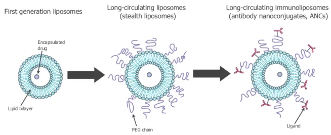
Figure 1.2. History of the development of liposomes for drug delivery: conventional liposomes (left); stealth
liposomes (center); immunoliposomes (right). Images adapted from[30].
to inefficient drug delivery (Figure 1.2, left). Therefore, at the end of the 80s, a second formula-tion of long-circulating liposomes has been developed to reduce the interacformula-tions between the MPS macrophages and the nanoparticles. Among these long-circulating liposomes, there are stealth
lipo-somes, which are nanocarriers obtained by modifying the surface with hydrophilic polymers such as
polyethylene glycol (PEG)[25,26] (see Figure1.2, center). The hydrophilic polymers possess a flex-ible chain that occupies the space around the liposome surface and excludes other macromolecules from this space, inhibiting the interaction between the macrophages and the liposomes. As a re-sult, long-circulating liposomes can passively accumulate inside other tissues and organs, taking advantage of the leaky vasculature in correspondence of the tumor. This phenomenon, called
pas-sive targeting, is particularly evident in tumors undergoing angiogenesis[27]. Finally, starting from
the 90s, efforts have been focused on developing stealth-liposomes that actively target tumor sites [28]. These stealth-immunoliposomes are obtained by coupling specific ligands to the liposomal
surface that bind tumor cell-specific receptors (Figure1.2, right). For instance, anti-Her2 monoclonal antibodies can be used as ligands to target Her2 receptors[29,3].
Doxil®(pegylated liposomal doxorubicin) is the first FDA approved nanodrug (1995)[31]. It is
used in the treatment of some types of cancer, such as metastatic ovarian cancer, metastatic breast cancer and AIDS-related Kaposi’s sarcoma.
As stated before, passive and active targeting are the main strategies that allow intravenously injected liposomes to reach the tumor site (Figure1.3). Here below, we explain the main features of these two mechanisms.
1.1. Biological background 7
Passive targeting
Big molecules, such as nanoparticles, liposomes or macromolecular drugs, tend to accumulate in tumor tissues much more than they do in healthy tissues. This phenomenon is called Enhanced Permeability Retention (EPR) effect[27]. Solid tumors are characterized by leaky vasculature that
is necessary for the development of the tumor (angiogenesis). In addition, solid tumors lack of an adequate lymphatic drainage, leading to a limited circulatory recovery of the molecules that go through the vasculature gaps. As a results, macromolecules and nanoparticles accumulate in the tumor microenvironment. Long circulating liposomes improve the EPR effect, since they have longer interaction with the target and are more likely to pass through the vasculature gaps.
Important parameters that affect passive targeting through the EPR effect, apart from the PEGy-lated liposomes, are the size of the liposomes, the composition and the charge on the surface of the nanoparticles[32]. In particular, the permeation of the nanoparticles within the tumor is highly
in-fluenced by the size of the nanoparticles. Small nanoparticles (∼10 nm) rapidly diffuse throughout the tumor matrix, while large nanoparticles (∼100 nm) stay close to the vasculature [32]. Physical
and chemical properties of the nanoparticles affect their pharmacokinetics and, therefore, their tu-mor accumulation capacity[32]. The process involving cell internalization of liposomes is mainly
endocytosis [33]. Depending on their physical attributes, such as particle size, shape and surface
charge and on the cell line, they are subject to a particular cellular internalization route or a com-bination of processes. These include, for example, phagocytosis, clathrin-mediated endocytosis (or receptor-mediated endocytosis), clathrin-independent endocytosis, caveolae and macropinocytosis.
Active targeting
Actively targeted liposomes have been designed to improve tumor specificity. They are prepared by engrafting targeting moieties, such as molecule ligands, peptides or monoclonal antibodies, on the liposomal surface. When liposomes accumulate in the tumor microenvironment, they are endocy-tosed into the cells by interacting with specific cell surface receptors (receptor-mediated endocytosis) [34]. However, a comparison between passive and active targeting reveals a lack of significant
8 Chapter 1. Introduction
Passive targeting
2. Expected improvements in pharmacokinetics with nanoparticles
2.1. Achieving longer exposure through decreased clearance
In vivo elimination of conventional liposomes, and to a lesser extent for other nanoparticles, has been extensively studied and reported in the past (Zamboni, 2005). Briefly, it depends on upstream interactions with specific proteins in plasma and the activity of Mononuclear Pha-gocyte System (MPS) (Zamboni, 2005). Macrophages indeed play a major role; 80%–90% of nanoparticles will get engulfed in the liver or spleen and degraded. Although this process occurs rapidly, liposomes show longer stay in the body as compared with free drugs (Gabizon et al., 1989). Even if first-generation liposomes displayed reduced clearance as compared with standard drugs, different strategies have been further developed next to limit organ uptake and immune system-related clearance. The most common strategy consists of masking the nanoparticle through surface pegylation, thus generating stealth, or second-generation nanoparticles (Fanciullino et al., 2013a). Second-generation liposomes are less likely to be recognized by MPS and ac-cumulate in the spleen and liver (Allen and Hansen, 1991), enabling the drug to stay in the blood stream longer, as demonstrated for instance in stealth liposomal doxorubicin (Gabizon et al., 1994).
2.2. Reducing toxicity via higher tumor specificity
Solids tumors present a leaky vasculature, originally allowing nu-trient supply necessary for sustained tumor growth. This anarchical organization has been defined by Maeda as enabling Enhanced Permeability and Retention (EPR) effect (Maeda, 2001). Liposomal nanoparticles could passively target the tumor by going through the vasculature gaps (i.e., 200 nm (Sawant and Torchilin, 2012)) and be retained near the tumor because of deficient lymphatic drainage (Fig. 2). In preclinical studies, radiotherapy has been sometimes used to enlarge these gaps by depleting the pericytes; further enhancing per-meation (Kobayashi et al., 2013) and thus tumor accumulation (Lammers et al., 2008). Developing stealth agents (e.g., PEG, see above) is another strategy to increase the EPR effect since the longer nano-particles stay in the blood, the more they will pass through the
vasculature gaps to target tumor tissue. This could explain why some liposomal nanoparticles display both decreased clearance and a higher volume of distribution (Vd) with limited drug accumulation in healthy tissues (Fanciullino et al., 2013a).
In spite of a more specific delivery to the tumor, the EPR effect alone usually achieves less than a 2-fold increase in tumor accumulation (Nakamura et al., 2016). Efforts have thus been made to develop third generation liposomes that display more active targeting. This is mostly achieved by grafting an agent on the surface that will specifically re-cognize cancer cells (Fig. 3). Many moieties (e.g., small-molecule li-gands, peptides and monoclonal antibodies) have been used over recent years to this end - including targeting EGFR (Xu et al., 2013), folate and transferrin receptors, tumor antigens (Deshpande et al., 2013) and neo-antigens that appear on the surface of irradiated tumor cells (Lowery et al., 2011).
Third generation liposomes can also be coupled to another up-coming strategy; triggered systems that can be functionalized with heat, ultrasound, light, enzymes or pH. Among them, the preclinical devel-opment of temperature sensitive liposomes exploits temperature or local hyperthermia, improving tumor targeting and cytotoxicity (Shin et al., 2016).
2.3. Main parameters driving nanocarrier pharmacokinetics 2.3.1. Size
As discussed since 1999 (Nagayasu et al., 1999), size is a major factor that impacts liposomal nanoparticle behavior, once adminis-tered. The smaller nanoparticles are, the less they will be recognized by MPS and be eliminated from the body (Liu et al., 1992). However it has been demonstrated that nanoparticles < 8 nm are mostly eliminated by the kidneys (Choi et al., 2007), not to mention a loss of stability in plasma and therefore quicker clearance below a given size. Being too big (i.e., > 200 nm) is also a major drawback, since it prevents nano-particles from benefitting from the EPR Effect. Several preclinical stu-dies have shown how size can affect the distribution phase within tumor tissue and does indeed matter for tumor accumulation. When testing three different batches of stealth liposomes of 5-FU of varying size (i.e., 70–250 nm) in mice bearing resistant breast tumors, data showed that the smaller the liposomes, the greater the tumor uptake
Fig. 2. Schematic representation of the EPR Effect. Tumor tissue Normal tissue
Lymphoid vessels
Early released drug
Gaps < 200 nm Encapsulated drug Normal vasculature Leaky vasculature Liposomes < 200 nm Ineffective lymphatic drainage
Active targeting
(Fanciullino et al., 2014). Consequently, better efficacy and longer survival were achieved in animals treated with smaller liposomes, thus demonstrating how size can impact tumor tissue distribution and ulti-mately efficacy. Similar results were found by Charrois et al. who stu-died the influence of liposome diameter on tumor distribution in mice bearing mammary carcinoma (Charrois and Allen, 2003). Statistically significant lower accumulation and reduced efficacy were evidenced for bigger liposomes. Overall, those experimental results confirmed older studies reporting that the optimal size for nanoparticles is in the 100–200 nm range, most probably because of the EPR Effect (Mayer et al., 1989).
2.3.2. Composition
The use of stealth or a targeting agent deeply modifies the phar-macokinetics of the nanoparticle. The choice of components is critical since it will modulate the stability of the nanoparticle in its systemic circulation by affecting the RES recognition and subsequently the drug release. Unstable liposomes will display increased plasma clearance and reduced circulation times compared to stable ones. The Gregoriadis group extensively studied the major role of composition in phospholi-pids and cholesterol in the early 1980’s (Senior and Gregoriadis, 1982); subsequent studies have further addressed the issue of lipids and cho-lesterol ratios required to achieve the most stable liposomes with op-timal controlled release, especially the critical role cholesterol plays (Senior and Gregoriadis, 1982). Indeed, cholesterol’s inclusion in the lipid bilayer of a liposome stabilizes its structure and decreases drug leakage and the risk of opsonization, thus extending circulation time (Ait-Oudhia et al., 2014;Briuglia et al., 2015).
2.3.3. Electric charge
The zeta potential of liposomal nanoparticles is another major factor influencing stability and therefore pharmacokinetics. Of note, this po-tential depends on the components used to synthesize liposomes. Geng et al. studied the impact of cholesterol on the stability of a doxorubicin PEGylated liposome - this time focusing on electric charge (Geng et al., 2014). Using two cholesterol derivatives (i.e., positively charged VS. neutral), pharmacokinetic studies in rats showed that neutral choles-terol liposomes displayed higher stability than positively charged ones. Similarly, the Torchillin group evaluated the clearance of liposomes displaying different surface properties in mice (Levchenko et al., 2002).
Different charged lipids were tested, with or without surface pegyla-tion. Charged liposomes showed higher clearance, especially the ne-gatively charged ones, which were preferentially found in the liver. Adding PEG-750 helped counter-balance the higher clearance of posi-tively charged liposomes but not for the negaposi-tively charged ones. Conversely, PEG-5000 partly reduced negatively charged liposome clearance, thus highlighting how complicated the combined impacts of electric charge and pegylation on nanoparticle pharmacokinetics can be. Recent studies have confirmed the deleterious impact of a negative charge on liposome clearance, and shown how pegylation can help improve their pharmacokinetics (Zhang et al., 2016). Additionally, other studies focused on the impact of positively charged nanoparticles on tumor uptake. For instance, Campbell et al. have studied the bio-distribution of cationic liposomes in human colon cancer bearing mice (Campbell et al., 2002). The impact of the cationic lipid ratio on dis-tribution was investigated: increasing cationic lipids by 10% decreased spleen uptake, while a further increase did not further reduce liposome accumulation in the spleen. Regarding tumor uptake, although differ-ences in the total tumor accumulation were not statistically significant, intravital microscopy revealed that cationic charges specifically target tumor vasculature. Increasing the charge content led to the doubling of neo-vessels uptake, suggesting its impact on tumor distribution and the benefit of using charged lipids for increased tumor specificity. Indeed, as compared with neutral liposomes, the cationic ones display a higher tumor uptake that can be hindered when pegylated. Positive charge and pegylation are then two opposite characteristics that can modulate tumor specificity. Both can be combined to achieve adequate targeting. For instance, Li et al. evaluated the quantitative relationship between these parameters on pancreatic cancer cells (Li et al., 2011), using li-posomes with alterable zeta potential and using a methoxy-analog of PEG-DSPE to reduce the electric charge. Data showed that each mol % of PEG could be compensated with a 4 mV increase, thus suggesting the existence of a balance between those two parameters to maximize stealth while ensuring tumor internalization of cationic liposomes. Once again, this highlights the complexity of how nanocarrier compo-sition must be finely tuned to ultimately optimize their pharmacoki-netics, especially at the tumor level.
2.3.4. Protein corona
When in biological fluids, the liposome surface attracts proteins and Fig. 3. Schematic representation of active tumor cells targeting for immunoliposomes.
A. Rodallec et al.
3. tL-receptor complex is internalized
2. tL binds to receptor
5. Tumor cell apoptosis 1. Targeted liposome
(tL) in plasma
4. Encapsulated drug is released
Figure 1.3. Passive targeting (top) and active targeting (bottom): mechanisms of drug delivery of the
im-munoliposomes. Images adapted from[37].
1.1.3
Development of antibody-nanoconjugates against breast cancer
Several efforts have been made to reduce the toxicity and improve the specificity of chemoterapy treatments of breast cancer. The research group SMARTc has developed a liposomal formulation that combines docetaxel and trastuzumab to target breast cancer cells [3]. These nanocarriers are
1.1. Biological background 9
currently in a preclinical trial. In this section, we explain the main characteristics of this kind of nanoparticles and how they are obtained.
Docetaxel is a chemotherapy drug belonging to the family of taxanes. It is an anti-mitotic drug
that arrests the mitosis cycle at the metaphase/anaphase stabilizing the microtubules. Then, since the microtubules don’t disassemble in presence of docetaxel, they accumulate inside the cell lead-ing to cell apoptosis. Trastuzumab is a monoclonal antibody that binds Her2 receptors, induclead-ing an immune-mediated response that causes internalization and downregulation of Her2. Docetaxel com-bined with trastuzumab is widely used to treat Her2 positive breast cancers, (neoadjuvant and/or adjuvant[38], both for advanced and early-stage diseases).
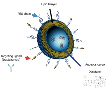
The high cytotoxicity of docetaxel affects all dividing cells in the human body, leading to severe adverse effects. To overcome this issue, the chemotherapy drug has been incorporated into stealth liposomes engrafted with trastuzumab on the surface to target better tumor cells and to spare healthy tissues. These immunoliposomes are called antibody-nanoconjugates (ANCs). A schematic of ANCs is shown in Figure1.4.
The ANCs developed by Rodallec et al. are made of natural lipids [39] and the diameter of
the nanoparticle is 140 nm. Other details on the encapsulation of docetaxel and the trastuzumab engraftment on the liposome surface are detailed in[39].
Figure 1.4. Stealth liposome with targeting ligand: the liposome is composed by a lipid bilayer and it contains
the chemoterapy drug (docetaxel). On the surface, the PEGylated chain impairs the interactions with the macrophages and the ligand (trastuzumab) targets Her2 receptors on breast cancer cells. Figure adapted from[40].
1.1.4
Open questions
The main challenge of nanoparticles for drug delivery is to enhance the drug efficacy at the site of action while reducing toxicity. The quantification of the efficacy of ANCs is not a trivial task due to the higher complexity associated with the nanoformulation compared with the standard treatments. Indeed, several parameters might affect the journey of the nanoparticles as well as the cellular up-take. Furthermore, the characteristics of the tumor tissue - such as interstitial fluid pressure, the permeability of the vessel walls, and tumor size - can impact the absorption of the nanoparticles. Moreover, the heterogeneity of the tumor microenvironment impairs a homogeneous distribution of the NP (e.g., some regions of the tumor might have high enhanced permeability retention, while others might show a limited vascular permeability), implying also a heterogeneous response in the in vivo experiments.
The quantification of the biodistribution properties of the nanoparticle is of fundamental im-portance to ameliorate the ANC design. Moreover, efficacy studies allow patient-specific therapy
scheduling.
The following points are the main questions that we want to address in this study:
• How is it possible to predict the tumor size and the NP efficacy? Which is the optimal treament scheduling? Which is the dose for first clinical studies?
• How does the tumor microenvironment (such as interstitial fluid pressure, vessel density, tu-mor size or Her2 expression) affect the transport of nanoparticles? How could it be possible to personalize the drug dose and scheduling from histology data about the tumor microenvi-ronment?
• Which is the optimal design of ANCs (size, trastuzumab graft rate) to increase their concen-tration in the tumor tissue?
Mathematical modeling is a powerful tool to (i) improve our understanding of biological processes of cancer and (ii) to help physicians and experimental design. For the first purpose, mechanistic models are built under given assumptions and tested against experimental data. On the one hand, this methodology allows us to strengthen or reject the initial theory and helps in formulating new hypotheses. On the other hand, the quantification of the treatment efficacy and predictions of the tumor progression are necessary for clinical decision management. Furthermore, in a preclinical context, they provide insights of new therapeutic options including optimal scheduling that can eventually be translated into clinical applications.
In this thesis, we focus on the two purposes: (i) we investigate the efficacy of the nanoparticles as function of the concentration to schedule an optimal treatment and (ii) we develop a continuum model to understand how nanoparticles penetrate into the tumor tissue.
1.2. State of the art 11
1.2
State of the art
1.2.1
Overview of mathematical models in cancer nanomedicine
Nanomedicine offers promising therapeutical and diagnostic options, but there are several chal-lenges that need to be investigated. Low and heterogeneous nanoparticle accumulation are the major limitations of nanomedicine[35]. NP physicochemical parameters, tumor models and cancer
types affect the low delivery efficiency. Therefore, it is necessary to define techniques and tools for a quantitative analysis. For example, computational tools and mathematical modeling can help in understanding tumor interactions and response to drugs and nanoparticles, coupling models of the physical microenvironment of the tumor with convection-diffusion equations for drug transport[41].
Several challenges must nevertheless be addressed to have a successful fruition of nanomedicine [42]: improving loading efficacy and on-command release, modulating recognition and
sequestra-tion by immune cells and maximizing accumulasequestra-tion at biological targets. This can be achieved with a joint collaboration between experimental and computational scientists.
Several mathematical modeling approaches can be employed according to the nanoparticle prop-erties that have to be investigated[43]. Different time and length scale models can be considered
to address questions about the biochemical interactions of the nanoparticle in the organism and the tumor deliverability[43] (Figure1.5). Molecular simulations, such as Monte Carlo, molecular dy-namics and coarse-grained simulations, can be employed to maximize the loading efficiency, namely the ratio between the mass of encapsulated drug and the total mass, while controlling the release (see, for example,[44, 45]). Molecular simulations can be also applied to analyze the interaction
of blood proteins with nanoparticles in order to optimize the surface features [46]. Indeed, it has
been noted that nanoparticles exposed to blood tend to be covered by different molecules forming a protein corona, affecting the bioavailability and the therapeutic performances of nanomedicines [47]. Discrete modeling has been adopted to investigate the nanoparticle internalization,
highlight-ing the process of endocythosis by the cells[48,49]. At the tissue scale, continuum models highlight
the macroscopic behavior of nanoparticles. Gentile et al. [50] investigated how the vessel
perme-ability and blood rheology impact the nanoparticle diffusion in the blood vessels using a diffusion-convection equation. Importantly, continuum models are increasingly validated against imaging data [51]. Continuum models of spheroids have been employed to determine the diffusion coefficient of
the nanoparticles and the cellular uptake [52, 53]. Several studies have analyzed the diffusion
co-efficient according to the properties of the nanoparticles. Size, shape, and density were found to impact the nanoparticle wall-deposition in the blood vessels[54,55]. In particular, small
nanopar-ticles improve the margination rates; lighter parnanopar-ticles marginate singificantly more compared to NP with larger density; non-spherical nanoparticles showed higher margination compared to spherical particles. Moreover, intratumoral pharmacokinetics of nanoparticle has been studied using intravital imaging and linked to the treatment efficacy [56]. Furthermore, the transport of nanoparticles in
tumor tissues can be modeled with continuum mechanics approaches and particle based systems [57].

![Figure 1.6. Left: scheme of solid tumor (from [ 61 ] ): blood vessels, interstitium and cancer cells](https://thumb-eu.123doks.com/thumbv2/123doknet/14549734.725585/32.892.132.777.152.497/figure-left-scheme-solid-tumor-vessels-interstitium-cancer.webp)