HAL Id: tel-01722820
https://tel.archives-ouvertes.fr/tel-01722820
Submitted on 5 Mar 2018
HAL is a multi-disciplinary open access
archive for the deposit and dissemination of
sci-entific research documents, whether they are
pub-lished or not. The documents may come from
teaching and research institutions in France or
abroad, or from public or private research centers.
L’archive ouverte pluridisciplinaire HAL, est
destinée au dépôt et à la diffusion de documents
scientifiques de niveau recherche, publiés ou non,
émanant des établissements d’enseignement et de
recherche français ou étrangers, des laboratoires
publics ou privés.
Reversal strategies within the lateral habenula to
ameliorate depressive-like behaviors
Anna Tchenio
To cite this version:
Anna Tchenio. Reversal strategies within the lateral habenula to ameliorate depressive-like behaviors.
Neurons and Cognition [q-bio.NC]. Université Pierre et Marie Curie - Paris VI, 2017. English. �NNT :
2017PA066301�. �tel-01722820�
Université Pierre et Marie Curie
Ecole doctorale n°138
« Cerveau Cognition Comportement »
Doctoral thesis title:
Reversal strategies within the lateral
habenula to ameliorate depressive-like
behaviors
Presented by Anna Tchenio
Directed by Dr. Manuel Mameli
Team « Synapses and pathophysiology of reward »
Institut du Fer à Moulin
Presented the 08th decembre, 2017
Members of the jury:
Dr. Philippe Faure – président
Pr. Paul Slesinger– rapporteur
Pr. Stephan Lammel–rapporteur
Dr. Alberto Bacci –examinateur
1
Abstract
Prolonged exposure to aversive stimuli leads to cellular and circuit adaptations that contribute
to the emergence of neuropsychiatric disorders such as depression. Interactions between the
dopaminergic (DA) and the serotoninergic (5HT) systems have been implicated in these
pathological adaptations ultimately influencing motivated behaviors. Interestingly, the lateral
habenula (LHb), an ethologically well-conserved epithalamic region, directly and indirectly
controls DA and 5HT systems, and its activity is modulated by aversive events in both
humans and animals. Moreover, the activity of the LHb increases in animal models of
depression and depressed human patients. Conversely, strategies that locally target the LHb
have been shown to reverse depressive-like symptoms both in animal models and in humans.
Altogether, this led to the hypothesis that LHb dysregulation could play a role in the
emergence of depressive like symptoms. However, little is known about the early cellular and
molecular adaptations that occur within the LHb after exposure to aversive events. Moreover,
most of the animal models employed to interrogate the LHb role in depressive states used
acute painful stimuli; whether LHb function becomes aberrant after chronic exposure to
painless stressors remain elusive. In my thesis work, I explored the precise cellular and
molecular adaptations of LHb neurons following exposure to different kind of unpredictable
aversive experiences, and their importance for the expression of depressive like symptoms.
More precisely, I will present the results of an initial work aiming to identify early cellular
and molecular adaptations within the LHb following unpredictable stimuli and their
importance for the emergence of depressive symptoms. This study shows that unpredictable
foot-shocks lead to decreased surface expression and function of the gamma-aminobutyrate
receptor (GABA
BR), a metabotropic receptor that hyperpolarizes LHb neurons through the
activation of the G protein-coupled inwardly-rectifying potassium channels (GIRKs). This
decrease of GABA
BR-GIRK signaling went along with an upregulation of the activity of the
protein phosphatase 2 (PP2A), which is a well-known down-regulator of GABA
B-GIRK
complex surface expression. GABA
BR-GIRK signaling tightly controls LHb activity, and its
downregulation consequently leads to aberrant hyperexcitability of LHb neurons. Using
specific strategies to restore the GABA
BR-GIRK signaling within the LHb, such as GIRK
overexpression, or local pharmacological inhibition of PP2A activity, we were able to
ameliorate depressive like states following unpredictable foot-shocks.
The second study allowed instead to establish the cellular adaptations within the LHb
following a chronic non-painful aversive experience and during a critical developmental
2
period. I showed that exposure to maternal separation in childhood (MS mice) also leads to
depressive like symptoms together with a hyperexcitability of LHb neurons. This
stress-driven increase in LHb activity was causally linked to a decrease of the GABA
BR-GIRK
signaling. Moreover, using diverse reversal strategies such as chemogenetics or a
therapeutically-relevant intervention such as Deep Brain Stimulation (DBS), we could
selectively decrease LHb neuronal activity and consequently ameliorate the depressive like
symptoms, suggesting a causal link between these two phenotypes.
Altogether, the work presented in this thesis suggests that LHb neuronal hyperexcitability
could represent a common substrate necessary for the expression of certain aspects of the
depressive like state and further supports its relevance as a potential target in the treatment of
this disorder.
3
Résumé
L’agression de l'organisme par un agent physique, psychique ou émotionnel déclenche une
réponse physiologique et comportementale qui permet à un individu de se prévenir du danger
et de maintenir sa survie. Le système nerveux central a depuis longtemps été identifié comme
un majeur acteur de cette réponse adaptative. Cependant, une exposition prolongée au stress
conduit à des adaptations cellulaires et des réadaptations de circuits neuronaux qui contribuent
à l'émergence de troubles neuropsychiatriques. L’interaction entre le système dopaminergique
(DA) et sérotoninergique (5HT) a été impliquées dans ces réarrangements physiologiques et
pathologiques qui influencent les comportements motivationnels de l'individu face à une
menace. Fait intéressant, l'habenula latérale (Hbl), une région du cerveau très conservée entre
les espèces, contrôle directement et indirectement les systèmes DA et 5HT, et son activité est
modulée par des stimuli aversifs chez les humains et les animaux. De plus, l'activité de la Hbl
est augmentée chez des modèles animaux de la dépression ou lors de l'induction d'un épisode
dépressif chez des patients humains. Inversement, l’emploie de stratégies ayant pour cible la
Hbl permettent d’améliorer certains symptômes dépressifs à la fois chez les modèles animaux
de dépression et chez l’homme. Ainsi, la dérégulation de l’Hbl pourrait jouer un rôle dans
l'apparition de symptômes dépressifs. Cependant, les changements moléculaires et cellulaires
précoces qui occurrent au niveau de Hbl suite à l'exposition continue à un environnent
aversifs restent peu connus. De plus, la plupart des modèles animaux utilisés pour interroger
le rôle de Hbl dans l'état dépressif implique une exposition répétée de l’animal à des stimuli
douloureux. Si la fonction de l’Hb est aberrante lors d’une exposition chronique à d’autre type
de stress reste méconnu. Dans mon travail de thèse, je me suis intéressée aux adaptations
cellulaires et moléculaires au niveau des neurones Hbl suite à l’exposition de différents types
d'expériences aversives et leurs relatives importances pour l'expression de symptômes
dépressifs.
Plus précisément, je présente dans ce manuscrit, les résultats d'un premier travail qui vise à
identifier les adaptations cellulaires et moléculaires de Hbl suite à l’exposition de souris à des
chocs électriques et leurs rôles dans l'émergence de symptômes dépressifs. Cette étude montre
que l’exposition à de brefs aléatoires chocs électriques entraine une diminution de
l’expression de surface de récepteur métabotropiques gamma-aminobutyrate B (GABA
BRs),
et par conséquent une diminution de leur fonction au niveau des neurones de l’Hbl. GABA
BR
4
par l'activation du canal potassique GIRK. La diminution de la signalisation GABA
BR-GIRK
est accompagnée par une augmentation de l'activité de la protéine phosphatase 2 (PP2A),
reconnue pour induire l’endocytose du complexe GABA
BR-GIRK. GABA
BR-GIRK contrôle
étroitement l'activité Hbl, et par conséquent une diminution de leurs fonctions conduit à
l’hyperexcitabilité des neurones de l’Hbl. En adoptant des stratégies visant à restaurer
spécifiquement la signalisation GABA
BR-GIRK dans l’Hbl, telle que la surexpression GIRK,
ou l'inhibition pharmacologique locale de l'activité PP2A, nous avons observé une
amélioration de certains symptomes « dépressifs », établissant ainsi un lien causal entre
l’aberrante diminution du signal GABA
BR et certain aspect de l’état dépressifs.
La deuxième étude permet d'établir l'effet d'une expérience aversive chronique non
douloureuse sur la fonction Hbl. Dans ce travail, la séparation de petits et de leur mères
(souris MS) conduit à posteriori au développement de symptômes dépressifs accompagnés par
une hyperexcitabilité des neurones de la Hbl. Cette augmentation d’activité est, au moins en
partie, expliquée par une diminution de la signalisation GABA
BR-GIRK. De plus, prenant
avantage de la technologie chimio-génétique ou des récentes avancées en terme thérapeutique
telle que la stimulation cérébrale profonde (DBS), j’ai été en mesure de réduire localement
l’activité de l’Hbl des souris MS. D’autre part, l’utilisation de ces diverses stratégies étaient
suffisante pour améliorer les symptômes dépressifs des souris MS, suggérant un lien de
causalité entre ces deux phénotypes.
Dans l'ensemble, ces travaux suggèrent que l'hyperexcitabilité des neurones Hbl pourrait
représenter un substrat commun nécessaire à l'expression de symptômes dépressifs et ainsi
constituer une cible potentielle pour traiter certains aspects de la dépression.
5
Aknowledgements
At the end of this research, I am convinced that the thesis is far from being a lonely work.
Indeed, I could never have carried out this doctoral work without the support of several
people whose generosity, good humor and interest in my research allowed me to progress and
learned a lot throughout these 3 years.
In the first place, I am grateful to my thesis supervisor, Manuel Mameli, for the trust he has
given me despite my limited knowledge in his field when I started. I would like to thank him
by accepting to supervise this doctoral work, for its multiple advices and challenges that make
me progress and advance, for all the hours he has devoted to directing this research. I would
also like to tell him how much I appreciated his high availability not only by providing an
indispensable support and good ideas in my research over almost 3 years, but also for all the
hours devoted to re-reading this thesis, for his abundant advices and its emotionally support
through the rough road to finish this thesis. Thank you for listening and understanding. As he
could say, “work hard/play hard”, I thank him for the amazing energy and dynamism that he
brings in the lab and the care that he has for his people. I greatly enjoy these 3 years in the
team and dedicated to this work. Thank you for this great and memorable opportunity.
I want to thank also my friends and colleagues: Frank (the professor) which try to teach me
Dutch proverbs although I must confess to have forgotten every word since… Kristina (the
mum) which often made sure none of us starved, Salvatore (the sailor) which made sure none
of us went thirsty (or sober) and Massimo (the “geek”) that kept us entertained with his huge
repertoire of anecdotes and funny video. Guys, I could thank you in many way, you are
amazing people, and it is a pleasure to work in a so crazy/funny lab… Among everything,
thank you for all this discussions inside or outside the lab, around a barbecue or a glass of
wine, for sharing with me your knowledge and experience, and your unlimited support in the
good and bad moments, that pushed me and help me a lot during these years. Thanks also for
all the nice moments spent together, celebrations, dinners, travelling and also to share the
motivation to “try” to stay healthy (swimming/ running / badminton/ climbing and hiking)…
not always simple…
I also like to thank all the Fer a moulin, starting by its director,
Jean-Antoine Girault, that
maintain a wonderful atmosphere in his department, full of conviviality, creativity and
exchanges which create a perfect environment to growth as a young scientist. Of course, it
will be nothing without all the people that composed it, so thank to all the “feramoulinos”
people. A particular thank to Sana, Emily, Jessica, Ferran, Marie, Fanny, Benoit... among
many others, that not only permit to have some nice constructive scientific discussions, but
also with who I shared a lot of fun in gypsy concert, jazz swinging, driving bike in the middle
of the night in Paris ….
I also would like to thank the help of Megan Creed for her tips for the DBS and the
researchers of the IFM for their scientific advice and helpful discussions.
Finally, I would like to thank my family, for their love and unrestricted support, which have
permit me to achieve this work. In particular, I would like to thank my mother Sylvie and my
grandmother Jeanine, as well as my brothers and sister Jonathan, Gwenael, and Chloe (and
also Aylan, Reda and Sophie) for their love and their skills to always find the good word (or
smile for Aylan) to motivate and guide me. Last but not least, I would like to thank my father
Paul, that transmit me his love for science, to always have trust in me, and always
understanding the things that I said or not.
It is impossible to summarize what all these people have done for me and how much they
contribute to this work, so THANK you.
6
Contents
Abstract ... 1
Résumé ... 3
Aknowledgements ... 5
Contents ... 6
Table of figures ... 7
List of abbreviations ... 8
Introduction ... 10
Depression ... 11
I. A brief history of depression ... 11
II. Treatment of major depressive disorder ... 13
III. Studying depression in laboratory animals ... 15
A. Learned helplessness model ... 17
B. The chronic mild stress ... 18
C. Social defeat ... 19
D. Maternal separation ... 20
Neural circuits of depression ... 21
Emerging role of LHb in depression ... 23
I. Lateral habenula as a node to control midbrain regions ... 24
II. Lateral habenula function in processing negative information... 26
III. Lateral habenula modifications in depression ... 28
GABA
Breceptors:
master of the neuronal activity ... 30
I. GABA
Breceptor structure and function ... 30
II. GABA B receptors trafficking... 32
III. GABA
Breceptor regulation ... 33
Methodological section ... 37
Experimental subjects and stress paradigms ... 37
Behavioral tests ... 38
Drug/intervention ... 39
Surgery ... 40
Electrophysiology ... 40
Analysis and drugs. ... 42
Context and objectives of the study I: GABA
Breceptors trigger LHb hyperactivity, an early marker of
depressive like symptoms ... 43
Context and objectives of the study II: LHb hyperactivity as a long term adaptation underlying
MS-driven depressive like symptoms... 68
Discussion ... 90
7
II. Induction mechanism of GABA
Breceptor plasticity following different kind of stress ... 93
III. Circuit specificity of GABA
Breceptor regulation in the LHb ... 95
IV. What is the functional relevance of GABA
Breceptor dependent hyperexcitability for the
emergence of depressive like symptoms ... 96
V. Targeting LHb hyperexcitability to treat depression? ... 97
A. Caveat of DBS intervention: ... 97
B. PP2A inhibitors as potential antidepressant drugs ... 98
C. Consequences of LHb hyperexcitability on downstream circuitries ... 99
Conclusion ... 102
Publications and contributions ... 104
References ... 105
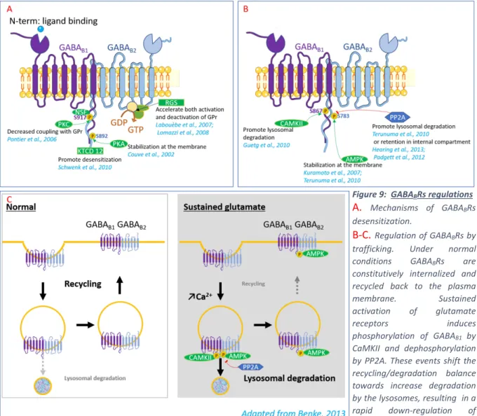
Table of figures
Figure 1 Epidemiology of depression ………... 12
Figure 2 Depression treatments ...……….………...……... 14
Figure 3 Rodent models of depression and behavioral assay to assess the depressive
phenotype ... 16
Figure 4 Simplified schematic of neural circuits implicated in depression ... 22
Figure 5 Lateral habenula anatomy: a hub between forebrain structures and neuromodulatory
nuclei ……... 25
Figure 6: Lateral habenula encodes of aversive information and drive motivated behaviors ... 27
Figure 7 Cellular and molecular adaptations within the habenula in a rodent model of depressive-like
states. ... 29
Figure 8 GABA
BR subunits and functions... 32
Figure 9 GABA
BR regulations... 35
Figure 10 Potential mechanisms underlying LHb GABA
BR-GIRK reduction in rodent model of
depression... 94
8
List of abbreviations
5HT
5-hydroxytriptamine
AMPAR
α-amino-3-hydroxy-5methyl-4-isoxazolepropionic acid receptor
AMPK
adenosine monophosphate-activated protein kinase
Ca
2+calcium ion
(β)CaMKII (beta)-calcium calmoduline-dependent protein kinase type II
ChR2
channel rhodopsin 2
CMS
chronic mild stress
DA
dopamine
DBS
deep brain stimulation
DREADD designer Receptor Exclusively Activated by Designer Drugs
DSM
Diagnostic and statistical manual of mental disorders
ECT
electroconvulsive treatment
EPN
entopeduncular nucleus
FS
foot-shock
FST
forced swim test
GABA
gamma-aminobutyric acid
GABAA R gamma aminobutyric acid type A receptor
GABA
BR gamma aminobutyric acid type B receptor
GABA
B1a,b/2gamma aminobutyric acid type B receptor subunit 1a,1b,or 2
GIRK
G-protein inwardly-rectifying potassium channel
GIRK1-4
G-protein inwardly-rectifying potassium channel subunit 1 to 4
GluA1-4
glutamate AMPA receptor subunits type 1-4
GPCR
G-protein coupled receptor
GRIN-lenses Graded-Index lenses
HPA
Hypothalamic–pituitary–adrenal
I-baclofen current evoked by baclofen application
KCTD
potassium channel tetradimerization Domain containing proteins
KO
knock-out
LDT
laterodorsal tegmentum
LH
lateral hypothalamus
9
LHp/(cLHp) learned helplessness / (congenital learned helplessness)
LTD
long term depression
LTP
long term potentiation
MAOI
monoamine oxidase inhibitor
MS
maternal separation
MHb
medial habenula
mPFC
medial prefrontal cortex
NA
noradrenaline
NAc
nucleus accumbens
NDRI
noradrenaline and dopamine reuptake inhibitors
NRI
noradrenaline reuptake inhibitors
NSF
N-ethylmaleimide-sensitive fusion
NMDA
N-methyl-D-aspartic acid
PFC
prefrontal cortex
PKA
protein kinase A
PKC
protein kinase C
PP1
protein phosphatase type 1
PP2A
protein phosphatase type 2
RMTg
rostromedial tegmental nucleus
RGS
regulator of G protein signaling
S
serine residue
SD
social defeat
SNX 27
Sorting nexin 27
SSRI
serotonin reuptake inhibitors
TCA
tricyclic antidepressant
TST
tail suspension test
VGCC
voltage-gated calcium channel
VP
ventral pallidum
10
Introduction
Physiological and behavioral adaptations of an individual facing a real or potential threat are
required for the well-being or the survival. This highly adaptive response depends on the
orchestration of several neuronal circuits ultimately allowing to assign specific negative
valence to an event and subsequently guide optimal behaviors such as avoidance.
Monoaminergic systems have been implicated in this immediate encoding of aversive event.
Indeed both serotoninergic (5HT) neurons of the raphe and dopaminergic (DA) neurons of the
ventral tegmental area (VTA) have been described to phasically respond to acute aversive
stimuli and influence negative reinforcement or avoidance learning both in human and animal
studies (Brischoux et al., 2009; Cohen et al., 2015; Crockett et al., 2009; Deakin and Graeff,
1991; Hayes et al., 2014; Matsumoto and Hikosaka, 2009a; McCutcheon et al., 2012;
Puglisi-Allegra and Andolina, 2015; Schweimer and Ungless, 2010; Ungless et al., 2010). In contrast,
exposure to severe or chronic aversive stimuli, particularly when coupled with a lack of
predictability or lack of control (Maier and Seligman, 1976; Short and Maier, 1993), elicits
detrimental, long-lasting effects on brain function, that could precipitate the emergence of
neuropsychiatric disorders, including mood disorders (Anisman and Matheson, 2005; Kessler,
1997; de Kloet et al., 2005). Mood disorders are associated with deficits in both rewarding
and punishment encoding, leading to the assumption that dysfunction in neural circuits
including the DA and 5HT systems may underlie depressive-like symptoms (Nestler and
Carlezon Jr, 2006; Owens and Nemeroff, 1994; Russo and Nestler, 2013). However, although
the role of DA and 5HT in depression have received particular attention, these systems are
interconnected and tightly controlled by a much broader circuit (Ogawa et al., 2014;
Pollak Dorocic et al., 2014). Considering that persistent negative events have deleterious
effect in both DA and 5HT system (Owens and Nemeroff, 1994; Russo and Nestler, 2013) it
is likely that dysregulation of common upstream structures may be critical for the
development of neuropsychiatric disorders. I therefore became interested in studying the
potential modifications within neuronal circuits that directly control monoaminergic nuclei.
The lateral habenula (LHb) is an epithalamic region that directly and indirectly controls DA
and 5HT neurons (Lammel et al., 2012; Pollak Dorocic et al., 2014; Stamatakis and Stuber,
2012; Weissbourd et al., 2014). Interestingly, a clinical study reported the potential
therapeutic benefit of deep brain stimulation (DBS) in the LHb for the treatment of depression
in a single case in human (Sartorius et al., 2010). This work was one of the first of a long
series of studies supporting the hypothesis of the potential role of LHb in depression (Lecca et
11
al., 2014; Li et al., 2011, 2013; Seo et al., 2017; Shabel et al., 2014). Despite this growing
body of evidence, whether LHb dysfunction can be a common substrate underlying specific
aspects of depressive-like state independently of the events that trigger it remains unknown.
In my thesis work, I will first introduce the complexity of the depressive disorder, then I will
briefly summarize key pathological reorganization of the mesolimbic system in
depressive-like state to give a general context of how aversive and reward-related circuits can underlie
certain aspect of this disease. Next, I will present how LHb can control the mesolimbic system
and I will discuss the current literature around the cellular properties - from the GABA
BR to
the hyperactivity - of the LHb, which can be instrumental for the expression of depressive like
symptoms.
Depression
I. A brief history of depression
Historical documents throughout the ages point to the long-standing existence of depression
as a health problem for human beings. Already in the ancient Greeks, the Hippocrates school
described the “Melancholia”, a condition associated with “aversion to food, despondency,
sleeplessness, irritability, restlessness”. Although they believed that an excess of black bile in
the spleen caused depression, the similarities between the Greek descriptions of depression
and those of the modern age are striking. The following centuries led to a dark period of
obscurantism where notably the religions refuted the natural cause of mental illness and
associated it to witchcraft or demonic possession. Indeed, it was not until the XIXth century,
that mental illness was reevaluated and successively considered to have psychological and
social causes or to be an inherited disease. The industrial revolution notably led to the
building of psychiatric institution, and the brain became the focus of efforts to understand the
pathophysiology of this disorder. The Second World War marked a turning point, where the
mental illness started to be considered as a real problem of society. Along with the evolution
of the psychiatry, the discovery of the first antidepressants greatly ameliorated the condition
of the depressed patients. Moreover, a new manual categorizing the various mental problems
were established in the United States, the Diagnostic and statistical manual of mental
disorders (DSM) (Frader, 1987).
In a new version of this manual, the major depressive episode is characterized by a persistent,
unreactive low mood and a loss of interest in pleasure. These symptoms are associated with a
range of other symptoms such as significant weight loss, insomnia or hypersomnia,
12
psychomotor agitation or retardation, fatigue or loss of energy, feelings of worthlessness or
excessive or inappropriate guilt, diminished ability to think or concentrate, or indecisiveness,
as well as recurrent thoughts of death or suicide (Diagnostic and statistical manual of mental
disorder, 5th edition). However, depression is recognized to be a highly heterogeneous
disease, with the presence of variable set of symptoms across individuals. Due to this
complexity, clinicians and scientists are still trying to discriminate and study specific aspects
of the disease, ranging from anhedonia, behavioral despair, and social withdrawal to learning
deficits. This different endophenotypes point out to the diversity of functional alterations: the
anhedonia, is a dysfunction in reward encoding, which leads to the loss of motivation for
natural rewards such as food or sex. Alternatively, behavioral distress or hopelessness imply
impairments in aversive-stimuli encoding, leading to the inability to cope with a negative
situation. This diversity of symptoms suggests that discrete neural circuits could be implicated
in different aspects of the disease.
Figure 1. Epidemiology of depression
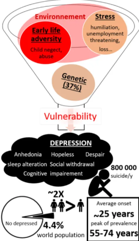
A large part of high individual vulnerability can be explained by interactions between genetic predisposition which has been estimated at 37% (Sullivan et al., 2000), and environmental factors (Anisman and Matheson, 2005; Kendler et al., 1999; Kessler, 1997). Epidemiologic studies provide evidence that early life adversity increases the risk to develop depression (Agid et al., 1999; Norman et al., 2012) as well as stress in adulthood. Indeed, major adverse life events such as humiliation, threatening or loss (Farmer and McGuffin, 2003; Paykel, 2003; Slavich et al., 2010) as well as accumulation of chronic stressful events (Brown and Harris, 1978; Hammen et al., 2009) precipitate depression.
Depression is among the most important cause of disability according to a report of the WHO (2017), it is a complex disorder characterized by a diversity of symptoms that can be categorized in “endophenotype” such as anhedonia, behavioral despair, hopeless, sleep disturbance, social withdrawal and cognitive impairment. In its most severe conditions depression can lead to suicide. It affects around 4.4 % of the global population and it is twice more common in females than males. The prevalence globally increases with the age and the average age onset is the middle of 20s’ years old with a peak of prevalence at 55-74 years old.
Considering the heterogeneity of etiology and symptoms, a general therapeutic treatment
taking into account all the variables is difficult to achieve.
In the next chapter, I will summarize the currently available treatments and their limitations as
well as newly emerged interventions offering potential promising improvements for
treatment.
13
II. Treatment of major depressive disorder
Antidepressant treatment emerged by serendipity in 1950 with the finding that the
antitubercular agent iproniazid (a monoamine oxidase inhibitor (MAOI)) and that the
imipramine (tricyclic antidepressant (TCA)) arising from antihistamine research were very
efficient in relieving depressive symptoms (Kuhn, 1957; Loomer et al., 1957). MAOIs inhibit
the breakdown of 5HT, noradrenaline (NA), and DA thus increasing the availability of these
neurotransmitters. TCAs increase the synaptic concentration of 5HT and NA by inhibiting
their reuptake. These discoveries were concomitant with the finding that reserpine (that
irreversibly blocks the vesicular monoamine transporter (VMAT)) induces depressive-like
symptoms (Muller et al., 1955). Overall, these data led to the monoaminergic theory of
depression, suggesting that an imbalance, mainly in 5HT, NA and DA neurotransmission, is at
the core of the pathophysiology of depression (Bunney and Davis, 1965; Delgado, 2000;
Hirschfeld, 2000; Schildkraut, 1965).
This theory led to the development of new several classes of antidepressants with similar way
of action, aiming to correct the monoaminergic deficiency. Among them the selective
serotonin, noradrenaline, and/or dopamine reuptake inhibitors, such as citalopram or
fluoxetine (SSRI; NRI; NDRI) that are still nowadays the most prescribed antidepressants
(O’Leary et al., 2014, 2015). Modern antidepressant molecules differ from the older drugs as
they aim to reduce their deleterious side effects and narrow the neurochemical targets.
However, even if their tolerance has improved, little ameliorations have been reported in
terms of the slow onset of clinical effect or their efficacy (Baghai et al., 2011; Holtzheimer
and Mayberg, 2011a). Indeed, standard treatment still presents a high percentage of
pharmacological resistance (±30%) or relapse (±50%) (Al-Harbi, 2012; Holtzheimer and
Mayberg, 2011a; Trivedi et al., 2006). Furthermore, the mechanism of action of such
molecules on brain wiring, neuronal activity or plasticity remains still elusive. Thus, the
monoaminergic theory appears to be too restrictive, and it looks obvious that the therapeutic
effects of antidepressants cannot be solely explained by the facilitation of 5HT and NA
neurotransmission (Hindmarch, 2002; Massart et al., 2012).
Other alternative strategies of the drug treatments have been developed, and are prescribed
notably in treatment resistant patients such as the electroconvulsive therapy (ECT). This
procedure showed a remission of 75% in resistant depression with the advantage of its
immediate effect (Sienaert, 2011). However, the action of ECT is broad, its mechanism is
poorly understood, and it presents side effects such as confusion and memory loss (Bolwig,
14
2011; Ingram et al., 2008). Alternatively, DBS of the prefrontal cortex (PFC) (Mayberg et al.,
2005), Nucleus Accumbens (NAc) (Bewernick et al., 2010; Schlaepfer et al., 2007) or more
recently LHb (Sartorius et al., 2010) also have reported amelioration in groups of treatment
resistant depressed patients. All these structures were primarily selected based on
neuroimaging studies showing their aberrant activities in depressed patients and/or their
important role in reward circuits correlated with anhedonia (one of the prominent symptoms
of depression) (Delaloye and Holtzheimer, 2014). Despite their promising therapeutic interest,
these studies remain still preliminary. Indeed, the way of action of the DBS is nowadays still
enigmatic. The observation that DBS and lesions could have similar behavioral effect led to
the theory that DBS inhibits neuronal activity. However, mixed pattern of neuronal excitation
and inhibition have been described following DBS in certain brain regions and also raise the
question of a local effect (Maks et al., 2009; Mcintyre et al., 2004; Vitek, 2002). Further
studies, looking at the effects of DBS in particular brain regions are thus warranted to refine
and better understand this intervention.
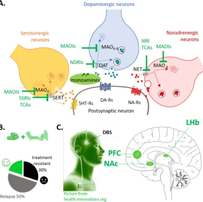
Figure 2. Depression treatments
A. Main action of pharmacological therapies:
Antidepressants aim to restore "imbalanced" brain chemistry. They boost monoaminergic neurotransmitters by blocking their reuptake (tricyclic antidepressants -TCAs-, Selective serotonin reuptake inhibitors -SSRIs- Norepinephrine–dopamine reuptake inhibitors -NDRIs-, Norepinephrine reuptake inhibitors -NRIs-) or by preventing their metabolism (monoaminoxidase inhibitors (MAOis).
B. Current treatments consist of psychological
treatments (such as psychotherapy) and/or antidepressant medication (such as selective serotonin reuptake inhibitors [SSRIs] and tricyclic antidepressants [TCAs]). However 30% of patients exhibit treatment-resistant, and less than the half shows remission.
C. Deep Brain Stimulation (DBS) as alternative strategy to treat depression. DBS is a promising
alternative intervention in the case of treatment-resistant depression.This approach involving the bilateral placement of electrodes at specific neuroanatomical sites. The prefrontal cortex (PFC), the Nucleus accumbens (NAc) and the lateral habenula (LHb), are nowadays the target of clinical trials. These regions have been selected based on previous neuroimaging studies that report their aberrant metabolicactivity as well as loss of brain volume in treatment resistant patients.
Overall major depression affects more than 300 million people according to a recent report by
the World Health Organisation, (WHO 2017-GENEVA). It is a leading cause of disability
worldwide, with a total annual cost estimated at 118 billion euro in Europe (Sobocki et al.,
2006), and 210 $-billion per year in US (Greenberg et al., 2015). Given the public health
15
significance of this pathology, novel treatments are needed. Indeed, current available
treatments fail to provide significant improvement nearly half of the time (Al-Harbi, 2012;
Holtzheimer and Mayberg, 2011a; Trivedi et al., 2006). One explanation is still the lack of
understanding of the specific brain circuits and molecular changes underlying depressive
symptoms. The focus on particular symptoms and novel treatment interventions is a
necessary step to permit a better understanding of the neurobiology of the illness and achieve
larger improvements.
III. Studying depression in laboratory animals
Using brain imaging approaches and neuroendocrine responses in humans provided valuable
information about the brain circuitry implicated in mood disorders. Nevertheless, the detailed
investigation of physiological responses and molecular modifications in the human brain are
limited by obvious practical and ethical difficulties. To circumvent these limitations, animal
models have emerged as an alternative and valuable tool to understand the pathophysiology of
depression. Three sets of criteria have been proposed for assessing animal models of human
mental disorders: construct validity, face validity, and predictive validity (Nestler and Hyman,
2010; Willner, 1984). Construct validity refers to the relevance of the mechanism in which the
model is based. So far, no genetic strong determinants were found for depression (Dunn et al.,
2015) limiting genetic model in replicating multifaceted phenotype and strong pharmacology
validity. Hence, the main animal models of depression are based on the epidemiological
evidence that severe or chronic stress and adverse psychosocial experience often precede the
depressive episode. The face validity corresponds to whether the model resembles depression
in respect to the presence of ethological similarities and biomarkers. Even if symptoms such
as guilt, suicidality, and sad mood are typical human features, other endophenotypes can be
reproduced and measured in laboratory animals. These include notably the helplessness
(referring to hopelessness in human), anhedonia, social withdrawal and behavioral despair,
respectively indicated by the increased failure and latency in the shuttle box test, decrease in
sucrose preference and social interaction, and increased immobility in forced swim test or tail
suspension test (see Fig. 3). Finally, the predictive validity is when the animal model exhibits
symptoms’ amelioration with pharmacological and non-pharmacological treatments for
depression. Here, I will give examples of different models currently used in the literature:
16
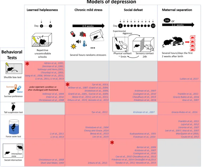
Figure 3:Rodent models of depression and behavioral assay to assess the depressive phenotype
Schematic representing the main models of depression describe in this thesis, and their associate phenotype. Different assays used to assess the depressive phenotype are listed below:
Shuttle box test: The subjects are place in a box with 2 compartments. The test consists of several trials where mice are exposed to escapable foot-shocks, as soon as they escape in the opposite compartment the shock stop. If the animal is not escaping after a pre-determined period, it is considered as failure. Latency to escape and number of failures are the two parameters measure in this paradigm. This test is routinely used to screen learned helplessness vulnerable mice(Chourbaji et al., 2005; Vollmayr and Henn, 2001)
The tail-suspension test (TST) (Steru et al., 1985) and the forced-swim test (FST), (Porsolt et al., 1977): Rodents are exposed to an acute, unescapable, short-duration stress: suspend by the tail or placed in an transparent tank filled with water respectively. Time spent performing active “escaping”-behavior (struggling/swimming) is quantified relative to time spent immobile. Higher immobility has been interpreted as a sign of behavioral despair or passivity. Although this interpretation can be debated (Nestler and Hyman, 2010), these tests have the advantage to permit a rapid, easy screening of potential antidepressant drugs, or depressive-like behavior in rodent (O’Leary and Cryan, 2013).
The sucrose preference test is one of the most common test to assess anhedonia. This test evaluated in a two-bottle choice paradigm the consumption of a sucrose solution in respect to the water consumption. Decreased consumption of sucrose is assumed to be indicative of a reduction in the motivation for the rewarding solution. This sucrose preference is attenuated by a diversity of chronic stressors, notably the chronic mild and unpredictable stress and it is commonly accepted to reflect anhedonia(Willner et al., 1987).
Social interaction/avoidance test: This test consists to place an unfamiliar congener (usually juvenile) in the home cage of the experimental subject or in a neutral environment. Social exploration is measured by the time spent around the congener as well as the amount and duration of “social” behaviors (e.g. sniffing, following, grooming, biting, mounting, wrestling…). Social interaction has been described to be rewarding (Krach et al., 2010; Trezza et al., 2011) and is affected in model of mood disorder notably in the social defeat model (Krishnan et al., 2007).
17
A.
Learned helplessness model
First described by Seligman (1967), the learned helplessness model (LHp), is based on the
idea that a repetition of uncontrollable/inescapable negative experience leads to
“helplessness” where the individual would not avoid future escapable adverse situations (they
give up because they learned that it is useless to avoid). This phenotype has also been reported
in human, and it is considered as a marker of depression (Abramson et al., 1978; Maier and
Seligman, 1976).The LHp paradigm in rodents (rats and mice) consists of two “training”
sessions where subjects are exposed to repetitive uncontrollable and unpredictable stress, such
as foot-shocks, followed by a testing day, where the animals are exposed to escapable
stressors. About 20% of the rodents become helpless and subsequently fail to escape, as
demonstrated by an increasing number of failure and latency to escape in the shuttle box test
(see Fig. 3). This phenotype persists for approximately ten days (Chourbaji et al., 2005;
Vollmayr and Henn, 2001). Because the percentage of helpless animals is quite low, and the
vulnerability to helplessness is highly heritable, breeding of rats presenting helplessness has
been done. This congenital learned helplessness (cLHp) rats are more vulnerable and show a
deficit in coping strategy without the training phase (Henn and Vollmayr, 2005). Aside the
deficit in coping strategy, the learned helpless animals share several characteristics with
depressed humans. Indeed, this model presents decreased food consumption and loss of
weight (Dess et al., 1988; Weiss, 1968). Different studies also reported an “anhedonia-like”
phenotype with a decreased motivation for various kinds of rewards such as sucrose (Enkel et
al., 2010; Vollmayr et al., 2004), reduced libido ((Henn and Vollmayr, 2005), reduced social
interaction (Christianson et al., 2008; Short and Maier, 1993), or reduced intracranial
self-stimulation of brain regions associated with reward such as the NAc or medial forebrain
bundle (Zacharko et al., 1983). Furthermore, other aspects of depressive states have also been
described such as behavioral despair (Li et al., 2011, 2013), disrupted sleep (Adrien et al.,
1991), cognitive impairments, such as spatial learning deficits (Song et al., 2006) as well as
biological markers of depression such as altered hypothalamic–pituitary–adrenal (HPA) axis
(Greenberg et al., 1989). Depression-like syndrome induced by acute or cLHp can be reduced
by pharmacological or non-pharmacological treatment. Indeed, treatment with imipramine has
been reported to ameliorate coping strategies in shuttle box and to reduce behavioral despair
in the forced swim test (FST) (Chourbaji et al., 2005; Li et al., 2013). Marked reversal of LHp
phenotype was also described after ECT (Sherman et al., 1982). Finally, DBS within the LHb,
decreases both immobility time in the FST and failure rate in the shuttle box (Li et al., 2011).
18
Overall, the LHp presents in some measures a good construct and face validity. However one
of its weaknesses is that it shows short-lasting depressive phenotype and a fast action to
antidepressants which does not reflect the clinical effect of these agents (Pfau and Russo,
2014).
B.
The chronic mild stress
The chronic mild stress (CMS) aims to mimic human everyday life stressors of different
nature that have been reported to be a factor of vulnerability for depression. Based on
previous work by Katz and colleagues (Katz, 1982), Willner developed the CMS model which
consists of exposing rodents (mice or rat) for several hours to different kind of
micro-stressors, schedule in relatively unpredictable sequence for several weeks. The mild stressors
include short periods of food/ water deprivation, small temperature reductions, change of cage
mates, cage tilt, overnight illumination, intermittent white noise among other similar but
unpredictable manipulations. One of the core symptoms of this model is the anhedonia,
illustrated by the gradual decrease in sucrose preference (or intake) over the stress exposure
(Willner, 2017; Willner et al., 1987), but also by the increased threshold current required to
support intracranial self-stimulation in VTA (Moreau, 2002). In addition to the decrease in
reward responsiveness, the CMS also presents deficits in other motivated behaviors such as
social interactions (Erburu et al., 2015), aggressive behavior (Strekalova et al., 2004) or
sexual behavior (Grønli et al., 2005) as well as behavioral despair as suggested by the
decreased performance in the tail-suspension test (TST) or the FST (Chang and Grace, 2014;
Lim et al., 2015; Strekalova et al., 2004; Tye et al., 2013; Venzala et al., 2013). Moreover,
studies have also reported other behavioral similarities with depression such as sleep
disturbances, with an increase in REM sleep and decreased waking (Grønli et al., 2004),
increased anxiety in the elevated plus maze (Erburu et al., 2015; Grønli et al., 2004) and
cognitive impairment such as new object recognition or spatial learning deficits (Erburu et al.,
2015; Song et al., 2006; Venzala et al., 2013). Concerning its predictive validity, the CMS
model responds to chronic, but not acute, administration of a wide range of established
antidepressant drugs (Garcia et al., 2009; Willner, 2017; Willner et al., 1987). In addition to
this, alternative strategies such as ECT (Moreau, 2002) or DBS (Lim et al., 2015) have also
demonstrated its efficiency to ameliorate the depressive like symptoms triggered by CMS. To
summarize, the CMS model present excellent face validity and predictive validity since it
induces the emergence of a wide range of depressive-like symptoms, which can be reversed
by chronic antidepressant treatment or alternative strategies. However, the model has two
19
significant drawbacks: 1) CMS rodents spend a long part of their lifespan under physical
stress somehow not reflecting the true nature of stress exposure in human. 2) CMS
experiments are labor intensive and demand space that can be sometimes difficult to carry out
and can trigger environmental or experimental bias.
C.
Social defeat
Considering that stress stimuli in humans leading to a psychopathological state are often of
social nature, animal models based on social-stress, such as social defeat (SD) present in some
measures good construct validity. The SD model consists of introducing an experimental male
rodent in the home cage of an aggressive, dominant male usually of bigger size and
belongging to a strain with a relatively higher level of aggression. The intruders are rapidly
attacked and defeated by the residents, assuming a submissive posture. After this brief
physical experience (several minutes), residents and intruders are usually separated by a
plastic divider with holes, which allows sensory contacts for the remainder of the 24-h period.
Each day, defeated rodents are exposed to a different resident aggressor, and the procedure
usually lasts around ten days (Kudryavtseva et al., 1991). Following repeated defeat, subjects
exhibit social avoidance behaviors when subsequently exposed to an unfamiliar mousethat
persist even 4 weeks after the last exposure (“susceptible” mice) (Berton et al., 2006).
Importantly to note, this long-term effect of SD effect are observed only in single housed but
not in socially housed animals (Von Frijtag et al., 2000). Around 40% of the defeated mice
never developed the behavioral symptoms (“resilient” mice) and which gives the advantages
to study the mechanism behind the resilience to chronic stress (Friedman et al., 2014).
Moreover, SD induces several other behavioral changes compared to the control, such as
increased anxiety in the open field or on the elevated plus-maze (Krishnan et al., 2007;
Kudryavtseva et al., 1991; Venzala et al., 2013). This paradigm also triggers a transient
decrease in body weight (Krishnan et al., 2007; Venzala et al., 2013) and reduced preference
for sucrose solution (Chaudhury et al., 2013; Covington et al., 2010; Friedman et al., 2014;
Krishnan et al., 2007). A study from Friedman and co-workers, also reported behavioral
despair in susceptible mice assessed in the FST (Friedman et al., 2014), although no such
behavior has been observed in other studies (Krishnan et al., 2007; Venzala et al., 2013).
Depressive phenotypes are rescued only after chronic, but not acute, administration of
antidepressants, and not after non-specific antidepressant treatment such as benzodiazepine
providing predictive validity (Berton et al., 2006; Cao et al., 2010; Kudryavtseva et al., 1991).
One concern is that only male rodents can be used for model, since female rats or mice do not
20
fight each other in a resident–intruder confrontation. Yet, epidemiologic studies report a
higher incidence of depression in woman (Ferrari et al., 2013).
D.
Maternal separation
A wide range of studies point out the importance of early-life stress in shaping adult behavior
(Kendler et al., 2002; de Kloet et al., 2005). The maternal separation (MS), is one of the most
used early-life stressors to model depression and child neglect (Vetulani, 2013). The paradigm
consists of separating the pups from both their mother and littermates and temporarily housing
them in a new environment during the first two weeks of postnatal life. Two experimental
protocols are usually employed: a prolonged 24h of maternal deprivation or periodic briefer
periods of maternal separations (3-8h for 1-2 weeks). The separation duration, frequency, and
its time windows vary among the studies and can be crucial for the behavioral outcomes. The
consequences of MS on the stress axis have been extensively studied, revealing that profound
disruption of the natural patterning dam-offspring interaction induced a persistent
hyper-responsiveness of the HPA axis. (for review (Heim et al., 2008; Holmes et al., 2005; de Kloet
et al., 2005)). Accordingly, studies reported that MS rodents present an increased basal
plasma corticosterone levels as well as an increase of CRF mRNA in paraventricular nucleus
as well as an excessive increase of adrenocorticotropic hormone and coticosterone plasma
level in responses to an acute stressor (Aisa et al., 2007; Lajud et al., 2012). In parallel to this
pathophysiological effect, MS induces anxiety phenotype (Franklin et al., 2010; Lee et al.,
2007) and a depressive-like profile including anhedonia (Aisa et al., 2007; Franklin et al.,
2010; Gracia-Rubio et al., 2016), helplessness (Lukkes et al., 2017) and behavioral despair
(Aisa et al., 2007; Couto et al., 2012; Franklin et al., 2010; Gracia-Rubio et al., 2016; Lajud et
al., 2012; Lee et al., 2007; MacQueen et al., 2003) as well as memory impairment (Aisa et al.,
2007; Couto et al., 2012; Llorente et al., 2011). Chronic antidepressant treatment can
ameliorate the adaptations triggered by MS (Couto et al., 2012; MacQueen et al., 2003) which
confirms the predictive validity. Overall, MS is a model with a good face, predictive and
strong construct validity, and have the advantage to look at psychosocial stress effect in both
male and female individuals. However, such paradigm did not take into account subtle
environmental influences, and genetic predispositions and the high variability in the
separation protocols used have led to some discrepancies in the behavioral outcomes
particularly in the observation of anxiety behaviors (Tractenberg et al., 2016). Despite this,
some MS paradigms seem to trigger a robust vulnerability to develop some core symptoms of
21
depression, which validated this model to study particular aspects of the pathophysiology of
depressive symptoms (Vetulani, 2013).
Taken together, these studies show that different models induce distinct subsets of depressive
phenotypes (see Fig.3) that are also heterogeneous in human (Anisman and Matheson, 2005;
Monteggia et al., 2014; Nestler and Hyman, 2010). Aside from the inherent limitations in
translating human affective disorders into relevant modeling in rodents, the diverse array of
useful animal models with their relative strengths can be combined with the recent advances
in technologies permitting precise circuit identification and manipulation to expand our
understanding of mechanisms underlying depression.
Neural circuits of depression
The neuronal circuits underlying precise depressive symptoms are complex, and there is not a
good association between aberrant function of defined circuits and their behavioral relevance.
However, research in the field starts to unravel particular brain structures or circuits that could
be implicated in discrete symptoms of depression.
Initial studies firstly focused onto regions that are affected by stress due to its relevance in the
etiology of depression (Chattarji et al., 2015; de Kloet et al., 2005). Accordingly, a large body
of work centered their attention particularly in hippocampus and PFC. These two regions
reported abnormal activity as well as morphological changes in human patients (Chattarji et
al., 2015; Fales et al., 2008; Manji et al., 2001; Mayberg et al., 2005) and in animal models
(Chattarji et al., 2015; Covington et al., 2010; Kim and Diamond, 2002; Venzala et al., 2013).
Moreover, optogenetic manipulation of PFC (Covington et al., 2010; Warden et al., 2012) and
hippocampal regions (Ramirez et al., 2015) have been shown to produce an antidepressant
effect in rodents. Notably, the hippocampus is mainly recognized for its role in memory
formation (Kim and Diamond, 2002) while the PFC is mainly implicated in executive
function (Dalley et al., 2004), both aspects being altered in depression (Beck, 2008; Elliott et
al., 1996; Marvel and Paradiso, 2004; Wang et al., 2008). However, even if the hippocampus
and PFC are likely to be involved in certain aspects of depression such as impaired cognitive
function, it is unlikely that these regions account for all symptoms of the disorder (Nestler et
al., 2002). As described in previous paragraphs, most depressed patients exhibit a reduced
ability to experience pleasure (anhedonia) and a loss of motivation. This led to a growing
amount of research (including this thesis work) to investigate the role of reward and
motivational circuits in depression.
22
DA neurons in the ventral tegmental area (VTA) and its projections to the NAc have a central
role in the motivational circuit. Using single unit recording in monkey, Shultz and colleagues,
have described that VTA DA neurons respond to unpredicted rewards (Schultz, 2007; Schultz
et al., 1997)
.
This leads to DA release in the NAc necessary for the rewarding properties and
motivation aspect of natural stimuli such as food and sex (Di Chiara et al., 1999; Schultz,
2007; Wise, 2004)
.
Conversely, aversive and stressful stimuli have been described to inhibit
DA neurons phasically (Ungless et al., 2004)
.
Thus, this VTA–NAc circuit is crucial for the
recognition of salient stimuli in the environment and for initiating adequate behavioral
response that aimed at acquiring rewards and avoiding punishment.
NAc
LHb
mPFC
RMTg
Hipp
VTA
Raphe
Dopaminergic
Serotoninergic
Glutamatergic
GABAergic
Unknown
Figure 4 Simplified schematic of neural circuits implicated in depression.
Based on the action of antidepressant and chronic stress effects on the brain, different structures have been implicated in depression such as serotonin nuclei in the raphe, the hippocampus and mPFC and have been the object of considerable amount work (Chattarji et al., 2015; De Deurwaerdère et al., 2017; Duman and Monteggia, 2006; McEwen et al., 2016; Metzger et al., 2017; Owens and Nemeroff, 1994; Turner et al., 2006). However, considering the complexity and the array of symptoms described in this disease it is unlikely that these structures encompassed all aspect of depressive states. Based on the fact that the loss of motivation is a major symptom of depression, researchers get interest to the role of reward/motivational circuits in mood disorders, comprising the VTA and its projection to the Nac (Russo and Nestler, 2013) as well as its direct and indirect input from the LHb and through the RMTg (Aizawa et al., 2013; Lecca et al., 2014; Proulx et al., 2014). Of course, all these various brain areas cannot be considered as distinct, since they are establishing highly overlapping and interacting circuits. (Picture modified from Russo and Nestler, 2013).
23
One possible explanation for the information-processing biases characterizing depression
(anhedonia or behavior despair) is that hypofunction of this VTA-NAc circuit would lead to
the decrease of rewarding experiences and the exaggerated responses to aversive events.
Consistent with this idea, human study, documented abnormal functioning of the VTA–NAc
in depression, as a reduced activation of the NAc in response to rewarding stimuli (Pizzagalli,
2014). Interestingly, in vivo recordings in VTA DA cells in rodents reported altered burst
activity (Friedman et al., 2008; Tye et al., 2013)
and decreased spontaneously active neurons
(Chang and Grace, 2014)
.Moreover, bidirectional opto-manipulations of DA cells in
behaving animal gives further support for the causal link between depressive states and the
modulation of mesolimbic DA system. Indeed, inhibition of VTA neurons transiently induced
depressive symptoms in naive mice. Conversely, acute phasic bursting stimulation of DA
neurons transiently reverse depressive phenotype of CMS mice at the level of the control (Tye
et al., 2013). Although, circuit-specific alterations have not been established, local infusion of
DA receptors antagonists in the NAc were able to prevent the restorative effect of the
stimulation suggesting that VTA neurons projecting to the NAc seem partly involved.
Altogether, these data suggest that alteration of mesolimbic system have a crucial role in
depression. Although, the hypodopaminergic hypothesis is nowadays debated (Cao et al.,
2010; Chaudhury et al., 2013; Friedman et al., 2014; Krishnan et al., 2007), there is large
agreement regarding the fact that alterations in the mesolimbic system represent a specific
substrate for discrete depressive symptoms (Cao et al., 2010; Chang and Grace, 2014;
Chaudhury et al., 2013; Friedman et al., 2008, 2014; Krishnan et al., 2007; Tye et al., 2013).
Given this fact, the research field started investigating the role of brain structures located
upstream the DA system and capable to directly and indirectly influence its activity. In this
context, particular attention was focused on the LHb, an ephitalamic region that strongly
modulates DA neuron activity (Christoph et al., 1986; Lammel et al., 2012, 2014; Proulx et
al., 2014; Stamatakis and Stuber, 2012).
Emerging role of LHb in depression
The habenula is an epithalamic region that is highly phylogenetically conserved (Bianco and
Wilson, 2009)
.
It is located close to the midline surrounded by the thalamus and the third
ventricle and can be divided in two different main subnuclei, the medial habenula (MHb) and
the LHb. These subregions present anatomical and morphological differences and differ in
their connectivity (Aizawa et al., 2011; Andres et al., 1999).
24
Functional and anatomical studies provide evidence that the LHb is mainly composed of
long-range projecting glutamatergic neurons (Aizawa et al., 2012; Lammel et al., 2012; Li et al.,
2011; Pollak Dorocic et al., 2014; Weiss and Veh, 2011). Although a small population of
GABAergic neurons has been also described in the medial part, their functional connectivity
is still unspecified (Smith et al., 1987; Zhang et al., 2016b).
The focus of the present thesis in the LHb is primarily due to the growing amount of evidence
highlighting its central role in the motivational system and consequently its potential
implication in the etiology of mood disorders. Here, I will first provide a brief introduction on
how LHb controls midbrain circuits, and its particular role in aversion encoding. Then, I will
summarize the current literature supporting the evidence that LHb is hyperactive in
depression.
I. Lateral habenula as a node to control midbrain regions
The LHb is a hub connecting forebrain regions and monoaminergic systems in the midbrain.
It mainly receives information from the stria medullaris and projects input through the
fasciculus retroflexus.
The main functionally identified outputs from the LHb are the rostromedial tegmental nucleus
(RMTg), (Stamatakis and Stuber, 2012) the DA neurons of VTA (Lammel et al., 2012) as
well as the 5HT nuclei of raphe (Pollak Dorocic et al., 2014) and the Laterodorsal tegmentum
(LDT) ; (Yang et al., 2016)).
Using single-unit recording techniques in anesthetized rats, stimulation of the LHb was
described to powerfully suppress both the activity of DA neurons in the substantia nigra and
VTA
(Christoph et al., 1986; Ji and Shepard, 2007). Conversely, pharmacological inhibition
of LHb neurons, in behaving animals transiently increased DA release in the NAc,
dorsolateral striatum, and to a lower extent in the PFC suggesting that LHb exerts a tonic
inhibitory control over the function of DA neurons (Lecourtier et al., 2008)
.
Considering that
LHb is mainly glutamatergic, these studies indicate the presence of an inhibitory relay. In line
with this hypothesis, tracing experiments reveal the existence of a GABAergic nuclei, caudal
to the VTA, the RMTg, which receives a strong input from the LHb (Jhou et al., 2009a;
Kaufling et al., 2009). The RMTg projects mainly to the VTA DA cells (Balcita-Pedicino et
al., 2011) exerting a strong inhibition onto this neuronal subpopulation (Lecca et al., 2011).
Consistently, an optogenetic study suggests opto-stimulation of LHb ChR2-expressing
efferent fibers in the midbrain evokes excitatory postsynaptic currents almost exclusively onto
GABAergic neurons, rather than DA VTA neurons (Stamatakis and Stuber, 2012).
25
Nevertheless, direct functional glutamatergic connection between the LHb and a
subpopulation of DA VTA neurons that specifically project to the medial prefrontal cortex
(mPFC) has also been described (Lammel et al., 2012).
Altogether, these data suggest that LHb exerts a strong inhibitory control of the mesolimbic
system implying a fundamental contribution of this structure in encoding motivational states.
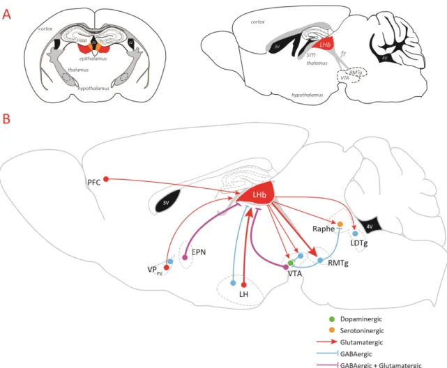
Figure 5: Lateral habenula anatomy: a hub between forebrain structures and neuromodulatory nuclei.
A. Coronal (left) and sagittal (right) section representing the habenula, epithalamic region that can be subdivided into two differents nuclei the medial habenula (in orange, MHb) and the lateral habenula (in red, LHb). This complex, located beneath the hippocampus(Hipp), close to the third ventricle (3V) receives input via the stria medullaris (sm) and in turn, sends efferents to the midbrain through the fasciculus retroflexus (fr).
B. Optogenetic studies reported that lateral habenula receives glutamatergic input from the prefrontal cortex (PFC), the lateral hypothalamus (LH) and the parvalbumin neurons from the ventral pallidum (VPPV) and mixed GABAergic/Glutamatergic input from the entopeduncular nucleus (EPN) and the ventral tegmental area (VTA). Nevertheless, GABAergic current from VPPV and the LH have also been described.
In turn, LHb mainly contains long-range projecting glutamatergic neurons. It sends output in the midbrain controlling directly the monoaminergic nuclei (DA of the VTA and 5-HT neurons of the dorsal raphe) and indirectly through the activation of GABAergic relays (RMTg or GABAergic interneurons). Moreover, LHb has also been described to control GABAergic neurons in the LDT.