HAL Id: tel-03168164
https://tel.archives-ouvertes.fr/tel-03168164
Submitted on 12 Mar 2021HAL is a multi-disciplinary open access archive for the deposit and dissemination of sci-entific research documents, whether they are pub-lished or not. The documents may come from teaching and research institutions in France or abroad, or from public or private research centers.
L’archive ouverte pluridisciplinaire HAL, est destinée au dépôt et à la diffusion de documents scientifiques de niveau recherche, publiés ou non, émanant des établissements d’enseignement et de recherche français ou étrangers, des laboratoires publics ou privés.
neuroanatomy via a domain-specific language
Antonia Machlouzarides-Shalit
To cite this version:
Antonia Machlouzarides-Shalit. Development of subject-specific representations of neuroanatomy via a domain-specific language. Cognitive Sciences. Université Paris-Saclay, 2020. English. �NNT : 2020UPASG041�. �tel-03168164�
Thè
se de
doctorat
NNT : 2020UP ASG041Development of subject-specific
representations of
neuroanatomy via a
domain-specific language
Thèse de doctorat de l’université Paris-Saclay
École doctorale n◦ 580, Sciences et technologies de
l’information et de la communication (STIC) Spécialité de doctorat: Informatique
Unité de recherche: Université Paris-Saclay, Inria, Inria Saclay-Île-de-France, 91120, Palaiseau, France. Référent: faculté des sciences d’Orsay
Thèse présentée et soutenue en visioconférence totale, le 15-12-2020, par
Antonia Machlouzarides-Shalit
Composition du jury:
Olivier Coulon Président
Directeur de Recherche, Aix-Marseille Université
Roberto Toro Rapporteur et Examinateur
Directeur de Recherche, Institut Pasteur
Michel Dojat Rapporteur et Examinateur
Directeur de Recherche, Grenoble Institute des Neuro-sciences
Stephanie Forkel Examinatrice
Postdoc avancé/jeune chercheur, Université de Bordeaux
Philippe Ciuciu Examinateur
Chercheur expert senior, CEA, Université Paris-Saclay
Demian Wassermann Directeur de thèse
The field of brain mapping is rapidly expanding, providing further insights into the spatial organisation of the brain and the relationships of cortical features. The development of brain mapping tools is dependent on the profound grasp of the anatomy of the brain. We identified the need for the development of a brain mapping tool which is grounded in the detailed knowledge of individual sulci. In this thesis, we develop a new brain mapping tool called NeuroLang, which utilises the spatial geometry of the brain to demonstrate subject-specific representations of sulcal morphology.
We approach this challenge with two perspectives: firstly, we situate our theory firmly in the comprehensive characterisations of sulci from the literature, or ’classical neuroanatomy’. We survey the wealth of infor-mation on neuroanatomical characterisation, consider the organisation of cortical features, and examine brain-mapping techniques in neuroimaging and their capabilities. Secondly, we design and implement methods for sulcus-specific queries in our domain-specific language, NeuroLang. We lay out the theoretical bases for NeuroLang in terms of the representation of the brain as a 3D object and of the foundation of a domain-specific language grounded in first-order logic. We test our method on 52 subjects of the Human Connectome Project dataset and evaluate the performance of NeuroLang for population and subject-specific representations of neu-roanatomy. Then, we interpret the quantification of the results of sulcal identification as an evaluation of sulcal stability, and establish a novel, data-driven hierarchical organisation of sulci.
Finally, we review the relevance of using sulci as cortical markers, and the implication of a subject-specific sulcal mapping tool for representing cortical anatomy. We relate the significance of sulcal patterns as associative measures to other cortical features such as cytoarchitectonics. We examine the implications of NeuroLang, its applications and its limitations. To conclude, we summarise the implication of our method within the current field, as well as our overall contribution to the field of brain mapping. Our work paves the way for future development of neuroimaging tools which prioritise individual variability and neuroanatomical inference.
A C K N O W L E D G M E N T S
This work would not be possible without the invaluable contributions of my supervisor, Demian Wassermann. I thank him for his endless guidance, patience, and availability. I strive to improve skills I have learned from him, both directly and indirectly.
I thank the members of the jury: Roberto Toro, Michel Dojat, Olivier Coulon, Stephanie Forkel, and Philippe Ciuciu for taking the time to re-view this thesis and for their supportive and constructive comments. I extend my gratitude to our collaborator, Nikos Makris at Harvard Uni-versity, for insightful conversations and sharing the complex and beautiful world of classical neuroanatomy with me.
Science is a collaborative journey, and I thank all the members of the Parietal team at Inria Paris-Saclay and the Athena team at Inria Sophia-Antipolis for their help and friendship these past few years.
I owe a lifetime of gratitude to my parents, my family and my friends. Finally, I thank ANR NeuroRef for funding my thesis.
1 overview 1
2 résumé 3
I neuroanatomy in neuroimaging analyses
3 the study of neuroanatomy 6
3.1 The cortex 6
3.2 Literature review of sulci 10 3.3 Organisation of sulci 23
4 brain mapping in neuroimaging 27
4.1 Imaging the brain 27
5 first order logic 36
6 hypothesis of this work 43
II design and implementation of neurolang
7 development of neurolang 46
7.1 First Order Logic for the Construction of Queries 46 7.2 Built-in predicates in NeuroLang 54
7.3 NeuroLang queries 56
8 manual segmentations 66
8.1 Method for manual segmentations 66
8.2 Sulcal Atlas as a Source for Anatomical Labelling 67 8.3 Validation for NeuroLang queries 69
9 outcomes of neurolang experiments 73
9.1 NeuroLang on Mindboggle-extracted sulci 73 9.2 NeuroLang on manually segmented sulci 75 9.3 Sulcal patterns 76
9.4 Stability of sulci 83
9.5 Conclusion 91
III implications of sulcal mapping using queries
10 contribution to sulcal mapping 93
11 significance of sulcal representations 95
11.1 Cortical folding patterns 95 11.2 Sulci and Cytoarchitectonics 96
12 implications of neurolang 99
IV conclusion
13 conclusion 101
contents v 13.1 Limitations 101 13.2 Further work 101 13.3 Data sharing 103 13.4 Final note 103 references references 105 appendices a sulci in neurolang 122
a.1 Sulci with their Destrieux counterparts 122
a.2 Sulci examples with their queries in first-order logic 123 a.3 Manual segmentations in Blender 140
1
O V E R V I E W
The development of brain mapping tools is grounded in the extensive grasp of the anatomy of the brain. The ability for brain mapping tools to accurately and efficiently capture the individual variability of neu-roanatomical morphology, while remaining relevant for every brain, is a particularly difficult balance to strike. Typically, the most reliable way of conserving detailed individual morphology of sulci is through manual labelling, which is tedious, time-consuming work which requires exten-sive knowledge of classical neuroanatomy. This body of knowledge is also notoriously inconsistent in terms in naming and morphology, and therefore could manifest as inconsistencies depending on who is doing the labelling and according to which framework. To combat this, extensive work has been carried out on template-based brain mapping methods, which focus on the identification of landmarks relative to which a prede-termined set of brain regions are superimposed. We identified the need for the development of a brain mapping tool which is grounded in the detailed knowledge of individual variability of sulci.
In this thesis, we develop a new brain mapping tool called NeuroLang, which utilises the spatial geometry of the brain to demonstrate subject-specific representations of sulcal morphology. We approach this chal-lenge with two perspectives: firstly, we ground our theory firmly in the comprehensive characterisations of sulci from the literature, or ’classical neuroanatomy’. Secondly, we design and implement methods for sulcus-specific queries in the domain-sulcus-specific language NeuroLang. We evaluate the performance of NeuroLang for population and subject-specific repre-sentations of neuroanatomy. From our results, we determine a data-driven hierarchical organisation of sulci. In this chapter, we outline the structure of this manuscript.
In the introductory part, we survey the wealth of information on neu-roanatomical characterisation, consider the organisation of cortical features, and examine brain-mapping techniques in neuroimaging and their capa-bilities. We establish a literature-based hierarchical organisation of the sulci which provide the framework for our sulcal atlas, and establish the rules of spatial geometry and first-order logic which act as a framework for NeuroLang.
In the next part, we present our work on the design and implementation of NeuroLang. Firstly, we present our collaborative work on administering anatomical names to images in a dictionary of functional modes. Next,
overview 2
we demonstrate how we design and construct sulcus-specific queries and evaluate their performances. We present the results of our experiment using NeuroLang. We demonstrate how we validate the sensitivity of our results, the results on the population level, and subject-specific outcomes. Lastly, we interpret the quantification of the results of sulcal identification as an evaluation of sulcal stability, and determine a novel, data-driven method for the hierarchical organisation of sulci.
Finally, we review and discuss the relevance of using sulci as cortical markers, and the implication of a subject-specific sulcal mapping tool for representing cortical anatomy. We relate the significance of sulcal patterns as associative measures to other cortical features such as cytoarchitecton-ics. We examine the implications of NeuroLang, its applications and its limitations.
To conclude, we summarise the implication of our method within the current field, as well as our overall contribution to the field of brain map-ping. Our work paves the way for future development of neuroimaging tools which prioritise individual variability and neuroanatomical inference.
2
R É S U M É
La cartographie cérébrale est un domaine actif de recherche, en croissance rapide, apportant de plus en plus de données sur l’organisation spatiale du cerveau. Le développement des outils de cartographie cérébrale repose sur une compréhension approfondie de l’anatomie du cerveau. De fait, il est essentiel de comprendre les caractéristiques corticales communes d’une population, ainsi que les sources et degrés de variabilité qui contribuent aux spécificités individuelles. En règle générale, le moyen le plus fiable de conserver la morphologie individuelle détaillée des sulci est l’étiquetage manuel. Ce travail fastidieux et chronophage nécessite une connaissance approfondie de la neuroanatomie classique. De plus, cet ensemble de connaissances est notoirement incohérent en termes de dénomination et de morphologie. Ainsi, l’étiquetage peut dépendre de son auteur et de son cadre d’utilisation. Pour lutter contre cela, des travaux approfondis ont été menés sur des méthodes de cartographie cérébrale basées sur des modèles, qui se concentrent sur l’identification de points de repère par rapport auxquels un ensemble prédéterminé de régions cérébrales se superpose. Nous avons identifié la nécessité de développer un outil de cartographie cérébrale qui repose sur la connaissance détaillée de la variabilité individuelle des sulci.
Dans cette thèse, nous développons un nouvel outil de cartogra-phie cérébrale: le language NeuroLang, reposant sur les spécificités géométriques individuelles des sulci et la logique du premier ordre. Nous avons abordé cette question avec deux approches. Premièrement, nous ancrons fermement notre théorie dans la caractérisation complète des sulci de la littérature en neuroanatomie classique. Deuxièmement, nous concevons et implémentons des composants et des requêtes spécifiques au sulcus dans le langage NeuroLang. Nous avons évalué les performances de NeuroLang pour les représentations de la neuroanatomie spécifiques à la population et au sujet. Dans ce chapitre, nous décrivons la structure de ce manuscrit.
Dans la partie introductive, nous passons en revue la richesse des infor-mations sur la caractérisation neuroanatomique, considérons l’organisation des caractéristiques corticales et examinons les techniques actuelles de cartographie cérébrale en neuroimagerie et leurs capacités. Nous établis-sons une organisation hiérarchique des sulci basée sur la littérature qui a fourni le cadre de notre atlas sulcal, et établissons des règles de géométrie
résumé 4
spatiale et de logique de premier ordre qui agissent comme un cadre pour NeuroLang.
Dans la partie suivante, nous présentons nos travaux sur la conception et la mise en œuvre de NeuroLang. Premièrement, nous présentons notre travail collaboratif sur l’attribution de noms anatomiques aux images dans un dictionnaire de modes fonctionnels.Ensuite, nous montrons comment nous concevons et construisons des requêtes spécifiques aux sulcus et évaluons leurs performances. Nous présentons les résultats de notre ex-périence utilisant NeuroLang. Nous démontrons comment nous validons la sensibilité de nos résultats, les résultats au niveau de la population et les résultats spécifiques à la matière. Enfin, nous utilisons la quantification des résultats de l’identification des sulci comme un critère d’évaluation de stabilité, et établissons une nouvelle méthode guidée par les données pour l’organisation hiérarchique des sulci.
Enfin, nous passons en revue et discutons de la pertinence de l’utilisation des sulci comme marqueurs corticaux, et de l’implication d’un outil de cartographie sulcal spécifique au sujet pour représenter l’anatomie corti-cale. Nous relions l’importance des modèles sulcaux en tant que mesures associatives à d’autres caractéristiques corticales telles que la cytoarchitec-tonique. Nous examinons les implications de NeuroLang, ses applications et ses limites.
Pour conclure, nous résumons l’implication de notre travail dans le do-maine, ainsi que notre contribution globale au domaine de la cartographie cérébrale. Nos travaux ouvrent la voie au développement futur d’outils de neuroimagerie qui donnent la priorité à la variabilité individuelle et à l’inférence neuroanatomique.
N E U R O A N A T O M Y I N N E U R O I M A G I N G
A N A LY S E S
3
T H E S T U D Y O F
N E U R O A N A T O M Y
Form and function of the human brain are inextricably linked. An open and ongoing question in neuroscience is which is more dependent on the other. When it comes to deciphering the actions and outputs of the brain, it is vital to be able to locate oneself within its space; not only to give an output a dimension, but to associate a location with other brain characteristics such as structural relationships and connectivity with other regions.
3.1
the cortex
The brain is composed of white matter and grey matter. White matter is composed of the axons of neurons, which connect different brain regions to each other. They appear white due to the fatty myelin sheaths surrounding them as insulation for better conduction of electrical signals. Grey matter is largely comprised of the rest of the neuronal cell parts, the somas, dendrites and terminal endings. These appear grey because they do not have myelin (Catani and Schotten,2013).
The cerebral cortex is the outermost layer of the brain. A very thin layer of about only 2.5mm, it is composed primarily of grey matter (Heuer and Toro,2019). The cortex (plural, cortices) is necessary for carrying out numerous functions and actions, both directly and indirectly. Constituting the majority of the corticocortical connection terminals, the arrangement of the cortex is imperative to the deduction of structural and functional connectivity in neuroimaging analyses (Toro, Fox, and Paus,2008). Across mammalian species, the surface area of the cortex varies greatly and the variable folding patterns reflect this property (Heuer et al., 2019; Heuer and Toro,2019). During the course of human evolution, human brain size has remained relatively stable while the lobes expanded and connectivity increased (Rilling,2014). As a compensatory mechanism, the human cortex is among the most highly folded from all the species (Heuer et al.,2019), with a vast collection of sulcal labels with varying stability.
The types of cortices which exist are dependent on the organisation of the cell types which make them up, and the neocortex is the largest one (Kennedy et al.,1998). The cortex is made up of outer, concave protru-sions called gyri (single, gyrus) and inner, convex indentations, called sulci
(single, sulcus). We can think of the cortex of the brain as a 2D surface if we unfold and flatten this outer layer of elevations and valleys, much like flattening a tablecloth (Fischl, Liu, and Dale,2001; Lohmann, Von Cramon, and Colchester,2008). The cortex is so highly convoluted that 2/3 of the surface of the brain is actually hidden within the sulci (Van Essen and Drury,1997; Zilles et al.,1988). This folding process begins during gesta-tion and only slows significantly during young adulthood (Chi, Dooling, and Gilles, 1977). There is a general folding pattern which serves as a draft for the overlap of sulcal arrangements in the general population. This is because the first sulci to develop during gestation and infancy are among the deepest sulci and are the most stable and reliable in their location, depth and shape (Chi, Dooling, and Gilles,1977; Pizzagalli et al., 2019). These sulci are dictated chiefly by genetics and driven by earlier mechanical movements and are thus less susceptible to change over the course of neurodevelopment (Chi, Dooling, and Gilles,1977; Heuer and Toro, 2019; Pizzagalli et al.,2019).
the history of neuroanatomy For centuries brains were analysed
only ex vivo (Catani and Schotten, 2013). Gross neuroanatomy, on the macroscale, consisted of identification and labelling of brain regions visible to the naked eye. Soon, patterns began emerging of visible landmarks which delineated the brain into functional territories defined as lobes. Anatomists used these landmarks, soon to be characterised as primary sulci, to orientate themselves in the topography of the brain (Auzias et al.,2013). From these landmarks, relations implying relativity to the primary landmarks provided a method for characterising the remaining gyri and sulci. The directional terms developed in classical neuroanatomy refer to relative location in space without a precise coordinate system, much like north, south, east, west in cartography. Due to the lack of systematisation, there were eventually many terms to designate the same relation, such as anterior/rostral, posterior/caudal, superior/dorsal, and inferior/ventral (Catani and Schotten,2013). The English and Latin terms designate the same relationship in the central nervous system, but the Latin terms do not necessarily correspond to the same relationship when in reference to the body. For this reason and to conserve simplicity, we henceforth utilise only the English directional terms.
3.1.1 Importance of sulci in neuroscience
Sulci form landmarks on the cortex visible to the naked eye. Descriptive anatomy provides names for sulci based on their location on the cortex (Catani and Schotten, 2013). The classification of sulci is based on charac-teristics including location, depth and relationship to other brain regions
3.1 the cortex 8
(Duvernoy, 1999; Ono, Kubik, and Abernathey, 1990; Rademacher et al., 1992).
Primary sulci are among the first to develop during gestation and are the least variable across individuals, due largely to their dependence on genetics and their relationship to primary functional areas (Duvernoy,1999; Ono, Kubik, and Abernathey,1990). The further away a region is from primary functional areas, the more variable and unreliable the morphology of a region becomes due to the integrative nature of the functional area and the dependence of the region’s development on environmental factors, as well as genetic (Rademacher et al.,1993). This ’distance’ of a region can be both functional and physical. As an example, Margulies et al. (2016) showed how gradients of functionality are evenly dispersed across the cortex, where ’hubs’ of the default mode network are found at equidistant points from the primary functional areas.
Though still not fully understood, the relevance of cortical folding to other aspects of neurobiology are becoming increasingly clear with the evolution of advanced brain imaging techniques. It is postulated that folding patterns give rise to connectivity patterns (Toro, Fox, and Paus, 2008; Van Essen,1997), act as cytoarchitectonic markers and boundaries (Amunts et al., 1999; Eickhoff et al., 2005b; Kujovic et al., 2013; Vogt et al., 1995), and correlate with functional regions (Amunts et al., 2004; Eichert et al., 2020; Sprung-Much and Petrides, 2020). Variability exists for all sulci, to varying degrees. A number of factors contribute to the morphology of sulci, with varying ratios. Typically, the first sulci to form during foetal life are landmarks in cortical development and anatomy, and are considered by many as ’primary sulci.’ These sulci exhibit certain characteristics which make them good contenders as cortical landmarks for atlases. The following characteristics are favourable in primary sulci and thus make them excellent landmarks and reference points in atlases:
• Location:Primary sulci are used to chart the lobes of the hemispheres into frontal, temporal, parietal and occipital. Their locations do not fluctuate enough to not be discernible by eye as lobe delineators. They are considered as the most stable and reliable sulci of the brain in terms of their location (Duvernoy,1999).
• Depth: The first sulci to develop during gestation are among the deepest across the cortex (Chi, Dooling, and Gilles,1977; Ono, Kubik, and Abernathey,1990). With advanced neuroimaging techniques, it is common to ’inflate’ the cortex, to make deeper sulci clearer to distinguish and thus view lobular differences more clearly. The pri-mary sulci retain their places on the cortex even when very inflated, acting as lobular partitions, while the surrounding shallower sulci
which are more susceptible to inter-individual differences are mostly smoothed out (Fischl, Sereno, and Dale,1999).
• Shape: Most of the surface of a sulcus is hidden within itself, in the folds. On the surface, the fold creates a line shape which is distinguishable by eye. Beneath the surface, the sulcus may have a different shape, branching, or connections with surrounding sulci. Ono, Kubik, and Abernathey (1990) have extensively catalogued the various morphologies of sulci on the surface, while more advanced techniques have been used to model the shape of primary sulci within the folds (Cykowski et al.,2008). The reliable depth of primary sulci also suggests that there is less variability of the inner sulcus shape.
• Relationships to surrounding sulci: Supposing that primary sulci are used as primary localisers for cortical navigation, the cartogra-phy of the brain is charted relative to these landmarks. Other sulci typically develop later and are shallower or more irregular, which generates larger variability in their morphologies. Despite this, what remains stable is their relative spatial location to the primary sulci. Regardless of their absolute location, size and shape, their charac-terisation in relation to the primary sulci remains unchanged, and a defining feature of a sulcus.
• Susceptibility to influences:All sulci are subject to genetic, environ-mental, and mechanical influences to varying degrees. The ratio of influence is different for sulci which appear at different stages of life and at different locations on the cortex. The earliest appearances of evidence of cortical folding begins in the first trimester of gestation, but formed sulci do not appear until the last trimester (Chi, Dooling, and Gilles,1977). During brain growth, the cortical folding patterns are also susceptible to the mechanical properties of the brain; that is, the constrained volume and shape (Heuer and Toro,2019). The order in which the sulci form affects how susceptible the formation of the sulcus is to mechanical influences on morphogenesis. This susceptibility is another element which factors in to how stable and reliable the sulcus is on the cortex.
• Existence in all healthy brains:There exists a group of sulci which are always localised in humans. Across numerous literature sources, a group of about 20 sulci on the neocortex are consistently reported. They always include the interlobular, ’primary’ sulci, and include a subset of all the sulci found in each lobe. Typically, the sulci described in each lobe are the most continuous, deepest, and reliable in terms of their location. The most ’stable’ sulci are an embodiment of the properties mentioned here. A representation of the hierarchical
3.2 literature review of sulci 10
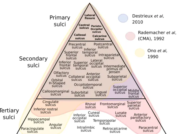
organisation of sulcal stability in 3 literature sources is depicted in Figure 3.1.
3.1.1.1 Importance of sulci in cytoarchitectonics
Sulci have long been used as visual cues to delineate various boundaries on the cortex. Those boundaries may be defined functionally, anatomically, cytoarchitectonically or myeloarchitectonically (Catani and Schotten,2013; Lohmann, Von Cramon, and Colchester,2008). Cytoarchitecture tells us a lot about how the composition of the layers of the cortex are structured. The cortex is composed of 6 layers, with varying cell types which dictate their functions (Amunts, Schleicher, and Zilles,2007; Brodmann,2007; Fischl et al., 2008). The cell types and their arrangements of their layers make up a large portion of the microstructural organisation of the brain. The functional anatomy of the brain is closely linked to the structural anatomy (Lohmann, Von Cramon, and Colchester, 2008). The microstructure, in turn, is closely linked to the functional organisation and connectivity of brain regions (Amunts et al.,2020).
It has been established that sulci can act as borders for cytoarchitectonic areas or functional territories (Rademacher et al.,1992), but the concept is still debated (Fischl et al.,2008). However, it is difficult to determine the cytoarchitectonic organisation of the cortex without invasive meth-ods. Probabilistic atlases, like the JulichBrain Cytoarchitectonic Atlas help (Amunts et al., 2020), but the identification of cytoarchitectonic regions of interest still heavily relies on the macroanatomical features (Amunts, Schleicher, and Zilles,2007; Auzias, Coulon, and Brovelli, 2016). Mapping the macroanatomical features on the individual level may complement atlases for cytoarchitectonics. But first, it’s important to ensure a compre-hensive understanding of sulcal morphology and their relationships to other cortical features.
3.2
literature review of sulci
A comprehensive overview of the anatomical traits of each sulcus is necessary in order to construct queries which are loyal to their classical neuroanatomy descriptions. Prior to designing the queries which search for sulci, or even the descriptors or relationships for the basis of our tool, we must gather information on sulcal characterisations from the literature. In this section, we present our extensive literature review of each sulcus included in Figure 3.1. We cover sulcal position on the cortex, develop-ment, relationships to surrounding sulci, segmentation, branching, and relationships to cytoarchitectonic areas, where relevant.
We refer the reader more interested in the application of this review to our organisation of the cortex to the next section, Organisation of Sulci, 3.3.
3.2.1 Primary sulci
lateral fissure The lateral, or Sylvian, fissure is the most characteris-tic, constant feature of the lateral surface, and is therefore regarded as the most reliable anatomical landmark (Ono, Kubik, and Abernathey,1990; Rademacher et al.,1992). It is distinctively one of the most deep fissures of the entire brain, and the first to appear in each hemisphere during gestation, at 14-19 weeks (Chi, Dooling, and Gilles, 1977; Ono, Kubik, and Abernathey,1990). It has a general longitudinal trajectory, coursing from the frontal opercula to the superior parietal lobule, acting as the main demarcator between the frontal, parietal and temporal lobes, the insula and the opercula (Catani and Schotten,2013; Destrieux et al.,2010). It exists as a main fissure with numerous extensions and side branches, many of which possess reliable appearances and have thus been charac-terised and others which have contrasting nomenclatures and appearances and are broadly described as generic side-branches (Ono, Kubik, and Abernathey, 1990; Rademacher et al., 1992). Anatomists have generally divided the lateral fissure between three and 10 parts (Destrieux et al., 2010; Ono, Kubik, and Abernathey, 1990; Rademacher et al., 1992). The posterior horizontal ramus is the main trunk of the fissure, and the deepest segment. It runs posteriorly and terminates posterior to the postcentral sulcus, between the parietal and temporal lobes (Rademacher et al.,1992). Here, the fissure may extend superiorly or inferiorly at an angle, and may possess side branches such as the terminal ascending segment (Ono, Kubik, and Abernathey,1990; Rademacher et al., 1992).
anterior ascending and horizontal rami of the lateral fissure
The anterior projections of the lateral fissure are the anterior ascending, anterior horizontal and sometimes the diagonal rami of the lateral fissure (Catani and Schotten, 2013; Destrieux et al.,2010; Ono, Kubik, and Aber-nathey, 1990). These project superiorly and supero-anteriorly from the lateral fissure into the inferior frontal gyrus of the frontal lobe (Catani and Schotten, 2013; Destrieux et al., 2010; Duvernoy,1999; Ono, Kubik, and Abernathey,1990; Rademacher et al.,1992). They may connect with the surrounding sulci, the inferior frontal sulcus or the inferior segment of the precentral sulcus (Ono, Kubik, and Abernathey,1990). Their locations are consistent as being anterior of the inferior portion of the precentral sulcus and inferior of the inferior frontal sulcus. The rami themselves act as boundaries to delineate the frontal operculum subdivisions into the
3.2 literature review of sulci 12
posterior pars opercularis, the middle pars triangularis and the anterior pars orbitalis (Brodmann, 2007; Rademacher et al., 1992). The positioning of these rami is fundamental in the placement of Brodmann areas 44 and 45, constituting Broca’s area (Amunts et al.,1999).
central sulcus The central sulcus, or the fissure of Rolando, is a
defining landmark on the lateral surface of the brain. Extending from the superior margin to the lateral fissure, it courses antero-ventrally in a continuous manner (Ono, Kubik, and Abernathey,1990; Rademacher et al., 1992). It ends near the superior aspect of the circular sulcus of the insula, where it often, but not always, meets the lateral fissure, where the frontal, temporal and parietal lobes converge like tectonic plates (Destrieux et al., 2010; Duvernoy,1999). It often, but not always, extends onto the medial surface (Duvernoy,1999; Ono, Kubik, and Abernathey,1990; Rademacher et al.,1992). The central sulcus is among the first sulci to develop in the em-bryonic brain, usually between 4 and 6 months/20-23 weeks of gestation (Chi, Dooling, and Gilles,1977; Cykowski et al.,2008; Toi, Lister, and Fong, 2004). Chi, Dooling, and Gilles, 1977observed its initial development first in the right hemisphere, followed by its appearance in the left hemisphere approximately a week later. Its early appearance in the foetal brain reflects its characteristic depth and continuity. Its depth may present asymmetri-cally according to various features, such as gender, age (Cykowski et al., 2008), handedness (Amunts et al.,2000) or musical abilities (Zilles et al., 1997). The central sulcus acts as a landmark to distinguish the frontal from the parietal lobes, as well as a cytoarchitectonic boundary between the gi-ant pyramidal cells without an inner granular layer of the precentral gyrus (Brodmann area 4), and the apyramidal granular layer of the postcentral gyrus (Brodmann areas 3, 1, 2) (Brodmann,2007; Pandya et al.,2015). parieto-occipital sulcus The parieto-occipital sulcus is one of deep-est sulci and one of the landmark sulci of the medial surface (Rademacher et al.,1992). Chi, Dooling, and Gilles, 1977 observed its appearance at the 16th gestational week. On the medial surface, the sulcus itself separates the parietal from the occipital lobes, and on the lateral surface, to which it almost always extends onto, its hypothetical line to the pre-occipital notch separates the occipital lobes from the parietal and temporal lobes (Catani and Schotten,2013; Duvernoy,1999; Ono, Kubik, and Abernathey,1990). It cleaves the medial surface, starting at the postero-superior junction of the superior hemispheric margin and coursing antero-inferiorly, terminating just inferior of the posterior part of the callosal sulcus (Ono, Kubik, and Abernathey,1990). Oftentimes, this inferior termination meets, or almost meets, the anterior termination of the calcarine sulcus, named the cuneal point (Duvernoy,1999; Ono, Kubik, and Abernathey, 1990; Rademacher
et al., 1992). Here, the parieto-occipital sulcus separates the cingulate gyrus, and thus the limbic lobe, from the cuneus, and the cuneus from the precuneus (Catani and Schotten,2013; Desikan et al.,2006; Margulies et al.,2009).
callosal sulcus A distinctive reference landmark on the medial
sur-face, the callosal sulcus gives reference to the location of the corpus callo-sum, separating it from the outer cingulate gyrus (Catani and Schotten, 2013; Chi, Dooling, and Gilles,1977; Destrieux et al.,2010; Duvernoy,1999). It is among the first sulci to develop during gestation, forming at the same time as the lateral fissure (Chi, Dooling, and Gilles,1977).
calcarine sulcus Among the first distinguishable sulci during
neu-rodevelopment is the calcarine sulcus (Chi, Dooling, and Gilles, 1977). It forms a deep, frequently continuous sulcus which reaches from the occipital pole to just anterior and inferior of the splenium of the corpus callosum (Ono, Kubik, and Abernathey, 1990; Rademacher et al., 1992). This conjunction point is referred to as the cuneal point, where the cal-carine sulcus often meets the parieto-occipital sulcus (Duvernoy,1999; Ono, Kubik, and Abernathey,1990; Rademacher et al.,1992). This is a principal landmark on the medial surface (Rademacher et al.,1992). Brodmann,2007 correlates the calcarine sulcus with the primary visual cortex (Brodmann’s area 17), remarking how much larger this area is than it appears due to the depth of the calcarine sulcus. It has been further distinguished cytoarchitecturally by the stripe of Gennari, a thick strip of horizontal fibres (Pandya et al.,2015).
3.2.2 Frontal lobe
frontomarginal sulcus The frontomarginal sulcus is a short sulcus
located on or near the frontal pole (Duvernoy, 1999). Ono, Kubik, and Abernathey,1990 reports it as mostly continuous (80% right hemisphere, 76% left hemisphere), but sometimes interrupted into two or three seg-ments. Often, it manifests as a continued stem of other frontal lobe sulci, namely the superior, middle or inferior frontal sulci (Duvernoy, 1999; Ono, Kubik, and Abernathey, 1990) . Bludau et al., 2014 describe it as part of the “Fp1” or frontal pole 1, area. Fp1 is on the lateral surface and has cytoarchitectonic features which distinguish it from surrounding and medial areas (Bludau et al.,2014).
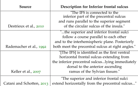
inferior frontal sulcus The inferior frontal sulcus emanates
ante-riorly from the precentral sulcus in a longitudinal manner, mirroring its superior counterpart. It acts as the limit between the middle and inferior
3.2 literature review of sulci 14
frontal gyri (Catani and Schotten,2013; Destrieux et al.,2010; Duvernoy, 1999; Rademacher et al.,1992). The junction with the precentral sulcus is generally perpendicular (Duvernoy,1999; Ono, Kubik, and Abernathey, 1990). The inferior frontal sulcus often has a true connection with the inferior part of the precentral sulcus, though Ono, Kubik, and Abernathey, 1990reported no junction at all in 12% of 50 hemispheres. They also re-ported frequent segmentation and branching of the inferior frontal sulcus, presenting as 2-4 segments in 44% and 60% of right and left hemispheres, respectively, and most commonly 2 upward side branches in the right hemisphere, 3 in the left hemisphere, and 2 downward side branches in the right and left hemispheres (Ono, Kubik, and Abernathey,1990). It can also connect with surrounding sulci, including the middle and superior frontal sulci, frontomarginal sulcus and the rami of the lateral fissure (Ono, Kubik, and Abernathey,1990).
superior frontal sulcus The superior frontal sulcus anteriorly
projects from the precentral sulcus in a longitudinal manner, mirroring its inferior counterpart. It acts as the limit between the superior and middle frontal gyri (Catani and Schotten,2013; Destrieux et al.,2010; Duvernoy, 1999; Rademacher et al.,1992). The junction with the precentral sulcus is generally perpendicular, shown by Ono, Kubik, and Abernathey,1990in 32% and 48% of right and left hemispheres, respectively, and no junction at all in 8% of right hemispheres. They also reported frequent segmentation and branching of the superior frontal sulcus, presenting as 2-4 segments in 60% and 68% of right and left hemispheres, respectively, which some-times overlapped (Ono, Kubik, and Abernathey,1990). Furthermore, they reported many upward and downward side branches of this sulcus. The superior frontal sulcus can also connect with surrounding sulci, including the middle and inferior frontal sulci, and the frontomarginal sulcus (Ono, Kubik, and Abernathey,1990).
middle frontal sulcus The middle, or intermediate, frontal sulcus
is located inferiorly of the superior frontal sulcus and superiorly of the inferior frontal sulcus, coursing through the middle frontal gyrus (De-strieux et al., 2010). Its appearance varies from long and continuous to short and segmented (Destrieux et al.,2010; Ono, Kubik, and Abernathey, 1990). It may be independent or connected to surrounding sulci, including the precentral, superior frontal, inferior frontal and frontomarginal sulci (Catani and Schotten, 2013; Destrieux et al., 2010; Duvernoy, 1999; Ono, Kubik, and Abernathey,1990).
olfactory sulcus The olfactory sulcus is a constant landmark on the
as a shallow depression around 16 weeks of gestation, around the same time as primary sulci such as the parieto-occipital sulcus, and deepens gradually (Chi, Dooling, and Gilles,1977). It has a longitudinal trajectory, parallel and just lateral of the interhemispheric fissure, medial of the orbital sulci, and separates the orbital gyrus, gyrus rectus and olfactory gyrus (Catani and Schotten,2013; Chi, Dooling, and Gilles,1977; Destrieux et al., 2010; Rademacher et al.,1992).
orbital sulci The orbital sulci are a collection of several short sulci located on the ventral surface of the frontal lobe, including what Destrieux et al.,2010 have referred to as the medial and lateral orbital sulcus and what Ono, Kubik, and Abernathey, 1990 and Rademacher et al., 1992 have referred to as the transverse orbital sulcus. Though Ono, Kubik, and Abernathey,1990 have recorded it as always present, its originations and terminations seem arbitrary, as it has been often recorded as a continuation of the frontomarginal, inferior frontal or olfactory sulci (Duvernoy,1999; Ono, Kubik, and Abernathey,1990). The configuration of these short sulci can form an ‘H’ or ‘X’ shape (Catani and Schotten,2013; Destrieux et al., 2010; Ono, Kubik, and Abernathey,1990).
paracentral sulcus The marginal sulcus of the cingulate sulcus
reaches the superior hemispheric fissure, which also acts as the poste-rior boundary of the paracentral lobule (Catani and Schotten,2013). The paracentral sulcus anteriorly separates this lobule from the medial poste-rior supeposte-rior frontal gyrus (Desikan et al., 2006; Destrieux et al., 2010). The paracentral sulcus may arise as various manifestations, including as a superior projection of the cingulate, or an extension from the lateral surface (Catani and Schotten, 2013; Destrieux et al., 2010; Ono, Kubik, and Abernathey,1990). It has been described as not always distinguishable enough (Destrieux et al.,2010).
paracingulate sulcus The paracingulate sulcus presents as an outer – more anterior and more superior – parallel of the cingulate sulcus (Fornito et al., 2006; Paus et al., 1996). First described as a “double parallel” of the cingulate sulcus, first by Weinberg, 1905 and later by Ono, Kubik, and Abernathey, 1990, further investigation has correlated its existence with a relative expansion of the paracingulate cortex (Fornito et al.,2006; Paus et al.,1996). Its existence is infrequent. Ono, Kubik, and Abernathey, 1990reported its existence in 24% of 50 hemispheres, while Paus et al., 1996reported its existence in 37% and 54% in right and left hemispheres, respectively, in 494 hemispheres. Other notable findings of theirs were paracingulate asymmetry skewing towards the left hemisphere, with more prominent appearance, and more regular appearance in males (Fornito
3.2 literature review of sulci 16
et al., 2006; Paus et al., 1996). This asymmetry mirrors earlier findings by Weinberg, 1905, who reported the existence of a “doubling of the cingulate sulcus in its anterior part” in 40% and 84% of right and left hemispheres, respectively.
precentral sulcus The precentral sulcus is a consistent sulcus on the lateral surface of the brain, lying just anterior to the central sulcus and with a roughly parallel, antero-inferior course (Ono, Kubik, and Abernathey, 1990). Superiorly, it crosses over the superior hemispheric margin and is always present on the medial surface (Ono, Kubik, and Abernathey, 1990). It is frequently discontinuous, most often into two parts, generally designated as the superior and inferior segments of the precentral sulcus (Destrieux et al., 2010; Duvernoy, 1999; Germann et al.,2005; Ono, Kubik, and Abernathey,1990).
superior/inferior rostral sulci / suborbital sulcus The
ante-rior projections of the cingulate sulcus in the medial frontal lobe are the superior and inferior rostral sulci. The superior rostral sulcus, always present, originates at either the anterior subcallosal region, anterior cin-gulate sulcus or anterior paracincin-gulate and extends anteriorly towards the frontal pole where it may or may not reach the anterior hemispheric margin (Ono, Kubik, and Abernathey,1990; Paus et al.,1996; Rademacher et al.,1992). Its posterior termination may be continuous with the anterior parolfactory sulcus (Ono, Kubik, and Abernathey, 1990; Spasojevi´c et al., 2011). Additionally, this sulcus was described by Brodmann, 2007as the infero-medial border of Brodmann Area 10. Its inferior counterpart, the inferior rostral sulcus, is less constant (Ono, Kubik, and Abernathey,1990; Rademacher et al.,1992). Ono, Kubik, and Abernathey, 1990showed that while the superior rostral sulcus is continuous, the inferior rostral sulcus can present as either continuous or segmented into multiple sulci (20% of 50 hemispheres), as well as connections with the anterior parolfactory sulcus.
3.2.3 Temporal lobe
collateral sulcus The name collateral sulcus is used interchangeably with medial occipitotemporal sulcus by some (Catani and Schotten,2013; Destrieux et al.,2010; Duvernoy, 1999). This alternative name indicates its position more clearly: as medial of the lateral occipitotemporal sulcus, another landmark sulcus of the ventral surface. The collateral sulcus is frequently identified, but its various branches and connections with the lateral occipitotemporal sulcus make it not easily discernible from the surrounding sulci (Destrieux et al., 2010). The collateral sulcus usually
gives rise to other ventral sulci, including the transverse collateral sulci such as the rhinal and lingual sulci, and the parahippocampal ramus (Destrieux et al., 2010; Duvernoy, 1999; Ono, Kubik, and Abernathey, 1990).
inferior temporal sulcus The inferior temporal sulcus is located on
the lateral surface of the temporal lobe, inferior to the superior temporal sulcus and supero-lateral to the occipitotemporal sulcus, with a general longitudinal trajectory (Catani and Schotten,2013; Destrieux et al.,2010; Rademacher et al.,1992). It acts as the boundary between the middle and inferior frontal gyri (Catani and Schotten,2013; Destrieux et al.,2010). It is a discontinuous sulcus, with reports of between 2 and 7 segments (De-strieux et al.,2010; Duvernoy,1999; Ono, Kubik, and Abernathey,1990). These segments can also have antero-posterior extensions to the temporal tips and parietal and occipital lobes and frequently have connections with surrounding sulci, including the superior temporal sulcus, anterior occip-ital sulcus, lateral occipoccip-ital sulcus, inferior occipoccip-ital sulcus, preoccipoccip-ital notch, occipitotemporal sulcus or intraparietal sulcus (Catani and Schotten, 2013; Destrieux et al.,2010; Duvernoy,1999; Ono, Kubik, and Abernathey, 1990; Rademacher et al.,1992).
occipitotemporal sulcus The occipitotemporal sulcus is a major
landmark of the ventral occipito-temporal lobes (Chau, Stewart, and Grag-naniello,2014). Though its presence is widely regarded as constant, it is characterised as a frequently discontinuous sulcus, extending from the temporal to occipital lobes but without reaching as far anterior as the temporal pole (Chau, Stewart, and Gragnaniello, 2014; Destrieux et al., 2010; Ono, Kubik, and Abernathey,1990). This sulcus is found between the inferior temporal sulcus, laterally, and collateral sulcus, medially, acting as the division between the inferior temporal gyrus, laterally, and the fusiform gyrus, medially (Catani and Schotten,2013; Chau, Stewart, and Gragnaniello, 2014; Destrieux et al., 2010; Ono, Kubik, and Abernathey, 1990). Ono, Kubik, and Abernathey, 1990 describes this sulcus as more discontinuous than continuous, observing up to 4 segments. Notably, they also rarely observed (8% of right hemispheres, 4% of left hemispheres) a “double parallel type”, with segments running parallel to each other, in an overlapping fashion. These segments often have side branches, or connections with surrounding sulci, such as the rhinal sulcus, inferior temporal sulcus or collateral sulcus (Ono, Kubik, and Abernathey,1990).
rhinal sulcus The rhinal sulcus is a short, constant sulcus of the
ventral temporal lobe (Chau, Stewart, and Gragnaniello,2014). Its position is often correlated with that of the collateral sulcus and the anterior
3.2 literature review of sulci 18
boundary of the limbic lobe (Destrieux et al.,2010; Duvernoy,1999; Ono, Kubik, and Abernathey,1990). It is described as a antero-medial sulcus usually projecting from the anterior end of the collateral sulcus, with which it is sometimes continuous–28% by Ono, Kubik, and Abernathey, 1990, 50% by Chau, Stewart, and Gragnaniello,2014–and sometimes overlapping (Ono, Kubik, and Abernathey,1990 documented overlapping in 52% and 40% of right and left hemispheres, respectively).
superior temporal sulcus The superior temporal sulcus is inferior
to the lateral fissure and runs in a parallel course (Duvernoy, 1999). It extends from the temporal pole to the posterior end of the lateral fissure, where it turns superiorly and continues as the angular sulcus. The angular sulcus is sometimes a true continuation of the superior temporal sulcus, and sometimes a segmented branch (Duvernoy,1999). Another horizontal posterior segment can be the anterior occipital sulcus. Frequently pre-senting with such branching, segmentation is more common in the left hemisphere than the right (Ochiai et al., 2004). Asymmetries extend to depth as well; the right superior temporal sulcus is deeper than the left, complementing the earlier appearance by 1 to 2 weeks of the superior temporal sulcus in the right than the left hemispheres during gestation. It has been suggested that this depth asymmetry, since it does not extend to other mammals, is unique to the human cortical development, acting as a “species-specific perisylvian anatomical marker” (Leroy et al.,2015).
temporopolar sulcus The temporopolar sulcus is described
by Catani and Schotten, 2013 as a variable sulcus at the ventral tip of the temporal lobe, separating the fusiform gyrus from the temporal polar region. Destrieux et al.,2010describe the separation of the temporal pole by the convergence of the antero-lateral planum polare, the superior, mid-dle, inferior temporal sulci, anterior collateral sulcus and occipitotemporal sulcus.
3.2.4 Parietal lobe
angular sulcus The angular sulcus is aptly named as a descriptor
of its trajectory beginning from the posterior segment of the superior temporal sulcus, which can be a true continuation of it or an independent segment of it (Duvernoy,1999). It is collectively described as a superiorly-curving posterior branch of the superior temporal sulcus, angling around the posterior part of the lateral fissure, to bisect the angular gyrus (Catani and Schotten,2013; Ono, Kubik, and Abernathey, 1990; Rademacher et al., 1992).
intermediate primus of jensen The intermediate primus of Jensen is a inferiorly-projecting orthogonal sulcus from the intraparietal sul-cus (Destrieux et al., 2010). It is one of many downward-projecting branches of the intraparietal sulcus (Catani and Schotten,2013; Destrieux et al., 2010; Ono, Kubik, and Abernathey, 1990). We characterise the in-termediate primus of Jensen in particular because of its distinction in bisecting the inferior parietal lobule, acting as the border between the supramarginal (anteriorly) and angular (posteriorly) gyri (Destrieux et al., 2010).
intraparietal sulcus A landmark of the lateral surface of the
pari-etal lobe, the intraparipari-etal sulcus courses in a loosely longitudinal manner from the postcentral sulcus to the hemispheric margin, just superior of the lateral notch of the parieto-occipital sulcus (Duvernoy,1999). Its nature is concave, and can be distinguished by up to three segments: an anterior, as-cending segment, which may begin from the base of the postcentral sulcus, the horizontal intermediate segment, which clearly delineates the supe-rior and infesupe-rior parietal gyri, and the postesupe-rior descending route which crosses over into the occipital lobe, continuing as the superior occipital sulcus (Destrieux et al.,2010; Duvernoy,1999; Ono, Kubik, and Abernathey, 1990). It separates the superior and inferior parietal lobules (Catani and Schotten,2013).
callosomarginal sulcus The callosomarginal or simply ’marginal’
sulcus, is consistently defined as the continued posterior segment of the cingulate sulcus which extends superiorly to reach the interhemispheric fissure, just posterior of the superior termination of the central sulcus. At this position, it lies between the often-bifurcated superior termination of the postcentral sulcus (Ono, Kubik, and Abernathey,1990; Rademacher et al.,1992).
postcentral sulcus The postcentral sulcus is located in the parietal lobe. It is the first sulcus posterior to the central sulcus and follows a generally similar antero-inferior trajectory (Rademacher et al., 1992). While the central sulcus is almost always a single continuous sulcus, the postcentral sulcus has been reported as either continuous and segmented into 2 or 3 segments (Destrieux et al.,2010; Ono, Kubik, and Abernathey, 1990). Alternatively, a “double parallel” type has been reported by Ono, Kubik, and Abernathey, 1990, though not commonly. It is frequently connected to surrounding sulci, most notably the intraparietal sulcus, and frequently has side branches projecting into the surrounding gyri (Ono, Kubik, and Abernathey, 1990; Rademacher et al., 1992). A noteworthy characteristic of the postcentral gyrus is its common Y-shaped bifurcation
3.2 literature review of sulci 20
at its superior termination, which may or may not extend over the superior hemispheric margin into the medial surface (Ono, Kubik, and Abernathey, 1990; Rademacher et al.,1992).
subparietal sulcus The subparietal sulcus is a landmark sulcus of
the medial parietal lobe (Ono, Kubik, and Abernathey, 1990). On the medial surface, it is located postero-inferiorly to the posterior end of the cingulate sulcus, from which it is occasionally a continuation of, and limits the precuneus from the cingulate gyrus (Destrieux et al.,2010; Rademacher et al.,1992). It is composed of several branches, and usually appears as an H-shape or Y-shape which extends inferiorly (Destrieux et al.,2010; Ono, Kubik, and Abernathey,1990; Rademacher et al., 1992).
superior parietal sulcus The superior parietal sulcus is located in
the superior parietal lobule, posterior of the postcentral sulcus, anterior of the parieto-occipital sulcus, and superior of the intraparietal sulcus (Ono et al, 1990; Destrieux et al, 2010). It is described as inconstant or supplementary (Ono et al, 1990; Destrieux et al, 2010). It can arise as a branch of the intraparietal sulcus (Destrieux et al.,2010), and reported by Ono et al (1990) as having between 0 and 2 branches.
3.2.5 Occipital lobe
anterior occipital sulcus A rather elusive sulcus, the anterior occip-ital sulcus nonetheless appears in many studies but with varying lobular positions. Some place it as a posterior branch of either the superior tempo-ral sulcus (Ono, Kubik, and Abernathey,1990) or the inferior temporal sul-cus (Duvernoy,1999). Its position is ambiguously located in the area where the temporal, parietal and occipital lobes converge. Some localise it to the parietal lobe (Rademacher et al., 1992), some to the temporal lobe (Ono, Kubik, and Abernathey,1990) and others to the occipital lobe (Duvernoy, 1999).
cuneal sulcus Brodmann,2007 remarks the presence of the cuneal
sulcus when describing it as a landmark for the V1 striate area, though not necessarily a boundary of it. Its existence and location is consistent, though its appearance is not. It is a short, shallow sulcus on the medial surface of the cuneus, wedged between the parieto-occipital and calcarine sulci.
inferior occipital sulcus The inferior occipital sulcus is the sulcus which limits the lateral occipital lobe from the ventral (Destrieux et al., 2010). Its delineation can be arbitrary, and its descriptions are synonymous
with other occipital sulci such as the lateral or transverse occipital sulci (Rademacher et al., 1992).
lateral occipital sulcus The lateral occipital sulcus, sometimes re-ferred to as the middle occipital sulcus or prelunatus sulcus, is a constant sulcus which lies in the arbitrary junction between the occipital, temporal and parietal lobes (Bailey and Urbana,1951; Destrieux et al., 2010; Ono, Kubik, and Abernathey,1990). It acts as the limit between the superior and inferior occipital gyri (Rademacher et al.,1992). It may arise as a posterior projection of the superior temporal sulcus and may be connected to sur-rounding sulci including the anterior occipital sulcus, inferior temporal sulcus or lunate sulcus (Destrieux et al.,2010; Duvernoy,1999).
lingual sulcus The lingual, or intralingual, sulcus is so named
be-cause it courses through the lingual gyrus (Destrieux et al., 2010). It is commonly found and described as a sulcus emerging from the posterior third of the collateral sulcus, reaching postero-medially into the occipital lobe (Chau, Stewart, and Gragnaniello,2014; Destrieux et al.,2010; Ono, Kubik, and Abernathey,1990).
lunate sulcus Though a characteristic feature of the macaque brain,
the lunate sulcus proves rather elusive in the human brain (Martin and Bowden, 2000; Ono, Kubik, and Abernathey, 1990). When present in humans, it is positioned at a much more posterior location, and is much less prominent than in the great ape brain (Allen, Bruss, and Damasio,2006; Brodmann,2007). It is described as a short sulcus on the posterior medial surface of the occipital lobe, near the occipital pole, separating Brodmann areas 17 and 18/primary and secondary visual cortices (Brodmann,2007; Rademacher et al.,1993). Contrary to some other cortical functional areas, the architectonic transition between the primary and secondary visual cortices is abrupt (Brodmann,2007) . Eickhoff, Yeo, and Genon,2018have demonstrated how, at particular locations such as this, cyto- or myelo-architectonic transitions represent the border of different cortical areas. The lunate sulcus may act as this border. Ono, Kubik, and Abernathey,1990 noted its presence only in 40% and 36% of the right and left hemispheres, respectively. Allen, Bruss, and Damasio,2006 reported its variability, with asymmetries within the same brain to variations in shape.
retrocalcarine sulcus The retrocalcarine sulcus is not always
present (Ono, Kubik, and Abernathey, 1990), but when it is it exists as a separate small sulcus posterior of the calcarine sulcus, on the occipital pole (Iaria and Petrides,2007). It can often be included as part of the cal-carine sulcus as posterior ascending or descending rami (Destrieux et al.,
3.2 literature review of sulci 22
2010; Duvernoy, 1999; Iaria and Petrides,2007). Elliot Smith,1902gave emphasis to this sulcus in the posterior occipital cortex for its relevance in comparative anatomy and relationship to the primary visual cortices.
superior occipital sulcus The superior occipital sulcus often arises as a postero-inferior continuation of the intraparietal sulcus almost reach-ing the occipital pole (Catani and Schotten, 2013; Destrieux et al., 2010; Duvernoy,1999).
3.2.6 Limbic lobe
anterior parolfactory sulcus The parolfactory region lies
infe-rior to the genu of the corpus callosum (Catani and Schotten, 2013; Rademacher et al., 1992). Here there exists a sulcus called the anterior parolfactory sulcus, which may not be easily distinguishable from its surrounding sulci because it often exists as a branch or continuation of other medial sulci, including the cingulate or superior and inferior rostral sulci (Ono, Kubik, and Abernathey,1990). It has been postulated that the anterior parolfactory sulcus may act as an anterior border to Brodmann area 25, a region implicated in depression (Hamani et al.,2011).
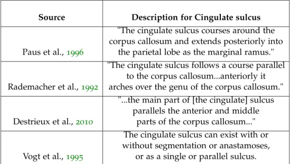
cingulate sulcus The cingulate sulcus is present on the medial
sur-face, separating the frontal lobe from the limbic lobe (Ono, Kubik, and Abernathey, 1990). It is the outer boundary of the cingulate gyrus, separat-ing it from the superior frontal gyrus and paracentral lobule (Catani and Schotten,2013). It begins in the subcallosal area and follows the course of the pericallosal sulcus, in a concentric manner, following the trajectory of the corpus callosum (Duvernoy, 1999). Posterior to the central and paracentral sulci, it curves superiorly, turning into the marginal sulcus, and terminates at the superior hemispheric margin, corresponding to the position of the postcentral sulcus on the lateral surface (Duvernoy, 1999). On the lateral surface, oftentimes the postcentral sulcus bifurcates just prior to its termination at the superior hemispheric margin. When this occurs, the termination of the marginal sulcus usually lies between these postcentral terminal bifurcations (Ono, Kubik, and Abernathey,1990; Rademacher et al.,1992).
hippocampal sulcus The hippocampal sulcus is located on the edge
of the limbic lobe, separating the hippocampus from the parahippocampal gyrus on the ventral temporal lobes (Duvernoy, 2005; Duvernoy, 1988; Rademacher et al.,1992). It can be seen from the ventral surface, medial to the collateral sulcus and with a similar, longitudinal trajectory but more convex (Ono, Kubik, and Abernathey,1990).
intralimbic sulcus The intralimbic sulcus, rarely present nor promi-nent, exists within the intralimbic gyrus, surrounding the Callosal sulcus (Destrieux et al.,2010; Duvernoy,1999; Paus et al.,1996)
3.3
organisation of sulci
In section 3.2, we covered the morphology of individual sulci as reviewed in numerous classical neuroanatomy sources. From a literature standpoint, we ascertain degrees of variability or instability of a sulcus. We gather this knowledge and infer the organisation of the cortex using sulci.
There exists a set of sulci in addition to the primary sulci which can be expected to be identified in all healthy brains, and have overlapping definitions in many neuroanatomy sources. These are referred to as the ’secondary’ sulci, or, more ambiguously, the ’major’ sulci. Folding patterns during neurodevelopment follow basic trajectories which form a rudimen-tary pattern for the ontogenesis of sulci and gyri on the macroscale. The folding patterns govern the cortical organisation, and the development of the primary sulci influence subsequent sulcal developments (Ono, Kubik, and Abernathey,1990; Toro and Burnod,2003).
We compiled sources of sulcal characterisation dating back centuries until present day, which included a vast array of anatomical techniques for identification and labelling. We identified a large proportion of overlap among the sources as well as an equally large proportion of inconsistencies in the identification of sulci and their namings. In many cases, sulci had multiple names across sources but the characterisation was the same. In the case of multiple names, we chose to include the name which is most reflective of its structural anatomy, as opposed to its functional relationship.
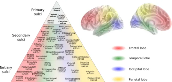
We present the NeuroLang atlas which has a universal set of 42 sulci. The schematic presented in Figure3.2is a representation of the accumulation of sulcal characterisations in a form which reflects the pattern of organisation of the sulci on the cortex in the literature. The positions of the sulci within each lobe are characterised in relation to the primary sulci, but are not dependent on sulci within other lobes. For this reason we distinguish the lobe to which the sulcus belongs in addition to assigning it a position on the hierarchical organisation. This representation suggests that there exists a gradient of stability within each lobe, in relation to the primary sulci. Therefore, the group of the ’secondary’ sulci have a vertical relationship to the ’tertiary’ sulci, but there are also 4 vertical relationships within the group of the secondary sulci, within each lobe. From the order in which sulci are identified per lobe, we derive a semi-hierarchical organisation of secondary and tertiary sulci, and this is what we depict in Figure 3.2.
3.3 organisation of sulci 24
Figure 3.1: Schematic depicting the overlap and disagreement in the sulci of 3 gold-standard sulcal atlases (Destrieux et al.,2010; Ono, Kubik, and Abernathey, 1990; Rademacher et al.,1992). The primary sulci are interlobular markers (except the calcarine) and are the most stable in their morphology and location. The secondary sulci were chosen are those represented independently in the Destrieux atlas, and the tertiary were included in at least one of the sources, or existed in the Destrieux atlas as sulci merged with their surrounding gyri. Tertiary sulci are more variable in their existence, morphology, location, relationships to other sulci, and relationships with other brain features.
Figure 3.2: Schematic depicting our semi-hierarchical organisation of the uni-versal set of sulci in NeuroLang. The position of the sulcus in the pyramid corresponds to its lobular location. The semi-hierarchical organisation signifies how the identification of a sulcus within a lobe may or may not be dependent on a previous identification of a different sulcus within the same lobe, but not necessarily on the identification of a sulcus in a different lobe, even if they are in the same tier or above. The primary sulci were manually selected, the secondary sulci are those included as independent regions in the Destrieux atlas template and could be verified with their Destrieux atlas counterparts, as we show in Appendix A.1 (Destrieux et al.,2010). The tertiary sulci comprise the remaining sulci, including those with irregular existence, morphology or location.
3.3 organisation of sulci 26
This semi-hierarchical organisation of sulci reflects the way in which classical neuroanatomists would navigate their way across the cortex in localising decreasingly reliable sulci. It therefore reflects the intuitive methodology for sulcal localisation based on the spatial geometry of the surface. It is this property of classical neuroanatomy descriptions we use in this work to devise a method for progressively localising cortical sulci. However, our literature-based semi-hierarchical ordering proves that characterisations of the sulci and sulcal reliability are quite arbitrary. To combat this, we would need our own set of ’ground truth’ neuroanatomy in which to base our own sulcal neuroanatomy localisation on and for the validation of our sulcal mapping queries in NeuroLang. While automated identification of sulci is a necessary step for the investigation of large populations, ’tedious anatomy’ provides the necessary accuracy and inter-individuality, a case neatly put forth by Devlin and Poldrack,2007.
4
B R A I N M A P P I N G I N
N E U R O I M A G I N G
4.1
imaging the brain
4.1.1 Templates
Before the advent of advanced neuroimaging tools, cortical anatomy took place largely on the gyral surface as the sulci were, for the most part, hidden (Zilles and Amunts,2010). Modern neuroscience has unlocked the prominence and relevance of the sulcal features for anatomical landmark-ing and correlations with structure and function. Brain atlases bridge the gap between information on and within the surface (Evans et al.,2012).
Modern brain mapping originated in the 1960s with the seminal work of Jean Talairach who introduced ’Talairach space’, the transformation of brain images into 3D coordinates (Talairach et al.,1967). This system was based on two landmark points on the sagittal plane of the brain, the anterior commissure (AC) and the posterior commissure (PC). This work culminated in the widely used anatomical spatial reference system described in Talairach and Tournoux (1988). The line between the two of these is the ACPC line and is the foundational reference point for linear alignment between brain images. Efforts to create standard ’templates’ for neuroanatomy through MRI blossomed across North America and Europe in parallel, as groups gathered data from various sources to try to build a standard model for brain mapping (Evans et al.,2012). Some of the difficulties and obstacles they encountered they overcame, others remain as drawbacks to this day. For example, the identification of the AC and PC points are not standard (Nowinski,2020). Among these are the extrapolation of cerebral morphology from a lack of sufficient data, such as mirroring the hemispheres, when it is commonly accepted that there are considerable left-right asymmetries which should be taken into consideration. It is a challenge to create a template which is based on a diverse enough dataset of human cerebral morphology but still applicable to the mass population.
The creation of a template in Talairach space paved the way for further standardisation techniques in the brain mapping field. An atlas derived from a single adult subject was created by Talairach and Tournoux in Talairach space (Talairach and Tournoux,1988). However, it posed its own limitations, such as its lack of anatomical variability (Iaria and Petrides,
4.1 imaging the brain 28
2007; Talairach and Tournoux, 1988). Soon, MNI space followed. Beginning with a template from manually labelled anatomical landmarks in the 1990s, images from a large set of young, healthy subjects were aggregated and averaged to a template, correcting for brain size and orientation, and resulted in the original MNI305 atlas (Evans et al.,1994). This evolved and was updated in the next decade to become the MNI 152 atlas, which remains a gold-standard anatomical tool today.
The benefits of a stereotaxic template system abound. It is incredibly practical to have a universal set of coordinates which groups working on different datasets, using different methods or pipelines can agree upon and understand each other (Nowinski,2020). It also allows for replication of studies with different subjects, a branch of neuroimaging studies which is consistently lacking (Poldrack et al.,2017).
4.1.2 Spaces in neuroimaging
There are many different coordinate spaces which can be used in neu-roimaging analyses. Brain images are represented using voxels, the 3-dimensional equivalent of pixels for 2-3-dimensional images. The voxel dimensions can vary but are typically 1mm x 1mm x 1mm, and are spaced equally on a 3-dimensional grid. Each voxel can be described in a data array by its index or by its coordinates. There are some spaces, including MNI space and ACPC space, which have their coordinates as stereotaxic locations in the form (x, y, z), relative to the origin point (0, 0, 0), fixed as the intersection of the anterior commissure with the interhemispheric fissure (Eickhoff et al.,2005a; Talairach and Tournoux,1988). The mapping between these two forms is by carried out by an affine transformation. The orientation is in the RAS form; the x-axis points to the right, the y-axis points to the anterior, and the z-axis points to the superior.
A subject’s MRI T1 (T1) scan without any correction or processing done to it is referred to as being in native volume space. Native space conserves individual head size and shape. T1 and T2 are pulse sequences commonly used in MRI acquisition. T1 images are used to identify cortical structures as they appear appear grey in T1, with the white matter lighter grey, and liquid such as cerebrospinal fluid appearing black.
These two types of images, generated by altering the relaxation (ET) and repetition (TR) times, can be used separately but also together. The contrast of the images can elucidate features of cortical anatomy otherwise unfounded, as well as revealing relative underlying cytoarchitecture and myeloarchitecture (Cohen-Adad,2014; Glasser and Essen,2011; Shams, Norris, and Marques, 2019). This feature is particularly useful due to the difficulty in acquiring exhaustive and flexible cytoarchitectonic and