AUTHOR COPY ONLY
CLINICAL STUDY
Adrenal involvement in MEN1. Analysis of 715 cases from the
Groupe d’e´tude des Tumeurs Endocrines database
B Gatta-Cherifi, O Chabre1, A Murat2, P Niccoli3, C Cardot-Bauters4, V Rohmer5, J Young6, B Delemer7,
H Du Boullay8, M F Verger9, J M Kuhn10, J L Sadoul11, Ph Ruszniewski12, A Beckers13, M Monsaingeon, E Baudin14, P Goudet15and A Tabarin
Service d’Endocrinologie, Diabe´tologie et Maladies Me´taboliques, Hoˆpital Haut Le´veˆque, Centre Hospitalier Universitaire de Bordeaux, Avenue de Magellan, 33600 Pessac, France,1Service d’Endocrinologie, Diabe`te et Maladies Me´taboliques, Centre Hospitalier Universitaire de Grenoble, Hoˆpital Michalon, Grenoble, France,2Clinique d’Endocrinologie, Centre Hospitalier Universitaire, Nantes, France,3Service d’Endocrinologie, Diabe`te et Maladies Me´taboliques, Centre Hospitalier Universitaire La Timone, Marseille, France,4Service de Medecine interne et Endocrinologie, Clinique Marc Linquette, Centre Hospitalier Regional et Universitaire, Lille, France,5Service d’Endocrinologie, Centre Hospitalier Universitaire, Angers, France,6Service d’Endocrinologie et des Maladies de la Reproduction, Assistance Publique-Hoˆpitaux de Paris Hoˆpital de Biceˆtre, Universite´ Paris-Sud, Le Kremlin Biceˆtre F-94276, France,7Service d’Endocrinologie, Hopital Robert Debre, Centre Hospitalier Universitaire de Reims, Reims, France,8Service d’Endocrinologie, Centre Hospitalier de Chambery, Chambery, France,9Endocrinologie et Metabolismes, Me´decine Interne, Nutrition, Centre Hospitalier de Pontoise, Pontoise, France,10Service d’Endocrinologie, Diabe´tologie et Maladies Me´taboliques, Centre Hospitalier Universitaire de Rouen, Hoˆpital de Bois-Guillaume, Bois-Guillaume, France,11Service de Medecine Interne et Endocrinologie, Centre Hospitalier Universitaire de Nice, Hoˆpital de l’Archet Nice, Nice, France, 12Service de Gastro-enterologie, Groupement Hospitalier Universitaire Nord, Hoˆpital Beaujon, Clichy, France,13Service d’Endocrinologie Clinique, Centre Hospitalier Universitaire de Liege, Liege, Belgium,14Service de Medecine Nucleaire et de Cance´rologie Endocrinienne, Institut Gustave Roussy, Villejuif Cedex, France and15Faculte´ de me´decine de Dijon, Centre d’Epide´miologie des Populations, Universite´ de Bourgogne, EA 4184 Dijon, France (Correspondence should be addressed to A Tabarin; Email: [email protected])
Abstract
Objective: Limited data regarding adrenal involvement in multiple endocrine neoplasia type 1 (MEN1) is available. We describe the characteristics of MEN1-associated adrenal lesions in a large cohort to provide a rationale for their management.
Methods: Analysis of records from 715 MEN1 patients from a multicentre database between 1956 and 2008. Adrenal lesions were compared with those from a multicentre cohort of 144 patients with adrenal sporadic incidentalomas.
Results: Adrenal enlargement was reported in 20.4% (146/715) of patients. Adrenal tumours (O10 mm in size) accounted for 58.1% of these cases (10.1% of the whole patient cohort). Tumours were bilateral and O40 mm in size in 12.5 and 19.4% of cases respectively. Hormonal hypersecretion was restricted to patients with tumours and occurred in 15.3% of them. Compared with incidentalomas, MEN1-related tumours exhibited more cases of primary hyperaldosteronism, fewer pheochromocytomas and more adrenocortical carcinomas (ACCs; 13.8 vs 1.3%). Ten ACCs occurred in eight patients. Interestingly, ACCs occurred after several years of follow-up of small adrenal tumours in two of the eight affected patients. Nine of the ten ACCs were classified as stage I or II according to the European Network for the Study of Adrenal Tumors. No evident genotype/phenotype correlation was found for the occurrence of adrenal lesions, endocrine hypersecretion or ACC.
Conclusions: Adrenal pathology in MEN1 differs from that observed in sporadic incidentalomas. In the absence of relevant symptoms, endocrine biology can be restricted to patients with adrenal tumours and should focus on steroid secretion including the aldosterone–renin system. MEN1 is a high-risk condition for the occurrence of ACCs. It should be considered regardless of the size of the tumour. European Journal of Endocrinology 166 269–279
Introduction
Multiple endocrine neoplasia type 1 (MEN1) is an autosomal dominant hereditary syndrome caused by germline mutations of the menin gene that predisposes the development of endocrine and non-endocrine tumours with variable penetrance (1–3). The most frequent MEN1 features are primary hyperparathyroid-ism, pancreatic endocrine tumours (PETs) and pituitary adenomas (4). Adrenal enlargement and tumours
related to MEN1 have been reported as early as 1960
(5). Its prevalence varies from 9 to 73% depending on series, radiological methods and criteria used to characterise adrenal enlargement (6–11). Three key issues with potential practical implications regarding adrenal involvement in MEN1 are: i) whether or not biological investigation of adrenal function should be performed in all patients to detect endocrine hypersecre-tion; ii) whether MEN1 predisposes the development of adrenocortical carcinomas (ACCs) and, if so, is a specific
European Journal of Endocrinology (2012) 166 269–279 ISSN 0804-4643
q 2012 European Society of Endocrinology DOI:10.1530/EJE-11-0679
AUTHOR COPY ONLY
follow-up required; and iii) is there any genotype– phenotype correlation that suggests the need to focus on the adrenal morphology and function of a subset of menin mutation-affected carriers. Data published on the prevalence of endocrine dysfunction and the occurrence of ACC are controversial: while the majority of adrenal tumours have been reported as non-hyperfunctioning, a 22% prevalence of hypersecreting lesions has been found in one study(10, 11, 12, 13). Similarly, no cases of ACC were identified in some series despite several years of follow-up contrasting with a 6% prevalence found in two series(5, 8, 12). Discrepant conclusions have also been published for genotype–phenotype relationships since the seminal finding that adrenal lesions were preferentially associated with mutations in exons 2 and 10 has not been confirmed (8, 11). Altogether, our limited knowledge concerning these keys issues prevented the establishment of a consensus for their management(13). Sample variability may be responsible for these discrepant results and the size of cohorts studied is a critical point to assess the prevalence and characteristics of rare endocrine and neoplastic events in a rare disease. Importantly, all information available to date on adrenal involvement in MEN1 are drawn from small cohorts with a median of 34 patients studied(6, 7, 14, 15). The aim of this study was to provide information from a large cohort that may serve to establish a rationale for the clinical management of MEN1-associated adrenal lesions. For this purpose, we analysed the multicentre database of the ‘Groupe d’e´tude des Tumeurs Endocrines’ (GTE) involving 715 MEN1 patients and compared the findings observed with that of 144 patients with sporadic adrenal incidentalomas.
Patients and methods
Patients and epidemiological methods
The GTE network for MEN1, created in February 1991, includes clinical centres that are scattered in 22 regions of France and Belgium, and associated with four genetic departments in charge of the diagnosis. Data from MEN1 patients who were prospectively followed from 1956 to 1991 in the various clinical departments involved in the management of MEN1 patients were incorporated into the GTE database in 1991. Since 1991, GTE members have been required to declare new cases for inclusion in the national MEN1 database. Moreover, the genetic departments regularly detect new cases and family trees are established for all familial cases. They are also used to identify affected family members.
To be included, patients had to fulfil specific diagnosis criteria according to International Guidelines(13, 16): i) patients with a MEN1 mutation and presenting at least one of the following symptomatic or silent lesions:
primary hyperparathyroidism, pancreatic or duodenal endocrine tumour, pituitary tumour, adrenal tumour, thymic neuroendocrine tumour bronchial neuroendo-crine tumour or gastric enterochromaffin-like tumour (ECLoma); ii) patients belonging to an already known MEN1 family (at least one first-degree relative affected) and presenting at least one of the aforementioned lesions; and iii) patients without positive genetic testing or family background presenting at least two of the three major MEN1 lesions, i.e. primary hyperparathyr-oidism, pancreatic or duodenal endocrine tumour, or pituitary adenoma. They were considered with caution and selected after a critical case-by-case analysis following rules already published by the GTE group
(17). The ‘first lesion’ corresponds to the lesion that was discovered first, whatever the other possible MEN1-associated lesions discovered during the following days or weeks. The patient’s age at the onset of a clinical feature is the age at the time of diagnosis for this feature. A patient was considered to have a family history of MEN1 when another MEN1 case was discovered or highly suspected in the same family during the pre-diagnosis period. The referent physician provided initial data for the patient. A copy of each patient’s file was obtained and stored in the Department of Epidemiology at the Burgundy School of Medicine. A computerised recording file (CRF) was created and filled in. This CRF comprised the following sections: identification data, pancreas gland, parathyroid glands, pituitary gland, adrenal glands, other endocrine tumours, thyroid, associated diseases, genetics and follow-up. For each lesion, the date of occurrence, biochemical and morphological tests, treatments and pathological reports were noted and then recorded in a computerised file. From copies of the patients’ medical files, infor-mation was collected on a regular basis and data were updated. According to International Guidelines, patients should be followed up on a regular yearly basis (16). When adrenal involvement was mentioned and/or informative data were missing an additional query form was sent to the physician in charge of the patient. The main centres were regularly visited by the surgeon in charge of the database (P Goudet).
For comparison purpose, a retrospective cohort of patients with sporadic adrenal incidentalomas was established. In accordance with guidelines of the French Society of Endocrinology, only incidentalomas O10 mm in size deserved further investigation(18). The six main endocrine departments of the GTE network contributing to the present series were asked to provide 144 cases of patients with adrenal incidentalomas randomly selected among cases explored during the 2002–2008 period. No control subject had MEN1 mutation testing. However, no patient had any other endocrine lesions except the adrenal tumour at the time of diagnosis or during the follow-up period, and had no family history of MEN1.
AUTHOR COPY ONLY
Imaging investigations
The type and size of adrenal lesions reported correspond to the evaluation made by radiologists in each centre. Only magnetic resonance imaging (MRI) and computed tomography (CT) scanning results were taken into consideration. Classification of adrenal lesions was performed according to initial radiologist reports. We arbitrarily classified adrenal lesions into two groups: adrenal hypertrophy for non-nodular enlargement or nodules !10 mm in size and tumours for nodules O10 mm in size. For analysis of follow-up records, tumour progression was arbitrarily considered signi-ficant when the largest diameter of the lesion increased by at least 10 mm.
Endocrine investigations
Diagnosis of non-secreting adrenal lesion was based on normal biological endocrine evaluations conducted in reference laboratories among the GTE network coupled with variable commercially available assays. A full set of analyses was considered mandatory to establish the secretory status and included: 24 h urinary free cortisol (UFC) and/or 1 mg dexamethasone suppression test; plasma ACTH concentration; 24 h urinary excretion of metanephrines or free catecholamines; plasma total testosterone and DHEA-S concentration; aldosterone concentration; and renin or plasma renin activity (PRA). Patients lacking more than one of these investigations were not included in the analysis of functionality of adrenal lesions. ACTH-independent Cushing’s syndrome was established by the presence of clinical features, increased UFC or lack of dexametha-sone suppression associated with suppressed plasma ACTH levels. Primary hyperaldosteronism was estab-lished in the presence of elevated urinary or plasma aldosterone concentrations associated with suppressed PRA or plasma renin concentrations.
MEN1 gene analysis
Exons 2–10 of the MEN1 gene were amplified from the genomic DNA of the patients’ blood lymphocytes by PCR using a set of specific primers, as described in a previous study (19). The sequencing reactions were performed directly from PCR-amplified DNA on automated sequencers (ABI 377, Applied Biosystems). A MEN1 cDNA partial sequence was produced by RT-PCR with forward and reverse primers at position 340 and 1325 respectively. This was used to search for MEN1 locus large rearrangements and/or deletions using Southern blot and FITC-labelled cDNA probes for fluorescence in situ hybridisation on metaphase kar-yotypes produced from in vitro Epstein–Barr virus immortalised lymphocytes of patients with MEN1. When no mutations were found, we concluded that further studies were needed on the menin locus to find
small intragenic deletions or complex inactivation pathways of the 50promoting sequences of the MEN1 locus, which has been recently identified(20).
Classification of adrenal lesions
Removed adrenal tissues were analysed using conven-tional histopathological procedures including Weiss score
(21). When ACC tissue was available a second Weiss score determination was performed. AWeiss score R3 has been associated with a significant increase in the risk of tumour recurrence after surgery and was therefore considered as consistent with the diagnosis of ACC (22, 23). The staging of ACCs was performed prospectively or retro-spectively according to the European Network for the Study of Adrenal Tumors (ENS@T) classification based on pre- and peri-operative findings(24).
Statistical methods
Results are expressed as meanGS.D. For quantitative
data, comparisons were made by the t-test. For qualitative data, comparisons between the groups were based on the c2test.
Results
Control group of sporadic adrenal
incidentalomas
The patients’ age and sex ratios were 55G0.9 years and 60 females/84 males respectively. One hundred and fifty-five adrenal tumours were found in the 144 patients with a mean size of 31 mm (range: 10–160). The overall prevalence of adrenal hyperactivity was 6.6%. No case of primary hyperaldosteronism was identified. Unequivocal Cushing’s syndrome and pheo-chromocytomas were found in 0.7% (1/144) and 5.5% (8/144) of patients respectively. Two ACCs were diagnosed (1.3% prevalence): a 150 mm left adrenal tumour (Weiss score: 4) associated with a lung metastasis occurred in a 44-year-old female who died 7 months later; and a 100 mm left adrenal tumour (Weiss score: 5) was found in a 39-year-old woman who was operated on and maintained on mitotane for 3 years.
Clinical presentation in MEN1
One hundred and eighty-six adrenal lesions occurring in 146 patients (84 females and 62 males) were reported (20.4% of the whole series,Fig. 1). The mean age at diagnosis of adrenal lesions was 46.1G1.4 years and ranged from 2 to 78 years. The age at which adrenal tumours (e.g. lesions O10 mm in size) were identified was significantly younger than in patients with sporadic incidentalomas (P!0.05) but sex ratios Adrenal lesions in MEN1 271
EUROPEAN JOURNAL OF ENDOCRINOLOGY (2012) 166
AUTHOR COPY ONLY
were similar between the groups. Adrenal lesion or the resulting endocrine syndrome was the first reported symptom of MEN1 in only 6% of cases. It was diagnosed concomitantly or after the diagnosis of MEN1 in 33 and 61% of cases respectively. Clinical symptoms of adrenal hyperfunction or abdominal pain revealed lesions in only 11% of patients and most were identified during systematic abdominal imaging for MEN1 (Fig. 2). Primary hyperparathyroidism, PETs and pituitary adenomas were reported in 93.1, 70.1 and 44.4% of MEN1 patients with adrenal involvement respectively. Among the PETs, there were 56 gastrinomas, 16 insulinomas, two somatostatinomas and 61 apparently non-functioning PETs. Apparently, non-functioning PETs and gastrinomas were the more frequent subtypes of PETs and occurred in 59.8 and 54.9% respectively. The prevalence of these main MEN1-related tumours was not different from that found in patients without adrenal involvement.
Adrenal imaging at diagnosis
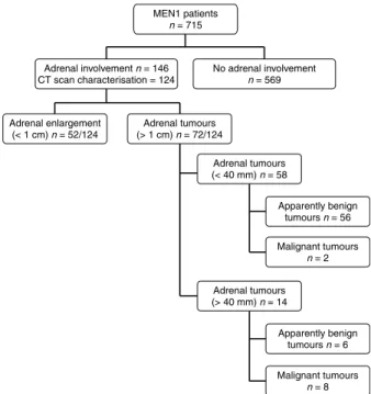
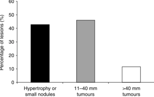
The characterisation of adrenal lesions at MRI or CT scanning was available is 124/186 lesions (Fig. 3). It ranged from slight enlargement to tumours of 120 mm in size. Adrenal tumours were found in 72 of the 124 (58.1%) patients reported with adrenal involvement corresponding to 10.0% (72/715) of the overall cohort. A total of 19.4% of the tumours were below 40 mm in size. The mean size of MEN1-associated tumours did not differ from that observed in incidentalomas. Of the 72 patients with adrenal tumours, 12.5% had bilateral lesions. This prevalence was not different from the 7.3%
prevalence found in incidental tumours (PZ0.24). The prevalence of declared adrenal lesions was similar before and after 1991 and 1998, these periods corresponding to the development of the spiral CT scan and the multidetector CT scan respectively.
Functionality of adrenal lesions
Seventy-three of the 146 MEN1 patients with radio-logical adrenal involvement underwent the biochemical evaluation required to characterise the functional status of adrenal lesions. Normal although incomplete endocrine evaluation was reported in 18.5% of patients while 31.5% of the 146 patients had no biological information available. None of these patients had clinical evidence of adrenal hypersecretion.
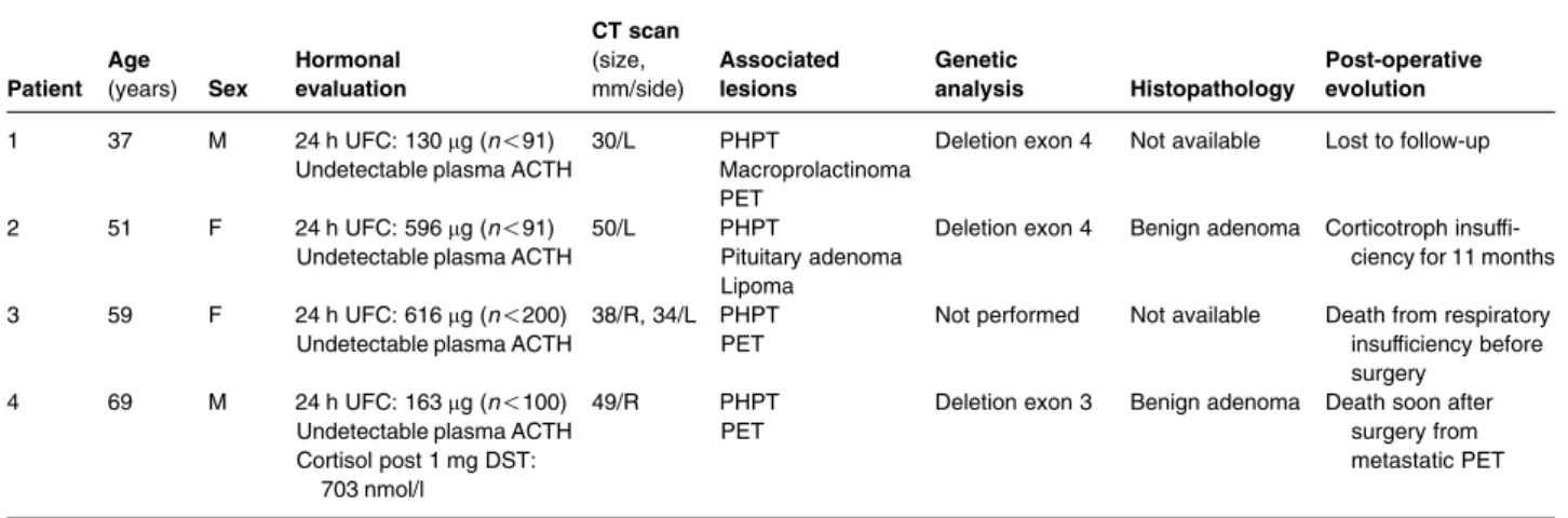
Eleven patients (7.5%) with radiological adrenal involvement had documented endocrine hypersecre-tion. These 11 patients had adrenal tumours and no case of endocrine hypersecretion was documented in patients that only had adrenal hypertrophy. The overall prevalence of endocrine hypersecretion among MEN1 patients with adrenal tumours was 15.3% (11/72) and was significantly increased compared with the 6.9% (10/144) prevalence found in adrenal incidentalomas (PZ0.03). Four cases of primary hyperaldosteronism were found among MEN1 patients (Table 1). Case no. 2 in which tumoural DNA analysis after surgical excision revealed loss of heterozygosity for polymorphic chromo-some 11 DNA markers, including those in the region of the MEN1 locus, has been previously published (25). ACTH-independent Cushing’s syndrome was diagnosed in four cases (Table 2). Two patients had clinical and biological hyperandrogenia that revealed an ACC (see below). One case of bilateral pheochromocytoma was reported in a patient with primary hyperparathyroidism and PET, hyperparathyroidism, acromegaly and familial cases of MEN1. However, no genetic analysis was performed and the patient also had obvious clinical features of neurofibromatosis type 1. The frequency of primary hyperaldosteronism and overt ACTH-independent Cushing’s syndrome was significantly increased in MEN1 patients with adrenal tumours compared with patients with sporadic incidentalomas (P!0.05). On the contrary, the prevalence of pheochromocytoma was lower in MEN1 patients (P!0.05).
MEN1 patients n = 715 No adrenal involvement n = 569 Adrenal involvement n = 146 CT scan characterisation = 124 Adrenal enlargement (< 1 cm) n = 52/124 Adrenal tumours (> 1 cm) n = 72/124 Adrenal tumours (< 40 mm) n = 58 Apparently benign tumours n = 56 Malignant tumours n = 2 Adrenal tumours (> 40 mm) n = 14 Apparently benign tumours n = 6 Malignant tumours n = 8
Figure 1 Distribution of adrenal involvement in 715 MEN1 patients.
Incidental or systematic screening 75% Surgery 3% Unknown 11% Clinical symptoms 11%
Figure 2 Circumstances of diagnosis of the adrenal lesions in MEN1 patients. The results are expressed in percentage of patients.
AUTHOR COPY ONLY
Malignancy
Forty-five MEN1 patients underwent resection of adrenal tumours. Benign cortical adenomas and/or hyperplasia were diagnosed in 35 cases and two cases of adrenal metastases of a somatostatinoma and a gastrinoma were identified. Ten ACCs were diagnosed in eight patients (1.4% of the whole cohort; seeTable 3). The prevalence of ACC in MEN1 patients with adrenal tumours was significantly higher than in patients with adrenal incidentalomas (13.8 vs 1.3%, P!0.05). Three MEN1 patients with ACC had no related clinical or biological abnormality, two complained of abdominal or dorsal pain and three patients had clinical mani-festations of endocrine hypersecretion (Table 3). Several features of ACCs deserve particular attention. First, five
of the ten ACCs had a small size !50 mm at diagnosis and nine of the ten ACCs displayed ENS@T stage I or II allowing apparently complete surgical resection in all but one case. Consequently, six of the eight patients were alive after 2–36 years of follow-up. Secondly, one patient (no. 5) had bilateral ACCs and one patient (no. 8) was diagnosed as having two distinct small-sized ACCs within the same adrenal gland after careful examination by two independent referent pathologists. Thirdly, ACCs developed during the follow-up of relatively small adrenal nodules in two patients. In the first patient (no. 8), two nodules of 8 and 13 mm on the right gland were discovered at the age of 21 years. Unenhanced attenuation values were both 38 UH. Five years later an increase in size of the two right adrenal lesions to 29 and 30 mm prompted to proceed with surgery and identification of two distinct ACCs. A 25 mm calcified adrenal lesion with an unenhanced attenuation of 40 UH was identified in patient no. 7 at initial evaluation. After 4 years of follow-up, the tumour size had increased to 40 mm. After surgical excision, two independent expert pathologists concluded to a rare case of adrenocortical sarcomatoid carcinoma. Of note, this patient had a history of gastro-intestinal stromal tumour as reported earlier (26). Fourthly, another noteworthy observation is the occurrence of ACC in a 3-year-old patient.
Genetics
No genotype–phenotype correlation was evident for the occurrence of adrenal lesions, adrenal tumours and occurrence of endocrine hypersecretion or ACC.
60 50 40 30 20 10 0 Hypertrophy or small nodules P ercentage of lesions (%) 11–40 mm tumours >40 mm tumours
Figure 3 Size of adrenal lesions in MEN1 patients. The size is divided into three categories and results are expressed in percentage of lesions in each category.
Table 1 Characteristics of MEN1 patients with primary hyperaldosteronism.
Patient Age
(years) Sex Symptoms
Hormonal evaluation CT scan (size, mm/side) Associated lesions Genetic analysis Histopathology Post-operative evolution 1 33 F Hypertension Supine PA:
1600 pmol/l (n!300) Undetectable PR 10/right Hyperparathyr-oidism, PET and prolacti-noma Negative Benign adenoma Recovery from hypertension 2 55 F Hypertension Hypokalemia Supine PA: 4230 pmol/l (n!430) Undetectable PRA 22/left Prolactinoma Hyperpara-thyroidism
LOH for polymorphic chromosome 11 DNA markers in the tumours No germline mutation Benign adenoma Recovery from hypertension and hypoka-lemia 3 57 M Hypertension Hypokalemia Supine PA: 312 pmol/l (n!300)
11/left None Exons 3–10 Duplication Benign adenoma Reduction of antihyperten-sive drugs Undetectable PRA Recovery from hypokalemia 4 50 F Hypertension Hypokalemia Supine PA: 861 pmol/l (n!300) Undetectable PRA 12/left Hyperparathyr-oidism PET Exon 3 Insertion Non-operated Mineralocorti-coid receptor antagonist treatment M, male; F, female; PA, plasma aldosterone; PR, plasma renin; PRA, PR activity; PET, pancreatic endocrine tumour; LOH, loss of heterozygosity.
Adrenal lesions in MEN1 273
EUROPEAN JOURNAL OF ENDOCRINOLOGY (2012) 166
AUTHOR COPY ONLY
Specifically, there was no statistical difference for the occurrence of adrenal lesions between patients with germline mutations in exons 2 and 10 and those with mutations in other exons in the menin gene.
Follow-up
Records of follow-up were available for 53 patients with adrenal lesions for a mean duration of 7.4G0.8 (3–72) months. During follow-up, 13 of the 53 patients (24.5%) showed a significant increase in size of the adrenal lesion and two (4%) developed a contralateral lesion of more than 10 mm in size. As mentioned above, three ACCs were identified in two patients at patho-logical examination of adrenal nodules that increased in size after 4 and 8 years of follow-up.
Discussion
This study analyses the findings observed in a multi-centre database involving 715 MEN1 patients and compares it with those found in a multicentre control cohort of 155 sporadic adrenal incidentalomas. There-fore, this is the largest study on adrenal involvement in MEN1. Since cases originate from 22 areas of France and from Belgium, one would expect a reduced selection bias compared with monocentric reports(5, 8, 9, 10, 11, 12). As previously reported, clinical symptoms of adrenal hyperfunction or abdominal pain seldom revealed the adrenal lesions and systematic abdominal imaging contributed to the diagnosis in 89% of MEN1 patients
(6, 11, 15). The prevalence of declared adrenal enlargement in our large series was 20.3% and unilateral in the majority of cases. As found in the few series giving enough detail on the size of adrenal lesions
(6, 7, 11), only half of declared lesions were adrenal tumours O10 mm in size (10% prevalence in our
series). One weakness of the study is the fact that imaging was heterogeneous as to technique and radiological equipment, since the study covers a 40-year span, and was not submitted to central review. Underestimation of slight enlargement or small nodules of the adrenal glands is therefore plausible and could explain the differences with series acknowledging a prevalence of adrenal enlargement between 35 and 55%(5, 9, 11, 12, 14). Indeed, one of the three adrenal lesions identified by endoscopic ultrasound sonography (EUS) may be missed by CT(11)and, in a recent study using exclusively EUS, up to 73% of MEN1 patients had evidence of adrenal enlargement. The pathological/ clinical significance of mild adrenal enlargement in MEN1 patients is unknown but, similarly to what has been described in sporadic and asymptomatic inciden-talomas (27), no case of adrenal enlargement below 10 mm was associated with endocrine dysfunction or evolution towards ACC.
Fifteen per cent of the 73 (11/73) patients in whom hormonal workup was performed had evidence of adrenal hyperactivity. This prevalence is increased compared with the 0–6% prevalence of adrenal hypersecretion usually quoted in the literature (5, 9, 12, 14). It is noteworthy that only a minority of published series included a detailed hormonal evalu-ation and thus underestimevalu-ation of adrenal hyperactivity in previous studies may be hypothesised. We cannot exclude that the prevalence of pheochromocytoma could be underestimated since accurate biological diagnostic tools were probably lacking in the early years of the registry. On the contrary, overestimation of the prevalence of endocrine hypersecretion in our series is probable since 50% of patients with adrenal lesions who were clinically asymptomatic from an endocrine perspective did not undergo a complete biological evaluation and therefore were not included in the prevalence calculation. Of note, all patients in our series Table 2 Characteristics of MEN1 patients with Cushing’s syndrome.
Patient Age (years) Sex Hormonal evaluation CT scan (size, mm/side) Associated lesions Genetic analysis Histopathology Post-operative evolution 1 37 M 24 h UFC: 130 mg (n!91) 30/L PHPT Deletion exon 4 Not available Lost to follow-up
Undetectable plasma ACTH Macroprolactinoma PET
2 51 F 24 h UFC: 596 mg (n!91) Undetectable plasma ACTH
50/L PHPT
Pituitary adenoma Lipoma
Deletion exon 4 Benign adenoma Corticotroph insuffi-ciency for 11 months 3 59 F 24 h UFC: 616 mg (n!200)
Undetectable plasma ACTH
38/R, 34/L PHPT PET
Not performed Not available Death from respiratory insufficiency before surgery
4 69 M 24 h UFC: 163 mg (n!100) Undetectable plasma ACTH Cortisol post 1 mg DST:
703 nmol/l
49/R PHPT PET
Deletion exon 3 Benign adenoma Death soon after surgery from metastatic PET PET, pancreatic endocrine tumour; PHPT, primary hyperparathyroidism; M, male; F, female; UFC, urinary free cortisol; DST, dexamethasone suppression test; L, left; R, right.
AUTHOR COPY ONLY
Table 3 Characteristics of MEN1 patients with adrenocortical carcinomas.
Patient Age
(years) Sex Symptoms
Biological evaluation CT scan findings (size in mm/spontaneous density (UH)/side) Associated lesions at diagnosis Mutation ENS@T stage/ Weiss
score Surgery Follow-up
1 3 M Precocious puberty High urinary 17-Ketosteroids
NA None Mis
Exon3
II/NA Apparent complete surgical excision
Adjuvant chemotherapy and radiotherapy
36 years, alive
Development of PHPT, PET and prolactinomas
2 19 F Virilism High plasma
tes-tosterone and DHEA-S
66/39/L PHPT Del
Exon 10
II/6 Apparent complete surgical excision Maintained on mitotane 4 years, alive 3 44 F Hypertension Hypokalemia No hypercortisolism Suppressed PRA 120/NA/R PHPT PET UK Familial history of MEN1
II/4 Apparent complete surgical excision
Maintained on mitotane Development of liver metastasis Death 3 years after the
diag-nosis despite chemotherapy
4 66 M Abdominal pain NS 100/NA/L PHPT Del
Exon 4
IV/NA Non-resectable adrenal tumourCmultiple liver metastasis
Death in the month after surgery
5 32 F None NS 28/32/L 25/35/R PHPT Cushing’s disease Stop codon Exon 5
I/3&5 Apparent complete surgical excision
Mitotane stopped because of adverse effects
3.5 years, alive
6 26 M Dorsal pain NS 80/NA/L PHPT
PET
Del Exon 2
II/3 Apparent complete surgical excision Development of benign contralateral tumour (10 mm) 14 years, alive 7 50 M None NS 45/40/L PHPT PET Acromegaly Del Exon 10 I/sarco-matoid tumour
Apparent complete surgical excision 3 years, alive 8 27 F None NS 25/35/R 29/35/R PHPT PET Del Exon 9
I/3&5 Apparent complete surgical excision
Left adrenalectomy 1 year later for benign adenoma 3 years, alive
M, male; F, female; NS, non-secreting; NA, not available; L, left; R, right; UK, unknown; PHPT, primary hyperparathyroidism; PET, pancreatic endocrine tumour; IE, initial evaluation; PRA, plasma renin activity; IE, initial evaluation; Del, deletion.
Adr enal lesions in MEN1 275 EUR OPEAN JOURN AL OF ENDOCRINO LOGY (2012) 166 www. eje-online.org
AUTHOR COPY ONLY
with adrenal hypersecretion had adrenal tumours and no case of endocrine hypersecretion was documented in patients that only displayed adrenal hypertrophy. Altogether our data confirm that most of MEN1-associated lesions are hormonally silent and suggest that, in the absence of evocative clinical features, endocrine investigations should be restricted to patients with tumours O10 mm in size.
Adrenal hyperfunction included primary hyperaldos-teronism (nZ4), Cushing’s syndrome (nZ4), excessive androgen secretion (nZ2) and pheochromocytoma (nZ1). Primary hyperaldosteronism in the setting of MEN1 has seldom been described, usually as isolated case reports (25), and was unusually frequent in this series. This discrepancy with previous reports might be related to the difficulty in diagnosing primary hyper-aldosteronism spontaneously (28) or in patients that are, in our practice, often already treated with antihypertensive agents that interfere with the evalu-ation of the renin–aldosterone system and may mask hypokalemia. Evaluation of the renin–aldosterone system may also have been overlooked in some series
(5, 6, 11, 12). In addition, the prevalence of primary aldosteronism was significantly increased compared with the expected low prevalence that we found in sporadic incidentalomas(29). Another point of import-ance is that primary hyperaldosteronism was predomi-nantly related to unilateral lesions, potentially curable with surgery, and not to bilateral hyperplasia with the limits of statistical comparison of small numbers. These findings suggest that aldosterone-producing adenomas could be a more common component of the MEN1 adrenal phenotype than previously thought and that caution should be paid to the renin–aldosterone system evaluation in hypertensive patients with MEN1.
ACTH-independent Cushing’s syndrome was found in 2.7% of patients with adrenal enlargement and 5.5% of patients with adrenal tumours. Interestingly, Cushing’s syndrome was due to a unilateral disease in three of the four of our cases with a frank asymmetry in size in favour of the adenoma allowing unilateral adrenalect-omy. The prevalence of Cushing’s syndrome in our series was increased compared with that found in sporadic incidentalomas. The 1% prevalence found in our control group was significantly lower to that quoted in series of the literature with a calculated median of 7.9% in a 2003 review (30). However, in the overwhelming majority of published series, patients do not display overt Cushing’s syndrome but are diagnosed as having ‘subclinical’ Cushing’s syndrome, e.g. poorly active adenomas associated with mild and often dissociated biological abnormalities of the hypothalamus-pituitary-adrenal (HPA) axis (18, 30). Owing to the lack of uniformity in the endocrine investigations between centres and the absence of consensus on diagnostic criteria, the diagnosis of subclinical Cushing’s syndrome was not considered in the MEN1 and sporadic incidentalomas groups(31, 32, 33). At variance with
the expected 5% prevalence of pheochromocytoma in sporadic incidentalomas (18, 34, 35), only one MEN1 patient had a bilateral pheochromocytoma but the responsibility of the menin dysfunction cannot be established since this patient had clinically obvious neurofibromatosis type 1. Fewer than 10 cases of pheochromocytoma in MEN1 patients have been reported(2, 8, 9, 36, 37). In cases where DNA testing was performed, patients were found to have germline MEN1 mutations, as well as loss of heterozygosity around the MEN1 gene in the pheochromocytoma tumour tissue(38). These findings associated with the development of bilateral pheochromocytomas in some genetically engineered mice models of MEN1 (39)
suggest that pheochromocytoma is part of the MEN1 syndrome. However, our data issued from a very large cohort together with data from the literature suggest that the occurrence of pheochromocytoma in patients with MEN1 is a very rare event.
Lastly, hyperandrogenism due to an excessive secretion of androgens, an almost specific feature of malignant adrenal secreting tumours(31), revealed an ACC in two cases.
Two credits of our study are to highlight the epidemiology of ACCs with the statistical power provided by a large cohort and to provide additional information on ACCs that is almost always lacking in the literature (CT scanning details, staging, Weiss score and survival). The overall prevalence that we found (1.4% of the whole MEN1 population) is in accordance with the 0–6% prevalence found in other series of the literature. However, this prevalence reached 13.8% in patients with adrenal tumours. Such prevalence significantly exceeded the 1.3% prevalence of ACC found in our control series, which is concordant with that found in a recent review and meta-analysis of sporadic adrenal incidentalomas(29). Another import-ant finding is that three of the ten ACCs were identified after growth of small adrenal tumours. This scenario has already been described in MEN1 (8, 9, 11) but is hardly ever encountered in follow-up series of adrenal incidentalomas (18). Interestingly, elevated spon-taneous CT attenuation values at previous evaluations before spontaneous increase in size were not consistent with benign, lipid-rich adenomas (18, 35, 41). The occurrence of growth after 4–8 years of follow-up may suggest that these were slowly growing ACCs. Another important finding is that nine of the ten ACCs were classified ENS@T stage I or II allowing apparently complete surgical resection in all but one case, an unexpected feature in sporadic ACCs (31, 42). Else-where, the Weiss score was %4 in four of the seven ACCs in which it was available. The stage of ACCs as well as the Weiss score are important prognostics factors for ACC (42). Our findings suggest that MEN1-related ACCs may represent a specific entity with reduced aggressiveness compared with sporadic ACCs. However, this hypothesis should not eclipse the fact that, as others
AUTHOR COPY ONLY
reported in the literature, two patients of our series died of their ACCs(8, 11, 43). An alternative explanation for our findings is that programmed imaging studies in the abdominal workup of MEN1 may have helped to identify ACCs at an early stage.
Altogether our observations combined with the demonstration of LOH at 11q13 in most MEN1-related ACCs analysed(9, 10), and the occurrence of ACCs in mouse models of MEN1(39), strongly suggest that the loss of the antioncogenic menin protein constitutes a predisposing condition for the development of adrenal malignancy that may develop after the occurrence of another somatic genetic event(2, 4, 40).
The pathogenesis of MEN1-associated adrenal lesions has been questioned and the hypothesis of a stimulating role of growth factors and peptides secreted by PET on adrenal tumorigenesis has been debated(5, 9, 11, 14). This issue was outside the scope of our clinical study but it should be noted that there was no evidence of PET in w30% of patients of our series with adrenal involvement. However, and as discussed earlier, we found a number of differences between sporadic inciden-talomas and MEN1 tumours suggesting that they represent two different entities. Unfortunately, we were not able to find any clear genotype/phenotype correlation for the occurrence of adrenal tumours, hypersecreting lesions or ACCs that could be of help to focus investigations and follow-up on a subset of MEN1 patients. Previous attempts to identify genotype/phenotype corre-lation for different MEN1-related tumours have been so far disappointing(4).
Conclusions
Altogether our study suggests that, similarly to what has been described in adrenal incidentalomas, slight enlarge-ment is usually exempt of clinical relevance and no additional endocrine investigation is needed if there is no clinical suspicion of endocrine hyperactivity. On the contrary, a MEN1-associated adrenal tumour differs in many ways from sporadic incidental adrenal tumours. In MEN1, endocrine investigations should focus on steroid hormone secretion since pheochromocytoma appears to be extremely rare and attention should be paid to the investigation of the aldosterone–renin system. Although rare, the occurrence of ACC should also be considered in all patients with adrenal tumours lacking CT charac-teristics of benign cortical adenoma even if their size is below 40 mm. Finally, lifelong imaging of adrenal tumours should be part of the follow-up agenda of patients with MEN1 as has been recommended for PET. The precise frequency of these examinations has to be determined. A significant increase in size of adrenal lesions eventually associated with a change in spon-taneous attenuation values at CT scanning or lipid content at MRI should prompt towards surgery.
Declaration of interest
The authors declare that there is no conflict of interest that could be perceived as prejudicing the impartiality of the research reported.
Funding
This research did not receive any specific grant from any funding agency in the public, commercial or not-for-profit sector.
Acknowledgements
The authors acknowledge the following doctors who provided information about their patients for this study: C Ajzenberg, J J Altman, T Aparicio, F Archambeaud, J R Attali, C Badet, J Barbier, M Barthet, B Bauduceau, H Becheur, P Bernades, X Bertagna, J Bertherat, O Ble´try, P Boissel, J M Boyaval, L Bresler, J F Bretagne, J Bringer, H Brixi-Benmansour, L Brunaud, J Burger, A Calender, P Carenco, B Carnaille, B Cathebras, M Celerier, D Chadenas, P Chanson, D Charitanski, J A Chayvialle, C Colmar, Montiel, J M Comas, B Conte Devolx, A Cortot, E Cosson, P Cougard, P Cubertafond, P D’Anella, P Darsy, T Defechereux, F Delecourt, J Denis, C Derrien, D Dewailly, A S Dramais, C Droumaguet, C Dubost, F Duron, B Emperauger-Beauvais, P Emy, S Gauthier, A P Gimenez Roqueplo, B Goichot, D Goldfain, M Gosselin, F Grunenberger, A M Guedj, P J Guillausseau, P Hamon, J F Henry, P J Jaquet, C Jublanc, V Kerlan, J M Khun, B Knebelmann, J L Kraimps, A Krivtzky, F Lagarde, J D Lalau, P Lecomte, J J Legros, D Levoir, N Le´vy-Bohbot, C J Lips, B Maizeray-Cailliau, D Malet, M Malinski, G Mantion, B Maroy, C Mathe, M Mathonnet, D Melliere, F Me´ne´gaux, E H Metman, M Meurisse, E Miraillie´, R Modigliani, C Montiel, C Naouri, C Oliver, F Olivier, J Orgiazzi, M Parneix, C Partenski, F Pattou, J L Peix, A Penfornis, A Pradignac, C Pouget, M Pugeat, M L Raffin-Sanson, M Rodier, P Roger, P Rougier, H Rousset, J Roy, J L Sadoul, E Sarfati, J L Schlienger, M Schlumberger, F Sebag, P Seve, D Simon, I Sobhani, O Soubrane, J C Soule, P Thieblot, C Thivolet, P Thomopoulos, P Valensi, M C Vantighem, M F Verger, O Verier-Mine, E Verlet, B Vialettes, R Viard, S Walter, A Warnet, B Wechsler, J L Wemeau, G Weryha, B Woehl-Kremer and the AFCE members (Association Franc¸aise de Chirurgie Endocrinienne), G Turpin, G Chabrier, F Borson-Chazot and P Bouchard.
References
1 Chandrasekharappa SC, Guru SC, Manickam P, Olufemi SE, Collins FS, Emmert-Buck MR, Debelenko LV, Zhuang Z, Lubensky IA, Liotta LA, Crabtree JS, Wang Y, Roe BA, Weisemann J, Boguski MS, Agarwal SK, Kester MB, Kim YS, Heppner C, Dong Q, Spiegel AM, Burns AL & Marx SJ. Positional cloning of the gene for multiple endocrine neoplasia-type 1. Science 1997 276 404–407. (doi:10.1126/science.276.5311.404) 2 Marx S, Spiegel AM, Skarulis MC, Doppman JL, Collins FS &
Liotta LA. Multiple endocrine neoplasia type 1: clinical and genetic topics. Annals of Internal Medicine 1998 129 484–494.
3 Wermer P. Genetic aspects of adenomatosis of endocrine glands. American Journal of Medicine 1954 16 363–371. (doi:10.1016/ 0002-9343(54)90353-8)
4 Thakker RV. Multiple endocrine neoplasia type 1 (MEN1). Best Practice & Research. Clinical Endocrinology & Metabolism 2010 24 355–370. (doi:10.1016/j.beem.2010.07.003)
5 Burgess JR, Harle RA, Tucker P, Parameswaran V, Davies P, Greenaway TM & Shepherd JJ. Adrenal lesions in a large kindred with multiple endocrine neoplasia type 1. Archives of Surgery 1996 131 699–702.
6 Schaefer S, Shipotko M, Meyer S, Ivan D, Klose KJ, Waldmann J, Langer P & Kann PH. Natural course of small adrenal lesions in multiple endocrine neoplasia type 1: an endoscopic ultrasound imaging study. European Journal of Endocrinology 2008 158 699–704. (doi:10.1530/EJE-07-0635)
Adrenal lesions in MEN1 277
EUROPEAN JOURNAL OF ENDOCRINOLOGY (2012) 166
AUTHOR COPY ONLY
7 Whitley SA, Moyes VJ, Park KM, Brooke AM, Grossman AB, Chew SL, Rockall AG, Monson JP & Reznek RH. The appearance of the adrenal glands on computed tomography in multiple endocrine neoplasia type 1. European Journal of Endocrinology 2008 159 819–824. (doi:10.1530/EJE-08-0516)
8 Langer P, Cupisti K, Bartsch DK, Nies C, Goretzki PE, Rothmund M & Roher HD. Adrenal involvement in multiple endocrine neoplasia type 1. World Journal of Surgery 2002 26 891–896. (doi:10.1007/ s00268-002-6492-4)
9 Skogseid B, Larsson C, Lindgren PG, Kvanta E, Rastad J, Theodorsson E, Wide L, Wilander E & Oberg K. Clinical and genetic features of adrenocortical lesions in multiple endocrine neoplasia type 1. Journal of Clinical Endocrinology and Metabolism 1992 75 76–81. (doi:10.1210/jc.75.1.76)
10 Skogseid B, Rastad J, Gobl A, Larsson C, Backlin K, Juhlin C, Akerstrom G & Oberg K. Adrenal lesion in multiple endocrine neoplasia type 1. Surgery 1995 118 1077–1082. (doi:10.1016/ S0039-6060(05)80117-5)
11 Waldmann J, Bartsch DK, Kann PH, Fendrich V, Rothmund M & Langer P. Adrenal involvement in multiple endocrine neoplasia type 1: results of 7 years prospective screening. Langenbeck’s Archives of Surgery 2007 392 437–443. (doi:10.1007/s00423-006-0124-7)
12 Barzon L, Pasquali C, Grigoletto C, Pedrazzoli S, Boscaro M & Fallo F. Multiple endocrine neoplasia type 1 and adrenal lesions. Journal of Urology 2001 166 24–27. (doi:10.1016/S0022-5347(05)66068-5)
13 Brandi ML, Gagel RF, Angeli A, Bilezikian JP, Beck-Peccoz P, Bordi C, Conte-Devolx B, Falchetti A, Gheri RG, Libroia A, Lips CJ, Lombardi G, Mannelli M, Pacini F, Ponder BA, Raue F, Skogseid B, Tamburrano G, Thakker RV, Thompson NW, Tomassetti P, Tonelli F, Wells SA Jr & Marx SJ. Guidelines for diagnosis and therapy of MEN type 1 and type 2. Journal of Clinical Endocrinology and Metabolism 2001 86 5658–5671. (doi:10.1210/jc.86.12. 5658)
14 Gibril F & Jensen RT. Zollinger–Ellison syndrome revisited: diagnosis, biologic markers, associated inherited disorders, and acid hypersecretion. Current Gastroenterology Reports 2004 6 454–463. (doi:10.1007/s11894-004-0067-5)
15 Pieterman CR, Schreinemakers JM, Koppeschaar HP, Vriens MR, Rinkes IH, Zonnenberg BA, van der Luijt RB & Valk GD. Multiple endocrine neoplasia type 1 (MEN1): its manifestations and effect of genetic screening on clinical outcome. Clinical Endocrinology 2009 70 575–581. (doi:10.1111/j.1365-2265.2008.03324.x) 16 Marini F, Falchetti A, Del Monte F, Carbonell Sala S, Gozzini A,
Luzi E & Brandi ML. Multiple endocrine neoplasia type 1. Orphanet Journal of Rare Diseases 2006 1 38. (doi:10.1186/1750-1172-1-38)
17 Goudet P, Bonithon-Kopp C, Murat A, Ruszniewski P, Niccoli P, Menegaux F, Chabrier G, Borson-Chazot F, Tabarin A, Bouchard P, Cadiot G, Beckers A, Guilhem I, Chabre O, Caron P, Du Boullay H, Verges B & Bauters C. Gender-related differences in MEN1 lesion occurrence and diagnosis. A 734-case cohort study from the GTE (Groupe d’etude des Tumeurs Endocrines). European Journal of Endocrinology 2011 165 97–105. (doi:10.1530/EJE-10-0950) 18 Tabarin A, Bardet S, Bertherat J, Dupas B, Chabre O, Hamoir E,
Laurent F, Tenenbaum F, Cazalda M, Lefebvre H, Valli N & Rohmer V. Exploration and management of adrenal incidentalo-mas. French Society of Endocrinology Consensus. Annales d’Endocrinologie 2008 69 487–500. (doi:10.1016/j.ando.2008. 09.003)
19 Wautot V, Vercherat C, Lespinasse J, Chambe B, Lenoir GM, Zhang CX, Porchet N, Cordier M, Beroud C & Calender A. Germline mutation profile of MEN1 in multiple endocrine neoplasia type 1: search for correlation between phenotype and the functional domains of the MEN1 protein. Human Mutation 2002 20 35–47. (doi:10.1002/humu.10092)
20 Fromaget M, Vercherat C, Zhang CX, Zablewska B, Gaudray P, Chayvialle JA, Calender A & Cordier-Bussat M. Functional
characterization of a promoter region in the human MEN1 tumor suppressor gene. Journal of Molecular Biology 2003 333 87–102. (doi:10.1016/j.jmb.2003.08.001)
21 Weiss LM, Medeiros LJ & Vickery AL Jr. Pathologic features of prognostic significance in adrenocortical carcinoma. American Journal of Surgical Pathology 1989 13 202–206. (doi:10.1097/ 00000478-198903000-00004)
22 Gicquel C, Bertagna X, Gaston V, Coste J, Louvel A, Baudin E, Bertherat J, Chapuis Y, Duclos JM, Schlumberger M, Plouin PF, Luton JP & Le Bouc Y. Molecular markers and long-term recurrences in a large cohort of patients with sporadic adrenocortical tumors. Cancer Research 2001 61 6762–6767. 23 de Reynies A, Assie G, Rickman DS, Tissier F, Groussin L,
Rene-Corail F, Dousset B, Bertagna X, Clauser E & Bertherat J. Gene expression profiling reveals a new classification of adrenocortical tumors and identifies molecular predictors of malignancy and survival. Journal of Clinical Oncology 2009 27 1108–1115. (doi:10.1200/JCO.2008.18.5678)
24 Fassnacht M, Wittekind C & Allolio B. Current TNM classification systems for adrenocortical carcinoma. Der Pathologe 2010 31 374–378. (doi:10.1007/s00292-010-1306-1)
25 Beckers A, Abs R, Willems PJ, van der Auwera B, Kovacs K, Reznik M & Stevenaert A. Aldosterone-secreting adrenal adenoma as part of multiple endocrine neoplasia type 1 (MEN1): loss of heterozygosity for polymorphic chromosome 11 deoxyribonucleic acid markers, including the MEN1 locus. Journal of Clinical Endocrinology and Metabolism 1992 75 564–570. (doi:10.1210/ jc.75.2.564)
26 Papillon E, Rolachon A, Calender A, Chabre O, Barnoud R & Fournet J. A malignant gastrointestinal stromal tumour in a patient with multiple endocrine neoplasia type 1. European Journal of Gastroenterology & Hepatology 2001 13 207–211. (doi:10.1097/00042737-200102000-00021)
27 Thompson GB & Young WF Jr. Adrenal incidentaloma. Current Opinion in Oncology 2003 15 84–90. (doi:10.1097/00001622-200301000-00013)
28 Stowasser M. Update in primary aldosteronism. Journal of Clinical Endocrinology and Metabolism 2009 94 3623–3630. (doi:10. 1210/jc.2009-1399)
29 Cawood TJ, Hunt PJ, O’Shea D, Cole D & Soule S. Recommended evaluation of adrenal incidentalomas is costly, has high false-positive rates and confers a risk of fatal cancer that is similar to the risk of the adrenal lesion becoming malignant; time for a rethink? European Journal of Endocrinology 2009 161 513–527. (doi:10.1530/EJE-09-0234)
30 Barzon L, Sonino N, Fallo F, Palu G & Boscaro M. Prevalence and natural history of adrenal incidentalomas. European Journal of Endocrinology 2003 149 273–285. (doi:10.1530/eje.0.1490273) 31 Abiven G, Coste J, Groussin L, Anract P, Tissier F, Legmann P, Dousset B, Bertagna X & Bertherat J. Clinical and biological features in the prognosis of adrenocortical cancer: poor outcome of cortisol-secreting tumors in a series of 202 consecutive patients. Journal of Clinical Endocrinology and Metabolism 2006 91 2650– 2655. (doi:10.1210/jc.2005-2730)
32 Chiodini I, Morelli V, Masserini B, Salcuni AS, Eller-Vainicher C, Viti R, Coletti F, Guglielmi G, Battista C, Carnevale V, Iorio L, Beck-Peccoz P, Arosio M, Ambrosi B & Scillitani A. Bone mineral density, prevalence of vertebral fractures, and bone quality in patients with adrenal incidentalomas with and without subclinical hypercorti-solism: an Italian multicenter study. Journal of Clinical Endo-crinology and Metabolism 2009 94 3207–3214. (doi:10.1210/jc. 2009-0468)
33 Terzolo M, Reimondo G, Gasperi M, Cozzi R, Pivonello R, Vitale G, Scillitani A, Attanasio R, Cecconi E, Daffara F, Gaia E, Martino E, Lombardi G, Angeli A & Colao A. Colonoscopic screening and follow-up in patients with acromegaly: a multicenter study in Italy. Journal of Clinical Endocrinology and Metabolism 2005 90 84–90. (doi:10.1210/jc.2004-0240)
34 Mantero F, Terzolo M, Arnaldi G, Osella G, Masini AM, Ali A, Giovagnetti M, Opocher G & Angeli A. A survey on adrenal
AUTHOR COPY ONLY
incidentaloma in Italy. Study Group on Adrenal Tumors of the Italian Society of Endocrinology. Journal of Clinical Endocrinology and Metabolism 2000 85 637–644. (doi:10.1210/jc.85.2.637) 35 Terzolo M, Stigliano A, Chiodini I, Loli P, Furlani L, Arnaldi G,
Reimondo G, Pia A, Toscano V, Zini M, Borretta G, Papini E, Garofalo P, Allolio B, Dupas B, Mantero F & Tabarin A. AME position statement on adrenal incidentaloma. European Journal of Endocrinology 2011 164 851–870. (doi:10.1530/EJE-10-1147) 36 Carty SE, Helm AK, Amico JA, Clarke MR, Foley TP, Watson CG &
Mulvihill JJ. The variable penetrance and spectrum of mani-festations of multiple endocrine neoplasia type 1. Surgery 1998 124 1106–1113 discussion 1113–1114. (doi:10.1067/msy. 1998.93107)
37 Trump D, Farren B, Wooding C, Pang JT, Besser GM, Buchanan KD, Edwards CR, Heath DA, Jackson CE, Jansen S, Lips K, Monson JP, O’Halloran D, Sampson J, Shalet SM, Wheeler MH, Zink A & Thakker RV. Clinical studies of multiple endocrine neoplasia type 1 (MEN1). QJM 1996 89 653–669. 38 Schussheim DH, Skarulis MC, Agarwal SK, Simonds WF,
Burns AL, Spiegel AM & Marx SJ. Multiple endocrine neoplasia type 1: new clinical and basic findings. Trends in Endocrinology and Metabolism 2001 12 173–178. (doi:10.1016/S1043-2760(00)00372-6)
39 Crabtree JS, Scacheri PC, Ward JM, Garrett-Beal L,
Emmert-Buck MR, Edgemon KA, Lorang D, Libutti SK,
Chandrasekharappa SC, Marx SJ, Spiegel AM & Collins FS.
A mouse model of multiple endocrine neoplasia, type 1, develops multiple endocrine tumors. PNAS 2001 98 1118–1123. (doi:10. 1073/pnas.98.3.1118)
40 Bertherat J & Bertagna X. Pathogenesis of adrenocortical cancer. Best Practice & Research. Clinical Endocrinology & Metabolism 2009 23 261–271. (doi:10.1016/j.beem.2008.10.006)
41 Hamrahian AH, Ioachimescu AG, Remer EM, Motta-Ramirez G, Bogabathina H, Levin HS, Reddy S, Gill IS, Siperstein A & Bravo EL. Clinical utility of noncontrast computed tomography attenuation value (hounsfield units) to differentiate adrenal adenomas/hyper-plasias from nonadenomas: Cleveland clinic experience. Journal of Clinical Endocrinology and Metabolism 2005 90 871–877. (doi:10. 1210/jc.2004-1627)
42 Fassnacht M, Libe R, Kroiss M & Allolio B. Adrenocortical carcinoma: a clinician’s update. Nature Reviews. Endocrinology 2011 7 323–335. (doi:10.1038/nrendo.2010.235)
43 Simonds WF, Varghese S, Marx SJ & Nieman LK. Cushing’s syndrome in multiple endocrine neoplasia type 1. Clinical Endocrinology 2012 In press. (doi:10.1111/j.1365-2265.2011. 04220.x)
Received 2 August 2011
Revised version received 25 October 2011 Accepted 14 November 2011
Adrenal lesions in MEN1 279
EUROPEAN JOURNAL OF ENDOCRINOLOGY (2012) 166