HAL Id: tel-02984778
https://tel.archives-ouvertes.fr/tel-02984778
Submitted on 1 Nov 2020
HAL is a multi-disciplinary open access archive for the deposit and dissemination of sci-entific research documents, whether they are pub-lished or not. The documents may come from teaching and research institutions in France or abroad, or from public or private research centers.
L’archive ouverte pluridisciplinaire HAL, est destinée au dépôt et à la diffusion de documents scientifiques de niveau recherche, publiés ou non, émanant des établissements d’enseignement et de recherche français ou étrangers, des laboratoires publics ou privés.
Hypothalamic control of hippocampal area CA2 activity
Vincent Robert
To cite this version:
Vincent Robert. Hypothalamic control of hippocampal area CA2 activity. Neurons and Cognition [q-bio.NC]. Université Sorbonne Paris Cité, 2018. English. �NNT : 2018USPCB232�. �tel-02984778�
1
Université Paris Descartes
Ecole doctorale 158 : Cerveau, Cognition, Comportement
Centre de Psychiatrie et Neurosciences / Equipe Chevaleyre - Piskorowski
Hypothalamic control of hippocampal
area CA2 activity
Par Vincent ROBERT
Thèse de doctorat de Neurosciences
Dirigée par Rebecca PISKOROWSKI, PhD & Vivien CHEVALEYRE, PhD
Présentée et soutenue publiquement le 16/05/2018
Devant un jury composé de :
Liset Menendez DE LA PRIDA, PhD (Rapporteur) Paul SALIN, PhD (Rapporteur)
Claire LEGAY, PhD, PU (Président du jury) Audrey MERCER, PhD (Examinateur)
Philippe ASCHER, PhD, Professeur émérite (Examinateur) Jean-Christophe PONCER, PhD (Examinateur invité) Karim BENCHENANE, PhD (Examinateur invité)
2
Acknowledgements
It is with sincere enthusiasm that I thank all the persons involved in the completion of this thesis.
I thank Rebecca Piskorowski and Vivien Chevaleyre for taking me on as a PhD student, teaching me many technical and intellectual aspects of research, supervising my work, and guiding me through these years. Thank you for your availability, your encouragements, your insightful advices and for setting such a pleasant atmosphere in the lab. I am proud to have learnt science from you. During these years, I have not only received an invaluable training from you but also found exceptional mentors who never failed to help me move forward through good and hard times with thoughtfulness and friendship. Rebecca, thank you for your patience, your support, your trust, and your enthusiasm in passing on your expertise. I can only hope to become a researcher like you in the future.
I thank the members of my thesis committee, Claire Legay, Liset Menendez de la Prida, Paul Salin, Audrey Mercer, Philippe Ascher, Jean-Christophe Poncer and Karim Benchenane. It is an honor to have you evaluate my work. I thank Manuel Mameli for being part of my progress committee.
I thank all the present and past members of the lab for useful discussions, help with experiments, and all the fun times we had. In particular, I thank Ludivine Therreau and Kaoutsar Nasrallah. Thank you Ludivine for your invaluable help with surgeries, genotyping, and histology. Thank you Kaoutsar for helping me when I started and for your thoughtfulness and positive attitude that never failed to bring serenity in the lab, it was amazing working with you.
I thank the Ecole Normale Superieure de Cachan, the French Ministère de la Recherche, and the Fondation pour la Recherche Médicale (FDT20170437387) for funding my work.
I thank the members of the McHugh lab for providing the Csf2rb2-Cre mouse line, the AAV.Synapsin.DIO.hM4D(Gi).mCherry virus, and in vivo experiments.
3
I thank Mario Penzo and Bo Li who trained me and became my friends during the year I spent in Cold Spring Harbor labs, showing me that I was capable of doing science which was critical in my decision to go for a PhD.
I thank my family. In particular, I thank my mother for her never-failing support and care, and for her genuine curiosity about my topics of interest. Thank you Julie for your positive attitude. I thank my father for his attention and interest in my work. I thank my grandparents for their encouraging attitude. Thank you Françoise for helping me grow up and sparkling my scientific curiosity.
I thank my friends, who were always there for me through good and hard times, having your friendship is truly inspirational. Thank you Clément for the amazing experiences and exchanges we had together, for always pushing me forward and never letting me down. Thank you Poppy for all the happy times and music we shared, for understanding who I am and picking me up every time I fall. Thank you Joss for your unshakable positivity, genuine altruism and for always helping to keep balance in my life be it with laughter or comfort. Thank you Coralie-Anne for all the fun times and fascinating scientific discussions we shared during our thesis years, and for always looking up what is best for me. I thank Mélanie Tobin for the years of our lives that we shared during this thesis. Thank you Benoit, Arnaud, Loulou, Elsa, Lucas, Mathilde, Victor, Charles, Benjamin, Julien, Théo, Anil, Thibault, Antoine, Edith, Agathe, Pierre, Audrey, Cécile, Jean-Marie, Guillaume, Gustavo, Alex, Daphnée.
I thank Mélanie for her kindness, thoughtfulness, tolerance, joyfulness, generosity, and confidence in me, which never fail to bring me happiness and help me overcome difficulties.
4
I dedicate this work to my aunt, Françoise Mantz
5
Table of contents
ACKNOWLEDGEMENTS ... 2
TABLE OF CONTENTS ... 5
LIST OF ABBREVIATIONS ... 8
LIST OF FIGURES AND TABLES ... 11
I-INTRODUCTION ... 13
I.1-ROLES OF THE HIPPOCAMPUS AND THE HYPOTHALAMUS IN MEMORY ... 13
I.2-BRAIN CIRCUITS RELEVANT TO EPISODIC MEMORY ... 14
I.2.a - Flow of information in the canonical hippocampal loop ... 15
I.2.a.i - Hippocampal anatomy and cellular composition ... 15
I.2.a.ii - Inter-regional connectivity and information processing ... 20
I.2.b - Integration of area CA2 inputs and outputs in the hippocampal circuit ... 23
I.2.b.i - Intra-hippocampal connectivity of area CA2 ... 23
I.2.b.ii - Area CA2 long-range afferences and efferences ... 26
I.3-PHYSIOLOGICAL FEATURES OF HIPPOCAMPAL-DEPENDENT MEMORY FORMATION ... 30
I.3.a - Network activity patterns underlying learning and memory ... 30
I.3.a.i - Theta and gamma oscillations ... 30
I.3.a.ii - Sharp wave / ripples ... 34
I.3.b - Mnemonic functions of the hippocampus and hypothalamus ... 35
I.3.b.i - Spatial coding and memory ... 35
I.3.b.ii - Emotional, novel and social aspects of memory ... 42
I.4-GOALS OF THE THESIS ... 43
II-MATERIAL AND METHODS ... 45
II.1-ANIMALS ... 45
II.2-STEREOTAXIC VIRAL INJECTIONS ... 46
II.3-ACUTE HIPPOCAMPAL SLICES PREPARATION ... 47
II.4-ELECTROPHYSIOLOGICAL RECORDINGS ... 47
II.5-OPTOGENETIC STIMULATION ... 48
II.6-IMMUNOCHEMISTRY AND CELL IDENTIFICATION ... 48
II.7-DATA ANALYSIS AND STATISTICS ... 50
II.8-EXPERIMENTAL STRATEGY ... 51
6
III.1-SPONTANEOUS NETWORK ACTIVITY OF HIPPOCAMPAL AREA CA2 UNDER CONDITIONS
OF ENHANCED CHOLINERGIC TONE ... 59
III.1.a - Introduction ... 59
III.1.b - Results ... 60
III.2-HYPOTHALAMIC CONTROL OF HIPPOCAMPAL AREA CA2 ACTIVITY BY DIRECT EXCITATION AND FEEDFORWARD INHIBITION ... 81
III.2.a - Introduction ... 81
III.2.b - Results ... 82
III.3-INFLUENCE OF SUM INPUTS ON HIPPOCAMPAL AREA CA2 DRIVE ONTO CA1 ... 115
III.3.a - Introduction ... 115
III.3.b - Results ... 117
IV-DISCUSSION AND FUTURE DIRECTIONS ... 131
IV.1-ROLES OF AREA CA2 IN HIPPOCAMPAL NETWORKS AND RHYTHMS ... 131
IV.1.a - Similarities and differences of network activity in area CA2 and CA3 ... 132
IV.1.b - Bursts of action potentials in CA2 PNs... 133
IV.1.c - Integration of area CA2 in theta and gamma hippocampal rhythms ... 134
IV.2-RELEVANCE OF SUM INPUTS TO HIPPOCAMPAL AREA CA2 PHYSIOLOGY ... 135
IV.2.a - SuM inputs to area CA2 form a microcircuit where PV+ basket cells strongly inhibit deep pyramidal neurons in area CA2 and CA3 ... 135
IV.2.b - SuM input and space coding in area CA2 ... 136
IV.2.c - SuM input and hippocampal oscillations ... 137
IV.2.d - Gating of area CA2 activity by PV+ INs and significance for pathologies .... 138
IV.2.e - Differential modulation of DG and area CA2 by SuM inputs and consequences on hippocampus-dependent memory formation ... 139
IV.2.f - Relevance of the SuM excitatory drive to CA2 PNs ... 140
IV.3-CONTROL OF HIPPOCAMPAL ACTIVITY AND OUTPUT BY SUM INPUTS ... 141
IV.3.a - Cholinergic modulation of SuM inputs ... 142
IV.3.b - Synchronization of CA2 activity by SuM inhibitory drive ... 143
IV.3.c - Transient silencing of CA1 activity by the SuM-CA2-CA1 circuit ... 144
V-REFERENCES ... 149
VI-APPENDICES ... 176
VI.1-CHRONIC LOSS OF CA2 TRANSMISSION LEADS TO HIPPOCAMPAL HYPEREXCITABILITY 176 VI.1.a - Introduction ... 176
7
VI.1.b - Paper ... 177
VI.2-HIPPOCAMPAL AREA CA2: PROPERTIES AND CONTRIBUTION TO HIPPOCAMPAL FUNCTION ... 178
VI.2.a - Introduction ... 178
VI.2.b - Paper ... 178
RÉSUMÉ ... 179
8
List of abbreviations
4-AP : 4-aminopyridine
Ication-Ca2+ : calcium-dependent non-selective cation current AAV : adeno-associated virus
ACSF : artificial cerebro-spinal fluid AHP : after-spike hyperpolarization Amp : ampicillin
AMPA : α-amino-3-hydroxy-5-methyl-4-isoxazolepropionic acid ANOVA : analysis of variance
AP : action potential
APV : (2R)-amino-5-phosphonovaleric acid; (2R)-amino-5 phosphonopentanoate BAC : bacterial artificial chromosome
BC : basket cell CA : Cornu Ammonis
CACNG5 : calcium voltage-gated channel auxiliary subunit gamma 5 CAV : canine adenovirus
CCh : carbachol CCK : cholecystokinin ChR2 : channelrhodopsin 2 CM : membrane capacitance
CNO : clozapine N-oxide Cre : Cre recombinase
Csf2rb2 : colony stimulating factor 2 receptor β 2 DAMGO : [D-Ala2, N-MePhe4, Gly-ol]-enkephalin DG : dentate gyrus
DIO : double inverted open reading frame DOR : δ opioid receptor
DPDPE : [D-Pen2,D-Pen5]enkephalin
DREADD : designer receptor exclusively activated by designer drug DSP : digital signal processor
EC : entorhinal cortex EEG : electroencephalogram EF1a : elongation factor 1 alpha
EGFP : enhanced green fluorescent protein
EGTA : ethylene glycol-bis(β-aminoethyl ether)-N,N,N',N'-tetraacetic acid EPSC : excitatory post-synaptic current
EPSP : excitatory post-synaptic potential EYFP : enhanced yellow fluorescent protein FLP : flipase
FRT : flipase recognition target GABA : γ-aminobutyric acid
9 GAD65 : glutamic acid decarboxylase 65kDa GC : granule cell
GH : growth hormone
HEPES : 4-(2-hydroxyethyl)-1-piperazineethanesulfonic acid hM4D(Gi) : modified human muscarinic receptor 4
Ih : hyperpolarization-activated cation current
IKsAHP : slow potassium AHP current
iLTD : long-term depression of inhibition IM : M current
IN : interneuron
INaP : persistent sodium current
IPSC : inhibitory post-synaptic current IPSP : inhibitory post-synaptic potential KO : knock-out
LED : light-emitting diode LFP : local field potential LIA : large irregular amplitude
mAchR : muscarinic acetylcholine receptor MF : mossy fibers
MOR : µ-opioid receptor
MPA3K15 : Mitogen-Activated Protein Kinase Kinase Kinase 15
NBQX : 2,3-dihydroxy-6-nitro-7-sulfamoyl-benzo[f]quinoxaline-2,3-dione NMDA : N-methyl D-aspartate
NMDG : N-methyl-D-glucamin NOS : nitric oxide synthase NPY : neuropeptide Y
N-unit : SWR non-positively modulated unit N-unit : SWR positively modulated unit PBS : phosphate buffered saline
PCP-4 : purkinje cell protein 4 PCR : polymerase chain reaction PH : posterior hypothalamus PN : pyramidal neuron PPR : paired-pulse ratio PSP : post-synaptic potential PV : parvalbumin PVN : paraventricular nucleus
RGS14 : Regulator of G-protein signaling 14 RM : membrane resistance
RPO : reticular nucleus pontis oralis SC : Schaffer collaterals
sEPSC : spontaeous excitatory post-synaptic current SIA : small irregular amplitude
10 sIPSC : spontaneous inhibitory post-synaptic current SL : stratum lucidum
SLM : stratum lacunosum moleculare SM : stratum moleculare
SO : stratum oriens SP : stratum pyramidale SR : stratum radiatum
SuM : supramammillary nucleus SWR : sharp wave / ripple SWS : slow wave sleep Syn : synapsin
TE : thorny excressence
TREK : potassium two pore domain channel subfamily K member 2 TTX : tetrodotoxin
vg : viral genome
VGAT : vesicular GABA transporter
VGluT2 : vesicular glutamate transporter isoform 2 VIP : vasoactive intestinal peptide
VM : membrane potential
VTA : ventral tegmental area
11
List of figures and tables
Figure I.2.1. Hippocampal anatomy in a transverse slice. ... 16
Figure I.2.2. Flow of information in the tri-synaptic loop of the hippocampus. ... 22
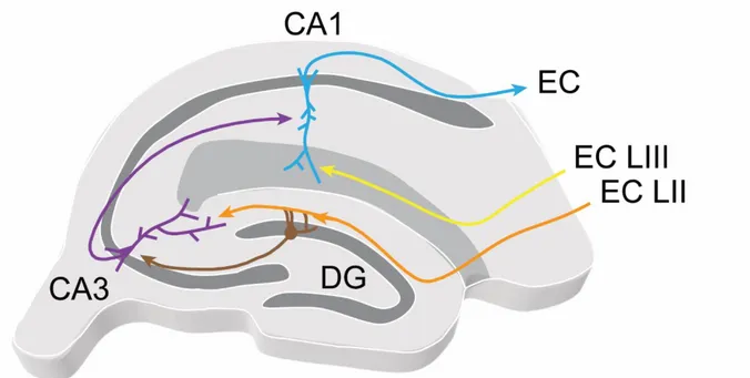
Figure I.2.3. Integrated view of area CA2 in the intra- and extra-hippocampal circuits. ... 29
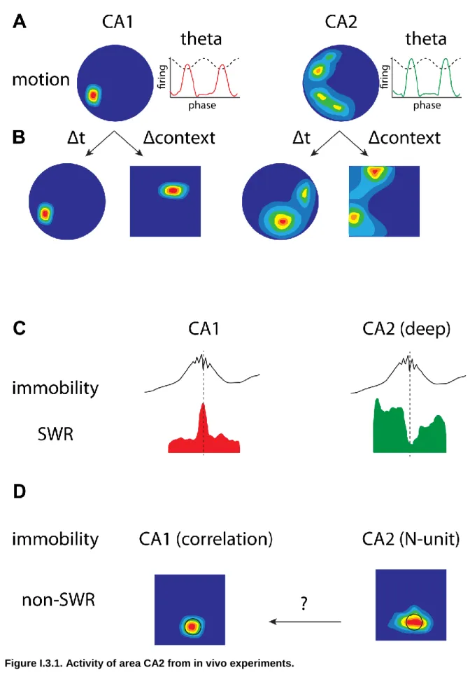
Figure I.3.1. Activity of area CA2 from in vivo experiments. ... 41
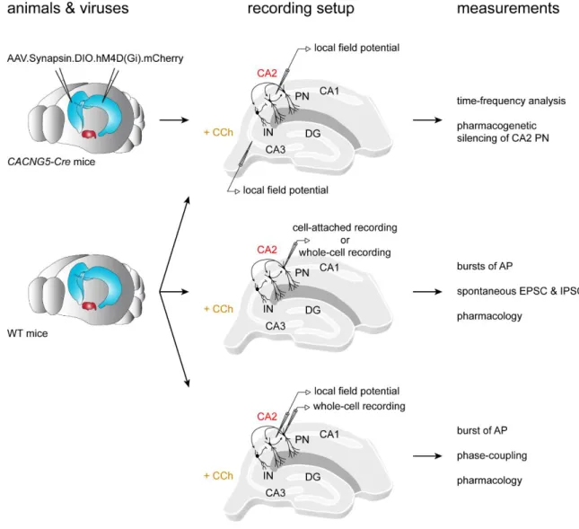
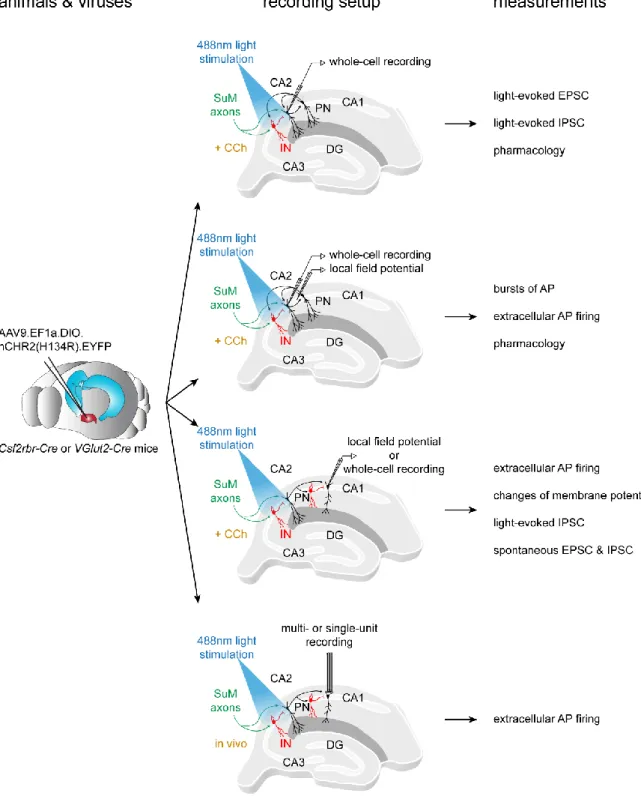
Figure II.8.1. Experimental workflow of the study on CCh-induced network activity... 53
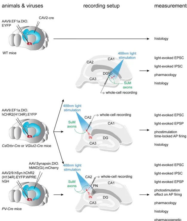
Figure II.8.2. Experimental workflow of the study on SuM input to area CA2. ... 56
Figure II.8.3. Experimental workflow of the study on the consequences of SuM activation on area CA2 and CA1 activity. ... 58
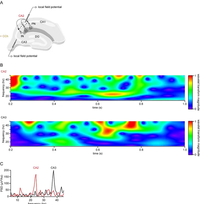
Figure III.1.1. Carbachol induces gamma-like oscillations in hippocampal area CA2 and CA3. ... 61
Figure III.1.2. Temporal relations of CCh-induced oscillations from area CA2 and CA3. ... 63
Figure III.1.3. Effects of chemogenetic silencing of CA2 PNs on CCh-induced oscillations. ... 65
Figure III.1.4. CA2 PNs fire bursts of APs following CCh application. ... 68
Figure III.1.5. Characteristics of charbachol-induced CA2 PN spontaneous activity. ... 71
Figure III.1.6. Effect of CCh on spontaneous excitatory and inhibitory events in CA2 PNs. ... 73
Figure III.1.7. Excitatory and inhibitory transmission shape bursts of action potentials in CA2 pyramidal cells. ... 77
Figure III.1.8. Bursts of action potentials in CA2 pyramidal cells are modulated by the ongoing field oscillation. ... 79
Figure III.2.1. Selective labeling of SuM neurons that project to hippocampal area CA2. ... 85
Figure III.2.2. CA2-projecting SuM neurons express VGluT2. ... 86
Figure III.2.3. SuM input provides excitatory glutamatergic transmission to diverse population of PNs in area CA2. ... 90
Figure III.2.4. Reconstruction of pre-synaptic SuM fibers and post-synaptic PNs in area CA2. ... 91
Figure III.2.5. SuM light-evoked mono-synaptic excitation as well as mono- and di-synaptic inhibition onto DG GCs. ... 95
Figure III.2.6. SuM axon stimulation recruits feedforward inhibition. ... 100
Figure III.2.7. Area CA2 PNs receive a net inhibitory drive from SuM that controls summation and AP firing properties. ... 104
12
Figure III.2.8. SuM inputs strongly excite basket cell interneurons in area CA2. ... 108
Figure III.2.9. SuM inputs provide excitation to Parvalbumin-expressing BCs. ... 110
Figure III.2.10. Parvalbumin-expressing BCs mediate the feedforward inhibition recruited by photostimulation of SuM fibers. ... 114
Figure III.3.1. Reduction of SuM excitatory and inhibitory transmission by carbachol. ... 119
Figure III.3.2. SuM control of CA2 PN bursting activity. ... 122
Figure III.3.3. Immediate consequences of SuM activation on CA1 activity. ... 125
Figure III.3.4. Delayed consequences of SuM activation on CA1 activity. ... 128
Figure IV.3.1. Integrated view of brain state-dependent SuM and area CA2 activity. ... 147
Table III.1.1. Characteristics of charbachol-induced CA2 PN spontaneous activity. ... 70
Table III.2.1. Electrophysiological properties of pyramidal neurons in SuM innervated area ... 88
Table III.2.2. Properties of deep and superficial pyramidal neurons in areas CA2 and CA3a ... 88
Table III.2.3. Characteristics of SuM light-evoked transmission onto PNs in area CA2. ... 92
Table III.2.4. Characteristics of SuM light-evoked responses in deep and superficial pyramidal neurons in areas CA2 and CA3a. ... 93
Table III.2.5. Electrophysiological properties of interneurons in SuM-innervated area. ... 106
Table III.2.6. Characteristics of SuM light-evoked transmission onto interneurons & pyramidal cells.106 Table III.3.1. Consequences of SuM photostimulation over area CA2 on CA1 PNs. ... 130
13
I - Introduction
I.1 - Roles of the hippocampus and the hypothalamus in memory
To form memories, the brain has the remarkable ability to encode, store and retrieve information. This cognitive function is critical for survival, adaptation to a changing environment, and life in society. Memory is a complex process that encompasses several systems spanning different structures of the central nervous system. This section briefly reviews important features of memory and introduces relevant brain regions, in particular the hippocampus and hypothalamus.
From human studies, much has been learned about how the brain forms memories. Memory is divided into short- and long-term based on the time course of the retained information. These two categories are handled by different parts of the brain, as evidenced by patients with long-term memory loss who can still perform well in a working memory task that requires keeping information available during a short period of time (Baddeley, 2003). Long-term memory is further divided in declarative and non-declarative based on the nature of the information retained. Declarative memory is the process in which items that can be explicitly expressed. Non-declarative memory regroups unconscious forms of memory (Cohen and Squire, 1980; Squire and Zola-Morgan, 1991). Non-declarative memory nevertheless implies learning such that a given input will reliably trigger a corresponding output. Reflex pathways, conditioned associations, priming processes and procedural abilities fall into the non-declarative class of memory (Squire and Zola-Morgan, 1991; Tulving and Schacter, 1990). Declarative memory applies to the process of memory formation and recall of conscious items, either semantic if they are general facts or episodic when they involve personal information. Episodic memory is therefore defined as the memory of events concerning the self that can be explicitly recollected. Strikingly, these different forms of long-term memory rely on specific brain structures that could be discovered in patients with lesions in the corresponding dedicated areas. Indeed, the seminal study of patient H.M. revealed a specific role for the hippocampus and related structures in episodic memory (Squire and Wixted, 2011). Following bi-lateral resection of medial temporal lobes to remove epileptic foci, H.M. developed a full anterograde and partial retrograde amnesia (Scoville and Milner, 1957). Remarkably, procedural and short-term memory were unaffected by H.M.’s lesions, and so was his retrograde long-term memory to the extent remote times before the surgery (Sagaret al., 1985; Squire and Wixted, 2011). Combined
14
with clinical observations from other amnesic patients and experimental work in monkeys and rodents, these findings firmly establish a selective role for the hippocampus in episodic memory (Mishkin, 1978; Mumby, 2001; Scoville and Milner, 1957; Zola-Morgan et al., 1986; Zola‐ Morgan et al., 1994). Furthermore, the anterograde nature of H.M.’s amnesia revealed that the hippocampus is necessary for learning and forming new episodic memories.
Although these findings converged on a central role for the hippocampus in episodic memory, it also appeared that the hippocampus was not the sole brain structure responsible for learning and memory. Indeed, the preserved past knowledge of H.M. proved that the hippocampus is not the ultimate storage site for long-term episodic memories. Moreover, H.M.’s lesions were not restricted to the hippocampus and additional work in monkeys and rodents reported that hippocampal lesions alone leave certain aspects of memory unaffected (Mishkin, 1978; Mumby, 2001; Squire and Wixted, 2011; Zola‐Morgan et al., 1994). Perhaps not too surprising given their close relations with the hippocampus, the entorhinal cortex and amygdala are also involved in episodic memory (Mishkin, 1978; Squire and Wixted, 2011). Another brain structure implicated in memory, notably in humans, is the hypothalamus. This diencephalic area suffers extensive damage in a pathological condition called Korsakoff’s syndrome (Mairet al., 1979). Often caused by chronic alcoholism, Korsakoff’s syndrome is characterized by damage to the thalamus and the hypothalamic mamillary region, resulting in memory impairments in humans and mice models of the disease (Daniel, 2005; Mair et al., 1979). Interestingly, the hippocampus itself is mostly spared in Korsakoff’s syndrome (Daniel, 2005) suggesting that injuries to brain structures connected to the hippocampus can cause similar memory deficits as hippocampal lesions. Indeed, the mamillary region of the hypothalamus is closely associated with the hippocampus as the mamillary bodies receive hippocampal projections and the suprammillary nucleus innervates the hippocampus (Pan and McNaughton, 2004a). However, even with this, there is paucity of physiological investigations of this hypothalamo-hippocampal circuit.
I.2 - Brain circuits relevant to episodic memory
In order to understand how episodic memory can be formed, stored and mobilized in the central nervous system, one needs to examine the brain structures involved, how they are connected and what neurons they host. Therefore, this section describes the anatomy, global and local connectivity, and cellular composition of the hippocampal formation with emphasis on area CA2.
15
I.2.a - Flow of information in the canonical hippocampal loop
I.2.a.i - Hippocampal anatomy and cellular composition
The hippocampus is a bilateral archicortical structure folded under the cerebral cortex and is widely conserved in numerous species including humans and rodents. In mice, the hippocampus is a prominent structure that occupies a large volume of the brain extending on a rostro-caudal and dorso-ventral axis from a septal pole to a temporal pole in a curved shape. Besides its local circuitry, the hippocampus is connected to several brain structures. The main glutamatergic afference to the hippocampus comes from layers II and III of the entorhinal cortex (EC) (Steward and Scoville, 1976; Witter et al., 1989). Another major glutamatergic input resides in the hippocampus itself, as left and right hippocampi are inter-connected (Blackstad, 1956). GABAergic neurons in the septum project to the hippocampus where they specifically innervate interneurons (Freund and Antal, 1988; Freund and Buzsáki, 1996). The septum also provides cholinergic inputs to the hippocampus (Shute and Lewis, 1963). Other neuromodulatory afferences to the hippocampus include noradrenaline from the locus coeruleus (Swanson and Hartman, 1975), serotonin from the raphe (Freund et al., 1990), dopamine from the ventral tegmental area and substantia nigra (Gasbarri et al., 1994). Additional projections to the hippocampus arise from the amygdala (Kemppainen et al., 2002), thalamus (Witter, 1996), and hypothalamus (Cui et al., 2013; Maglóczky et al., 1994). Efferent projections of the hippocampus are diverse as well. The major hippocampal output is the EC, thus establishing a reciprocal loop with this structure, either directly or via the subiculum (Naber et al., 2001). Reciprocal connections also consist of hippocampal projections to the septum, amygdala, and hypothalamus (Groen and Wyss, 1990; Swanson and Cowan, 1977). In addition, the hippocampus sends outputs to the neocortex olfactory bulb, nucleus accumbens and thalamus (Groen and Wyss, 1990; Gulyás et al., 1998; Jay et al., 1989; Swanson and Cowan, 1977). Like most regions of the brain, the hippocampus hosts two main kinds of neurons : the majority are principal cells and the minority interneurons (INs). Classically, principal cells are excitatory as they use the neurotransmitter glutamate while interneurons release GABA and are therefore inhibitory (excepted during the development of the central nervous system (Rivera et al., 1999)). Principal cells can drive excitation within and across regions and are therefore considered as the main communication units of the brain. Interneurons are involved in local interactions but also drive inhibition between brain areas through long-range projections (Caputi et al., 2013). In the hippocampal formation, neurons are segregated in different regions
16
that have anatomical and functional specificities : the dentage gyrus (DG), and Cornu Ammoni (CA) areas CA3, CA2 and CA1 (Lorente de No, 1934). These regions are further subdivided in their radial axis corresponding to the different parts of the principal cells parallel dendritic arborization. Indeed, principal cells are organized in a laminar way such that all somas are aligned, thus forming the pyramidal layer (stratum pyramidale, SP). The dendrites of these principal cells extend in a preferential direction perpendicular to SP and are therefore aligned parallel to each other on either side of SP. The outer layer formed by basal dendrites of pyramidal neurons (PNs) is called stratum oriens (SO), and the apical dendrites fill the inner layers of the hippocampus. The juxta-somatic inner layer in area CA3 is called stratum lucidum (SL) and corresponds to axonal tracks of the dentate gyrus granule cells, the mossy fibers (MF). Axons from area CA3 PNs, the Schaffer collaterals, run through the proximal part of the apical dendrites forming a layer called stratum radiatum (SR). Finally, the distal part of the apical dendritic arbor is contacted by afferent fibers from the entorhinal cortex (EC) in the stratum
lacunosum moleculare (SLM).
Figure I.2.1. Hippocampal anatomy in a transverse slice.
Drawing of Golgi stain illustrating the cytoarchitecture and connections within the different
hippocampal subfields with highlighted laminar organization (adapted from Ramon y Cajal, 1911).
Within this common structural organization, principal cells from each region display specific characteristics in terms of morphology, dendritic arborization, electrophysiological properties, connectivity, and gene expression profiles (discussed here and in Section I.2.a.ii). Principal
17
cells of the DG, the granule cells (GCs), have small ovoid cell bodies and a highly polarized dendritic tree that arborizes only on their basal side in stratum moleculare (SM) (Claiborne et al., 1990). Their axons emerge on the apical side in the hilus and extend towards area CA3 in SL. Pyramidal neurons (PNs) of area CA3 are large cells with basal dendrites in SO and bifurcated apical ones in SL, SR and SLM. The dendritic portion in SL displays thorny excrescences (TEs) where MF from the DG make synaptic contact. Preferential axonal projections of CA3 PNs varies according to their location on the CA3a (closer to CA2) – b – c (closer to DG) axis. CA3a PNs give rise to extensive recurrent connections and project to proximal CA1 (closer to CA2). CA3c PNs contribute less to the recurrent system and project more towards distal CA1 (closer to the subiculum) (Ishizuka et al., 1990; Li et al., 1994). CA1 PNs have smaller somas and a different apical dendritic organization than CA3. In contrast to CA3, CA1 PNs apical dendrites do not bifurcate in two apical branches but instead arborize extensively in SR where CA3 afferent axons project, and more modestly in SLM (Bannister and Larkman, 1995). CA2 PNs differ from both CA3 and CA1 in that they have large soma, bifurcated apical dendrites that do not display TEs and arborize very little in SR but dramatically in SLM where inputs from EC form synapses (Chevaleyre and Siegelbaum, 2010; Mercer et al., 2007; Piskorowski and Chevaleyre, 2012). Electrophysiological properties of CA2 PNs are also unique with respect to CA1 and CA3 PNs. CA2 PNs show a low membrane resistance, hyperpolarized resting membrane potential, low hyperpolarizing sag current, and no after spike hyperpolarization (Chevaleyre and Siegelbaum, 2010; Piskorowski et al., 2016; Sun et al., 2017). Besides variations on the proximo-distal axis, PNs vary according to their somatic location on the radial axis of the hippocampus. In area CA1, deep cells closer to SO and superficial cells closer to SR differ in terms of genetic expression, afferent innervation, efferent projections, and physiological functions (Danielson et al., 2016; Lee et al., 2014; Masurkar et al., 2017; Mizuseki et al., 2011; Valero et al., 2015). In particular, CA2 PNs drive stronger excitation on deep compared to superficial CA1 PNs (Kohara et al., 2013; Valero et al., 2015). Interestingly, in vivo functional differences between deep and superficials PNs have recently been reported in area CA2 (Oliva et al., 2016a), however the cellular and circuit mechanisms underlying these observations are not yet elucidated.
Besides excitatory pyramidal neurons, the hippocampus contains inhibitory interneurons that gate the inputs and outputs of PNs, shape network oscillations and participate in synaptic plasticity. In contrast with pyramidal neurons that form a relatively homogeneous population, interneurons are extremely diverse in terms of morphology, electrophysiological properties and
18
expression of genetic markers, reflecting their involvement in various distinct functions (Freund and Buzsáki, 1996; Somogyi and Klausberger, 2005). INs can be classified based on morphological features that allow functional predictions such as the location of their soma, the extension of their dendritic tree, and the targets of their axonal projections. Additional genetic and electrophysiological criteria provide further refinement of often overlapping classifications. A first functional group of INs target the dendrites of PNs and are therefore likely to control their afferent synaptic inputs. O-LM cells have their soma and dendrites located in SO and project their axon all the way to SLM (Maccaferri et al., 2000; McBain et al., 1994), thus potentially controlling EC inputs onto distal PNs or INs dendrites (Somogyi and Klausberger, 2005). O-LM cells express somatostatin (SOM) and fire action potentials quite regularly (Maccaferri et al., 2000). Bi-stratified INs also target PNs and INs dendrites but in SO and SR where they potentially control afferent excitatory inputs from CA3 on proximal dendrites (Buhl et al., 1994; Somogyi and Klausberger, 2005). They have a soma in SP or SO, dendrites extending in SO and SR, express SOM and show little accommodation in their firing pattern (Maccaferri et al., 2000; Pawelzik et al., 1999). SC-associated cells show a similar targeting of their axon to SR but have their soma located in SR, dendrites spanning SLM through SO, express cholecystokinin (CCK) and significantly accommodate their AP firing (Pawelzik et al., 2002; Vida et al., 1998). The other broad class of INs have axons targeting the soma or axon initial segment of PNs and are therefore in position of controlling their ouput. It is especially the case for axo-axonic cells whose axon targets axon initial segment of PNs and are thus likely to affect action potential generation in these cells (Somogyi and Klausberger, 2005). Axo-axonic cells extend their dendrites from SO through SLM, express parvalbumin (PV), and fire action potentials with either little or marked accommodation (Buhl et al., 1994a; Somogyi and Klausberger, 2005). The major IN type controlling PN output is the basket cell (BC) population, named after the dense peri-somatic net formed by axons from these cells (Buhl et al., 1994a). BCs have their soma in SP or SR, their dendrites spanning all CA strata, and express either PV or CCK (Maccaferri et al., 2000; Pawelzik et al., 2002). Remarkably, PV-expressing BCs are mostly fast-spiking whereas CCK-expressing BCs are mostly regular-spiking (Pawelzik et al., 2002).
Regardless of their type, INs can be involved in different type of wiring in hippocampal networks. The source of INs excitation can be local PNs that are subsequently inhibited by the INs : this scenario is called feedback inhibition. Feedback inhibition controls the duration and spread of excitation in PNs and can therefore pace their activity (Bartos et al., 2011).
19
Alternatively, feedforward inhibition happens when a common excitatory inputs targets both PNs and INs that can inhibit the PNs even before their AP discharge. Feedforward inhibition curtails excitation in post-synaptic PNs thus increasing the temporal precision of their AP firing (Pouille and Scanziani, 2001). In addition, INs can be engaged in mutual inhibition if they target one another, much like recurrent excitation amongst PNs but with opposite effect. Finally, certain populations of INs such as BCs are electrically coupled by gap junctions allowing for the propagation of membrane potential variations amongst these networks of INs.
Like the pyramidal neurons in area CA2, there is mounting evidence that the interneurons in this region have unique properties and potentially play significant roles in memory formation and disease. Studies performing hippocampal-wide comparison of different subclasses of interneurons based on immunological markers have revealed that CA2 comprises a peculiar composition of interneurons as compared to CA1 and CA3 (Botcher et al., 2014). While most immunohistological studies overlook area CA2 when quantifying interneuron density, recent studies have performed detailed quantification of interneuron densities along with concomitant staining of CA2-specific proteins. One very thorough study performed in rat hippocampus examined the density and stratum-localization of interneurons by staining for GAD-67, parvalbumin (PV), CCK, caretinin, calbindin, reelin, somatostatin, NPY and VIP, all with CA2 borders well-defined by either PCP-4 or alpha-actinin 2 staining (Botcher et al., 2014). This study and others found that area CA2 contains the highest density of PV-expressing interneurons (Botcher et al., 2014; Piskorowski and Chevaleyre, 2013). Furthermore, area CA2 contains the highest density of reelin-expressing neurons in the hippocampus (Botcher et al., 2014), which is very interesting given the various signaling roles of reelin in the adult hippocampus (Kupferman et al., 2014).
Using sharp recordings, Mercer et al. performed elegant and detailed analysis of individual CA2 interneurons in hippocampal slices from rats. Their work revealed unexpected morphological and electrophysiological properties of basket cells and bistratified cells in this region (Mercer et al., 2012a; 2012b). Area CA2 hosts two types of PV-expressing (PV+) basket cells: a minority resemble classical CA1 basket cells with narrow dendritic arborization in the septo-temporal axis, no sag potential and non-adapting fast spiking firing pattern, while the majority have a broad dendritic arborization, substantial sag potential and an adapting firing pattern normally encountered in CA1 O-LM interneurons (Mercer et al., 2007). Wide-arbor basket cells further differed by displaying excitatory post-synaptic potentials (EPSPs) elicited by pre-synaptic CA2 pyramidal neuron stimulation that were less depressing and longer in duration
20
due to a NMDAR component (Mercer et al., 2012a). Strikingly, similar observations were made regarding bistratified cells in area CA2, which consisted of narrow and wide-arbor subpopulations, indicating that the width of dendritic arborization, rather than axonal targeting, could serve as a reliable determinant of electrophysiological properties of interneurons in area CA2 (Mercer et al., 2007; 2012a). Finally, a novel type of interneuron was described in area CA2 with similar electrophysiological properties and morphologies as wide-arbor bistratified cells but with axons restricted to SR (SP–SR interneurons) (Mercer et al., 2012b). How these unusual types of basket cells, bistratified cells and SP–SR interneurons participate in controlling information flow in the hippocampal network remains to be understood.
I.2.a.ii - Inter-regional connectivity and information processing
As described above, the hippocampal formation comprises different regions with specific connectivity allowing for information processing. Inputs from layers II of the medial and lateral entorhinal cortex (EC) transfer information to the DG that then targets area CA3 proximal dendrites through the mossy fibers (MF) in the stratum lucidum (SL), CA3 subsequently makes synaptic contact on the proximal dendrites of area CA1 through the Schaffer collaterals in SR and CA1 returns information to the EC, thus forming the tri-synaptic loop (Andersen et al., 1971). Additional routes running through the SLM allow the EC to directly impinge on the distal dendrites of pyramidal neurons in all CA regions (Ruth et al., 1982; 1988; Steward and Scoville, 1976). Much can be learnt about the functions of this tri-synaptic loop by considering the physiology of each region and the computation performed at each step of the circuit. Afferent information to the hippocampus come from EC inputs that convey multimodal sensory (Canto et al., 2008), metric (Hafting et al., 2005) and directional signals (Taube et al., 1990). Axons from the EC run through the perforant path and make glutamatergic synapses onto the dendrites of granule cells in stratum moleculare (SM) (Deller et al., 1996). This information is processed by the dentate gyrus to allow pattern separation, i.e. the discrimination of similar inputs by sending a specific output. Indeed, dentate gyrus lesions impair spatial pattern separation in rats (Gilbert et al., 2001). This remarkable function of the dentate gyrus can be explained by the underlying physiology of its network. Although electrotonically compact, granule cells have a hyperpolarized resting membrane potential (Spruston and Johnston, 1992) making it potentially difficult for EC excitatory inputs to elicit action potential firing in post-synaptic granule cells. In addition, both EC inputs and granule cells excite dentate gyrus interneurons, thus recruiting feedforward and feedback inhibition, respectively (Freund and Buzsáki, 1996; Han et al., 1993; Kneisler and Dingledine, 1995; Sloviter, 1991). Altogether,
21
these characteristics confer low excitability to dentate gyrus granule cells (Jung and McNaughton, 1993; Penttonen et al., 1997). Therefore, excitatory transmission from EC inputs is likely to result in the activation of only a sparse population of granule cells. This is believed to allow pattern separation in the dentate gyrus as similar EC inputs would result in different granule cells outputs.
Axons from the dentate gyrus granule cells form the mossy fibers (MF) that run through SL towards the very proximal part of CA3 PNs. The synapse between mossy fibers and CA3 PNs consists of a mossy presynaptic bouton and a large postsynaptic differentiation called thorny excrescence. A single granule cell axon contacts thorny excrescences of a limited number of postsynaptic CA3 PNs (Acsády et al., 1998), thus keeping the pattern separated information orthogonalized. Efficacy of transmission at these synapses can be high and can reliably trigger action potential firing in CA3 PNs provided pre-synaptic granule cells fire repeatedly (Henze et al., 2002). In addition, mossy fibers also project onto CA3 interneurons (Acsády et al., 1998) that in turn drive feedforward inhibition onto CA3 PNs. Much like the dentate gyrus, CA3 PNs and INs receive EC inputs in SLM (Kiss et al., 1996; Witter et al., 1989). From the information conveyed by these two upstream inputs, area CA3 performs pattern completion, i.e. builds a coherent output from partial inputs (Rolls, 1996). Pattern completion in area CA3 has been evidenced by the reactivation of CA3 ensembles after exposure to similar contexts (Vazdarjanova and Guzowski, 2004), and by impaired recall in mice lacking NMDARs in CA3 PNs (Nakazawa et al., 2002). A notable feature of the CA3 network that likely underlies pattern completion is the prominent recurrent excitation between CA3 PNs (Miles and Wong, 1986). These recurrent excitatory connections between CA3 PNs, together with sparse but strong input from the dentate gyrus (Geiger and Jonas, 2000; Henze et al., 2002) and the recruitment of interneurons that drive feedback inhibition (Miles, 1990), are thought to allow the reactivation of “meaningful” CA3 PNs ensembles from sparse dentate gyrus inputs. It should be noted that pattern completion is highly specific as small changes in context can lead to orthogonalization of spatial representation in area CA3 (Leutgeb et al., 2004).
CA3 PNs in turn project onto CA1 PNs proximal dendrites via the Schaffer collaterals in SR. The CA3 – CA1 PN synapses have been shown to bring about modest levels of excitation (Larkman et al., 1991; Sayer et al., 1990). Afferent CA3 axons also recruit feedforward inhibition onto CA1 PNs by exciting local CA1 interneurons (Lacaille, 1991). Entorhinal inputs provide another source of excitation (Yeckel and Berger, 1990) to CA1 PNs and to interneurons that can contribute in feedforward inhibition (Kiss et al., 1996). However, excitatory synapses
22
formed in SLM by EC axons are inefficient in driving CA1 PNs action potential firing because of EPSPs generated in SLM are dampened by filtering on their way from the distal dendrites to the soma (Andreasen and JDC, 1998; Golding et al., 2005; Magee, 1998). Therefore, EC inputs require summation with other sources of excitation to drive activity in postsynaptic CA1 PNs. This puts area CA1 in a good position to act as an integrator of current experience through EC inputs and past experience “kept in memory” relayed by the DG – CA3 – CA1 path. Furthermore, excitatory recurrent connections are rare in area CA1 (Deuchars and Thomson, 1996) while feedback inhibition is prominent (Ali and Thomson, 1998; Ali et al., 1998; Buhl et al., 1994a). Altogether, these properties of the CA1 network are thought to allow weighted representations of past and present experience to be formed and sent to outside of the hippocampus. Efferent projections from CA1 PNs target the entorhinal cortex, directly or via the subiculum, hence closing the loop (Naber et al., 2001).
Figure I.2.2. Flow of information in the tri-synaptic loop of the hippocampus.
Diagram illustrating the EC – DG – CA3 – CA1 – EC circuit.
The accumulated knowledge of the functions of the tri-synaptic loop led to the idea that it supports memory formation in the hippocampus through pattern separation in the DG, pattern completion in area CA3, and integration in area CA1. However, in vivo testing of this view by disruption of the transmission between CA3 and CA1 led to unexpected results. Indeed, major aspects of hippocampal-dependent spatial memory were unaffected by lesions or genetic block
23
of the CA3 – CA1 transmission (Brun et al., 2002; Nakashiba et al., 2008). The mono-synaptic EC – CA1 loop was proposed to rescue mnemonic functions of the hippocampus in the absence of the tri-synaptic loop. However, this interpretation is in contradiction with the inability of EC inputs to efficiently drive CA1 PNs. Shortly thereafter, the discovery of a di-synaptic loop bypassing the DG and area CA3 but rather involving area CA2 provided an alternative hypothesis for the rescue of hippocampal memory formation in the absence of transmission between CA3 and CA1 (Chevaleyre and Siegelbaum, 2010).
I.2.b - Integration of area CA2 inputs and outputs in the hippocampal circuit
I.2.b.i - Intra-hippocampal connectivity of area CA2
To understand and support the role of area CA2 in information transfer through the hippocampus, one musts characterize its connections with other hippocampal areas. The intra-hippocampal inputs to area CA2 have been studied using tracing, optogenetics and electrophysiology in vivo and ex vivo. Area CA2 receives direct input in SLM from the EC (Bartesaghi and Gessi, 2004; Chevaleyre and Siegelbaum, 2010; Cui et al., 2013; Hitti and Siegelbaum, 2014; Kohara et al., 2013). In vivo recordings from anesthetized guinea pigs reported that area CA2 is the first CA region to be active in response to EC input stimulation (Bartesaghi and Gessi, 2004), indicating a strong synaptic connection. This synaptic input was further elucidated with acute mouse brain slice recordings, revealing that mild electrical stimulation of these inputs is sufficient to make CA2 pyramidal neurons fire action potentials (APs) (Chevaleyre and Siegelbaum, 2010). This strong synaptic connection in area CA2 contrasts with distal CA1 cortical inputs, which has both high levels of feedforward inhibition as well as a large Ih current that prevents strong excitation of CA1 PNs (Chevaleyre and
Siegelbaum, 2010; Nolan et al., 2004). Thus, it is possible that cortical inputs directly excite CA2 PNs, which then project to CA1 PNs, forming a di-synaptic hippocampal circuit. The EC – CA2 connection was further examined using conventional retrograde tracing from area CA2 in mice. Cui et al. reported afferent projections from primarily layer II EC cells, with a few layer III cells (Cui et al., 2013). Further, the generation of two different transgenic mouse lines expressing Cre recombinase specifically in CA2 pyramidal neurons allowed Hitti and Siegelbaum with the Amigo2-Cre line and Kohara et al. with the MAP3K15-Cre line to examine this input in a highly controlled manner. Both studies confirmed a strong layer II EC
24
with sparcely labeled layer III cells following retrograde rabies tracing (Hitti and Siegelbaum, 2014; Kohara et al., 2013).
DG granule cell mossy fibers form functional synapses with CA2 pyramidal neurons, as shown by optogenetic stimulation (Kohara et al., 2013). Mossy fiber inputs onto CA2 PNs have been shown to differ from the well-studied DG – CA3 synapse. As originally observed by Lorenté de No, CA2 pyramidal neurons lack the thorny excressences that are stereotypical for the mossy fiber – CA3 synapse (No 1934) (Kohara et al., 2013). Furthermore, the DG – CA2 synapse has relatively much lower levels of excitatory transmission as compared to CA3 (Sun et al., 2017). This input is capable of driving feedforward inhibition by recruitment of CA2 interneurons (Kohara et al., 2013). Interestingly, it has been proposed from immunohistological studies that newborn DG granule cells project their axons to area CA2 (Llorens-Martín et al., 2015). This synapse merits further examination, as the DG was unlabeled in retrograde studies performed in the rat (Cui et al., 2013), indicating a potential species-specific difference. Furthermore, in the Amigo2-cre line, this connection was not identified with retrograde tracing (Hitti and Siegelbaum, 2014) but was with experiments performed in the MAP3K15-Cre line (Kohara et al., 2013). These differences may be due to technical differences between vectors and transgenic lines.
Conventional and rabies-based retrograde labeling in mice showed that CA2 pyramidal neurons receive inputs from both CA3 PNs as well as CA2 PNs (Cui et al., 2013; Hitti and Siegelbaum, 2014). Interestingly, stimulation of CA3 Schaffer collaterals in SR on acute slices from mice revealed that this input is dominated by a strong feedforward inhibitory drive in area CA2, unlike in CA1 (Chevaleyre and Siegelbaum, 2010). This strong feedforward inhibitory transmission between CA3 and CA2 strongly contrasts with the strong direct excitation and limited feedforward inhibition observed following distal EC input stimulation (Chevaleyre and Siegelbaum, 2010). In the context of information transfer, this large feedforward inhibition recruited by area CA3 onto CA2 indicates that area CA2 may not be recruited by CA3 neurons and may be bypassed in the tri-synaptic circuit. This alternative pathway for information flow had been previously proposed based on field recordings and voltage-sensitive dye imaging studies of entire hippocampal slices (Sekino et al., 1997). This study found that, in the majority of instances, no activity was detected in area CA2 following stimulation of the hilus. However, in some instances, a delayed activation of CA2 was detected that led to a larger and delayed signal propagation of area CA1. This early and insightful work may be revealing a potential role of area CA2 in hippocampal network function. Remarkably, the PV+ population of
25
interneurons is the substrate of a long-term depression of the CA3 to CA2 feedforward inhibition (Piskorowski and Chevaleyre, 2013) that subsequently allows CA2 to be recruited by CA3 inputs (Nasrallah et al., 2015). Hence, the inhibitory gating of area CA2 activity is also unique and shows dysfunctions in schizophrenia due to a loss of PV+ interneurons (Piskorowski et al., 2016).
The intra-hippocampal outputs of area CA2 have been investigated with tracing experiments,
ex vivo electrophysiology and genetically restricted optogenetics. Interestingly, by selectively
infecting EC neurons with rabies viruses in mice and conventional retrograde tracing in both mice and rats, Rowland et al. showed that area CA2 sends a return projection to EC layer II neurons (Rowland et al., 2013). Although this observation stands alone from other studies using either conventional or genetically targeted anterograde viral tracing from area CA2 (Cui et al., 2013; Hitti and Siegelbaum, 2014), the retrograde method used by Rowland et al. is not restricted to a specific pre-synaptic cell type and is potentially more efficient in labeling. This intriguing finding merits replication and further investigation. Using unilateral viral anterograde labeling of CA2 neurons in mice, Cui et al. reported ipsi- and contra-lateral projections to areas CA1, CA2 and CA3 (Cui et al., 2013). Paired recordings from CA2 and CA1 pyramidal neurons on acute hippocampal slices from mice directly proved an excitatory monosynaptic connection from CA2 to CA1 that appeared several-fold stronger than the CA3 to CA1 connection (Chevaleyre and Siegelbaum, 2010). Taking advantage of the Amigo2-Cre transgenic mouse line, Hitti et al. also reported axons of CA2 pyramidal neurons projecting to every CA area (Hitti and Siegelbaum, 2014). The CA2 to CA1 inputs were further examined with optogenetics using the MAP3K15-Cre mouse line, revealing that CA2 PNs have different excitatory drive along the radial CA1 axis, providing a much stronger excitation to deep CA1 pyramidal neurons (Kohara et al., 2013). This is quite interesting, as the deep and superficial CA1 pyramidal neurons project to different cortical and limbic regions (Lee et al., 2014). Using a different CA2-specific mouse Cre line driven by the calcium voltage-gated channel auxiliary subunit gamma 5 (CACNG5) promoter region, Boehringer et al. examined CA2 pyramidal neuron output to areas CA1 and CA3 in acute slices. This study showed that, while area CA2 recruits direct excitation and feedforward inhibition in both areas CA1 and CA3, the inhibitory drive of CA2 predominates in CA3 (Boehringer et al., 2017). In summary, by projecting to every CA subfield, CA2 pyramidal neurons are poised to act on the entire hippocampal network: there is a reciprocal control of E/I balance between areas CA2 and CA3, whereas CA2 acts very
26
strongly to preferentially excite deep CA1 pyramidal neurons, thereby influencing hippocampal output.
I.2.b.ii - Area CA2 long-range afferences and efferences
In addition to forming the hub of an intrinsic hippocampal network, area CA2 is connected with several extrahippocampal structures. Retrograde tracing from area CA2 and anterograde labeling of vasopressinergic neurons in the paraventricular nucleus of the hypothalamus (PVN) revealed that area CA2 is targeted by inputs containing vasopressin (Cui et al., 2013). The PVN is strongly activated in stressful and social situations and results in the release of oxytocin and vasopressin throughout the brain (reviewed by Iovino et al., 2017), thus area CA2 is likely to be directly modulated during these circumstances. Additional long-range inputs to area CA2 have been described from the medial septum, diagonal band of Broca and median raphé that were labeled after retrograde marker injections in area CA2 of mice (Cui et al., 2013). Projections from these 3 regions were confirmed with rabies-based retrograde tracing from CA2 pyramidal neurons in the Amigo2-Cre and MAP3K15-Cre mouse lines (Hitti and Siegelbaum, 2014; Kohara et al., 2013).
Another hypothalamic projection to area CA2 arises from neurons in the supramammillary nucleus (SuM) (Haglund et al., 1984; Vertes, 1992). Albeit a relatively small area, the SuM has a significant role in the central nervous system through its connections with numerous brain structures. It receives inputs from other hypothalamic nuclei and also from the raphe, the habenula, the central gray, the septum and cortical regions (Pan and McNaughton, 2004). The SuM outputs are diverse as well and include reciprocal connections several hypothalamic areas, the raphe, the central gray, the septum and the cingulate and infralimbic cortex (Pan and McNaughton, 2004; Vertes, 1992). In addition, the SuM sends projections to the locus coeruleus, the thalamus, the amygdala, the EC and the hippocampus (Pan and McNaughton, 2004; Vertes, 1992). Neurons in the SuM are chemically diverse and have been reported to express calretinin, CCK, VGluT2, VGAT, dopamine, VIP, substance P and nitric oxide synthase (NOS) (Pan and McNaughton, 2004). In the hippocampus, afferent axons originate from calretinin-, VGluT2-, VGAT-, NOS-, VIP-, CCK- and substance P-expressing neuronal populations in the SuM (Berger et al., 2001; Borhegyi and Leranth, 1997; Haglund et al., 1984; Kiss et al., 2000; Pedersen et al., 2017). Interestingly, a subpopulation of SuM neurons has been shown to project both to the hippocampus and to the septum (Borhegyi et al., 1997; Vertes and McKenna, 2000). In contrast, inputs to area CA2 and the DG are likely provided by different populations of SuM neurons (Borhegyi and Leranth, 1997; Soussi et al., 2010). Indeed, SuM
27
afferent axons to the DG co-express glutamatergic (VGluT2) and GABAergic markers (VGAT or GAD65) (Boulland et al., 2009; Soussi et al., 2010). These axons co-release glutamate and GABA onto DG granule cells (Pedersen et al., 2017) and potentially interneurons (Leranth and Nitsch, 1994; Nitsch and Leranth, 1996). Transmission from the SuM to the DG has been shown to facilitate population spikes evoked by performant path stimulation of EC inputs, potentially by increasing activity in granule cells and decreasing it in interneurons (Mizumori et al., 1989). In contrast, area CA2 is targeted by VGluT2-expressing SuM axons only that presumably form synapses exclusively on pyramidal neurons in the rat (Kiss et al., 2000; Maglóczky et al., 1994; Soussi et al., 2010). These CA2-projecting SuM axons also express calretinin and substance P (Berger et al., 2001; Borhegyi and Leranth, 1997; Nitsch and Leranth, 1993). This projection has also been reported in mice (Cui et al., 2013; Hitti and Siegelbaum, 2014; Kohara et al., 2013) and primates (Berger et al., 2001), where it is present in prenatal hippocampus and proposed to play a role in hippocampal rhythms necessary for proper development. Until now, functional investigation of the glutamatergic SuM – CA2 transmission has never been carried out, and neither has the neuromodulation of hippocampal networks by substance P released from SuM.
Neurons in area CA2 also send projections outside the hippocampal circuit to a number of brain regions, sometimes establishing reciprocal connections. Tracing studies in rodents indicate that this is the case for the SuM, the medial septum and the diagonal band of Broca (Cui et al., 2013). The lateral septum has also been reported as a long-range output of area CA2 by anterograde tracings in mice (Cui et al., 2013). These extrahippocampal projections were not observed when using the Amigo2-Cre specific of CA2 pyramidal neurons mouse line (Hitti and Siegelbaum, 2014) and are thus potentially due to a different population of neurons in area CA2, either long-range projecting interneurons or a subpopulation of pyramidal neurons not labeled by this Cre line.
The tendency of long-range reciprocal connections between area CA2 and other cerebral structures raises the possibility of mutual influences between these regions and the hippocampal network via area CA2. However, functional characterizations of these bi-directional connections are still missing and should provide a more comprehensive understanding of the interplay between area CA2 and the rest of the brain.
In summary, the cellular composition, neuronal physiology, intrinsic and extrinsic connectivity patterns of area CA2 make it uniquely positioned in the hippocampal network. A general feature of area CA2 is its low overall excitability conferred by a high density of INs and hyperpolarized
28
non-resistive PNs. As opposed to CA1, the net input received by CA2 PNs from CA3 is dominated by feedforward inhibition. Interestingly, this CA3 – CA2 feedforward inhibition is highly plastic whereas the CA3 – CA2 excitatory synapse is not. Also contrasting with CA1, distal EC inputs efficiently provide excitation to CA2 PNs thus potentially bypassing the tri-synaptic loop. Area CA2 strongly drives excitation in area CA1 and provides inhibition back to area CA3, thereby possibly isolating the di-synaptic loop from the tri-synaptic one. Interestingly, area CA2 receives and sends long range inputs and outputs that may favor or prevent activity of CA2 PNs thus influencing the rest of the hippocampal network by switching between CA3- and CA2-dominated loops.
29
Figure I.2.3. Integrated view of area CA2 in the intra- and extra-hippocampal circuits.
A. Diagram illustrating the input and output connections of area CA2 and the distribution of interneurons
30
neurons in intrinsic excitability. C. Example traces of post-synaptic potentials recorded from CA1 and CA2 pyramidal neurons in response to EC input stimulation. D. Examples traces of post-synaptic potentials recorded from CA1 and CA2 pyramidal neurons in response to CA3 input stimulation. E. Examples traces of unitary excitatory post-synaptic responses recorded from CA1 pyramidal neurons in response to CA3 or CA2 pre-synaptic cell stimulation.
Illustrations in C, D and E are inspired by (Chevaleyre and Siegelbaum, 2010).
I.3 - Physiological features of hippocampal-dependent memory formation
While being conceptually useful, the information transfer theory derived from neuronal connectivity within hippocampal networks provides only a “static” view of the system that does not reflect its operational states. To alleviate this limitation, this section details physiological activity patterns in the hippocampus and related functions with highlights on the contributions of area CA2.
I.3.a - Network activity patterns underlying learning and memory
I.3.a.i - Theta and gamma oscillations
PNs of the hippocampus are organized laminarly with their somas aligned and their dendrites parallel, thus forming electrical dipoles. In such a network, synchronous changes in membrane potential from populations of neurons generate an electrical field potential that can be recorded extra-cellularly either locally (local field potential, LFP) or superficially (electroencephalogram, EEG). This allows the examination of activity in neuronal networks in
vivo and led to the discovery of several patterns occurring during different brain states : theta,
beta, gamma, sharp waves / ripples (SWR), large and small irregular amplitude (LIA and SIA) (Vanderwolf, 1969). Theta and gamma rhythms are present during active wake and REM sleep while SWR occur during quiet wake and slow wave sleep (SWS) when LIA is predominant (Buzsáki, 2015).
Theta waves correspond to rhythmic oscillations ranging from 6 to 12 Hz (in rodents) encountered in the hippocampus and several related brain structures, either as proper field potential oscillations or as rhythmic AP discharges in this frequency band. These include a number cortical areas, the amygdala, septum, hypothalamus, thalamus, VTA and raphe (Buzsáki, 2002; Vertes and Kocsis, 1997). In the hippocampus, theta oscillations are prominent in all layers and subregions although their amplitude is largest in CA1 (Buzsáki, 2002). Of note, hippocampal networks are current generators of theta oscillations but not their sole pacemaker,
31
as isolation of the hippocampus from the septum abolishes the hippocampal theta (Vertes and Kocsis, 1997). Indeed, rhythmic activity in the theta range exists in brain structures afferent to the hippocampus : namely the septum, EC and SuM (Alonso and García-Austt, 1987a; 1987b; Kocsis and Vertes, 1994; Stewart and Fox, 1990). EC inputs impinging on distal dendrites bring about rhythmic excitation generating current sinks in SLM (Buzsáki, 2002). In addition, the septum is critical for hippocampal theta waves as it provides both permissive cholinergic and pacemaking GABAergic inputs (Buzsáki, 2002; Pan and McNaughton, 2004; Vertes and Kocsis, 1997). Remarkably, hippocampal neurons possess a repertoire of conductances allowing intrinsic resonance in the theta and gamma frequency range (Wang, 2010). Therefore, although septal cholinergic afferents to the hippocampus are diffuse and do not contribute to the post-synaptic potentials underlying theta, acetylcholine released by these fibers causes modifications in these conductances leading to a global depolarization of hippocampal neurons (Madison et al., 1987). This effect of elevated cholinergic tone setting hippocampal networks in a theta prone state led to the definition of an atropine-sensitive theta, named after the cholinergic antagonist atropine that abolishes it as opposed to an atropine-resistant theta (Kramis et al., 1975). Further contribution of the septum to hippocampal theta is brought about by rhythmic inhibition of local INs by hippocampus-projecting GABAergic septal INs (Freund and Antal, 1988; Hangya et al., 2009; Tóth et al., 1997). Peri-somatic targeting hippocampal INs entrained by periodic septal inhibition in turn trigger rhythmic inhibitory post-synaptic potentials in PNs, thus generating a current source in SP (Buzsáki, 2002). Finally, rhythmic activation of the intra-hippocampal CA3 – CA1 SC contribute to another current sink in CA1 SR (Buzsáki, 2002). Altogether, specific sets of conductances and rhythmic excitatory and inhibitory synaptic inputs on different dendritic compartments trigger oscillations of the membrane potential of hippocampal neurons at theta frequencies. This sets windows of excitability that differ depending on the neuronal population, thus leading to action potential firing by PNs and INs at specific preferential phases of the theta cycle (Fox et al., 1986; Klausberger et al., 2003; 2005). Indeed, average firing rates of PNs are highest around the negative peak of the CA1 SP theta and are preceded by INs from the pyramidal layer whose maximal discharges occurs 60° earlier (Csicsvari et al., 1999). It should be noted that these are only indicative values as preferred theta phases vary across cell types and depends on behavior (as discussed later in Section I.3.b.i - ). Still, PNs of areas CA3, CA2 and CA1 of the hippocampus seem to form a relatively homogeneous population with regards to theta oscillations : recent in vivo studies that examined PN properties in different CA regions reported either no differences (Kay et al., 2016; Mankin et al., 2015), or slightly stronger theta
32
modulation and earlier preferred theta phase of CA3 PNs compared to CA2 and CA1 PNs (Oliva et al., 2016a).
Besides the septum and EC, the SuM is of particular interest for the generation and control of theta oscillations in the hippocampus by extra-hippocampal sources. It has been described as the first nucleus where neurons display phasic rather than tonic firing in the so-called “ascending theta synchronizing path” that also comprises the reticular nucleus pontis oralis (RPO), posterior hypothalamus (PH), septum and hippocampus (Kirk, 1998). Indeed, neurons in the SuM fire in bursts at theta frequency coherent with hippocampal theta in anaesthetized rats (Kirk et al., 1996; Kocsis and Vertes, 1994). The SuM is in close relation with both the hippocampus by direct projections and the septum through reciprocal connections (Borhegyi and Freund, 1998; Leranth and Kiss, 1996; Vertes, 1992; Vertes and McKenna, 2000). Of note, theta activity in the SuM is not solely relayed by its interactions with the septum as it remains upon inactivation of the septum (Kirk, 1997; Kirk et al., 1996). Under anesthesia, the SuM influence over hippocampal theta is massive as evidenced by a reduction in frequency and amplitude of theta oscillations in the hippocampus upon disruption of SuM activity by pharmacological agents or lesions (Kirk and McNaughton, 1993; McNaughton et al., 1995; Thinschmidt et al., 1995). Although less pronounced, this effect remains in freely behaving animals (McNaughton et al., 1995; Pan and McNaughton, 1997; 2002). Altogether, these findings point out the SuM as a critical actor in the regulation of hippocampal theta oscillations, either through its direct projections to the hippocampus or via the septum.
Often concurring with theta, gamma waves are higher-frequency oscillations (30-100 Hz) also occurring throughout hippocampal layers and subregions, as well as in other brain areas including the cortex (Buzsáki and Wang, 2012; Colgin, 2016). Detailed analysis of gamma oscillations reveal that they can be subdivided based on their frequency, origin, amplitude-phase coupling relation to theta, and laminar organization (Belluscio et al., 2012; Bragin et al., 1995; Colgin et al., 2009; Schomburg et al., 2014). First, fast-gamma (also referred to as epsilon) spans frequencies from 90 to 150 Hz, is largest at the theta trough, and is confined to CA1 SP (Belluscio et al., 2012). Next, mid-gamma occupies the 50 to 90 Hz band of frequencies, shows maximal amplitude at the peak of theta, and is localized in SLM and DG consistent with its entorhinal origin (Belluscio et al., 2012; Bragin et al., 1995; Chrobak and Buzsáki, 1998; Colgin et al., 2009; Csicsvari et al., 2003; Schomburg et al., 2014). Finally, slow-gamma ranges from 30 to 50 Hz, is highest during the descending phase of theta, originates in area CA3 and therefore predominates in CA1 SR (Belluscio et al., 2012; Bragin et
33
al., 1995; Chrobak and Buzsáki, 1998; Colgin et al., 2009; Csicsvari et al., 2003; Schomburg et al., 2014).
Much like theta oscillations, cellular and circuit mechanisms underlying gamma oscillations have been examined using various approaches. Modelling studies based on physiological data indicate that gamma oscillations can emerge in neuronal networks consisting either of interneurons only engaged in mutual inhibition and driven by a common excitatory source (I-I or ING model), or of pyramidal cells and interneurons through recurrent excitation and feedback inhibition (E-I or PING model) (Buzsáki and Wang, 2012; Wang, 2010). In support of the latter, in vivo examination of action potential firing relative to the gamma cycle revealed that PNs discharge before INs in the recurrent CA3 network, but also drive CA1 INs consistent with feedback and feedforward inhibitory circuits respectively (Csicsvari et al., 2003). Given the intrinsic nature of slow gamma in hippocampal networks, several studies took advantage of the possibility to induce gamma oscillations in acute slices to further elucidate the mechanism involved. Indeed, elevation of the cholinergic tone by bath application of carbachol (CCh) induces gamma oscillations ex vivo that resemble the slow-gamma seen in vivo (Fisahn et al., 1998; Mann et al., 2005). These approaches notably allowed to validate the central role of fast-spiking PV-expressing basket cells in gamma oscillations, as they generate an active current source in SP through rhythmic perisomatic inhibition (Gulyás et al., 2010; Mann et al., 2005). In addition, they confirmed that intra-hippocampal gamma oscillations build up through recurrent excitation and feedback inhibition in area CA3 and are subsequently transferred to area CA1 by feedforward inhibition (Mann et al., 2005; Oren et al., 2006; Zemankovics et al., 2013).
Area CA2 has yet to be incorporated in this scheme although its intra- and extra-hippocampal connections indicate that it is involved in gamma oscillations. Indeed, area CA2 displays recurrent excitation (Cui et al., 2013; Hitti and Siegelbaum, 2014) and feedback inhibition (Mercer et al., 2012a; 2012b) much like CA3 and is therefore endowed with the necessary features for generating gamma oscillations. Furthermore, its strong excitatory drive from EC (Chevaleyre and Siegelbaum, 2010), reciprocal inhibitory influences with CA3 (Boehringer et al., 2017), and potent excitatory output onto CA1 (Chevaleyre and Siegelbaum, 2010; Kohara et al., 2013), places area CA2 in an ideal position to participate in mid-, slow-, and fast-gamma respectively. In addition, one can predict a role for area CA2 in theta phase coupling of gamma amplitude as it receives theta-locked inputs from the SuM (Kirk et al., 1996; Kocsis and Vertes, 1994). Finally, in vivo and ex vivo work describe the initiation site of slow-gamma waves in