UNIVERSITÉ DU QUÉBEC À MONTRÉAL
PROTECTIVE ROLE OF MILO THERMOTOLERANCE (40°C) AGAJNST -APOPTOSlS INDUCED BY OXlDATIVESTRESS
TI--IESIS PRESENTED
IN PARTIAL FULLFILLMENT OF THE REQUIREMENTS FOR THE DEGREE OF DOCTOR OF PHJLOSOPHY IN BIOCHEMISTRY
BY
PRAGATI-!I PALLEPATI
Avertissement
La diffusion de cette thèse se fait dans le rèspect des droits de son auteur, qui a signé le formulaire Autorisation de reproduire et de diffuser un travail de recherche de cycles supérieurs (SDU-522 - Rév.01-2006). Cette autorisation stipule que «conformément à l'article 11 du Règlement no 8 des études de cycles supérieurs, [l'auteur] concède à l'Université du Québec à Montréal une licence non ·exclusive d'utilisation et de publication de la totalité ou d'une partie importante de [son] travail de recherche pour des fins pédagogiques et non commerciales. Plus précisément, [l'auteur] autorise l'Université du Québec à Montréal à reproduire, diffuser, prêter, distribuer ou vendre des copies de [son] travail de recherche à des fins non commerciales sur quelque support que ce soit, y compris l'lnternE?t. Cette licence et cette autorisation n'entraînent pas une renonciation de [la] part [de l'auteur] à [ses] droits moraux ni à [ses] droits de propriété intellectuelle. Sauf entente contraire, [l'auteur] conserve la liberté de diffuser et de commercialiser ou non ce travail dont [il] possède un exemplaire.»
UNNERSITÉ DU QUÉBEC À MONTRÉAL
ROLE PROTECTEUR DE LA THERMOTOLERANCE ( 40°C) CONTRE L'APOPTOSE INDUITE PAR LE STRESS OXIDA TIF
THÈSE PRÉSENTÉE
COMME EXIGENCE PARTIELLE DU DOCTORAT EN BIOCHIMIE
PAR
PRAGATHI PALLEPATI
lll
ACKNOWLEDGEMENTS
1 would like to thank my supervisor Dr. Diana Averill-Bates, for accepting me as a student and promoting me into the field of cancer research. I greatly appreciate her constant foliow up and long lasting encouragement on ali aspects of this work. Finaliy her positive attitude towards life and science has enabled me to complete the pro gram.
1 would like to thank my husband Subhash Rauthu for ali his encouragement and kind support to me in joining the Ph.D. program. I would also like to thank, him for his cooperation in sharing household duties equaliy with my young daughter along with his own Ph.D. program. It would not have been possible to complete my Ph.D. without his continous help. Thanks to my daughter Shreya Rauthu for her cooperation by being nice most of the times.
I would like to thank my parents Paliepati Surender Rao and Laxmi and my twin sister Pranitha for their love towards me and their endless efforts. Without their efforts I would not have been able to reach this position.
I would like to thank my colleagues Melanie Grondin, Andre Tanel, Ahmed Bettaieb, Julie Roy, Yulia Zilber, Catherine Lauzon, Isabelle Robinson, Audrey Glory and ali my colieagues working in the department for their friendliness, encouragement and advice throughout the progress of my studies.
I would like to thank Denis Flipo and Bertrand Fournier for their technical support and statistics.
Finaliy I would like to thank CRSNG for financial support (research grant award to D. Averill-Bates) and Université du Québec à Montréal for awarding me with the internai scholarships (FARE).
TABLE OF CONTENTS
LIST OF FIGURES ... vii
LIST OF TABLES ... xii
LIST OF SCHEMES ... xiii
LIST OF ABREVIATIONS ... xiv
RÉSUMÉ ... xix CHAPTERI INTRODUCTION ... .1 1.1 Oxidative Stress ... 1 1.2 Antioxidants ... 5 1.2.1 Enzymatic antioxidants ... 5 1.2.2 Non-enzymatic antioxidants ... 7
1.3 Overview of Apoptosis and Ce11 Dea th ... 8
1.3.1 Caspases ... ll 1.3.2 Signa1ing pathways of Apoptosis: ... l5 1.3.2.1 Mitochondrial (intrinsic) pathway ... 16
1.3.2.2 Death receptor (extrinsic) pathway ... 20
1.3 .2.3 Endoplasmic reticulum (ER) stress (intrinsic) pathway ... 26
1.3.2.3.1 ER Stress ... 28
1.3.2.3.2 ER Stress-mediated apoptosis ... 30
1.4 The role ofp53 in apoptosis: ... 35
1.4.1 Induction of apoptosis by p53 ... 35
v
1.4.3 Role ofp53 in apoptosis: ... 39
1.4.4 p53 and oxidative stress ... 44
1.5 Protective/adaptive mechanisms against stress-induced apoptosis ... .45
1.5.1 The serine/threonine protein kinase Akt ... .47
1.5.2 Inhibitor of Apoptosis Proteins (IAPs) ... .47
1.5.3 FLIP ... 49
1.5.4 Bcl-2 Family proteins ... .49
1.5 .5 Thermotolerance (heat pre-conditioning) ... 51
1.5.5.1 Heat shock proteins: ... 52
1.5.5.2 Hsps: role in apoptosis ... 56 1.6 Presentation ofProject ... 59 1.6.1 Hypothesis ... 60 1.6.2 General objectives: ... 60 1.6.3 Specifie objectives: ... 61 CHAPTER II 2.1 PREFACE ... 62 2.2 AR TIC LE I MILD THERMOTOLERANCE INDUCED AT 40°C INCREASES ANTIOXIDANTS AND PROTECTS HeLa CELLS AGAINST MITOCHONDRIAL APOPTOSIS INDUCED BY HYDROGEN PEROXIDE: ROLE OF P53 ... 65
2.3 ARTICLE II MILD THERMOTOLERANCE INDUCED AT_ 40°C PROTECTS HeLa CELLS AGAINST ACTIVATION OF DEATH RECEPTOR-MEDIATED APOPTOSIS BY HYDROGEN PEROXIDE ... 109
2.4 ARTICLE III
ACTIVA TI ON OF ER STRESS AND APOPTOSIS BY HYDROGEN PEROXIDE IN HeLa CELLS: PROTECTIVE ROLE OF MILD HEAT
PRECONDITIONING AT 40°Co .. 0 .. 0 .... 0 .. 0 .. 0 .. 0 .. o .. 0 0 .. 0 0 0 .. 0 .. o .. 0 0 .... o .. o .. 0 0 . . . . 0142
CHAPTER III
DISCUSSION AND CONCLOSlON 0 0 0 0 0 0 0 0 0 0 0 0 0 0 0 0 0 0 0 0 0 0 0 0 0 0 0 0 0 0 0 0 0 0 0 0 0 0. 0 0 0 0 0. 0 0 0 0 0 0 0 .. 0 0 0. 0 0 0 0 0 180
APPENDIXA
ACROLEIN INDUCES A CELLULAR STRESS RESPONSE AND TRIGGERS MITOCHONDRIAL APOPTOSIS IN
A549 CELLSooooooo ... o ... o ... o ... o .. oooooo ... o ... oo ... o .. ooooooooooo ... ol98
APPENDIX B
ACROLEIN INDUCES APOPTOSIS THROUGH DEA TH RECEPTOR
PATHWAY IN A549 LUNG CELLS: ROLE OF P53 ... o .. ooooooooooo0213
VIl
LIST OF FIGURES
1.1 The mitochondrial formation ofROS and RNS ... 2
1.2 Damaging effects of ROS on key cell components ... 5
1.3 Role of enzymatic antioxidants in ROS detoxification ... 7
1.4 Different modes of cell death ... 1 0 1.5 Caspase activation by proteolysis ... 12
1.6 Downstream substrates that are targeted by active effector caspases ... 15
1. 7 Caspase-dependent and -independent pathways of apoptotic cell death ... 17
1.8 Effects ofROS on cardiolipin oxidation and cytochrome c release into the cytosol. ... 19
1.9 Fas- mediated death receptor pathway of apoptosis .. ... 22
1.10 Apoptosis-mediated by the dea th receptors ... 25
1.11 Functions of ER ... 27
1.12 Signaling the ER stress response (UPR) pathway ... 29
1.13 Mechanism of the ER mediated apoptosis ... 31
1.14 Role of Bcl-2 family pro teins in ER-mediated apoptosis ... 33
1.15 Activation of p53 and cellular response ... 36
1.16 Regulation ofp53 ... 38
1.17 p53-apoptotic pathways ... .40
1.18 p53 and Bcl-2 family proteins at the level of mitochondria ... 42
1.19 p53-associated pathways involved in apoptotic cell death ... 43
1.20 Levels of ROS regulate p53 function ... .44
1.21 The role of anti-apoptotic proteins (Akt, lAPs, FLIP, Bcl-2, Bel-XL, ROS scavengers and Hsps) in the regulation of major pathways of apoptosis ... 46
1.23 State of Bcl-2 family proteins during normal and apoptotic conditions ... 50 1.24 Diagram illustrating different stress conditions that induce the
heat shock response ... 53 1.25 Induction of Hsps inhibits apoptosis and promo tes cell survival.. ... 54 1.26 Events regulated by Hsps in the intrinsic (mitochondrial) and
extrinsic ( death receptor) pathways of apoptosis ... 58
ARTICLE I
1 A mild temperature of 40 °C induces enzymatic activity and
protein expression of antioxidant MnSOD ... 94 2 Mild heat treatment at 40 °C increases activity and protein
expression ofH202 scavenger catalase ... 95 3 Mild thermotolerance at 40 °C increases intracellular levels
of antioxidant glutathione and yGCS ... 96 4 ROS generation increases during induction of mild
thermotolerance at 40 °C ... 97 5 Thermotolerance developed at 40 °C protects cells against H202
-induced mitochondrial Bax translocation and mitochondrial
membrane depolarization ... 98 6 H202-induced release of cytochrome c from mitochondria is
decreased by mild thermotolerance ( 40 °C) ... 99 7 Mild thermotolerance ( 40 °C) inhibits activation of caspase-9 and
caspase-3 by H202 ... 100 8 Mi1d thermotolerance diminishes H202 -induced P ARP cleavage ... 1 01 9 Mild thermotolerance at 40 °C protects against H20rinduced
10 Inhibition of glutathione synthesis during thermotolerance partially decreases the development of resistance to H202
-lX
induced P ARP cleavage ... 1 03 11 Thermotolerance induced at 40 °C inhibits H202-induced
translocation of AIF from mitochondria to the nucleus ... 104 12 H20rinduces phosphorylation of p53 at ser 15 and ser 46 ... 105 13 Inhibition of p53 by pifithrin-a decreases induction of PUMA
and Bax translocation by H20 2 ... 106 14 Effect ofpifithrin-a on induction of chromatin condensation and
cytotoxicity by H202 ... 107
ARTICLE II
1 Protective effect ofmild thermotolerance against H202-induced
chromatin condensation and cytotoxicity ... 128 2 Mild thermotolerance protects cells against induction of Fas
ligand expression by H20 2 ... 130 3 H202 causes translocation of the adaptor protein F ADD to the
plasma membrane: abrogation by mild thermotolerance
developed at 40 °C ... 131 4 Caspase-8 plays a key role in H202-induced apoptosis:
abrogation by mild thermotolerance ... 132 5 Mild thermotolerance ( 40°C) inhibits H202-induced caspase-2
activation ... 133 6 H202-acivates the Bid-mediated cross-talk pathway:protection
by thennotolerance at 40 °C ... 134 7 Roles of caspase-8 and caspase-2 in H20rmediated cleavage ofBid ... 135 8 Caspase-9 activation by H20 2 is mediated by the cross-talk pathway ... 136
9 Role of the Fas in apoptosis mediated by H20 2 ... 137
10 Role ofp53 as an upstream factor in H202-mediated death receptor apoptosis ... 13 8 11 p53 inhibition decreases execution-phase events ofH20rinduced apoptosis ... 139
ARTICLE III 1 Mild heat pretreatment at 40°C causes resistance to H20r induced apoptosisa and clonogenic cell killing ... 164
2 Induction and activation of PERK in cells at a mi1d temperature of 40°C ... 165
3 Mild thermal stress ( 40°C) causes induction and phosphorylation of eiF2a ... 166
4 Protein expression of chaperone protein Bip (GRP78) in non-thermotolerant (37°C, 3 h) and themotolerant (40°C, 3 h) cells ... 167
5 H20 2 causes activation of PERK: role of mild thermotolerance ... 168
6 H20 2 causes eiF2a phosphorylation: role of thermotolerance at 40°C ... 169
7 H202 causes IRE 1 a activation: inhibition in thermotolerant cells ... 170
8 H202 causes cleavage of ATF6: role ofmild thermotolerance ... 171
9 Effect ofH202 on expression ofBip ... 172
10 H202 increases CHOP expression in HeLa cells: protective role of thermotolerance ( 40°C) ... 1 73 11 H20 2 causes calpastatin cleavage: inhibition by mild thermotolerance ... 174
12 H20 2 induces calpain activation and translocation to the plasma membrane: abrogation by mild thermotolerance developed at 40°C ... 175
13 Activation of caspase-4 and caspase-12 by H202: inhibition by mild thermotolerance ... 176
Xl
14 Roles of calcium, cal pain and caspase-7 in H202-induced
activation of caspase-4 and caspase-12 ... 177 15 H202-induced activation ofcaspase-9, caspase-3 and apoptosis is
diminished by apoptotic protease inhibitors ... 1 78 16 Knockdown of PERK by siRNA causes partial revers al of resistance
LIST OF TABLES
1.1 Characteristic morphological features of distinct modalities
of cell death ... 9
1.2 Subfamily members of the caspase family ... 13
Xlll
LIST OF SCHEMES
ARTICLE I
1 Mild thennotolerance protects against induction of mitochondrial
apoptosis by pro-oxidant H2020 0 0 0 0 0 0 000 0 0 0 0 0 00 0 . . 0 0 0 0 0 0 0 0 0 0 0 0 0 0 0 0 0 0 0 0 0 0 000 0 0 0 0 0 0 0 0 00 0 0 0 0 0 0 0 0 0 0 0 0 0 0 0 0 0 oooo 108
ARTICLE II
1 Schematic representation of the p53-dependent death receptor pathway of apoptosis induced by hydrogen peroxide and mediated
by Fas in HeLa cellso 0 0 0 ooooooooooooo 0 0 0 0 00 000 00 00 0 0 0 0 0 0 0 0 0 0 ooooooooooooooooooooooooooooooooooo 0 0 0 0 0 0 0 0 0 0 0 0 000 0 0 140
2 Summary of the protective role ofmild thennotolerance developed at 40 °C in the regulation of the death receptor pathway of apoptosis
LIST DES ABREVIATIONS
AFC Amino trifluorocoumarin
AMC Amino methylcoumarin
AIF Apoptosis-inducing factor
APAF-1 Apoptosis protease activating factor-1
Ac-DEVD-AMC Ac-Asp-Glu-Val-Asp-amino-4-methylcoumarin Ac-LEHD-AFC Ac-Leu-Glu-His-Asp-AFC
Ac-LEVD-AFC Ac-Leu-Glu-Val-Asp-AFC ATF6 Activating transcription factor-6
BAX Bcl-associated X protein
BCL-2 B celllymphoma 2
BSA Bovine serum albumin
BAPT A-AM 1, 2-Bis(2-aminophenoxy)ethane-N ,N ,N' ,N' -tetraacetic acid tetrakis (acetoxymethyl ester)
CuZnSOD Copper-zinc superoxide dismutase CAD Caspase activated DNase
CARD Caspase recruitment domain
CYT C Cytochrome c
CHAPS 3 -[ (3-cholamidopropyl) dimethylammonio ]-2-hydroxy-1-propanesulfonic acid
DIABLO/SMAC Direct IAP Binding Protein with Low PVSecond Mitochondria-derived Activator of Caspases
DNA Deoxyribose nucleic acid
D-MEM Dulbecco's modification ofEagle's medium
DISC Death-inducing signalling complex
DTT Dithiothreitol
EndoG Endonuclease G
ER Endoplasmic reticulum
ETC Electron transport chain
ERAD ER-associated protein degradation
eiF2a Eukaryotic initiation factor 2 alpha
EDTA Ethylenediaminetetraacetic acid
EGTA Ethylene glycol (2-aminoethyl ether)-N,N,N'N' -tetraacetic acid
ECL Electrochemiluminescence
FBS F etal bovine serum
FCCP P-trifluoromethoxy-phenyl-hydrazone
FasL Fas ligand
Fas Fas receptor
FADD Fas-associated death domain
c-FLIP Cellular FLICE inhibitory protein
FMK Fluoromethyl ketone
FACS Fluorescent activated cell sorter
GRP Glucose related protein
Gpx Glutathione peroxidise
GST-n:1 Glutathione S-transferase-n:1
y-GCS Gamma-glutamyl cysteine synthetase
GAPDH Glyceraldehyde-3-phosphate dehydrogenase
HtrA2/0mi High-temperature requirement A2 serine protease
HeLa Human cervical carcinoma cells
H202 Hydrogen peroxide
Hsps Heat shock proteins
IRE1 Inositol-requiring protein -1
lAPs Inhibitor of apoptotic proteins
JNK c-Jun N-terminal kinase
JC-1 5,5' ,6,6' :-tetrachloro-1, 1 ',3,3 '-tetraethylbenzimidazolcarbocyanine
L-BSO L-buthionine sulfoximine
LC3 Light chain 3
MPT Mitochondrial permeability transition
MnSOD Manganese superoxide dismutase
MOPS 3-(N-morpholino)-propane sulfonic acid
MOMP Mitochondrial outer membrane permeabilization
NF-KB Nuclear transcription factor KB NaCl Sodium chloride
NAD PH PUFA PERK PARP PBS PMSF PEG PI PTP PDI PVDF ROS RNS RIP1
Nicotinamide adenine dinucleotide phosphate Pol y unsaturated fatty acids
Protein kinase RNA (PKR)-like ER kinase PolyADP-ribose polymerase
Phosphate-buffered saline Phenylmethylsulfonyl fluoride
Polyethylene glycol Propidium iodide
Permeability transition pore Protein disulphide isomerise Polyvinylidene difluoride Reactive oxygen species Reactive nitrogen species Receptor-interacting kinase 1
SDS-PAGE Sodium dodecyl sulfate-polyacrylamide gel electrophoresis SEM
SOD t-Bid TNF TRAIL
Standard error of mean Superoxide dismutase Truncated bid
Tumor-necrosis factor
Tumor necrosis factor-related apoptosis-inducing ligand
TRAIL-R1 Trail receptor-1
TNFR
TRADD
TRAF-2
UPR
XIAP
Tumor necrosis factor receptor
TNF-receptor-associated death domain
TNF-receptor -associated factor-2
Unfold protein response
X-linked inhibitor of apoptosis protein
Z-IETD-AFC Z-Ile-Glu-Thr-Asp-AFC
Z-VDV AD-AFC Z-Val-Asp-Val-Ala-Asp-AFC
Xl X
RÉSUMÉ
Une faible exposition à des stress tels qu'un choc thermique, du stress oxydatif ou des radiations peut induire une réponse adaptative qui permet aux cellules et aux organismes qu'elles constituent de continuer à fonctionner normalement lorsqu'ils sont confrontés à un stimulus négatif. Ces réponses adaptatives impliquent de nombreux changements dans l'expression de gènes et de protéines, dont l'induction de défenses cellulaires (an ti oxydants, protéines de choc thermique (heat shock pro teins (Hsps)), etc.) qui permettront à la cellule de survivre. Si une réponse adaptative ne parvient pas à contrer les effets de l'exposition au stress, les cellules sont éliminés via des processus de mort cellulaire comme l'apoptose ou la nécrose. Une exposition préalable à des températures modérées, de l'ordre de 40°C, induit une réponse adaptative (la thermotolérance) qui permet aux cellules de résister à une agression toxique ultérieure. La thermotolérance induite par des températures modérées de l'ordre de 39-40°C (ce qui correspond physiologiquement à un état fiévreux) a été peu étudiée et est mal comprise. Les deux objectifs principaux de cette étude sont: (i) d'évaluer si l'induction d'un système de défense par une température modérée et non-létale ( 40°C) peut permettre de protéger la cellule contre l'activation de la cascade apoptotique par le stress oxydatif, et (ii) de comprendre les mécanismes moléculaires détaillés de l'apoptose induite par le stress oxydatif. L'apoptose est évaluée au niveau des voies de signalisation médiées par les mitochondries, des recepteurs de mort et le réticulum endoplasmique (RE) dans des cellules HeLa.
L'objectif premier est de déterminer si un choc thermique modéré peut induire un mécanisme de défense cellulaire autre que les Hsps. En effet, une thermotolérance modérée ( 40°C, 3h) induit une augmentation du taux de plusieurs antioxydants et protéines de stress du RE. L'expression protéique et l'activité enzymatique de la manganèse superoxyde dismutase (MnSOD) et de la catalase sont augmentées dans les cellules thermotolérantes, comparées aux contrôles (37°C). Les niveaux intracellulaires de glutathion et de y-glutamylcysteine synthétase, l'enzyme qui synthétise le glutathion, sont aussi augmentés. Ceci peut être expliqué par l'augmentation de la production de dérivés réactifs de l'oxygène (reactive oxygen species, ROS) dans les cellules thermotolérantes. De plus, l'expression des protéines de stress du RE, PERK, p-PERK, eiF2a et p-eiF2a, est accrue dans ces cellules. Cela démontre que la thermotolérance modérée accroit les effets pro-survie de la voie PERK/eiF2a de réponse aux mauvais repliements de protéines (unfolded protein response, UPR).
Le second objectif a pour but de déterminer les mécanismes de l'apoptose induite par le stress oxydatif. Lorsqu'elles sont exposées à du peroxyde d'hydrogène (H202), les cellules entrent en apoptose via les voies des mitochondries, des recepteurs de
mort et du RE. L'activation de la cascade mitochondriale de l'apoptose implique la translocation de Bax à la mitochondrie, la dépolarisation de la membrane mitochondriale, la libération du cytochrome c, l'activation des caspases -9 et -3 et la condensation de la chromatine du noyau. De plus, H202 cause l'activation de p53 et l'induction de sa protéine cible PUMA, ainsi que l'apoptose caspase-indépendante impliquant le facteur d'induction de l'apoptose (AIF). Ces évènements sont tous inhibés dans les cellules thermotolérantes, ce qui suggère qu'une exposition préalable à des températures modérées et physiologiques peut protéger les cellules contre l'apoptose mitochondriale déclenchée par le stress oxydatif, qu'elle soit caspase-dépendante ou -incaspase-dépendante.
De plus, H202 active la voie d'apoptose du Fas recepteur de mort par l'induction du ligand FasL, le recrutement de la protéine F ADD à la membrane plasmique et l'activation des caspases -8 et -2. Ceci mène au clivage de Bid et à la voie mitochondriale de l'apoptose. Tous ces évènements sont diminués dans les cellules thermotolérantes, ce qui démontre une fois de plus le rôle anti-apoptotique de cette réponse adaptative. L'induction de FasL, l'activation des caspases -8 et -2 et l'apoptose sont inhibées par pifithrin-a, un inhibiteur de p53, ce qui suggère que p53 agit en amont dans l'activation de la voie des death receptors par H202.
Une exposition courte (15 min) des cellules au H202 donne lieu à l'activation de l'UPR, comme le confirment l'augmentation de l'expression de p-PERK, eiF2a, p-IRE 1 a et du clivage de ATF6. Une exposition plus longue (1-3 h) au H202 induit l'apoptose liée au RE, durant laquelle l'expression de CHOP ainsi que l'activité enzymatique de la calpaïne et des caspase-7, -4, -12 et -3 augmentent. Tous ces évènements pro-apoptotiques sont diminués dans les cellules thermotolérantes. Il a été montré que le calcium, la calpaïne et la caspase-7 agissent en amont de l'activation de l'apoptose liée au RE par H202. De plus, la réponse adaptative (UPR) prédomine lors d'expositions courtes au H202 (stress léger) tandis que lors d'expositions plus longues (stress sévère), c'est l'apoptose liée au RE qui est la plus fréquente.
En conclusion, cette étude permet d'ajouter aux connaissances sur l'effet anti-apoptotique et protecteur de la réponse adaptative induite par un stress modéré, tel une fièvre, sur le stress oxydatif. Cette étude améliore également notre compréhension de l'activation de la cascade apoptotique par le pro-oxydant H202, cascade qui a d'importantes répercussions sur la santé humaine si l'on considère le rôle des ROS dans les pathologies majeures que sont le cancer, le diabète, les maladies cardiovasculaires ou encore les maladies neurodégénératives.
1 -
-XXI
ABSTRACT
Low dose exposure to stresses such as heat shock, oxidative stress and radiation can induce adaptive responses, which allow cells and organisms to continue normal function in the face of adverse stimuli. Adaptive responses involve multiple changes in gene and protein expression, including induction of cellular defences ( antioxidants, heat shock proteins (Hsps), etc.) to enable the cell to survive. If an adaptive response cannot counteract an adverse stress exposure, then cells are eliminated by death processes such as apoptosis or necrosis. Pre-exposure to mild temperatures such as 40°C induces an adaptive response (thermotolerance), whereby cells resist subsequent exposure to a toxic insult. The phenomenon of thermotolerance induced at moderate temperatures such as 39-40°C, within the physiological fever range, has received little attention and is not well understood. The two main objectives of this study are: (i) To evaluate whether induction of defense systems by a mild nonlethal temperature ( 40°C) can afford cellular protection against activation of the apoptotic cascade by oxidative stress, and (ii) To understand the detailed molecular mechanisms of apoptosis induced by oxidative stress. Apoptosis is evaluated at the levels of signalling pathways mediated by mitochondria, death-receptors and the endoplasmic reticulum (ER) in HeLa cells.
The first objective determines whether mild heat shock can induce other cellular defences besides Hsps. Indeed, mild thermotolerance ( 40°C, 3h) induced an increase in levels of several antioxidants and ER stress proteins. The protein expression pattern and enzymatic activity of manganese superoxide dismutase (MnSOD) and catalase were increased in thermotolerant cells, compared to controls (37°C). There were also increases in levels of intracellular glutathione and y-glutamylcysteine synthetase, the glutathione synthesis enzyme, in these cells. This could be explained by increased generation of reactive oxygen species (ROS) in thermotolerant cells. Furthermore, expression of the ER stress proteins PERK, p-PERK, eiF2a and p-eiF2a was increased in thermotolerant cells. This shows that mild thermotolerance enhanced the pro-survival effects of the PERK/eiF2a branch of the unfolded protein response (UPR).
The second objective was to determine the mechanisms of apoptosis induced by oxidative stress. When exposed to hydrogen peroxide (H202), cells underwent apoptosis through the mitochondrial, death receptor and ER pathways. Activation of the mitochondrial cascade of apoptosis entailed translocation of Bax to mitochondria, mitochondrial membrane depolarisation, cytochrome c release, activation of caspases-9 and -3, and nuclear chromatin condensation. In addition, H202 caused activation of p53 and induction of its target protein PUMA, as well as caspase-independent apoptosis involving apoptosis-inducing factor (AIF). These events were
aU inhibited in thermotolerant ceUs, which suggests that pre-exposure to mild physiological temperatures can protect ceUs against both caspase-dependent and caspase-independent mitochondrial apoptosis triggered by oxidative stress.
Moreover, H202 activated the Fas death receptor pathway of apoptosis, which was evident by induction of death ligand FasL, recruitment of adaptor protein Fas-associated death domain (FADD) to the plasma membrane, and activation of caspases-8 and -2. This led to activation of the cross-talk pathway mediated by Bid cleavage, and activation of the mitochondrial apoptotic pathway. AU of these events were diminished in mild thermotolerant ceUs, once again demonstrating the antiapoptotic role of this adaptive response. FasL induction, activation of caspases-8 and -2 and apoptosis were inhibited by p53 inhibitor pifithrin-a, implicating p53 as an upstream factor in activation of the death receptor pathway by H202.
A short exposure (15 min) of ceUs to H202led to activation of the UPR, revealed by increased expression of p-PERK, p-eiF2a, p-IRE 1 a, and A TF 6 cleavage. Longer exposure (1-3 h) to H202 induced ER-mediated apoptosis, whereby expression of CHOP increased, as weU as activity of calpain, caspase-7, -4, -12 and -3. AU of these pro-apoptotic events were diminished in thermotolerant cells. Calcium, calpain and caspase-7 were shown to be upstream factors in the activation of ER-mediated apoptosis by H202. Moreover, the adaptive response (UPR) dominates at shorter exposure times to H202 (milder stress), whereas ER-mediated apoptosis occurs at longer exposure times (more severe stress).
In conclusion, this study advances knowledge about the protective anti-apoptotic effect of the adaptive response induced by exposure to mild stresses, such as fever temperatures, against oxidative stress. Furthermore, this study improves understanding about activation of the apoptotic cascade by the pro-oxidant H202, which has important repercussions for human health, given the role of ROS in major physiopathologies such as cancer, diabetes, cardiovascular diseases and neurodegenerative disorders.
CHAPTERI
INTRODUCTION
1.1 OXIDATIVE STRESS
Oxygen plays a major role in the life of aerobic organisms. lt is used as a terminal electron acceptor during the production of ATP in the mitochondria by a process known as oxidative phosphorylation. This process results in the generation of bath reactive oxygen and nitrogen species (ROS, RNS). The majority of ROS are products of the mitochondrial respiratory chain. The three major types of ROS include the superoxide anion (Oz.-), hydrogen peroxide (HzOz) and the hydroxyl radical (OH} In most cases, Oz.- is the first ROS species generated in living cells. The one-electron reduction of molecular oxygen produces a relatively stable intermediate, the superoxide anion (Figure 1.1) (Orrenius et al, 2007). Apart from the mitochondrial respiratory chain, other sources of Oz.-include enzymes such as cytochrome P450 in the endoplasmic reticulum (ER), lipooxygenases, cyclooxygenases, xanthine oxidase and NADPH oxidase.
DNA Respiratory chain Complex 1 and Complex Ill
02
~~
NO(
0 ·-2 Superoxide anion SOD (Mn or Mg) \ Catalase H 0 ~2 H202<---...
Hydrogen ~ GSSG peroxide GSH \ peroxydase GSSG!
\
reductase \ or thloreoxln GSH .,.--réductase Peroxynitrite ONOO· •OH Hydroxy/ radical\_ RNS and ROS)
r-
~
OVERPRODUCTION Damages induced on Proteins Lipids NORMAL PRODUCTION Physiological signalling , l_ 'JJ..'
T
,._
Growth Hormone Inflammation synthesis
Figure 1.1 The mitochondrial formation of ROS and RNS. Figure adapted from Bellance et al, 2009 (see text for details).
The superoxide anion acts as a major precursor for the generation of various other reactive pro-oxidants, where dismutation of superoxide anion by the mitochondrial manganese-superoxide dismutase (MnSOD) produces hydrogen peroxide.
Under stress conditions, an excess of superoxide releases "free iron" from
iron-containing molecules (Liochev et al, 1994). The released iron (Fe2+) can participate in the Fenton reaction. Generation of the hydroxyl radical by the Fenton reaction takes place in two steps:
3
Thus, under stress conditions, 02.- acts as an oxidant of iron-sulfur [ 4Fe-4S] cluster-containing enzymes and facilitates OH. production from H202 by making Fe2+ available for the Fenton reaction.
The highly reactive OH. radical can also be generated by the metal catalysed Haber-Weiss reaction:
Fe Il/Fe III
Reactive nitrogen species (RNS) include •NO and their related species such as peroxy nitrite anion (ONOO -). Ni tric oxide is a reactive and transient radical molecule that can diffuse across the cell membrane. It is generated from guanido nitrogen of L-arginine by three distinct isoforms ofnitric oxide synthase (NOS) in the cells (Pacher et al, 2007). 02. - undergoes a reaction with •NO to produce ONOO- (Figure 1.1). Peroxynitrite is relatively long-lived, stable molecule, which has an ability to reach critical targets of cells, as it is membrane permeable (Glebska and Koppenol, 2003). •NO related species in mitochondria including ONOO-, N02, N203 and S-nitrosothiols (from •NO and thiol reaction) cause slow, nonselective, weak, but irreversible or slowly reversible inhibition of many mitochondrial components including proteins, lipids and mt-DNA. Moreover, the inhibition observed in most cells in response to •NO or expression of inducible nitric oxide synthase (iNOS or NOS2) appears to be largely competitive with 02 (Brown and Borutaite, 2007).
1) L-arginine ~
NOS --:.•NO
citrulline
eNOS (endothelial NOS)
iN OS {inducible NOS)
nNOS (neuronal NOS)
2) NO+ 02 ---:.~RSNO
3) NO+ o2·--~)~0NOO- + H ~ --:.•ONOOH --:.•N03
ROS species can also interact with diverse macromolecules such as proteins, lipids and DNA, resulting in their damage (Figure 1.2) (Ott et al, 2007). Out of various free radical-induced biochemical processes, lipid peroxidation is the most extensively studied pathway. The methylene ( -CH2-) groups of polyunsaturated fatty acids (PUF A) are highly susceptible to oxidation. The oxidation of membrane phospholipids in the plasma membrane, as well as within internai organelle membranes such as mitochondria, leads to biophysical changes that disrupt membrane and organelle function (Valko et al, 2006). Lipid pero xi dation in tum yields additional reactive species ( e.g., 4-hydroxynonenal ( 4-HNE), malonaldehyde), which may contribute to toxicity and/or cellular signaling (Franco and Cidlowski, 2009).
5
Cell death
..-ti on
N dama e
Oxid
t"on of
poteins
C Il
divisio
~
~
-
lm · ired
ref·pi.-... tion
Figure 1.2 Damaging effects of ROS on key cell components. ROS cause damage to cell constituents, which include DNA, proteins and lipids. Depending on the levels of ROS, the cell either stimulates or inhibits cellular proliferation resulting in the initiation
of cell death processes. Figure adapted from Ott et al. 2007.
1.2 ANTIOXIDANTS
1.2.1 Enzymatic antioxidants
Cells possess various defense mechanisms to prevent the damage caused by ROS. The
major defense mechanisms include the enzymatic antioxidants superoxide dismutase
(1) Cytoplasmic Copper/Zinc (Cu/Zn)-SOD, mitochondrial MnSOD, and extracellular SOD, catalyze conversion of 0 2"- to H20 2. H20 2 is lipid soluble and it can diffuse
through membranes and generate the hydroxyl radical (OH") at localized Fe2+- or Cu+- containing sites. It is also the precursor ofhypochlorous acid (HOCl), a powerful oxidizing agent that is produced by phagocytic cells. Leukocytes and macrophages release myeloperoxidase that converts H202 to HOCl (Klebanoff,
1999).
(2) Catalase, present in peroxisomes, can neutralize hydrogen peroxide to water and oxygen (2 H202 -7 H20 + 02).
(3) Glutathione peroxidase (Gpx) can reduce cytoplasmic hydrogen peroxide to water with the help ofreduced glutathione (GSH), which gets oxidized to glutathione di-sulfide (GSSG). The later can be reconverted to the reduced form by the enzyme glutathione reductase using a molecule ofNADPH produced by the pentose phosphate pathway.
7
Antioxidants
[;]
~
>
1"
n
,
o
,
1;.
,.
~1
2H
,
O
1
Fenton reaction P 1 GSH 1 1 esse 1~Fe3•~ j PmtdM
~
~r
lb~
~
DNA 1··
D
•
{;lutathionc pcroxidasc 1 Lipids 1Figure 1.3 Role of enzymatic antioxidants in ROS detoxification. Generation of ROS
can be triggered by various environmental factors (e.g. UV, IR radiations, and toxins) and by normal metabolic processes. ROS are kept in check by antioxidant defenses, which
include detoxifying enzymes such as catalase, GSH peroxidase and SOD. If these antioxidant defence mechanisms are too weak, ROS-mediated damage to cellular
macromolecules will eventually lead to cell death.
1.2.2 Non-enzymatic antioxidants
Non-enzymatic antioxidants like glutathione (GSH), carotenoids, flavonoids, une
acid, bilirubin, a-tocopherol (vitamin E), ascorbic acid (vitamin C) and retinol (vitamin
A) can prevent the oxidation of various cel! components (e.g. unsaturated fatty acids) by
free radicals such as superoxide and hydrogen peroxide by acting in one of the following ways: 1) they directly neutralise ROS, 2) they act as a chelators of transition metals such
as iron and copper (e.g. urie acid) to decrease ROS production, or 3) they oppose the
toxic actions of lipid peroxides and limit the amount of cellular lipid peroxide formation,
and they also help by maintaining membrane integrity and homeostasis by repairing
The balance between the generation of ROS and the cellular antioxidants determines the level of oxidative stress. When the intracellular oxidative activities overwhelm the reducing equivalents, it shifts cells into a condition of oxidative stress, leading to the activation of several stress signaling pathways (mitogen-activated protein kinases (MAPKs)/Extracellular signal-regulated kinases (ERK) pathway, stress-activated protein kinase (SAPK) pathways) and transcription factors (eg: NFKB, HSF-1, p53) that govem
specifie gene expression patterns (reviewed by Martindale and Holbrook, 2002; Finkel and Holbrook, 2000). Although the molecular mechanisms by which ROS initially activate these pathways are not yet clear, once activation occurs, these pathways lead to
various cellular consequences, including proliferation, growth arrest, senescence and apoptosis.
1.3 OVERVIEW OF APOPTOSIS AND CELL DEA TH
The term apoptosis was first introduced by Kerr and colleagues in 1972 (Kerr et al., 1972). It was named after an ancient Greek word, referring to the process of leaves falling from trees. Apoptosis is a genetically-controlled process of cell death, essential for the development and life of multicellular organisms (Prindull, 1995). It is an active, complex process with a characteristic set of morphological and biochemical features whereby cells undergo a cascade of self-destruction. Multicellular organisms often need to discard cells that are superfluous and potentially harmful, having accumulated mutations or become infected by pathogens. Thus, proper regulation of apoptosis is essential for maintaining normal cellular homeostasis. However, the deregulation of
apoptosis is an underlying factor in many pathological conditions where excessive apoptosis of neurons due to toxic protein aggregates and oxidative stress has been
implicated in Parkinson's and Alzheimer's diseases, and failure to execute apoptosis contributes to chronic inflammation, atherosclerosis and cancer (Thompson, 1995). Thus, it is highly important to understand the molecular mechanisms behind the regulation of
apoptosis and determine which proteins are involved in this process.
-9
The current classification of different types of cell death was reviewed recently by (Kroemer and Galluzzi 2009; Galluzzi et al, 2007). There are many observable morphological differences between the cell death phenotypes for apoptosis, necrosis, autophagy and mitotic catastrophe (Figure 1.4; Table 1.1 ). The mechanism (i.e. apoptosis, necrosis or autophagie cell death) by which a cell dies depends on different exogenous factors, the severity and duration of stress, the cell type, as well as the ability of the cell to handle the stress exposure. The important objective of this study is to understand the molecular mechanisms of apoptotic cell death induced by oxidative stress; hence it has been elaborated in detail.
Cell death mode Apoptosis (Type 1)
Autophagy (Type 2)
Necrosis (oncosis) (Type 3)
\~itotic catastrophe
Characteristic morphological aspects • Rounding up of the cell
• Reduction of cellular and nuclear volume (pyknosis)
e
Retraction of pseudopodes• Nuclear fragmentation {karyorrhexis) • Little modification of cytoplasmic organelles • Plasma membrane blebbing
• Lack of chromatin condensation • Massive vacuolization of the cytoplasm
(double-membraned autophagie vacuoles) • Cytoplasmic swelling
• Rupture o1 plasma membrane • Swelling of c~~toplasmic organelles • 1/loderate chromatin condensation • Micronucleation
• Multinucleation
Table 1.1 Characteristic morphological features of distinct modalities of cell death. Table adapted from Galluzzi et al, 2007.
Figure 1.4 Different modes of cell death. Characteristic morphological features of distinct modes of cell death, seen by transmission electron microscopy. (a) H1975
(human non-small celllung cancer) cell undergoing apoptotic cell death is represented by
reduction of cellular and nuclear volume (pyknosis). (b) HeLa (human epithelial cancer) cell undergoing autophagie cell death is observed by the massive vacuolization of the
cytoplasm and accumulation of double-membraned autophagie vacuoles (indicated by
arrows). (c) HCT116 (human colon carcinoma) cell responding to a necrotic stimulus. The nucleus (N) remains intact and bas clearly defined nucleoli, while the plasma
membrane and cytoplasm are extensively damaged. (d) HCT116 cell exhibiting
hallmarks of mitotic catastrophe, including aberrant multipolar mitotic figures (AM) and
11
Apoptosis can be triggered by a variety of exogenous factors (Wyllie, 201 0). These include death signais, which can be physiological messenger molecules ( e.g. cytokines, hormones), and stress induced by DNA damaging agents (e.g. UV and gamma irradiations), serum deprivation, ROS, reactive nitrogen species (RNS), hypoxia, heat shock, viral infection, as weil as chemical (e.g. pesticides, environmental pollutants) and pharmacological agents ( e.g. chemotherapeutic drugs ). Furthermore, many apoptotic stimuli can themselves increase generation of ROS and RNS, leading to oxidative damage of cellular constituents and eventually cell death by apoptosis. A family of enzymes known as caspases plays a major role in the induction of apoptotic cell death.
1.3.1 Caspases
The family of cysteine proteases known as "Caspases" (ç_ysteinyl ~spartate-~pecific nroteases), play an important role in apoptosis and inflammation (Pop and Salvesen, 2009). "C" in the term caspase stands for cysteine protease and "aspase" reflects their ability to cleave substrates at aspartic acid (Asp) residues (Lavrik et al, 2005) in the context of tetrapeptide motifs. They are synthesized as catalytically inactive zymogens. These proteases typically lie dormant in the healthy cell and, in response to cell-death stimuli, are converted, either by proteolytic cleavage or by recruitment into large complexes, into active enzymes. The constitutively expressed procaspases consists of three domains: an NH2-terminal pro-domain, a large (~20 kDa) and a small (~10 kDa) domain. The large 20 kDa domain contains the active site cysteine within a conserved QACXG motif. The activation of caspases requires the cleavage of specifie aspartic acid residues, which separates the N-terminal prodomain, the plO domain and the p20 domain. When processed, the two subunits (pl 0 and p20) will associate and form a heterodimer. The active form of caspase is a heterotetrameric enzyme consisting of two large and two small subunits (Figure 1.5) with two active catalytic sites per molecule (Kohler et al, 2002).
plO
'>- Act•ve
caspase
Nature Reviews 1 Microbiology
Figure 1.5 Caspase activation by proteolysis. Caspases are synthesized as inactive zymogens/procaspases that are activated by proteolysis. Caspase activation requires proteolytic processing (two cleavages) at specifie aspartic acid (Asp) residues. The first divides the proenzyme into large and small caspase subunits, and the second removes the N-terminal prodomain. The resulting functional caspase is a tetramer of two large (p20)
and two small (plO) subunits. Figure adapted from Clarke et al, 2009.
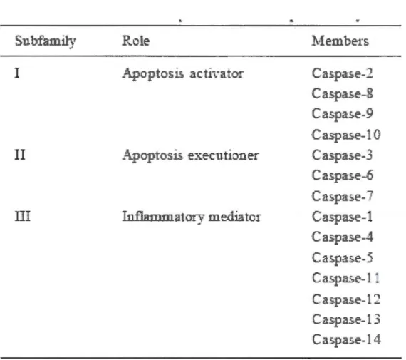
Fourteen caspases have been identified so far, based on their homology in amjno acid
sequences and substrate specificity; these caspases can be classified into apoptotic
(caspases-2, -3, -6, -7, -8, -9, -10) and pro-inflammatory (caspases-1, -4, -5,-11,-12,-13 and -14) groups. Apoptotic caspases can be subdivided further into initiator (apical,
apoptosis activator) (caspases-8, -9 and -10) and effector (executioner, downstream)
S
:~.bfamil!v .;Rc
.1
.e
I
II
I
l.
Ca
:;,pas,
.
e-2
C
a
:;,p::s
.
e-8
C
a:::
pa:::
.
e-9
C
:..=;
.
p
a
::,e-
1
0
C
·
-
:::
.
p
-
-
.=::
e
-3
C- 1(asp.a.
=:
-
e-
._
.
C
.
-1 • :a ~pas.e- " Ca:;,
.
p
rF-
e-1
C
.::
=::.pa.::,e-4
C
:a: =:.
p,a;
=::.e-
S
C
.a ::
.
p
.
a=::
.
e-
1
C
a::
.
p
a:::.e-
12
C
:a
spa:::.e-
13
C.a
:::pa:;,e-1
.!_.
13
Table 1.2 Subfamilymembers ofthe caspase family. Table adapted from Fan et al, 2005.
The most significant structural difference regarding caspases involves the varying length of the N-terminal prodomain. The initiator caspases such as caspase-8 and -9 have longer prodomains that allow them to interact with death effector domains (DEDs) or caspase recruitment domains (CARDs) in adaptor proteins such as Fas-associated death domain (FADD) protein and apoptotic protease-activating factor -1 (Apaf-1). Effector caspases (such as caspase-3) contain short pro-domains and they function primarily to cause the morphological features of apoptosis (Kohler et al, 2002). In response to an
apoptotic stimulus, initiator caspases are the first to be activated. These active initiator caspases in tum process and activate effector procaspases. Once activated, they subsequent1y cleave many intracellular substrates, including structural proteins such as
lamin A, actin, gelsolin and fodrin, the DNA repmr enzyme poly (ADP-ribose) polymerase (P ARP), cell cycle regulators such as p21, Rb and mdm2, and the inhibitor of caspase-activated DNase (ICAD) (Figure 1.6) (Fan et al, 2005). Caspase-activated deoxyribonuclease (CAD) is one of the key substrates for activated caspase-3. It normally exits as an inactive enzyme bound to its inhibitor ICAD (inhibitor of caspase-activated DNase). Active caspase-3 cleaves ICAD, thereby releasing CAD, which cleaves cellular DNA, resulting in DNA fragmentation. Another important target for activated caspase-3 is the nuclear protein P ARP (molecular mass 116 kDa). P ARP pla ys a key role in the maintenance of chromatin structure as well as DNA repair. Active caspase-3 cleaves P ARP to an 85 kDa fragment, which hinders its DNA-repairing activity (Wang et al,
2005). Cleavage of ICAD, nuclear and cytoskeletal proteins leads to characteristic morphological hallmarks of apoptosis such as chromatin condensation, nucleosomal DNA fragmentation, nuclear membrane breakdown, extemalization of phosphatidylserine, and the formation of apoptotic bodies (Hengartner 2000). Apoptotic bodies are removed by phagocytic or neighbouring cells without releasing intracellular contents into surrounding tissue, thus avoiding induction of inflammatory responses. Addition to their function in cell death recent publications also provide increasing evidence that apoptotic caspases also participate in several non-apoptotic cellular processes such as cell proliferation, differentiation, shaping and migration (Kuranaga,
2011; Kuranaga and Miura, 2007).
The other important family of cysteine proteases that is calpains also plays an important role in initiation, regulation and execution of cell death (Harwood et al, 2005),
but their role is not as well characterized as that for the caspases. Calpains are also known to cleave many important regulators of apoptosis and to crosstalk with caspase cascades. All the ab ove mentioned intracellular pro teins, su ch as lamins, actins, fodrin, and P ARP, also act as substrates for active calpains and thus play an important ro1e in the execution of apoptosis (reviewed by Lopatniuk and Witkowski, 2011 ).
-15
Cellular stress
Caspase-6
Figure 1.6 Downstream substrates that are targeted by active effector caspases, resulting in the execution of characteristic morphological features of apoptosis. Figure adapted from Fan et al, 2005.
1.3.2 Signaling pathways of Apoptosis
Cells undergo apoptosis mediated by three major signaling pathways, involving death receptors ( extrinsic pathway), mitochondria (intrinsic pathway), and more recently, the
endoplasmic reticulum (ER) (intrinsic) (Strasser et al, 2009; Fulda et al, 201 0; Du prez et al, 2009).
1.3.2.1 Mitochondrial (intrinsic) pathway
Mitochondria are important intracellular organelles for the production of A TP by oxidative phosphorylation, which supplies energy for various cellular functions. In addition to supplying energy, mitochondria play a critical role in the initiation and
regulation of apoptosis. A variety of stress-inducing agents such as aldehydes (e.g. acrolein), chemotherapeutic agents, UV and gamma irradiations, heat shock and growth factor withdrawal can trigger apoptosis through the mitochondrial pathway (Jin et al.,
2005; Tanel and Averill-Bates, 2005; Bettaieb and Averill-Bates, 2005).
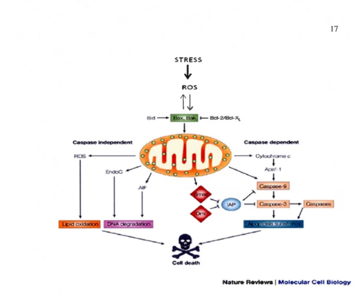
A cri ti cal cellular target of ROS is mitochondrial DNA (mtDNA), which could lead to lethal cell injury by disrupting electron transport, mitochondrial membrane potential and ATP generation (Ott et al, 2007). Therefore, ROS can cause mitochondrial dysfunction, leading to activation of the mitochondrial pathway of apoptosis (Orrenius et al, 2007). Induction of the mitochondrial permeability transition (MPT) and mitochondrial outer
membrane permeabilization are important features of apoptosis. A consequence of the MPT is swelling of the matrix and rupture of the outer mitochondrial membrane. These changes lead to the release of cytochrome c (Cyt c), and other pro-apoptotic mitochondrial proteins such as DIABLO/SMAC, Omi/Htr2, apoptosis inducing factor
(AIF) and endonuclease G (Endo G) into the cytoplasm, resulting in cell death by both
17
STRESS
J,
ROS
ROS+
-Natur. Revt.ws 1 Moi6Cular Cell Biology
Figure 1.7 Caspase-dependent and -independent pathways of apoptotic cell death.
Various apoptotic stimuli (e.g., ROS) cause permeabilization of the mitochondrial outer
membrane and the release of proapoptotic proteins. In the cytosol, cytochrome c, Apaf-1
and dATP form the apoptosome complex, to which initiator procaspase-9 is recruited and activated. Caspase-9-catalyzed activation of effector caspase-3 executes the final steps of apoptosis. Caspase activation is further enhanced by neutralization of caspase inhibitors by apoptogenic proteins such as Srnac/Diablo and Orni/HtrA2 that are released from
mitochondria. In addition, rnitochondrial proteins such as AIF and Endo G prornote
caspase-independent apoptosis through nuclear translocation and by mediating genornic
18
The haemoprotein cytochrome c plays an important role in caspase-dependent apoptosis. Under normal conditions, cytochrome c is a component of the respiratory
chain and is attached to the outer surface of the inner mitochondrial membrane (IMM),
mainly in association with the anionic phospholipid cardiolipin (CL) (Ott et al., 2007).
The oxidation of cardiolipin by ROS decreases cytochrome c binding and increases the level of soluble cytochrome c in the intermembrane space (Orrenius et al., 2007) (Figure
1.8). Once released, cytochrome c binds to Apaf-1 and procaspase-9, generating an
intracellular disc-like complex known as the "apoptosome". This leads to the
auto-activation of caspase-9, which can subsequently activate the executioner caspases such as
19
Figure 1.8 Effects of ROS on cardiolipin oxidation and cytochrome c release into the
cytosol. Normally cytochrome c (Cyt c) is bound to the mitochondrial inner membrane by an interaction with the anionic phospholipid cardiolipin (CL). Oxidation of cardiolipin by mitochondrial ROS (H202) decreases its binding affinity for cytochrome c, which results in cytochrome c detachment. Oxidized cardiolipin, together with pro-apoptotic proteins (e.g. Bid and Bax), forms a megapore channel within the mitochondrial outermembrane, which enables cytochrome c release from mitochondria into the cytosol.
In the cytosol, cytochrome c interacts with Apaf-1, procaspase-9 and dA TP, forming the
apoptosome complex, resulting in caspase-9 activation. Figure adapted from Circu and Aw 2010.
The pro-apoptotic proteins second mitochondrial activator of caspases (SMAC)/ direct inhibitor of apoptosis-binding protein with low pl (DIABLO), and high-temperature requirement A2 serine protease (HtrA2/0mi), which are released from mitochondria during apoptosis, facilitate caspase activation by binding to and neutralizing the a nti-apoptotic activity of inhibitors of apoptosis proteins (lAPs), which are potent caspase inhibitors (Figure 1. 7).
Once released from mitochondria, AIF and Endo G are involved in caspas e-independent apoptosis (Figure 1.7). These proteins can bind directly to DNA, which can stimulate their DNAse activity, leading to chromatin condensation and large-scale DNA
fragmentation. AIF appears to play a role in mediating cell death in severa} human disease states, including ischemia-reperfusion injury, neurodegenerative disorders and certain types of cancers (Norberg et al, 201 0).
1.3.2.2 Death receptor (extrinsic) pathway
The extrinsic pathway involves the binding of extemal ligands to death receptors located at the plasma membrane. These death receptors belong to the tumor necrosis factor receptor (TNF-R) super family and generally have several cellular functions including inflammation and apoptosis (Baud and Karin, 2001). The death receptors that are involved in apoptosis possess an intracellular death domain (DD). They include TNF receptor 1 (TNFR1 (also known as death receptor 1 (DR1)), Fas receptor (DR2, CD95 or AP0-1), DR3 (AP0-3 or TRAMP), TNF-Related Apoptosis-Inducing Ligand (TRAIL) receptor 1 (TRAIL-R1; also called DR4), TRAIL receptor 2 (TRAIL-R2; also called DR5 or KILLER) and DR6 (Russo et al, 2010).
The Fas ligand (FasL)/Fas system is the most widely studied system in death- receptor-mediated apoptosis. Fas-mediated apoptosis is activated by the binding of FasL to its
-21
specifie receptor Fas. This interaction results in clustering and trimerization of the receptor, which is required for formation of the DISC at the plasma membrane. The DD ofthe receptor interacts with the DD ofFADD, while the DED ofFADD interacts with the DED domains of procaspase-8 or procaspase-1 O. The association of procaspase-8 or procaspase-10 at the DISC leads to their auto-activation (Elrod et al, 2008). A significant level of caspase-8 activation directly activates effector caspases such as caspase-3, -6 and -7, whereas low caspase-8 activation mediates caspase-3 activation through an amplification loop involving mitochondria (Figure 1.9). Thus, the death receptor and mitochondrial pathways can be linked through caspase-8-mediated cleavage of pro-apoptotic Bcl-2 family protein Bid. The cleaved fragment, t-Bid, then translocates to mitochondria ap.d induces outer mitochondrial membrane permeabilization through its interaction with Bcl-2 family proteins such as Bax and Bak. This interaction results in the release of cytochrome c into the cytosol, which is a key step required for the induction of mitochondrial-mediated apoptosis (Y oule and Strasser, 2008) (Figure 1.9).
-•
Cf) ct u.J
_
...
.
/ Bax,Bak~
®
O
~
s~c-\
l
Apoptosome apoptosisFigure 1.9 Fas- mediated death receptor pathway of apoptosis. Figure adapted from Zhaoyu et al 2005 (see text for details).
23
Fas can also mediate apoptosis via death domain associated protein (Daxx (also known as Death-associated protein 6)), with subsequent activation of apoptosis signal-regulating kinase 1 (ASK-1) and c-Jun N-terminal kinase (JNK) (Dorion et al, 2002;
Salomoni and Khelifi, 2006; Fujisawa et al, 2007). During apoptosis, Daxx translocates from the nucleus to the plasma membrane and specifically binds to the death domain of Fas. Although Daxx does not contain a death domain, it plays a central role in Fas-mediated apoptosis through activation of ASK-1. ASK-1 is an upstream regulator of the JNK and p38 mitogen-activated protein kinases (MAPKs) during exposure to stressful stimuli such as hyperosmotic shock, oxidative stress, cold shock, UV irradiation, TNF-a and interleukin-1 (Fusijawa et al, 2007). JNK activation was shown to promote apoptosis by reducing the anti-apoptotic activity of Bcl-2 by causing its phosphorylation (Liu and Lin, 2005).
The induction of apoptosis by TRAIL is similar to that of Fas (Russo et al, 201 0; Jin and El-Deiry, 2005). The binding of TRAIL-R1 and -R2 to their respective receptors,
DR4 and DR5, triggers formation ofthe DISC by recruiting FADD and procaspase-8, or-10, followed by activation of caspase-3. TRAIL-induced apoptosis also involves amplification through the mitochondrial pathway, similar to Fas-induced apoptosis.
Unlike Fas and TRAIL signaling, the cytokine TNF-a does not induce cell death
spontaneously. TNF-R1-mediated intracellular signaling is more complex than that ofFas
or TRAIL-R1 and -R2 since it can activate both apoptotic and survival signais (Russo et al, 201 0). Upon activation by TNF-a, trimerization ofTNF-R1 is followed by recruitment of the adaptor protein TNFR-associated protein with death domain (TRADD). This results in the formation of two complexes that activate distinct downstream survival or
apoptotic signaling pathways. TRADD recruits signaling proteins like F ADD, TNF
-associated factor-2 (TRAF-2), and receptor-interacting protein (RIP). In complex I, the interaction of TNF-R1 with TRADD, RIP and TRAF-2 leads to the activation of
transcription factors such as nuclear factor kappa B (NF-KB) and JNK, which can promote cell survival and the activation of immune and inflammatory responses (Deng et al, 2003; Beyaert Ret al, 2002). Activation ofNF-KB by TNF-a up-regulates several anti -apoptotic factors, including c-FLIP, Bel-XL, MnSOD, X-linked inhibitor of apoptosis protein (XIAP), and c-lAP 1 and 2.
Complex II is comprised ofTNF-Rl, TRADD, FADD and procaspase-8 and promotes apoptosis through the direct activation of effector caspases such as caspase-3 (Russo et al, 2010) (Figure 1.10). The cellular balance between FLIPL and RIPl appears to be an important factor in determining whether TNF-Rl activation results in apoptosis or survival signalling. High concentrations of FLIPL inhibit procaspase-8 binding at complex II and prevent DISC formation, whereas caspase-8 mediated cleavage of RIPl dissembles complex I and promotes the formation of complex II that mediates apoptosis (Circu and Aw, 2010).
'·
ROS NFKB IKB/~
' - , ASKl ActLted : NFKB MnSOD -.!
JNK1
1l
Effector cas poses (Casp 3)+
25 . - - - -Casp9 DNA fragmentationFigure 1.10 Apoptosis-mediated by the death receptors. Stimulation of death receptors
such as Fas/FasL, TNF-Rl/TNFa, and TRAlL-Rl/TRAIL results in receptor aggregation
and recruitment of adaptor molecules FADD/TRADD and caspase-8. Once recruited, caspase-8 becomes activated and initiates apoptosis by direct cleavage of downstream
effector caspases. The mitochondrial pathway is initiated by stress signais through the
release of apoptogenic factors such as cytochrome c, AIF, or Smac/DIABLO.
Cytochrome c release into the cytosol triggers caspase-3 activation through formation of
the apoptosome complex. Smac/DlABLO promotes caspase activation by neutralizing the
inhibitory effects of lAPs, whereas AIF causes DNA condensation. The receptor and the mitochondrial pathways can be interconnected, for example, by Bid, which causes cytochrome c release upon cleavage by caspase-8. Activation of caspases is negatively
regulated at the receptor level by FLIP, which blocks caspase-8 activation, at the
mitochondria by Bcl-2 family proteins, and by lAPs. Activation of the NF-KB survival
pathway enhances transcription of antiapoptotic proteins such as FLIPL or MnSOD and
apoptosis blockade. At high ROS levels, failure to activate NF-KB promotes ASKl/JNK activation that triggers apoptosis. Figure adapted from Circu and Aw, 201 O.
Despite the early discovery of caspase-2, its physiological role remams unclear (Krumschnabel et al, 2009). Caspase-2 appears to have multiple roles in processes such
as apoptosis, DNA repair and tumor suppression. Caspase-2 activation has been demonstrated in apoptosis induced by different systems such as anti-Fas, cytokine deprivation, B-amyloid, etoposide and other stress stimuli (Zhivotovsky and Orrenius, 2005). Several studies have reported p53-mediated activation of caspase-2 by the PIDDosome, in response to DNA damage (Maiuri et al, 2007; Tinel et al, 2007). The PIDDosome is a temary complex that is composed of p53-induced protein with death domain (PIDD), RIP-associated Ich-1/Ced-3-homologue protein with a death domain (RAIDD) and procaspase-2. However, it was also shown that recruitment of caspase-8 to the DISC can lead to caspase-2 activation in the absence of PIDDosome formation (Lavrik et al, 2006; Manzl et al, 2009; Olsson et al, 2009). Thus, there exist different routes by which caspase-2 can be activated. Once activated, caspase-2 can trigger Bid-mediated activation of the mitochondrial pathway of apoptosis (Lavrik et al, 2006).
1.3.2.3 Endoplasmic reticulum (ER) stress (intrinsic) pathway
The ER is an organelle that is involved in lipid synthesis, protein folding and maturation (Breckenridge et al, 2003). Membrane and secretory proteins undergo folding
and modification into their mature conformation in the ER and then are transported to lysosomes, Golgi and the cell membrane. Protein chaperones such as Bip/glucose-related
protein 78 (GRP78), GRP94, GRP170, calnexin, calreticulin and the oxidoreductase protein disulphide isomerase (PDI) are present in the ER to assure correct folding of
Uf"R ~nsd..:en: (IR!: 1. Pï;RK,I\TF Bi El tl"
...
·.·.' H stt- s.s 11!111]' branchoo gl~·'""'n Malfolded prote in c".
,.
' STRESS•
27 ËR mcml1r=•• •
•
•
f'Riumon Ca'· 0•
• •
•
•
.•. Folded prote inFigure 1.11 Fonctions of ER. Nascent proteins are translocated into the ER lumen,
through protein channels in the ER membrane called translocons. Proper folding of nascent proteins is facilitated by ER chaperones including Bip, calnexin and protein disulphide isomerise (PDI). These chaperones facilitate proper folding of the nascent protein by preventing aggregation, and by maintaining polypeptides in appropriate conformations for folding and subunit assembly. During stress conditions, changes in the ER environment shift the balance from normal to improper folding, leading to accumulation of unfolded proteins in the ER lumen. This results in activation of the ER stress response pathway. Figure adapted from Faitova et al, 2006 (modified).
1.3.2.3.1 ER Stress
A variety of conditions including glucose deprivation, inhibition of protein glycolysation, disturbance of calcium homeostasis, hypoxia and excess ROS can perturb ER function leading to accumulation of unfolded proteins in the ER (Schroder and Kaufman, 2005; Fulda et al, 201 0). This phenomenon is known as ER stress. ER stress activates several signaling pathways, including the unfolded protein response (UPR) and ER-associated protein degradation (ERAD). The UPR is generally a survival response that aims to recover normal cellular function in the face of adverse conditions. However,
if protein damage is too severe, ER stress pla ys an important role in elimination of the damaged cell by apoptosis and/or autophagy (Schleicher et al, 2010; Verfaillie et al, 2010).
The ER stress response involves 3 distinct mechanisms: (i) translational attenuation to decrease the synthesis of new proteins; (ii) transcriptional activation of genes for ER chaperones ( eg: Bip, GRP94), ERAD molecules and antioxidants; and (iii) ERAD, which involves translocation of misfolded or aggregated ER proteins to the cytoplasm, where they are tagged by ubiquitin for proteosomal degradation (Shroder and Kaufman, 2005; Verfaillie et al, 2010).
The activation of the UPR is mediated by three distinct ER stress sensors: Inositol-requiring protein-1(IRE1), activating transcription factor-6 (ATF6) and protein kinase RNA (PKR)-like ER kinase (PERK) (Figure 1.12). In non-stressed cells, these sensors are retained in the ER lumen by interactions with Bip/GRP78 (Shroder and Kaufman,
2005). During ER stress, Bip releases these 3 sensors, leading to their activation, and then binds to unfolded proteins. PERK and IRE1 undergo homo-oligomerization and autophosphorylation during activation (Figure 1.12).
29
Figure 1.12 Signaling the ER stress response (UPR) pathway. Accumulation of
unfolded proteins in the ER leads to activation of three ER stress sensors PERK, IREl
and A TF6. Activation of these sensors is triggered by dissociation of Bip from their ER
luminal domains. Once activated, PERK phosphorylates the translation initiation factor
eiF2a to prevent further accumulation of unfolded proteins. Phosphorylation of NF-E2-related factor 2 (Nrf2) by PERK, promotes its dissociation from Kelch-like Ech-asspciated protein 1 (Keap1), leading to nuclear accumulation of Nrf2. Nrf2 binds to the
antioxidant response element (ARE) to activate transcription of genes encoding
detoxifying enzymes (antioxidants). Activated IRE1 splices the mRNA for XBP-1, which
allows the translation of mature XBP-1 protein, a transcription factor, responsible for the
upregulation of genes involved in ERAD, ER quality control and redox homeostasis.
ATF6 is activated by a two-step cleavage by site-1 and site-2 proteases (S 1P and S2P).
The cleaved fragment (p50) forms an active transcription factor that mainly mediates
expression of severa! components for ERAD and ER homeostasis. When functions of the ER are severely impaired, apoptosis is induced by the transcriptional induction of CCAAT/-enhancer-binding protein homologous protein (CHOP), which can be up-regulated by XBPl, ATF6 and PERK, and can mediate transcription of the pro-apoptotic BH3-only protein Bim. Figure adapted from Verfaillie et al, 2010.