HAL Id: hal-01821088
https://hal-univ-tours.archives-ouvertes.fr/hal-01821088
Submitted on 22 Jun 2018
HAL is a multi-disciplinary open access
archive for the deposit and dissemination of
sci-entific research documents, whether they are
pub-lished or not. The documents may come from
teaching and research institutions in France or
abroad, or from public or private research centers.
L’archive ouverte pluridisciplinaire HAL, est
destinée au dépôt et à la diffusion de documents
scientifiques de niveau recherche, publiés ou non,
émanant des établissements d’enseignement et de
recherche français ou étrangers, des laboratoires
publics ou privés.
immune tolerance induction
N. Deluce-Kakwata-Nkor, L. Lamendour, V. Chabot, A. Héraud, Z. Ivanovic,
F. Halary, F. Dehaut, F. Velge-Roussel
To cite this version:
N. Deluce-Kakwata-Nkor, L. Lamendour, V. Chabot, A. Héraud, Z. Ivanovic, et al.. Differentiation
of human dendritic cell subsets for immune tolerance induction. Transfusion Clinique et Biologique,
Elsevier, 2018, 25 (1), pp.90-95. �10.1016/j.tracli.2017.08.002�. �hal-01821088�
Please cite this article in press as: Deluce-Kakwata-nkor N, et al. Differentiation of human dendritic cell subsets for immune tolerance induction. Transfusion Clinique et Biologique (2017),http://dx.doi.org/10.1016/j.tracli.2017.08.002
ARTICLE IN PRESS
+ModelTRACLI-2956; No. of Pages 6
Research perspective
Differentiation of human dendritic cell subsets for immune tolerance
induction
Différenciation de cellules dendritiques humaines pour l’induction de la tolérance immune
N. Deluce-Kakwata-nkor
a, L. Lamendour
a, V. Chabot
b, A. Héraud
a, Z. Ivanovic
c, F. Halary
d,e,
F. Dehaut
b, F. Velge-Roussel
a,f,∗aEA 4245 cellules dendritiques, immuno-modulation et greffes, université Fran¸cois-Rabelais de Tours, UFR de médecine, 10, boulevard Tonnellé, 37032 Tours
cedex, France
bService recherche du laboratoire d’histocompatibilité et d’immunogénétique, établissement fran¸cais du sang Centre-Atlantique, 2, boulevard Tonnellé, BP 40661,
37206 Tours cedex 3, France
cEFS Aquitaine-Limousin, place Amélie-Raba-Léon, CS 21010, 33075 Bordeaux, France
dUMR 1064, Inserm, centre de recherche en transplantation et immunologie (CRTI), université de Nantes, Nantes, France
eInstitut de transplantation urologie néphrologie (ITUN), Hôtel-Dieu, CHU de Nantes, 30, boulevard Jean-Monnet, 44093 Nantes cedex 01, France fUFR des sciences pharmaceutiques, avenue Monge, 37000 Tours, France
Abstract
Objectives. – Since no further progress was achieved, in order to improve the long-term organ transplantation outcome, the immune tolerance
appears as an interesting therapeutic goal. Dendritic cells (DCs) are specialized cells participating in the homeostasis of the immune response. Moreover, subsets of DCs, identified in humans, appear to have their respective competences in immune response modulation. Our objective is to purify from PBMC or to differentiate DC subsets from monocytes using several strategies and evaluate their IL10 secretion.
Methods. – CD14+ cells were purified from peripheral blood mononuclear cell (PBMC) by affinity beads and cultured with cytokines up to 7 days.
The pDCs were purified with anti-BDCA-2 beads from PBMC fraction enriched by Percoll® gradient. The moDCs, pDCs and moLCs subsets were analyzed by phenotype labelling and FACS analyses and IL10 secretion measured by ELISA.
Results. – The moDCs were characterized by the CD209 expression and a lower expression of CD1a markers. Expression of CD207 and CD1a
markers characterized moLCs and CD123+/BDCA-2+ pDCs. Variable IL-10 secretions were shown between the three DC subsets, both at basal
and activated levels.
Conclusions. – As the several DC populations studied have different capacities of IL-10 synthesis, they might play, among others, distinct roles in
the induction of immune tolerance.
Keywords: Dendritic cells; DC subsets; Immuno-modulation; IL-10 Résumé
Objectifs. – L’induction de tolérance immune reste un challenge important dans le domaine de la transplantation d’organe. Les cellules dendritiques
(DCs), piliers de la réponse immunitaire, jouent un rôle crucial aussi bien dans l’induction d’une immunité spécifique que dans celle d’une tolérance immune. Chez l’homme, il existe au moins quatre types de DCs effectrices majeures, les DCs conventionnelles (cDC), les DCs plasmacytoïdes (pDCs), les DCs inflammatoires (MoDCS) et les cellules de Langerhans (LCs). L’objectif du projet est de préparer différents sous-types de DCs (moDCs, pDCs, moLCs) afin d’analyser leurs capacités à synthétiser de l’IL-10.
∗Corresponding author. EA 4245 cellules dendritiques, immuno-modulation et greffes, UFR de médecine, université Franc¸ois-Rabelais, 10, boulevard Tonnellé,
37032 Tours cedex, France.
E-mail address:[email protected](F. Velge-Roussel).
2 N. Deluce-Kakwata-nkor et al. / Transfusion Clinique et Biologique xxx (2017) xxx–xxx
Méthodes. – La différenciation des moDCs et moLCs est faite à partir de monocytes CD14+ isolés des PBMCs, en présence de cytokines spécifiques. La purification des pDC est faite avec des billes anti-BDCA-2 à partir des PBMC.
Résultats. – Les moLCs différenciées sont caractérisées par l’expression de la Langerine (CD207) et de CD1a. Les moDCs se caractérisent par
l’expression de CD209 et une plus faible expression de CD1a et les pDCs par l’expression conjointe de CD123 et BDCA-2. La synthèse d’IL10 est différente entre les trois sous-types de DCs étudiés au niveau basal comme activé.
Conclusions. – Les différentes populations de DC étudiées, parce qu’elles présentent des capacités variées, entre autres, pour la synthèse d’IL-10,
pourraient jouer des rôles distincts dans l’induction de tolérance immune.
Mots clés : Cellules dendritiques ; Sous-types de DCs ; Immunomodulation ; IL-10
1. Introduction
Dendritic cells (DCs) are professional antigen-presenting cells that induce immunity upon detection of pathogens, while maintaining tolerance in response to innocuous molecules due to their functional plasticity [1,2]. Two main DC subsets, at last, have been identified in the blood, spleen, tonsil and lymph nodes including conventional DCs (cDCs), consisting of either BDCA1/CD1c+ DCs or BDCA3/CD141+ DCs and plasma-cytoid DCs (pDCs) consisting of CD123+ cells. cDCs are effective at antigen-specific stimulation of CD4+ and CD8+ T
cells whereas pDCs specialize in producing type I interferons in response of virus motifs [3]. In the skin, three DC sub-sets have been described: Langerhans cells characterized by the expression of Langerin (CD207+)[4], CD1a+ dermal DCs, and CD14+ dermal DCs, which all migrate into skin-draining lymph nodes[5]. Additional revised taxonomy of human blood DC subsets have been described based on Single-cell RNA-seq including two subpopulations within BDCA1/CD1c+ DCs (CD1 C+-A and CD1 C+-B) which are distinguished by their strong signature of inflammatory genes[6]. Moreover, a new DC subset “AS DCs” which share properties with pDCs have been identified in human blood, nevertheless, pDCs remains as “the natural interferon-producing cells” with weaker T cell pro-liferation induction ability[6]. Since their discovery, DCs have proved to play a central role in regulating immune responses. Tolerant DCs are often characterized by a low expression of sur-face MHC II molecules and co-stimulatory molecules (CD40, CD80 and CD86) and low T cell stimulatory ability[7]. More-over, they usually show reduced IL-12 and increase IL-10. During the last decades, significant advances have been made in establishing methods to manipulate DCs in vitro to gen-erate tolerant DCs [8] using cytokines such as: Il-10 alone
[9] or in combination of transforming growth factor (TGF-ß1)[10]. Alternatively, pharmacological mediators including: 1,25-dihydroxyvitamin D3[11], histone deacetylase inhibitors (HDAC)[12]or immunosuppressive drugs such as mycophe-nolate mofetil or rapamycin [13,14]which modulate DCs in that sense. Despite the importance of Tol-DCs, the capacities of each DC subset to induce and promote immune toler-ance remain unknown. We explore DC subset capacities of secreting IL-10 versus pro-inflammatory cytokines using PRRs agonist.
2. Methods
2.1. Monocyte isolation and culture
Cytapheresis products were obtained from Centre Atlantic Transfusion Department (EFS-CA). They were issued from the healthy adult volunteers who had given their written informed consent and the university ethic committee approved the proce-dure. Mononuclear cells were obtained from peripheral blood of healthy donors using Ficoll (Dutscher) density gradient cen-trifugation. The monocytes were then purified by a positive selection using CD14 microbeads (Miltenyi Biotec) (> 90% of purity). For immature monocyte-derived DCs (moDCs), mono-cytes were differentiated for 6 days in RPMI 1640 (Dutscher) medium supplemented with 10% FCS (Dutsher), 66 ng/mL granulocyte macrophage colony stimulating factor (GM-CSF, Miltenyi Biotec) and 25 ng ng/mL IL-4 (Miltenyi Biotec). At day 6, cells were collected and flow cytometry analysis was performed. For human moLCs, monocytes were cultured for 7 days in RPMI 1640 medium supplemented with the 2% human albumin (HAB, Vialebex LFB), 50 ng/mL GM-CSF (Miltenyi Biotec), and 10 ng/mL TGF-ß1 (Miltenyi Biotec), renewed at day 3. At day 7, cells were collected and FACS analysis was performed. The pDCs were isolated by negative selection using plasmacytoid dendritic cells isolation kit II (Miltenyi Biotec), from an enriched DC cells fraction, which was obtained by gra-dient centrifugation of PBMCs, Percol® (Healthcare) gradient centrifugation of PBMCs. Cells were gated based on their char-acteristic pattern of SSC and FSC. Doublets were excluded based on forward scatter height (FSC-H) and forward scatter width (FSC-W) and analysed for the double expression of CD123+ (IL-3R+) and CD303 (CLEC4 C, BDCA-2). The purity of population is up to 98,1%.
2.2. Flow cytometry analysis
Cells (1× 10⁵/100L) were stained for 30 min at 4◦C with the following anti-human antibodies at the appropri-ate concentration or with the relevant isotypes: CD83-FITC (BD Biosciences), CD14-PE (Beckman coulter), CD86-PE (BD Biosciences), HLA-DR–APC (BD Biosciences), CD207-APC (Biolegend), CD1A-AF488 (Biolegend), CD123-CD207-APC (Biolegend), BDCA-2-APC (Biolegend), CD209-PE (Beckman
Please cite this article in press as: Deluce-Kakwata-nkor N, et al. Differentiation of human dendritic cell subsets for immune tolerance induction. Transfusion Clinique et Biologique (2017),http://dx.doi.org/10.1016/j.tracli.2017.08.002
ARTICLE IN PRESS
+ModelTRACLI-2956; No. of Pages 6
N. Deluce-Kakwata-nkor et al. / Transfusion Clinique et Biologique xxx (2017) xxx–xxx 3
coulter). The cells were then washed with 1× PBS (Dutscher) and viable cells analysed on a Canto I flow cytometer (BD Bio-sciences). Results were expressed as the ratio of MFI (mean of fluorescence) of the marker on the MFI of the isotype control and referred as MFI ratio.
3. ELISA
MoDCs and monocyte-derived Langerhans-like cells (moLCs) (1.106/mL) were stimulated by lipopolysaccha-rides (LPS, TLR-4 ligand) 50 ng/mL (Invivogen), and pDCs by Classe A CpG-ODN 2236 (TLR-9 ligand), 1g/mL (Invivogen) for 48 h and then IL-10, IL12-p70 and IFN-␣ ELISA measurement was performed on culture supernatants of each DC subset according to the manufacturer instructions (eBiosience). Data were expressed as means± SD of 3 donors for pDCs and moDCs and as means of duplicate± SD of 1 donor for moLCs.
3.1. Statistical analysis
Histograms represent the mean values± S.D. Statistical significance was determined by the unpaired nonparametric Kruskal–Wallis test. Difference was considered significant when
P < 0.05.
4. Results
4.1. Differentiation of DC subsets
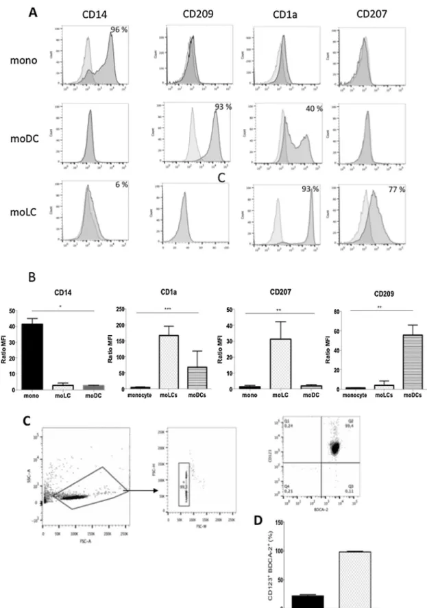
In order to obtain different DC subsets in sufficient quan-tities, we first explored the possibility of differentiating these DCs from monocytes using specific cytokine cocktail. Monocyte derived-cells were collected after a 6-day culture and identified using CD14, CD209, CD1a and CD207 as phenotypic mark-ers. Cells differentiated in the presence of IL4 and GMCSF as moDCs expressed DC-SIGN (CD209) marker up to 93% and among these, 40% are CD1a+(Fig. 1A and B). When cultured in the presence of GM-CSF and TGF-ß1, monocytes generated large number (60%) of dendritic cells as moLCs that expressed Langerin (CD207) as referred to the literature[15](Fig. 1A and B) but not CD209 compared to moDCs or CD14 and in minor levels compared to monocyte (Fig. 1A and 1B). The percent-age of phenotypic markers, which were less than 5%, has been considered as negative.
The pDCs were purified as described in methods (Fig. 1C). Cells were gated on the basis of double expression of CD123 and BDCA-2, pDCs characteristics markers. CD123+ BDCA-2+cells have been enriched > 4-fold from 22% before separation to > 96% after negative column isolation (Fig. 1D).
For the pDCs purification, 1.109PBMC have been used to obtain 1.5 106of purified pDCs. Concerning the moLCs, 1.107 monocytes have been used to obtain 6.106 moLCs (60% of CD207+).
4.2. Functional capacities of DC subsets
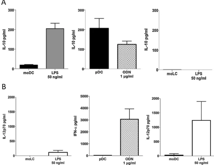
The capacity to secrete IL-10 for each subset obtained has been evaluated by ELISA (Fig. 2A) after 48 h of stimula-tion using TLRs ligand such as LPS for moDC and moLCs and CPG-A ODN, (TLR-9 ligand) for pDCs. MoDCs IL-10 secretion increased between un-stimulated and LPS stimulated stage from 18.53± 2.59 pg/mL to 204.91 ± 22.14 pg/mL. The immature pDCs have a basal secretion at 206.90± 70.43 pg/mL and reduce their Il-10 secretion when stimulated with ODN at 125.07± 23.24 pg/m. We found out, as a preliminary result, that un-stimulated moLCs had neither, IL-10 basal secretion (1.559± 0.004 pg/mL) nor with LPS stimulation. IL-12p70 secretion or interfon-␣ secretion as well as maturation marker have been evaluated after 48 h of stimulation by ELISA and flow cytometry respectively as control of responsiveness of each DC subsets due to stimulation. In presence of the pro-inflammatory stimulus, LPS or class A CPG-ODN, IL-12p70 or INF-alpha were secreted higher than non-stimulated DC (Fig. 2B) as well as the expression of maturation marker CD86, CD83 and HLA-DR (data not shown).
5. Discussion
As playing a key role on inducing immune response or immune tolerance, each DC subset might have different capac-ities of responsiveness due to PRRs stimulus. To study the capacity of each subset to induce immune tolerance, it is nec-essary to have a clear immuno-phenotypic definition of each DC population and the possibility to purify a sufficient amount of the phenotypically identified cells. The moDCs is a well-characterized model of DC differentiation and huge number of works published is related to this population[8,16]. It has also been used as therapeutic strategy in cancer for cell therapy[17]
and in graft tolerance[18]. The plasmacytoid DCs are not easily derived from CD34+progenitors but they could be purified from a PBMC fraction after an enrichment using a Percoll® gradient
[19]. In humans, pDCs do not express the CD11c molecule, but strongly express the IL-3 receptor chain␣ (CD123). The iden-tification of BDCA-2 markers, type C lectin and BDCA-4, the neuropilin-1 receptor, facilitated the identification of these cells in the blood[20]. The pDCs are the main source of virus-induced IFN-␣ in human peripheral blood. They migrate to different lym-phoid tissues[3]and are found to be elevated in some tumours, in the skin of systemic lupus erythematous (SLE) patients and in the nasal mucosa of allergic patients[21]. pDCs play a central role in SLE pathogenesis through their unique ability to pro-duce large amounts of type I interferon (IFN-I) upon TLR-7 or TLR-9 triggering[22]. The pDCs also promote immunological tolerance by inducing IL-10-secreting regulatory T cells (Treg). Indeed, after maturation, ICOS-L, which is overexpressed on pDCs, allows the development of IL-10 producing Treg lym-phocytes, within Th1 or Th2 responses[9]. If many aspects of their properties remain to be study, pDCs are clearly involved in cancer and autoimmune subversion mechanisms and they express specific surface regulatory receptors involved in negative
4 N.Deluce-Kakwata-nkoretal./TransfusionCliniqueetBiologiquexxx(2017)xxx–xxx
Fig.1.StrategiesforDCsubsetdifferentiation.MoLCsandmoDCssubsetscanbedifferentiatedfrommonocyteintheGM-SCFandTGF-ß1orGM-SCFandIL-4 respectivelyandtheyexhibitdistinctivesurfacemarkers.A.AphenotypicanalysisofmoDCsandmoLCsstainedwithapanelofantibodiesspecifictoDCrelated surfacemarkers.Flowcytometryresultplotsshowisotypecontrol(grey)andspecificmarker(black).B.MFIratioofCD1a,Langerin(CD207),CD14andDC-SIGN (CD209)expressionsbyflowcytometrydisplayedasratioofMFIusingMFImarkerbyMFIisotypecontrol.Theseresultsarerepresentativeof4independent experiments(n=4;Kruskal–Wallistest;*P<0.0145,**P<0.005;***P<0.0006).C.pDCswereisolatedusingLDcolumnsbynegativeselectionfromanenriched DCcellsfraction.CellsweregatedbasedontheircharacteristicpatternofSSC,andFSC.Doubletswereexcludedbasedonforwardscatterheight(FSC-H)and forwardscatterwidth(FSC-W)(middleplot)andanalysedforthedoubleexpressionofCD123andCD303(BDCA-2).D.ThepercentageofCD123+/CD303+cells
Pleasecitethisarticleinpressas:Deluce-Kakwata-nkorN,etal.Differentiationofhumandendriticcellsubsetsforimmunetoleranceinduction. TransfusionCliniqueetBiologique(2017),http://dx.doi.org/10.1016/j.tracli.2017.08.002
ARTICLE IN PRESS
+ModelTRACLI-2956; No.ofPages6
N.Deluce-Kakwata-nkoretal./TransfusionCliniqueetBiologiquexxx(2017)xxx–xxx 5
Fig.2.CytokinesecretionsofeachDCsubset.A.ELISAmeasuredofIL-10orB.IL-12p70andIFN-␣secretionin48hinthesupernatantsinun-stimulatedand LPSorCPG-AODNstimulatedDCs.Barrepresentedthemeanvalue±SD(pDCandmoDCsn=3,moLCsn=1).
regulationof IFN-␣secretionsuggestingtheir potential asan attractivetherapeutictarget[23].
LangerhanscellsarereadilydistinguishedfromotherDCby theexpressionofaC-typelectinLangerin(CD207),associated withcharacteristicintra-cytoplasmicvesicles,Birbeckgranules
[4].Langerhanscellsformastableself-renewingpopulationthat doesnotrequireCD34+ progenitorsinthe absenceof inflam-mation[24].It canbe differentiatedfrommonocytes[15,17], dermalresident CD14+ cells[25]andCD1c+ DC[26].Ithas becomeclearthatLCswerenotonlycapableoftriggering adap-tiveimmuneresponsesbutalsoofmediatingimmunetolerance toself-antigensinnon-inflammatoryconditions[27,28].Wealso demonstratedthatthebasalamountofIL-10secretionwas differ-entaccordingDCsubsets,withlowsecretionformoDCs,higher forpDCsandnosecretionformoLCs.Thisvariablecapacityto secreteIL-10couldberelatedtotheintrinsicpropertiesofeach subset to become pro-tolerantDCs. The activity level of the ERKpathwaystimulationishigherinmoDCsthaninpDCsand seemstocontrolIL-10secretion[29].
Largenumbersofworksillustratedthecapacityof moDCs to become pro-tolerant at the immature and mature state as wellas to inducein vitro andinvivo regulatory T cells [9]. Likewise,somepDCsphenotypesareabletoimpairTcell pro-liferation[11],activateTreg functions[21]andparticipate to
theimmuneresponsesubversion[22].Inregardtotheamount ofIL-10secretedbypDCsatthebasallevel,thesecellsmight havethecapacitytoswitchTcellphenotypetoregulatory pro-file, as domoDCs. Although,it remains to be demonstrated. Moreover,pDCsaresensitivetoTGF-[30]andPRRsligand activationleadingtotheinhibitionofitsIFN-␣secretion[23]. Langerhanscellsappeartobeinvolvedintheskinhomeostasis withhighsensitivitytoTGFandtoinducecross-tolerancein mice[31].ButtheircapacitytoorientatenaiveTlymphocytes intoregulatoryphenotyperemainsunknown.
Toconclude,wepresentedinthisworkourcapacitytoobtain severalDCsubsetsandtheircapacitytosecreteIL-10inorder toassesstheirpotentialtoinduceimmunetoleranceusingPRRs stimulus.
Authorcontributions
NDKco-elaboratedtheproject,designedandperformedthe experimentsandanalysedtheresults.AHandVCtrainedNDK inflowcytometryexperiments.FD,LL,VC,FHandZIwere involvedinmanuscriptreading,discussionaboutanalysisand interpretation of the results. FVR and FD co-elaborated the project,supervisedthedesignandtheanalysesofexperiments. NDKandFVRwrotethearticle.
6 N.Deluce-Kakwata-nkoretal./TransfusionCliniqueetBiologiquexxx(2017)xxx–xxx
Disclosureofinterest
Theauthorsdeclarethattheyhavenocompetinginterest.
Acknowledgments
FVR is supported by the EU Cost Action BM1406. This workhasbeenfundedwithsupportfromtheEFS-Centre Atlan-tique,theFrenchHigherEducationandResearchministryunder the program “ANRT” andthe Labex MabImprove ANR-10-LABX-53-01.AuthorsthankMrsLindaRouhaultforhercareful proofreading.
References
[1]Steinman RM. Dendritic cells: understanding immunogenicity. Eur J Immunol2007;37:S53–60,http://dx.doi.org/10.1002/eji.200737400.
[2]SteinmanRM.Thecontrolofimmunityandtolerancebydendriticcell.
PatholBiolParis2003;51:59–60.
[3]Segura E, Valladeau-Guilemond J, Donnadieu M-H, Sastre-Garau X, SoumelisV, AmigorenaS.Characterizationofresident andmigratory dendriticcells in humanlymph nodes.J Exp Med2012;209:653–60,
http://dx.doi.org/10.1084/jem.20111457.
[4]ValladeauJ,RavelO,Dezutter-DambuyantC,MooreK,KleijmeerM,
LiuY,etal.Langerin,anovelC-typelectinspecifictoLangerhanscells,
isanendocyticreceptorthatinducestheformationofBirbeckgranules.
Immunity2000;12:71–81.
[5]CollinM,McgovernN,HaniffaM.Humandendriticcellsubsets. Immunol-ogy2013;140:22–30,http://dx.doi.org/10.1111/imm.12117.
[6]Villani A-C,Satija R,Reynolds G,Sarkizova S,ShekharK,Fletcher J,etal.Single-cellRNA-seqreveals newtypes ofhumanblood den-dritic cells, monocytes and progenitors. Science 2017;356:eaah4573,
http://dx.doi.org/10.1126/science.aah4573.
[7]Horton C, Shanmugarajah K, Fairchild PJ. Harnessing the properties ofdendriticcellsin thepursuitofimmunologicaltolerance.BiomedJ 2017;40:80–93,http://dx.doi.org/10.1016/j.bj.2017.01.002.
[8]Amodio G, Gregori S. Human tolerogenic DC-10: perspectives for clinical applications. Transplant Res 2012;1:14, http://dx.doi.org/
10.1186/2047-1440-1-14.
[9]Ito T, Liu Y-J, Kadowaki N. Functional diversity and plasticity of human dendritic cell subsets. Int J Hematol 2005;81:188–96,
http://dx.doi.org/10.1532/IJH97.05012.
[10]SatoK,YamashitaN,BabaM,MatsuyamaT.Modifiedmyeloid den-dritic cells act as regulatory dendritic cells to induce anergic and regulatoryTcells.Blood2003;101:188–3581,http://dx.doi.org/10.1182/
blood-2002-09-2712.
[11]FerreiraGB.VitaminD3inducestoleranceinhumandendriticcellsby activationofintracellularmetabolicpathways.CellRep2015;10:711–25,
http://dx.doi.org/10.1016/j.celrep.2015.01.013.
[12]Arbez J, Lamarthée B, Gaugler B, Saas P. Histone deacetylase inhibitor valproic acid affects plasmacytoid dendritic cells pheno-typeand function.Immunobiology2014;219:637–43, http://dx.doi.org/
10.1016/j.imbio.2014.03.013.
[13]LagaraineC,HoarauC,ChabotV,Velge-rousselF,LebranchuY.Human
mycophenolicacidtreateddendriticcellshaveimmatureco-stimulary
abil-itiesbutmaturemigratoryphenotype.JLeukocBiol2005;17:351–63.
[14]Ciancio G, Sageshima J, Chen L, Gaynor JJ, Hanson L, Tueros L, etal.Advantage ofrapamycin overmycophenolatemofetilwhenused withtacrolimus forsimultaneouspancreaskidneytransplants: random-ized,single-centertrialat10years.AmJTransplant2012;12:3363–76,
http://dx.doi.org/10.1111/j.1600-6143.2012.04235.x.
[15]PicardaG,ChéneauC,HumbertJ-M,BériouG,PiletP,MartinJ,etal. Func-tionalLangerinhigh–expressinglangerhans-likecellscanarisefromCD1
highCD16–humanbloodmonocytesinserum-freecondition.JImmunol
2016;196:3716–28,http://dx.doi.org/10.4049/jimmunol.1501304.
[16]CauxC,Ait-YahiaS,CheminK,deBouteillerO,Dieu-NosjeanMC,
Homey B, et al. 17-Dendritic cell biology and regulation of
den-dritic cell trafficking by chemokines. Springer Semin Immunopathol
2000;22:345–69.
[17]Palucka AK, Ueno H, Fay JW, Banchereau J. Taming cancer by inducingimmunityviadendriticcells.ImmunolRev2007;220:129–50,
http://dx.doi.org/10.1111/j.1600-065X.2007.00575.x.
[18]Xia MJ, Shan J, Li YP, Zhou YN, GuoYJ, Sun GX, et al. Adop-tive transfusion of tolerant dendritic cells prolong the survival of renalallografts:asystematicreview.JEvid-BasedMed2013;6:250–64,
http://dx.doi.org/10.1111/jebm.12070.
[19]DuraesFV,LippensC,SteinbachK,DubrotJ,BrighouseD, Bendriss-Vermare N, et al. pDC therapy induces recovery from EAE by recruitingendogenouspDCtositesofCNSinflammation.JAutoimmun 2016;67:8–18,http://dx.doi.org/10.1016/j.jaut.2015.08.014.
[20]Dzionek a, Sohma Y, Nagafune J, Cella M, Colonna M, Facchetti F, et al. BDCA-2, a novel plasmacytoid dendritic cell-specific type II C-type lectin, mediates antigen capture and is a potent inhibitor of interferon alpha/beta induction. J Exp Med 2001;194:1823–34,
http://dx.doi.org/10.1084/jem.194.12.1823.
[21]Guéry L, Hugues S. Tolerogenic and activatory plasmacytoid den-driticcellsinautoimmunity.FrontImmunol2013;4:59,http://dx.doi.org/
10.3389/fimmu.2013.00059.
[22]Crow MK. Type I interferon in the pathogenesis of lupus. J Immunol Baltim Md 1950 2014;192:5459–68, http://dx.doi.org/
10.4049/jimmunol.1002795.
[23]Chappell CP, Giltiay NV, Draves KE, Chen C, Hayden-Ledbetter MS, Shlomchik MJ, et al. Targeting antigens through blood den-dritic cell antigen 2 on plasmacytoid dendritic cells promotes immunologictolerance.JImmunol2014;192:5789–801,http://dx.doi.org/
10.4049/jimmunol.1303259.
[24]Chopin M, Seillet C, Chevrier S, Wu L, Wang H, Morse HC, et al. Langerhans cells are generated by two distinct PU.1-dependent transcriptional networks. J Exp Med 2013;210:2967–80,
http://dx.doi.org/10.1084/jem.20130930.
[25]LarreginaAT,MorelliAE,SpencerLA,LogarAJ,WatkinsSC,Thomson AW,etal.Dermal-residentCD14+cellsdifferentiateintoLangerhanscells. NatImmunol2001;2:1151–8,http://dx.doi.org/10.1038/ni731.
[26]Martínez-Cingolani C, Grandclaudon M, Jeanmougin M, Jouve M, Zollinger R, Soumelis V. Human blood BDCA-1 dendritic cells differentiate into Langerhans-like cells with thymic stromal lym-phopoietin and TGF-. Blood 2014;124:2411–20, http://dx.doi.org/
10.1182/blood-2014-04-568311.
[27]KlechevskyE,MoritaR,LiuM,CaoY,CoqueryS,Thompson-Snipes L, et al. Functional specializations of human epidermal Langerhans cells and CD14+ dermal dendriticcells. Immunity 2008;29:497–510,
http://dx.doi.org/10.1016/j.immuni.2008.07.013.
[28]SeneschalJ,ClarkRA,GehadA,Baecher-AllanCM,KupperTS.Human epidermalLangerhans cellsmaintain immune homeostasis in skin by activatingskinresidentregulatory T cells.Immunity 2012;36:873–84,
http://dx.doi.org/10.1016/j.immuni.2012.03.018.
[29]Kaiser F, Cook D, Papoutsopoulou S, Rajsbaum R, Wu X, Yang H-T, et al. TPL-2 negatively regulates interferon- production in macrophagesandmyeloiddendriticcells.JExpMed2009;206:1863–71,
http://dx.doi.org/10.1084/jem.20091059.
[30]Bonnefoy F, Couturier M, Clauzon A, Remy-Martin J-P, Gau-gler B, Tiberghien P, et al. TGF- -exposed plasmacytoid dendritic cellsparticipate in Th17 commitment. JImmunol 2011;186:6157–64,
http://dx.doi.org/10.4049/jimmunol.1002497.
[31]FlacherV,TrippCH,Mairhofer DG,SteinmanRM,StoitznerP, Idoy-agaJ,etal.Murine Langerin+dermal dendriticcellsprime CD8+ T cellswhileLangerhanscellsinducecross-tolerance. EMBOMolMed 2014;6:1191–204,http://dx.doi.org/10.15252/emmm.201303283.