A survey on staging and treatment in uterine cervical carcinoma in the
Radiotherapy Cooperative Group of the European Organization for
Research and Treatment of Cancer
Philippe A. Coucke
a,*, Philippe Maingon
b, Ilja F. Ciernik
a, Huu Phuoc Do
caDepartment of Radiation Oncology, Centre Hospitalier Universitaire Vaudois, 1011 Bugnon, Lausanne, Switzerland bDepartment of Radiation Oncology, Centre George-FrancËois Leclerc, Dijon, France
cInstitut de Radiophysique AppliqueÂe, Lausanne, Switzerland
Received 16 March 1999; received in revised form 10 November 1999; accepted 22 December 1999
Abstract
Background: The treatment outcome of advanced stage uterine cervical carcinoma remains unsatisfactory. In order to elaborate a novel trial within The Radiotherapy Cooperative Group (RCG) of the European Organization for Research and Treatment of Cancer (EORTC), we conducted a survey in 1997±1998 to determine the variability of pre-treatment assessment and treatment options. The variability of choosing surgery, de®ned radiation therapy techniques and chemotherapy are investigated, as well as the center's choices of future treatment strategies. Methods: Fifty two of 81 RCG centers from the RCG have participated in the survey. As one would expect, there is a large variation in the techniques used for pretreatment evaluation and treatment options. There is no `standard' for reporting acute and late side effects. Chemotherapy is used neither systematically nor uniformly, and some centers continue to use neadjuvant chemotherapy modalities.
Results: Furthermore, the survey reveals that there is a strong demand for the reduction of overall treatment-time, for clinical investigation of novel combined modality treatment strategies, especially chemo±radiation therapy, and also for the use of new radiation sensitizers.
Conclusion: We conclude that a more homogeneous approach to the pretreatment evaluation as well as treatment techniques is required in order to allow adequate quality control in any future trial of the RCG in the EORTC. q 2000 Elsevier Science Ireland Ltd. All rights reserved.
Keywords: Cervix cancer; Pretreatment evaluation; Surgery; Chemo±radiotherapy
1. Introduction
The Radiotherapy Cooperative Group (RCG) of the European Organization for Research and Treatment of Cancer (EORTC) is conducting a variety of trials for differ-ent tumor localizations. However, gynecological tumors are currently not the subject of any ongoing trial, notwithstand-ing the fact that radiation therapy plays a major role in the treatment of cervical carcinoma. There is one ongoing inter-group trial (EORTC-GCCG) investigating the usefulness of the addition of cisplatin-based chemotherapy to postopera-tive radiation therapy in stages Ib and IIa cervical carcinoma after radical hysterectomy and complete lymphadenectomy. There is not a single trial addressing the question of adequate treatment for locally-advanced cervical cancer (LACC), i.e. FIGO stages IIb, IIIa and IIIb. This is dif®cult
to understand, facing the multitude of open questions related to the optimal investigational approach and therapeutic management of cervical carcinoma.
In order to de®ne whether there is an interest within the RCG to study new therapeutic concepts in LACC, we sent a questionnaire to radiotherapy groups which are listed as participating members of the RCG. This questionnaire is designed to determine the general feasibility of a trial in Europe, taking into consideration the decreasing number of patients with LACC addressed to radiation therapy depart-ments. Finally, we try to de®ne which questions have to be given priority in future trials. Special attention is dedicated to the concept of the association of chemotherapy and ionizing irradiation, the concept of the impact of treatment duration on local control, and to the use of `new' radio-sensitizers. The answers on this questionnaire are analyzed and discussed, taking into account the `guidelines' and/or `evidence' reported in currently published literature.
0167-8140/00/$ - see front matter q 2000 Elsevier Science Ireland Ltd. All rights reserved. PII: S0167-8140(00)00146-8
www.elsevier.com/locate/radonline
2. Methodology
The questionnaire was designed in 1997 and sent to 81 RCG centers. The questionnaire contains general questions related to the number of patients (on a yearly basis) treated in the individual RCG centers, their stage distribution, the techniques used in the pretreatment evaluation and the ther-apeutic options considered as `standard'. For the latter, speci®c questions address the relative role and sequence of the four basic therapeutic options, namely surgery, chemotherapy, external irradiation and brachytherapy. Regarding external irradiation, special attention is given to technical details related to the treated volumes, number of ®elds used, total dose and dose/fraction, and prescription point. The importance of brachytherapy is reported again with some details on dose-rate, number of applications, in vivo dosimetry and timing with external treatment. The questionnaire aims to de®ne the relative importance of chemotherapy, the type of chemotherapy, i.e. neoadjuvant, concomitant or sequential, and the most frequent drugs used. One question concerns the toxicity scale used to report the acute and late side effects.
Finally, bearing in mind the more general question raised by the RCG, i.e. the feasibility of a trial within the EORTC, the centers are asked if they will participate in a general effort to run such a trial. The RCG members are asked to state if they are interested in investigating the importance of the package-deal concept (the time factor), the importance of chemotherapy or the use of `new' sensitizers.
3. Results
Fifty two centers out of 81 (64%) answered the question-naire. The average number of patients/year addressed to the departments of radiation±oncology is 48 (median value 24, range 5±600). These patient numbers are listed in Table 1 according to stage distribution. Considering LACC (de®ned as IIb±IIIa±IIIb), we recorded a mean value of 25 (median 15, range 1±358) new patients/center per year.
3.1. Pretreatment evaluation and staging
One of the ®rst important questions is related to T and N staging. For the clinical evaluation of local tumor in®ltra-tion, routine cystoscopy is rated high (89%) alongside the digital gynecological examination, but only 56% of the centers mention routine rectoscopy. The use of transvaginal ultrasound has not been recorded in this questionnaire.
Ninety four percent of the centers reported practicing nodal staging. Evaluation of nodal involvement is mainly performed with computed tomography (CT) (85%) and nuclear magnetic resonance (NMR) (50%). Patients are less frequently submitted to surgical staging (35%). The technique used for surgical staging is laparatomy (23%), or coelioscopy (21%). Unfortunately, no data are available in order to determine how much of these surgical stagings are associated with a radical hysterectomy, and the rate of surgical staging in respect of stage.
Lymphangiography (LAG) as part of the routine pretreat-ment work-up has been reported by only seven centers (14%).
3.2. Treatment options and treatment sequence 3.2.1. Surgery
The possible treatment policies include surgery, external irradiation, brachytherapy and chemotherapy. As illustrated in Fig. 1a, the use of curative surgery as the initial approach to treatment decreases as the FIGO stage increases. The importance of radiotherapy (Fig. 1b) and brachytherapy as primary treatments (Fig. 1c) progressively increases with higher FIGO stages. Sixteen centers use surgery as part of the treatment for stage IIb, six for stage IIIa, ®ve for stage IIIb, and ten centers for stage IVa. Moreover, four centers record surgery as upfront treatment in IIb, one in IIIa and four in IVa. Surgery is the preferred ®rst-line treatment modality in stage Ia (83%), Ib (73%) and IIa (60%). Surgery includes at least hysterectomy with or without node sampling or node dissection. More details on the surgical procedure are not available.
3.2.2. External beam irradiation
External irradiation is more likely to be used as a ®rst-line treatment for IIb and higher stage disease. The use of radia-tion as a ®rst-line treatment modality or as part of a combined treatment-approach in respect of stage is summar-ized in Table 2.
Thirty out of 52 centers (58%) are using 2D planni®ca-tion, i.e. using one single CT-slice at the center of the ®elds, whereas 23 (44%) are using 3D (a series of CT-slices). Six out of 52 centers (12%) use neither 2D nor 3D for dose-calculation for external beam radiation therapy. Five out of these six centers are located in western Europe. Five centers use 2D or 3D according to the complexity of the case. The median dose for external irradiation is 45 Gy, but with a range of 36±70 Gy according to the center and tumor stage. The median number of fractions/week is ®ve (range 5±10) with a median dose/fraction of 2 Gy (range 1.5±2 Gy). This external treatment is usually given by a four-®eld box tech-nique on the pelvis (85%). However, in eight centers, an AP±PA ®eld is still used to cover the pelvic volume. If para-aortic nodes are considered as the target, these nodes are covered by AP±PA ®elds by the majority of the centers (67%). The median number of ®elds treated daily for the
Table 1
Distribution of number of patients/stage per year
Stage IA IB IIA IIB IIIA IIIB IVA IVB
Meana 7 14 6 9 4 12 3 3
Mediana 3 6 4 6 3 6 2 3
pelvic volume is four, and two for the para-aortic volume. The para-aortic nodes are never treated by ®ve centers (10%). Forty-six centers (89%) are covering those nodes
if they are found to be positive during pretreatment evalua-tion, whatever the technique used (CT, NMR or surgical staging), and 22 centers (42%) are including those nodes within the target volume if the pelvic nodes are positive. 3.2.3. Brachytherapy
Brachytherapy is used to boost external radiation espe-cially in stages IIb, IIIa and IIIb (58, 71 and 77%, respec-tively). In Table 3, we have summarized the percentage of centers using brachytherapy as part of the treatment accord-ing to FIGO stage. The dose speci®cation for the brachytherapy is `Point A' in 29 centers (56%), an isodose envelope in 18 centers (35%), and three centers (6%) do not really specify their reference point. Brachytherapy is essen-tially applied after the completion of the external irradiation (35 centers, 67%), but in 16 centers (31%) it is given during irradiation, and for seven of these centers there is at least one brachytherapy application after the end of the external irradiation. The centers have reported a range of fractions for the brachytherapy of 1±8, with a median value of two for LDR and three for HDR. The median cumulative dose at the reference point is tailored according to stage and to the external radiotherapy dose. Brachytherapy is subdivided according to the dose-rate, resulting in high dose-rate (HDR, .l2 Gy/h), medium dose-rate (MDR, 2±12 Gy/h) and low dose-rate (LDR, ,2 Gy/h). A majority of centers (64%) are using LDR, 31% use HDR, 4% use MDR and one center uses both LDR and HDR. The ranges and median values at the reference point for the individual stages Ia, Ib, IIa, IIb, IIIa, IIIB, IVa and IVb are listed in Table 4 according to the dose-rate. The progressive reduction in dose applied by brachytherapy for increasing stages re¯ects the growing importance of the external irradiation compo-nent. In vivo dosimetry at the level of the bladder and the rectum is rarely performed, 13 and 31% of the centers reporting it, respectively.
Table 2
Use of external irradiation as ®rst-line (A), or as a component of treatment (B), according to FIGO stagea
RTH Ia Ib IIa IIb IIIa IIIb IVa IVb
A ± ± ± 71 87 92 67 39
B 15 54 54 89 96 98 83 58
aNumber of centers reported in % of total number.
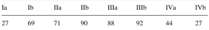
Table 3
Use of brachytherapy as ®rst-line or as a component of `curative' treatment according to FIGO stagea
Ia Ib IIa IIb IIIa IIIb IVa IVb
27 69 71 90 88 92 44 27
aNumber of centers reported in % of total number.
Fig. 1. Treatment sequence according to FIGO stage. The centers were asked to report the curative treatment modalities in function of priority. Places of (a), surgery; (b), external radiation therapy; (c), brachytherapy; and (d) chemotherapy in the treatment sequence.
3.2.4. Reporting radiation side effects
Radiation side effects are reported using different grading scales; the more recently published LENT/SOMA has not gained wide acceptance since six centers (12%) are using this scale for acute side effects and nine centers (17%) for late side effects. The French±Italian glossary [5] is more frequently used (acute effects: 19 centers, i.e. 37%; late effects: 24 centers, i.e. 46%). This is followed by the World Health Organization (WHO: l7 acute and 13 late) and the Radiation Therapy Oncology Group (RTOG: l4 acute and 13 late) grading systems. Fifteen centers use more than one grading system to report side effects, i.e. they mix up a scale for early effects from one group with a scale for late effects from another group. Thirty ®ve centers (67%) consistently use the grading system issued from the same group (mainly the French±Italian glossary) to report acute and late effects.
3.2.5. Chemotherapy
Chemotherapy is divided into neoadjuvant (prior to de®-nitive local treatment, such as surgery or radiotherapy), concomitant (synchronous combination with external irra-diation) and sequential (alternating with external radiation therapy). Neoadjuvant chemotherapy is used by 11 centers, concomitant chemo±radiation therapy by 15 and sequential by six. The indications for chemotherapy are FIGO stage-dependent. None of the centers report any form of chemotherapy in stage Ia compared to 23 centers, i.e. 44%, in the case of stage IVb. Neoadjuvant chemotherapy is more frequently applied in more advanced diseases. For example, seven centers report neoadjuvant chemotherapy for stage IIb, nine for stage IIIa and eleven for stage IIIb. There is a general trend to introduce more and more chemotherapy with higher stage disease. Cisplatin is the most commonly used chemotherapeutic agent.
3.3. New approaches
One of the primary aims of the present questionnaire is to determine the interest in the RCG within the EORTC in conducting a phase III trial. Forty one out of 52 centers (79%) are willing to participate to a prospective trial. Most of the latter are interested in a trial investigating the
importance of chemotherapy in LACC, especially in the setting of concomitant use of chemotherapy (33 centers 64%). It should be noted that the questionnaire was addressed to the centers before the data on the effect of cisplatin in LACC was available.
The question relating to the importance of total treatment duration (reducing the package-deal concept) was found to be a worthwhile test in a randomized trial in 25 centers (48%). The concept of innovative approaches and especially the testing of `new' sensitizers in a phase I/II trial was accepted by 29 centers (56%), with the majority (16/29) choosing taxol. Other possible `new sensitizers' generating interest in the questionnaire were gemcitabine (11 centers), CPT-11 (eight centers), iodo-deoxy-uridine (®ve centers), tirapazamine (four centers). Hydroxyurea was not reported as an interesting agent to be tested.
4. Discussion
This survey, established initially to evaluate the feasibil-ity of a trial within the EORTC-RCG in LACC, results in an interesting amount of information concerning staging and treatment habits in Europe. The ®rst relevant point is the rate of response to the survey. Sixty four percent of the centers have answered to the questions raised, and therefore this survey yields a representative estimation of staging and treatment habits in the RCG centers of the EORTC. The most important information is the large variation in patient care, and this, although expected, has not yet been docu-mented.
4.1. Pretreatment evaluation and staging in cervical cancer Tumor stage (T) is, with nodal stage (N), one of the most important prognostic factors. To assess T and N stage, an extensive local and regional examination is necessary. T can easily be assessed by digital rectal and vaginal examination under anesthesia. The clinician should not only report tumor stage, but also tumor size, because the latter behaves as an independent prognostic factor, as shown in a recently published multivariate analysis [1]. However, tumor size is rarely reported.
We initially expected a large majority of centers reporting
Table 4
Range and median values in Gy of brachytherapy doses according to stage and dose-ratea
Ia Ib IIa IIb IIIa IIIb IVa IVb
MDRb ± 21 21 24 24 24 24 ±
HDR 10±28 10±30 10±35 10±35 9±40 9±40 10±40 10±40
Median 17 17 18 18 18 20 18 17
LDR 20±70 20±80 20±70 10±70 10±60 10±60 10±50 19±60
Median 60 40 45 40 35 35 30 20
aThe ranges in total dose at the reference point are explained by variation of the relative importance of brachytherapy in the treatment protocol in the
different centers.
both cystoscopy and rectoscopy as routine procedures for clinical evaluation of local tumor extent. While cystoscopy is rated in nearly all centers, only slightly more than half of them are reporting rectoscopy as a routine procedure.
One of the most challenging problems in the diagnostic procedure is to detect lymph-node disease, as its presence changes the prognosis, and hence, treatment. The literature reports a 20±40% discrepancy between surgical and clinical staging [31], and some authors claim that there is a role for debulking of pelvic node metastases, as this debulking may result in increased cure-rates [9,31]. Most of the centers report a procedure for the detection of lymph-node metas-tases, but there is a large variation in the techniques used. Surgical staging is performed in about one quarter of the centers. If pelvic lymph-node staging is performed surgi-cally, retroperitoneal staging should be preferred as it is less morbid than transperitoneal staging [9,31]. Our ques-tionnaire does not allow us to assess whether lombo-aortic sampling is performed in centers which mention a surgical procedure.
If non-surgical techniques are used for the assessment of lymph-node stage, one has to mention that the lymphangio-graphy (LAG) remains the best available technique, although this procedure is rarely performed in the present day. This is mainly due to the lack of expertise, and there-fore the technique risks being discarded as part of the routine diagnostic work-up. Only a minority of centers (14%) in our study use LAG for nodal status evaluation, most of them use CT or NMR, which are known to be less sensitive and speci®c than LAG. However, as shown by the Gynecologic Oncology Group (GOG), LAG has a sensitivity of 79% and speci®city of 73%, whereas CT and ultrasound have sensitivities of 34 and 19%, and speci®ci-ties of 96 and 99%, respectively [15]. The importance and utility of LAG, especially if external radiotherapy is planned, is con®rmed by the work of Bonin et al., who demonstrated that bony landmarks are not an adequate substitute for LAG for the localization of pelvic lymph nodes [3]. Some authors are convinced that LAG remains the tool of choice in order to de®ne target volumes and ®eld limits for external radiation [3].
In conclusion there is a clear shift in diagnostic work-up, with a decrease of the importance of LAG to 14%. This can be compared with the 11±18% reported in the patterns of care study of Perez and Montana [25,27]. The reduction in the use of LAG corresponds with an increase to 85% in the use of CT (compared to 70% in the patterns of care study), although a poor correlation between CT and surgical staging has been documented (50±60%). In our study, 50% of the centers are using MRI, although the accuracy of MRI in detecting metastatic lymph nodes has not been established. Surgical nodal staging is performed in 18 centers, and hence, declining frequency in the use of LAG is not compensated by a more frequent use of surgical staging. The shift towards CT and/or MRI in the staging procedure, and away from LAG, results in a potential loss of accuracy
of the diagnostic work-up. This has to be considered in reviewing the therapeutic results and comparing these with the historical series, in which LAG was considered as standard.
4.2. Treatment options and treatment sequence 4.2.1. Surgery
Most of the centers are advocating curative surgery for early stage cervical cancer. Our results are therefore similar to a survey performed in the USA, highlighting a progres-sive increase in the implication of gynecological oncologi-cal surgeons in the primary treatment of early cervix cancer. In our survey, radiation therapy seems to be less frequently used for early stage disease, decreasing from 70 to 60.3%, as either the only treatment or in combination with other treat-ment modalities [34]. This trend in stage Ib±IIa cervical cancer seems not to be evidence-based, taking into account the randomized study reported by Landoni et al. [22]. That trial compares curative surgery to curative radiation therapy in early stage cervix cancer. The 5-year actuarial survival is identical in both arms of the trial (83%), but there is a signi®cant difference favoring external radiotherapy as far as severe morbidity, especially urological, is concerned (28 vs. 19%). The combination of surgery and radiotherapy yields the highest morbidity, especially regarding late-responding normal tissues. Therefore, optimal selection is required for surgery, leaving only premenopausal women with normal ovarian function and with cervical diameters of less than 4 cm as potential candidates for primary surgery, assuming there is no suspicion of nodal involve-ment [10,22]. Furthermore, emphasis should be put on total cost in clinical practice, with a net increase if hysterectomy and postoperative radiotherapy are compared to radiation therapy alone in stage Ib±IIb [8].
It is noteworthy that a number of centers report surgical options in LACC even as a ®rst-line curative treatment in the present survey. To our knowledge, there is, however, no evidence supported by any randomized trial suggesting that such an aggressive surgical approach would yield any bene-®t in patients who will need subsequent high-dose radiation therapy.
4.2.2. Radiotherapy
Three different aspects of radiation therapy deserve special attention: the de®nition of the target volume, the volumes to be treated by external radiation and the brachytherapy.
To obtain optimal treatment, one needs to adequately de®ne the target volume. A CT-guided planni®cation (either 2D or 3D), seems to be performed in nearly all centers. Only six centers are not using CT at all. This re¯ects the general quality of the treatment planning. Kim et al. have shown that the most common site of an inadequate margin is located at the position of the rectal block resulting in a geographical miss in 39±50% of the patients in stage Ib±IVa [20]. This is
followed by the posterior margin in 25±32% of the cases. If the anterior surface of the sacrum is included, a dramatic drop to almost no geographical misses at the posterior border of the lateral ®elds is observed, suggesting that a four-®eld box technique with customized blocking should not be used unless a dedicated CT is available [20]. In the present survey, six centers do not use CT-guided planni®ca-tion, and four of them are treating the pelvic volume with a four-®eld box technique. The posterior limit and the use of individualized blocks in the lateral ®eld have not been inquired in the present survey, and therefore we cannot really assess if there is an increased risk of geographical miss. A second question relates to the volumes to be treated with external radiation therapy, especially the irradiation of para-aortic nodes. The RTOG trial demonstrates a 10-year survival bene®t (44 vs. 55%) in bulky Ib (cancer diameter of more than 4cm) and IIa cervical carcinoma if para-aortic lymph nodes are included in the target volume, although there is no difference in disease-free survival nor in the control of local disease between pelvic irradiation and pelvic plus para-aortic irradiation [33]. The reported 10-year survival difference can be explained by a lower inci-dence of distant failures, eventually with a better salvage in complete responders who later failed locally, or a difference in the death-rate due to treatment-related complications (see late effects). One should be aware that 37% of those patients did not have their para-aortic nodes evaluated surgically or radiologically. The EORTC trial performed on more advanced cervix cancer (comparing pelvic plus para-aortic vs. pelvic volume in early clinical stages with positive LAG or histologically proven pelvic nodes, and LACC, i.e. IIb and III), does not show any bene®t in survival, local control or overall distant metastases [14]. There was a clear increase in the incidence of severe digestive complications in patients receiving para-aortic irradiation (9 vs. 4.8%) [14,41]. However, considering the reduced incidence of para-aortic and distant metastases in patients without recur-ring tumor at pelvic sites, if treated with extended ®elds, the authors proposed to treat para-aortic nodes where there is a high probability of local control [14]. In the present survey, about 90% of the centers are treating this large volume if they have arguments for positive para-aortic and/or pelvic lymph nodes.
A third important parameter relates to the quality of the brachytherapy. Brachytherapy is an essential component of treatment in LACC, although there is no consensus on the optimal dose-rate. A majority of centers are using LDR. A randomized trial comparing HDR vs. LDR showed similar 5-year survival rates, but a signi®cant difference in favor of LDR as far as toxicity is concerned (10% moderate to severe complications in HDR compared to 4% in LDR) [39]. The question on dose-rate is not de®nitely settled, with some authors con®rming the importance of dose-rate, although at a different level, i.e. comparing two low dose-rate levels [23], and others rejecting the conclusions of the Osaka trial stating that there is no increase in late toxicity [35].
4.2.3. Chemotherapy
The addition of chemotherapy to radiotherapy has become standard in the treatment of LACC [34]. Morris et al. demonstrated in a randomized trial the bene®cial effects of adding 5-¯uoro-uracil and cisplatinum to radiotherapy in LACC, resulting in a signi®cant improvement in survival (from 73 vs. 58%) [26]. The Gynecologic Oncology Group compared, in a randomized fashion, cisplatin vs. cisplatin plus hydroxyurea, and 5-¯uorouracil vs. hydroxyurea alone [32]. Cisplatin containing regimens improve the survival and progression-free survival in LACC [32].
One might expect major changes in treatment prescrip-tions following these randomized trials. The frequency of adjunctive chemotherapy already changed from 6% in 1984 to 24.8% in 1990. In the patterns of care study, 12% of the patients were treated with concurrent chemotherapy at the cost of a greater morbidity [25,27]. In view of an unproven bene®t at that time, it is interesting to note the frequency of use of chemotherapy and especially neoadjuvant chemotherapy. One could wonder why some patients with LACC are still submitted to neoadjuvant chemotherapy, as a suf®cient amount of data issued from randomized trials show a deleterious effect on survival [24,30,36,37]. A substantial reduction in survival from 39 to 23% at 5 years has been shown by Souhami et al., comparing radio-therapy to neadjuvant chemoradio-therapy followed by radiother-apy in patients with FIGO stage IIIb [37]. The issue of chemotherapy is not de®nitely settled and a lot more empha-sis will be put on concomitant chemo±radiotherapy, espe-cially after these two positive randomized trials. The advantages are spatial cooperation and additive or supra-additive effects on the primary tumor. For spatial coopera-tion, one should realize that distant metastases as the ®rst site of failure are rare (5±15%), and that distant metastases are not related to the stage. Regarding the effect on the locoregional control, most of the drugs are used because of their capacity to sensitize cancer cells to ionizing irradia-tion. Cisplatin is one of the most frequent agents reported to be used, together with radiotherapy, in the present survey. Indeed, clinical experience favors cisplatin as the drug of choice in various human tumors. However, some experi-mental data in vitro, especially on human cervix cancer cell lines, show that cisplatin leads to additive toxicity and not radiosensitization [4].
4.3. New approaches
Possible factors affecting the outcome in the treatment of LACC are intrinsic radio-resistance, tumor proliferation and hypoxia. Most of the centers of the RCG are willing to participate in trials addressing these issues. The highest rating is obtained for a trial combining chemotherapy as a concomitant sensitizer to radiotherapy, which obviously would require running phase I/II trials if drugs like taxol or gemcitabine have to be used, because the data indicate an increase in gastrointestinal toxicity.
The importance of reducing the overall treatment-time is also considered an important issue in the treatment of LACC. This re¯ects evidence issued from retrospective studies addressing the importance of the `time package deal' in LACC [6,11,12,21,28,29,40]. As stated by Eifel `a comparison between policies that emphasize early intra-cavitary treatment and short 5±6-week overall treatment-times with those that emphasize an initial course of pelvic treatment to achieve greater tumor regression before brachytherapy would be of considerable interest' [8].
Last but not least, hypoxia is certainly an issue in cervix cancer, as shown by different authors performing in vivo measurements and seeking a correlation between low tumor pO2 and outcome [16,17]. The hypoxia in cervix
tumors provides a physiological selective pressure for the expansion of HPV-infected epithelial variants that have lost their apoptotic potential, which is deemed to be a supple-mentary reason for radiation resistance [13,19]. The Radia-tion Therapy Oncology Group tested misonidazole as a hypoxic cell-sensitizer in a randomized trial; no improve-ment in survival was observed [2]. The Gynecology Oncol-ogy Group compared hydroxyurea vs. misonidazole with no improvement in survival [38].The Medical Research Coun-cil Working Party on advanced carcinoma of the cervix ran another randomized trial with Ro 03-8799 (pimonidazole) as a hypoxic cell-sensitizer. They observed no bene®t by adding pimonidazole to radiotherapy, and a truly adverse effect of the drug was a possible cause of failure in this trial [7]. A new approach is the use of bio-reductive drugs, such as tirapazamine (SR-4233), which in contrast to misonidazole and pimonidazole, offers a selective way of killing those resistant hypoxic cells.
The presence of HPV opens new directions for radiation biology research, as it is currently known that HPV onco-proteins E6 and E7 inhibit both p53 and Rb onco-proteins, both playing an essential role in the cell cycle machinery, and hence, in¯uencing the cell-cycle response to stress [18]. Mechanisms of inhibition of E6 and E7 expression could reinstore p53 functionality and modify radiation response. 5. Conclusions
Several questions are unresolved in cervix cancer. These uncertainties are re¯ected in the answers provided by the members of the RCG on questions related to staging, treat-ment options and techniques, and on innovative approaches to be tested within trials. However, a clear shift in patterns of care does occur in cervix cancer treatment. There seems to be a decrease in the number of earlier stages secondary to screening policy, but on the other hand, there is a suf®cient number of IIIa and IIIb disease, leaving some space for trials within Europe. Our survey re¯ects the interest within the RCG to launch and participate in a large prospective multi-center trial in Europe. However, we need an educational program to improve our `standards' of care, and to adhere
to the principles of evidence-based medicine. A comprehen-sive quality-control program is mandatory, especially due to the complexity of the combination of external irradiation and brachytherapy. As a matter of fact, this program is a prerequisite to launching any kind of trial in LACC. References
[1] Barillot I, Horiot J-C, Pigneux J, et al. Carcinoma of the intact uterine cervix treated with radiotherapy alone: a French cooperative study: update and multivariate analysis of prognostic factors. Int. J. Radiat. Oncol. Biol. Phys. 1997;38:969±978.
[2] Bauer M, Leibel S, Wasserman T, et al. Effect of misonidazole dose on survival in patients with stage IIIB±IVA squamous cell carcinoma of the uterine cervix. Int J. Radiat. Oncol. Biol. Phys. 1986;12:1101± 1103.
[3] Bonin S, Lanciano R, Corn BW, Hogan WM, Hartz WH, Hanks G. Bony landmarks are not an adequate substitute for lymphangiography in de®ning pelvic lymph node location for the treatment of cervical cancer with radiotherapy. Int. J. Radiat. Oncol. Biol. Phys. 1996;34: 167±172.
[4] Britten RA, Evans AJ, Allalunis-Turner MJ, Pearcey RG. Effect of cisplatin on the clinically relevant radiosensitivity of human cervical carcinoma cell lines. Int. J. Radiat. Oncol. Biol. Phys. 1996;34:367± 374.
[5] Chassagne D, Sismondi P, Horiot J-C, et al. A glossary for reporting complications of treatment in gynecological cancers. Radiother. Oncol. 1993;26:195±202.
[6] Delaloye J-F, Coucke PA, Pampallona S, De Grandi P. Effect of total treatment time on event-free survival in carcinoma of the cervix. Gynecol. Oncol. 1996;60:42±48.
[7] Dische S. On behalf of the MRC Working Party on advanced carci-noma of the cervix: a trial of Ro 03-8799 (pimonidazole) in carcicarci-noma of the uterine cervix: an interim report from the Medical Research Council Working Party on advanced carcinoma of the cervix. Radio-ther. Oncol. 1993;26:93±103.
[8] Eifel P, Thames HD. Has the in¯uence of treatment duration on local control of carcinoma of the cervix been de®ned? Int. J. Radiat. Oncol. Biol. Phys. 1995;32:1527±1529.
[9] Fine BA, Hempling RE, Piver MS, Baker TR, McAuley M, Driscoll D. Severe radiation morbidity in carcinoma of the cervix. Impact of pretherapy surgical staging and previous surgery. Int. J. Radiat. Oncol. Biol. Phys. 1995;31:717±723.
[10] Eifel PJ, Morris M. Irradiation alone or combined with surgery in carcinoma of the cervix. When will we know the answer? Int. J. Radiat. Oncol. Biol. Phys. 1995;31(4):1007±1008.
[11] Fyles A, Keane TJ, Barton M, Simm J. The effect of treatment dura-tion in the local control of cervix cancer. Radiother. Oncol. 1992;25:273±279.
[12] Girinsky T, Rey A, Roche B, et al. Overall treatment time in advanced cervical carcinoma: a critical parameter in treatment outcome. Int. J. Radiat. Oncol. Biol. Phys. 1993;27:1051±1056.
[13] Graeber TG, Osmanian C, Jacks T, et al. Hypoxia mediated selection of cells with diminished apoptotic potential in solid tumors. Nature 1996;379:88±91.
[14] Haie C, Pejovic MH, Gerbaulet A, et al. Is prophylactic para-aortic irradiation worthwhile in the treatment of advanced cervical carci-noma? Results of a controlled clinical trial of the EORTC radiother-apy group. Radiother Oncol. 1988;11:101±112.
[15] Heller PB, Malfetano JH, Bundy BN, Barnhill DR, Okagaki T. Clin-ical±pathological study of stage IIB, III and IVa carcinoma of the cervix: extended diagnostic evaluation for para-aortic node metas-tases - a Gynecologic Oncologic Group Study. Gynecol. Oncol. 1990;38:425±430.
[16] HoÈckel M, Knoop C, Schlenger K, et al. Intratumoral pO2predicts
survival in advanced cancer of the uterine cervix. Radiother. Oncol. 1993;26:45±50.
[17] HoÈckel M, Schlenger KH, Aral B, Mitze M, SchaÈffer U, Vaupel P. Association between tumor hypoxia and malignant progression in advanced cancer of the uterine cervix. Cancer Res. 1996;56:4509± 4515.
[18] Kapp DS, Giaccia AJ. New directions for radiation biology research in cancer of the uterine cervix. J. Natl. Canc. Inst. 1996;21:131±139. [19] Kim CY, Tsai MH, Osmanian C, et al. Selection of human cervical epithelial cells that possess reduced apoptotic potential to low-oxygen conditions. Cancer Res. 1997;57:4200±4204.
[20] Kim RY, McGinnis S, Spencer SA, Meredith RF, Jenelle RLS, Salter MM. Conventional four ®eld pelvic radiotherapy technique without CT treatment planning in cancer of the cervix: potential geographic miss. Radiother. Oncol. 1994;30:140±145.
[21] Lanciano RM, Pajak TF, Martz K, Hanks GE. The in¯uence of treat-ment time on outcome for squamous cell cancer of the uterine cervix treated with radiation: a patterns-of-care study. Int. J. Radiat. Oncol. Biol. Phys. 1993;25:391±397.
[22] Landoni F, Maneo A, Colombo A, et al. Randomized study of radical surgery versus radiotherapy for stage Ib±IIa cervical cancer. Lancet 1997;350:535±540.
[23] Lambin P, Gerbaulet A, Kramar A, et al. Phase III trial comparing two low dose rates in brachytherapy of cervix carcinoma: report at 2 years. Int. J. Radiat. Oncol. Biol. Phys. 1993;25:405±412.
[24] Leborgne F, Leborgne JH, Doldan R, et al. Induction chemotherapy and radiotherapy of advanced cancer of the cervix. A pilot study and phase III randomized trial. Int. J. Radiat. Oncol. Biol. Phys. 1997;37:343±350.
[25] Montana GS, Hanlon AL, Brickner TJ, et al. Carcinoma of the cervix. Patterns of care studies: review of 1978, 1983, and 1988±1989 surveys. Int. J. Radiat. Oncol. Biol. Phys. 1995;32:1481±1486. [26] Morris M, Eifel PJ, Lu J, et al. Pelvic irradiation with concurrent
chemotherapy compared with pelvic and para-aortic radiation for high-risk cervical cancer. N. Engl. J. Med. 1999;340:1137±1143. [27] Perez CA. Changing patterns of care in carcinoma of the uterine
cervix. Need for cost-bene®t studies. Int. J. Radiat. Oncol. Biol. Phys. 1995;32:1535±1537.
[28] Perez CA, Grigsby PW, Castro-Vita H, Lockett MA. Carcinoma of the uterine cervix. Impact of prolongation of overall treatment time and timing of brachytherapy on outcome of radiation therapy. Int. J. Radiat. Oncol. Biol. Phys. 1995;32:1275±1288.
[29] Petereit DG, Sarkaria JN, Chappell R, et al. The adverse effect of
treatment prolongation in cervical carcinoma. Int. J. Radiat. Oncol. Biol. Phys. 1995;32(5):1301±1307.
[30] Potish RA, Twiggs LB. On the lack of demonstrated clinical bene®t of neoadjuvant cisplatinum therapy for cervical cancer. Int. J. Radiat. Oncol. Biol. Phys. 1993;27:975±979.
[31] Potish RA. Surgical staging, extended ®eld radiation, and enteric morbidity in the treatment of cervix cancer. Int. J. Radiat. Oncol. Biol. Phys. 1995;31(4):1009±1010.
[32] Rose PG, Bundy NB, Watkins EB, et al. Concurrent cisplatin-based radiotherapy and chemotherapy for locally-advanced cervical cancer. N. Engl. J. Med. 1999;340:1144±1153.
[33] Rotman M, Pajak TF, Choi K, et al. Prophylactic extended-®eld irra-diation of para-aortic lymph nodes in stages IIb and bulky Ib and IIa cervical carcinomas. Ten-year treatment results of RTOG 79-20. J. Am. Med. Assoc. 1995;274:387±393.
[34] Russell AH, Shingleton HM, Jones WB, et al. Trends in the use of radiation and chemotherapy in the initial management of patients with carcinoma of the uterine cervix. Int. J. Radiat. Oncol. Biol. Phys. 1998;40:605±613.
[35] Sarkaria JN, Petereit D, Stitt J, et al. A comparison of the ef®cacy and complication rates of low dose-rate versus high dose-rate brachyther-apy in the treatment of uterine cervical carcinoma. Int. J. Radiat. Oncol. Biol. Phys. 1994;30:75±82.
[36] Shueng P-W, Hsu W-L, Jen Y-M, Wu C-J, Liu H-S. Neoadjuvant chemotherapy followed by radiotherapy should not be a standard approach for locally-advanced cervical cancer. Int. J. Radiat. Oncol. Biol. Phys. 1998;40:889±896.
[37] Souhami L, Gil RA, Allan SE, et al. A randomized trial of chemother-apy followed by pelvic radiation therchemother-apy in stage IIIb carcinoma of the cervix. J. Clin. Oncol. 1991;9:970±977.
[38] Stehman FB, Bundy BN, Thomas G, et al. Hydroxyurea versus miso-nidazole with radiation in cervical carcinoma: long-term follow-up of a Gynecologic Oncology Group trial. J. Clin. Oncol. 1993;11:1523± 1528.
[39] Teshima T, Inoue T, Ikeda H, et al. High-dose rate and low-dose rate intracavitary therapy for carcinoma of the uterine cervix. Final results of Osaka University Hospital. Cancer 1993;72:2409±2413. [40] Tsang RW, Fyles AW, Kirkbride P, et al. Proliferation measurements
with ¯ow cytometry Tpotin cancer of the uterine cervix. Correlation
between two laboratories and preliminary clinical results. Int. J. Radiat. Oncol. Biol. Phys. 1995;32:1319±1329.
[41] Vigliotti AP, Wen B-C, Hussey DH, et al. Extended ®eld irradiation for carcinoma of the uterine cervix with positive peri-aortic nodes. Int. J. Radiat. Oncol. Biol. Phys. 1992;23:501±509.