Journal Pre-proof
Cancer-associated fibroblasts in desmoplastic tumors: emerging role of integrins
C ´edric Zeltz, Irina Primac, Pugazendhi Erusappan, Jahedul Alam, Agnes Noel, Donald Gullberg
PII: S1044-579X(19)30038-0
DOI: https://doi.org/10.1016/j.semcancer.2019.08.004
Reference: YSCBI 1633
To appear in: Seminars in Cancer Biology
Received Date: 29 April 2019 Revised Date: 1 August 2019 Accepted Date: 5 August 2019
Please cite this article as: Zeltz C, Primac I, Erusappan P, Alam J, Noel A, Gullberg D,
Cancer-associated fibroblasts in desmoplastic tumors: emerging role of integrins, Seminars in
Cancer Biology (2019), doi:https://doi.org/10.1016/j.semcancer.2019.08.004
This is a PDF file of an article that has undergone enhancements after acceptance, such as the addition of a cover page and metadata, and formatting for readability, but it is not yet the definitive version of record. This version will undergo additional copyediting, typesetting and review before it is published in its final form, but we are providing this version to give early visibility of the article. Please note that, during the production process, errors may be discovered which could affect the content, and all legal disclaimers that apply to the journal pertain.
Journal Pre-proof
Review
Cancer-associated fibroblasts in desmoplastic tumors: emerging role of
integrins
Cédric Zeltz1,2, Irina Primac3, Pugazendhi Erusappan1,4, Jahedul Alam1, Agnes Noel3 and Donald Gullberg1,*
1 Department of Biomedicine and Centre for Cancer Biomarkers, University of Bergen, Bergen, Norway
2 Princess Margaret Cancer Center, University Health Network, Toronto, Canada
3 Laboratory of Tumor and Development Biology, GIGA-Cancer, University of Liege (ULiège), Liege, Belgium
4 Institute for Experimental Medical Research, Oslo University Hospital and University of Oslo, Oslo, Norway
*Corresponding author: Donald Gullberg, PhD University of Bergen Dept. of Biomedicine Jonas Lies vei 91 NO-5009 Bergen Norway
Tel: (+47) 55 58 63 32
E-mail: donald.gullberg@uib.no
ABSTRACT
The tumor microenvironment (TME) is a complex meshwork of extracellular matrix (ECM) macromolecules filled with a collection of cells including cancer-associated fibroblasts
Journal Pre-proof
(CAFs), blood vessel associated smooth muscle cells, pericytes, endothelial cells, mesenchymal stem cells and a variety of immune cells.In tumors the homeostasis governing ECM synthesis and turnover is disturbed resulting in abnormal blood vessel formation and excessive fibrillar collagen accumulations of varying stiffness and organization. The disturbed ECM homeostasis opens up for new types of paracrine, cell-cell and cell-ECM interactions with large consequences for tumor growth, angiogenesis, metastasis, immune suppression and resistance to treatments. As a main producer of ECM and paracrine signals the CAF is a central cell type in these events. Whereas the paracrine signaling has been extensively studied in the context of tumor-stroma interactions, the nature of the numerous integrin-mediated cell-ECM interactions occurring in the TME remains understudied. In this review we will discuss and dissect the role of known and potential CAF interactions in the TME, during both tumorigenesis and chemoresistance-induced events, with a special focus on the “interaction landscape” in desmoplastic breast, lung and pancreatic cancers. As an example of the multifaceted mode of action of the stromal collagen receptor integrin 111, we will summarize our current understanding on the role of this CAF-expressed integrin in these three tumor types.
Journal Pre-proof
ABCG2 ATP Binding Cassette Subfamily G Member 2
ADAM12 A disintegrin and metalloproteinase domain-containing protein 12
αSMA Alpha-smooth muscle actin
CAFs Cancer-associated fibroblasts
CAF-S Cancer-associated fibroblast subset type
cCAFs Cell cycle cancer-associated fibroblasts
CAV1 Caveolin-1
CD10 Cluster of differentiation 10, membrane metallo-endopeptidase (MME)
CD29 Cluster of differentiation 29, integrin 1
CD49c Cluster of differentiation 49c, integrin 3 subunit CD49e Cluster of differentiation 49e, integrin 5 subunit CD51 Cluster of differentiation 51, integrin v subunit CD105 Cluster of differentiation 105, endoglin
CD126 Cluster of differentiation 126, interleukin 6 receptor
CLCF1 Cardiotrophin-like cytokine factor 1
CLU Clusterin
CSC Cancer stem cell
dCAFS Developmental cancer-associated fibroblasts
dcn Decorin
DDR2 Discoidin domain receptor 2
DPP4 Dipeptidylpeptidase 4
ECM Extracellular matrix
EMT Epithelial-mesenchymal transition
EndoMT Endothelial-mesenchymal transition
ER Estrogen receptor
ERK Extracellular signal-regulated kinase
FAK Focal adhesion kinase
FAP Fibroblast activation protein
FGF Fibroblast growth factor
FSP-1 Fibroblast specific protein-1
GFPT2 Glutamin-fructose-6-phosphate transaminase 2
GLI1 Glioma-associated oncogene homologue 1
GPR77 G protein-coupled receptor 77
HER2 Human epidermal growth factor receptor 2 breast cancer subtype
HGF Hepatocyte growth factor
Hh Hedgehog
iCAFS Inflammatory cancer-associated fibroblasts
IGF Insulin-like growth factor
IGFBP3 Insulin-like growth factor-binding protein 3
IL-1 Interleukin-1
IL-6 Interleukin-6
IL-11 Interleukin-11
IL-33 Interleukin-33
IRAK-4 Interleukin-1 receptor-associated kinase 4
KPC KrasLSL.G12D/+; p53R172H/+; PdxCretg/+
Journal Pre-proof
Keywords: tumor microenvironment, cancer-associated fibroblast, fibrosis, TME-mediated chemoresistance, integrin
LTBP3 Latent transforming growth factor beta binding protein 3
LOXL1 Lysyl oxidase-like 1
LOXL2 Lysyl oxidase-like 2
mCAFS Matrix cancer-associated fibroblasts
MAPK MDSCs
Mitogen-activated protein kinase Myeloid-derived suppressor cells
MMTV Mouse mammary tumor virus
MRTF Myocardin-related transcription factor
MSCs Mesenchymal stem cells
myCAFS Myofibroblastic cancer-associated fibroblasts
NF Normal fibroblast
NG2 Neuron-glial antigen 2
NSCLC Non-small cell lung carcinoma
PDAC Pancreatic ductal adenocarcinoma
PDGFRα Platelet-derived growth factor receptor alpha PDGFRβ Platelet-derived growth factor receptor beta
pFAK Phosphorylated focal adhesion kinase
P-GP P-glycoprotein
PyMT
PTEN
Polyoma middle T
Phosphatase and tensin homologue
RTKs Receptor tyrosine kinases
RTKIs Receptor tyrosine kinase inhibitors
SCC Squamous cell carcinoma
SMOi Smoothened inhibitor
STAT3 Signal transducer and activator of transcription 3
STC1 Stanniocalcin-1
Taz TAM
Transcriptional coactivator with PDZ-binding motif Tumor-associated macrophage
TGFβ Transforming growth factor beta
TNBC Triple negative breast cancer subtype
TME Tumor microenvironment
vCAFS Vascular cancer-associated fibroblasts
WISP2 WNT1- inducible signaling pathway protein2
Journal Pre-proof
1. Introduction
The fibroblast is a cell type of paramount importance for extracellular matrix (ECM) production and remodeling in interstitial tissues[1]. Fibroblasts are central in wound healing, tissue fibrosis and tumor fibrosis and studies of molecular mechanisms have demonstrated that fibroblasts use similar “toolkits” to remodel the ECM in these different conditions [2-4]. In tumor biology the activated fibroblasts, often called cancer-associated fibroblasts (CAFs), act in the realms of the tumor microenvironment (TME) with consequences for tumor growth, formation of stem cell niches, immunosuppression, metastasis and chemoresistance[5,6]. In the current review we will focus on this important compartment of the tumor and discuss how fibrosis contributes to TME-mediated effects on tumor progression and chemoresistance. Box 1
In medicine, desmoplasia is the growth of fibrous or connective tissue. It is also called desmoplastic reaction to emphasize that it is secondary to an insult. Desmoplasia may occur around a neoplasm, causing dense fibrosis around the tumor, or scar tissue (adhesions) within tissues.
The complexity of tumor microenvironment in different tumor types is overwhelming and therefore we have decided to limit ourselves and try to give an overview of the role played by CAFs in cell-ECM and paracrine interactions in the TME of three desmoplastic tumor types: breast, lung and pancreatic cancer. We will summarize some interesting new developments (without any claims to cover all new interesting findings), including data suggesting that integrin 111 is a major CAF integrin in desmoplastic tumors [7-10].
2.Tissue Fibrosis Box 2a
Fibroblast- A poorly defined cell type of mesenchymal origin, which is vascular, non-inflammatory and non-epithelial. Fibroblasts play a major role to produce fibrillar collagens and other interstitial ECM components and to take active part in matrix remodeling via integrins and release of matrix metalloproteinases during tissue regeneration events[1,5]. The transcriptional profile of fibroblasts varies with the anatomical location[11]. Cell lineage tracing in mouse has clarified distinct origins of fibroblasts in the heart and skin. Mouse cardiac fibroblasts are derived from epicardium or endocardium[12] and a common multipotent progenitor of reticular and papillary skin fibroblasts has been identified in mouse skin where neonatal fibroblast subtypes are characterized by a dynamic biomarker expression pattern [13,14]. Further heterogeneity in skin fibroblasts is introduced by presence of hair follicles, different embryonic origins of dermal fibroblasts in face (neural crest), anterior part (lateral plate mesoderm) and the posterior part of body (dermomyotome). Closer examination of dermal fibroblasts comparing human and mouse skin confirms the dynamic expression of biomarkers in human dermal fibroblasts and identifies differences in biomarker expression between mouse and human dermal fibroblasts[15]. Several groups have defined multiple subtypes of human skin fibroblasts[15-17] and a protocol to isolate reticular and papillary fibroblasts based on FAP and CD90 expression exists[18]. In lung, transcriptional profiling
Journal Pre-proof
has identified six subtypes of fibroblasts [19] and in years to come additional tissue-specific fibroblast populations are likely to be described.Box 2b
Myofibroblast- An activated fibroblast considered to be contractile due to expression of the contractile isoform of actin, alpha smooth muscle actin (SMA)[20,21]. In some tissues known to express v1 integrin with a central role in TGF- activation in fibrotic conditions[22]. After completed wound healing myofibroblasts are usually depleted via apoptosis[21,23]. Mouse cardiac myofibroblasts have been observed to turn off SMA expression in the heart and form a cell type called matrifibrocyte with different properties than the undifferentiated pre-myofibroblasts[24]. Current data thus suggests that myofibroblasts display more plasticity than previously thought. The finding that subsets of mouse skin myofibroblasts under certain conditions can differentiate into adipocytes further stresses the plasticity of myofibroblasts[25].
Cancer-associated fibroblasts (CAFs)- Fibroblast-like cells, of different origins, present in the TME. Sometimes used as abbreviation for carcinoma-associated fibroblasts, to specifically denote cells associated with epithelial-derived tumors. Demonstrated to be surprisingly heterogeneous. A number of CAF subtypes have been defined within tumor stroma. Pioneer work has defined two major types of fibroblasts in pancreatic cancer, inflammatory CAF (iCAFs) and myofibroblastic CAFs (myCAFs)[26], and four major subclasses of CAFs in breast cancer (CAF-S1-S4), distinguished by different levels of SMA and fibroblasts activation protein (FAP) expression [27,28]. Due to plasticity and dynamic nature it has been suggested that the CAF subtypes do not represent fixed cell types, but rather represent fibroblast “states”[29]. Epigenetic changes do however result in more stable phenotypes[30,31]. Indirect evidence suggests that some subpopulations of CAFs are tumor-supportive whereas others are tumor-suppressive[32,33]. Demonstrated to act in a paracrine manner to affect different aspects of tumorigenesis, and via matrix synthesis and matrix remodeling to induce stiffness and hypoxia, which in turn also affect tumor growth.
Major challenges in all forms of fibrosis include characterizing the degree of fibroblast heterogeneity, defining the origin of pro-fibrotic cells (also the potential targets of anti-fibrosis therapy), and characterizing the dynamics of different biomarkers, which can be used to follow the fibrotic process as well as serve as potential therapeutic targets.
Gene technology developments have helped to clarify some of the issues related to the origin of fibroblasts in animal fibrosis models. In several experimental systems, cell lineage tracing has thus clarified “muddy waters” where epithelial to mesenchymal transition (EMT) and fibrocyte invasion were suggested to contribute significantly to fibrotic processes. The genetic-method based cell lineage tracing, often contested earlier immunohistochemistry-based studies (typically relying on antibodies with unclear specificity or reactivity) and instead showed major roles played by tissue myofibroblasts derived either from endogenous resident fibroblasts[12,13,34], pericytes[35,36] or Gli+-positive mesenchymal stem cells (MSC)[37]. Pericytes exist as a major cell type in pancreas and in liver in the form of stellate cells[38,39], which in fibrosis models become activated to myofibroblasts and in tumors into CAFs. The relative contribution of pericytes to fibrotic stroma in tissues like kidney, lung and breast is complex[40] and will need careful cell lineage tracing in different mouse models. Heart and skin are two examples where resident endogenous fibroblasts are major sources of the fibrotic stroma[12,15], but where also Gli+ MSC expansion has been shown to play a major
Journal Pre-proof
role in tissue fibrosis[37]. In the developing mouse heart EMT and endothelial mesenchymal transition (EndoMT) do play a role, but not in fibrotic conditions [12].As just mentioned, cell lineage analysis has thus clarified the origin of fibroblasts in skin and heart[12,13]. During scarring in the skin after injury or in heart after an infarction, endogenous fibroblasts migrate in and fill up the damaged area. The dynamics of the in-migration of two major types of fibroblasts in the wounded mouse skin occurs in two waves [41]. In a careful separate study these fibroblasts were further characterized into a “PDGFRhigh subset” and a “PDGFRlow subset”, which could be further subdivided into a several clusters [14]. As wounding is complete, wound fibroblasts disappear via apoptosis. In mouse skeletal muscle and skin ADAM12+/PDGFR+- perivascular cells appear to play an important role in tissue repair[36]. Recent data also demonstrate a role of Gli+ MSC in dermal wound healing[37]. In the heart the interstitial fibroblasts fill up the damaged area and during the repair phase express alpha smooth muscle actin (SMA) and are contractile. Interestingly, these fibroblasts then loose SMA expression and become quiescent [24]. Similar to the wound response in skin, Gli+ MSCs have been shown to contribute to cardiac fibrosis [37].
2.1 Tumor fibrosis
Major factors that drive tumor desmoplasia include: autocrine and paracrine signaling of growth factors, cytokines and chemokines. These factors affect cell proliferation and migration as well as CAF-mediated ECM protein secretion and crosslinking of fibrillar collagen matrices that eventually lead to increased tissue stiffness and hypoxia [4]. Furthermore, ECM reorganization and ECM remodeling are determinant factors that affect the properties of the TME.
Just as the origin of myofibroblasts in fibrosis varies, so does the origin of CAFs. Major sources of CAFs are the endogenous tissue fibroblasts, pericytes and ADAM12+ perivascular cells[4,36,42]. Applying cell lineage tracing methods in the polyoma middle T (PyMT) mouse model have demonstrated the contribution of mesenchymal, non-hematopoietic bone marrow cells to a PDGFR-, clusterin+- breast cancer CAF subpopulation (see 4.1 below)[43]. EMT contribution to CAF generation appears to be limited and EMT in the TME seems to be more involved in forming an invasive mesenchymal tumor cell type and in creating a niche for cancer stem cell formation[44]. However, active EMT processes in the tumor have indirect consequences for the stroma. In a recent study, EMT was studied in some detail in a genetic model Kras LSL-12GD/ p53 fl/fl /Lgr5CreER of squamous cell carcinoma (SCC) where tumors undergo spontaneous EMT[45]. These studies convincingly demonstrated that EMT occurs in stepwise manner leading to the generation of subpopulations of tumor cells in different intermediate states between epithelial and mesenchymal. Interestingly, as cells progressed towards EMT[45], the stroma changed in parallel, with regard to composition, presence of immune cells and localization. Most likely these changes are in part due to changes in the paracrine signaling of tumor cells undergoing EMT. A recent study also suggest that great care has to be taken when analyzing cells in invasive breast cancer[46]. Westcott et al studied the process of invasion and identified a switch of tumor cells state into a mesenchymal invasive state without the tumor cells actually undergoing EMT. The invasive cells leading the way in this initial invasive migration, so called trailblazer cells, were characterized by a mesenchymal seven gene signature composed of DOCK1, ITGA11, DAB2, PDGFRA, VASN, PPAP2B and LPAR1[46].
When trying to map the heterogeneity of tumor stroma, it is thus important to distinguish CAFs from: 1) cells undergoing EMT and expressing a variable degree of mesenchymal biomarkers, 2) trailblazer cells with a mesenchymal signature or 3) mesenchymal stem cells residing in the tissue. As of now, biomarkers clearly distinguishing these cells types are lacking.
Journal Pre-proof
2.2 Cell surface markers/biomarkers for fibroblasts, myofibroblasts and CAFs: Expression and biological function
Multiple reviews have focused on the different biomarkers, which are useful when studying the TME. For simplicity we think it is convenient to categorize the biomarkers into membrane proteins, cytoskeletal proteins, intracellular proteins and nuclear proteins. For excellent reviews of the different biomarkers we refer to [5,47,48]. We would just like to add a few facts as reminders of uses and pitfalls for some of the relevant CAF markers.
2.2.1 Membrane proteins
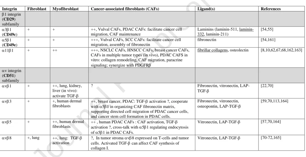
Integrins: We will divide the discussion into different subfamilies, the major ones involved on tumor stroma belonging to 1- or v-integrin subfamilies (Fig.1, Table 1).
1 integrin subfamily: The integrin heterodimers, which have emerged as candidates on CAFs to execute 1 integrin functions include 31, 51, 111 and v1. v1 will be discussed under v integrin subfamily.
1 integrin subunit (CD29): The integrin 1 subunit is shared by 12 different integrin heterodimers and is present on all nucleated cells [49]. The 1 subunit is expressed in excess compared to integrin chains in an intracellular pool. Cell surface expression of integrin heterodimers containing CD29 is determined by integrin chain expression. Due to the ubiquitous expression extreme care has to be taken when using CD29 as a CAF biomarker. Down-stream targets of 1 integrin signaling include the soluble tyrosine kinase FAK and the autophosphorylated FAK tyrosine residue Y397, as a general marker of active 1 integrin signaling[50]. In addition to this role of FAK in integrin outside -in signaling it has also been demonstrated to take part in adhesion strengthening and affect myofibroblast differentiation in an unforeseen manner[51,52]. [53].
3 integrin subunit (CD49c): Integrin with wide expression on cells in contact with basement membranes[49]. 31 binds different laminin isoforms[49]. In CAFs, first reported to be involved in facilitating tumor cell migration in a mixed artificial matrix composed of laminin-111 and collagen I[54]. 31 has later been shown to bind laminin-332 in pancreatic ductal adenocarcinoma (PDAC) CAFs and facilitate cell migration of PDAC cancer cells[55]. 5 integrin subunit (CD49e): Stromal integrin expressed on variety of cell types such as fibroblasts, endothelial cells, immune cells[56] and CAFs[57]. CAF integrin 51 is involved in assembly of fibronectin[58] and in enabling v3-mediated directional prostate and pancreas tumor cell migration[59]. In colon cancer 51 on CAFs cooperate with v3 to assemble fibronectin[60]. In a separate study it is shown that fibronectin-bound 51 integrin promotes tension-dependent malignant transformation through engagement of the synergy site that enhances integrin adhesion force. Ligation of the synergy site of fibronectin permits tumor cells to engage a zyxin-stabilized, vinculin-linked scaffold that facilitates nucleation of phosphatidylinositol (3,4,5)-triphosphate at the plasma membrane to enhance phosphoinositide 3-kinase (PI3K)-dependent tumor cell invasion[61]. The effect of fibronectin synergy site ligation by CAF 51 is unknown. In a careful study of PDAC CAFs in 3D environment 51 subcellular localization (and hence also activity) was controlled by v5 in a complex manner [57].
11 integrin subunit: Integrin11 is expressed on subsets of fibroblasts and mesenchymal stem cells [62-66]. Expression on subsets of stromal cells needs to be better characterized and is ongoing. Data obtained so far, based on studies of PDAC and head and neck squamous cell carcinoma (HNSCC), have failed to demonstrate co-expression of NG2 in 11-positive CAFs[67]. In an 11-positive subset of non-hematopoietic bone marrow-derived
Journal Pre-proof
mesenchymal stem cells, 11 expression correlates with osteogenic potential of these cells [62]. Recent screening of tumor tissue array reveal expression of 11 in CAFs in multiple solid tumors[67]. Studies using animals deficient in 11 expression in the tumor stroma reveal major attenuation of tumor growth and metastasis in non-small cell lung cancer and breast cancer in the absence of 11 [8,10,68]. v (CD51) integrin subfamily: The integrin v chain dimerizes with different integrin chains. v6 is an epithelial integrin and is in tissues like lung and kidney involved in activating TGF-β (via binding of RGD in LAP-TGF-complex) in fibrosis[69]. Overall, whereas the understanding of the role of v integrins in tissue fibrosis is increasing relatively little is currently known about the role of v integrins on CAFs in terms of tumor-stroma interactions. In the tumor context it is possible that the v6 on tumor cells could take over the activating role, resulting in TGF--dependent CAF activation[70]. v1 in myofibroblastic cells is involved in TGF- activation in the context of fibrosis[22]. It is likely that v1 has similar orchestrating role for tumor fibrosis in different types of CAFs[70]. No antibody specific to the v1 dimer exists, and expression of this integrin needs to be verified biochemically in immunoprecipitation studies[22]. In vitro studies suggest similar roles for v3 and v5 in TGF- activation on myofibroblasts, but in fibrosis models v1 seems to play a major role. v8 is an interesting integrin, which use MMP-14 as the mechanism to activate TGF-[71,72]. v8 is expressed on different cells in the tumor. When expressed on tumor cells it helps tumor cells evade host immunity by regulating TGF- activation in immune cells [71,72]. As mentioned above v3 has recently been demonstrated to assist 51 in fibronectin fibrillogenesis in CAFs to support directed cell migration[59]. 51 is the major receptor in mesenchymal cells for fibronectin assembly, but in early work on 1 -/- cells, v (in the absence of 51), was demonstrated to be relatively inefficient in assembling small and thick fibronectin fibrils in vitro [58]. It remains to be determined if the contribution of v3 to fibronectin assembly is a general feature of CAFs or a special feature of tissue-specific subsets of CAFs supporting metastasis.
Fibroblast activating protein (FAP): FAP is a serine protease with post proline exopeptidase activity as well as gelatinase activity [73]. Initial studies of FAP expression suggested expression during development but only rarely in adult tissues. However, FAP is highly upregulated at sites of active tissue remodeling, including wound healing, fibrosis and cancer. More recent studies have shown that FAP expression in healthy tissues might not be as restricted as previously thought, which paradoxically becomes a major concern when targeting FAP (reviewed in [73]). Global deletion of FAP leads to impaired hematopoiesis and cachexia [74]. In the tumor context, in vitro studies suggest that FAP affects an inflammatory secretome including IL-6 and factors stimulating angiogenesis[73]. Together with other biomarkers it is a useful biomarker to identify CAF subsets, but FAP may be not the ideal target in therapeutic strategies.
Cadherin-11: Classical cadherin expressed on multiple stromal cell types, including fibroblasts, macrophages and vascular smooth muscle cells. Increased expression on myofibroblasts (cadherin switch from expression of CDH2 to CDH11)[75]. One study has presented some evidence for an interaction between syndecan-4 and cadherin-11 and suggested that cadherin-11 regulates cell-matrix adhesion by binding syndecan-4[76]. This remains to be demonstrated in further studies but is certainly an interesting possibility. The role of cadherin-11 in skin and lung fibrosis has been suggested to be due to activation of TGF- signaling pathway[76-78]. Further support of a profibrotic role of cadherin-11 has been reported in studies of a homotypic cadherin-11-mediated interaction of macrophages and myofibroblasts suggesting this interaction as being important for TGF- activation and the
Journal Pre-proof
stability of the pro-fibrotic niche [79]. Cadherin-11 might be a useful marker for fibroblast subsets.PDGFR: PDGFR expression extends to multiple mesenchymal cell types. In addition to being expressed on pericytes it is also expressed on subsets of fibroblasts[80,81]. In a PyMT model of breast cancer PDGFR is expressed on bone marrow derived CAFs[43]. Careful studies have demonstrated prognostic significance of PDGFR in breast cancer[82-84]. The collaboration of PDGFR with 111 will be discussed in 4.2.
PDGFR: A biomarker for fibroblasts that should probably not be used as a marker to isolate all subsets of fibroblasts in a tissue. In careful studies of cell heterogeneity in breast cancer and wound healing PDGFR is expressed on distinct subsets of CAFs and fibroblasts, respectively [43] Prognostic value of PDGFR expression has been studied in breast cancer[82,83]. Interestingly, bone marrow derived mesenchymal stem cells differentiated into CAFs in breast tumors of PyMT mice were distinguished from other CAFs by lacking PDGFR expression[43].
2.2.2 Cytoskeletal proteins and cytosolic proteins
SMA (ACTA2): With increased awareness about CAF heterogeneity and the varying expression levels of SMA in different subsets of CAFs great care is needed when usingSMA as CAF marker for activated collagen-producing stromal cells [85].
Vimentin: The cytoskeletal protein vimentin is often regarded as a general stromal marker, but in TME it is not only expressed in CAFs, it is also a major intermediate filament protein in endothelial cells in blood vessels. Curiously, resident MSCs have been reported to be characterized by low expression of vimentin [47].
FSP-1: Fibroblast specific protein (FSP1; S100A4) is present in subsets of fibroblasts, but the expression in immune cells is a major concern when analyzing fibroblasts and CAFs[86-88]. With this in mind, studies using FSP1-Cre to delete CAFs probably has to be re-interpreted as it is becoming clear that it involves depletion of a subset of CAFs in addition to immune cells and other cell types[89].
2.2.3. Secreted proteins
Tenascins: Tenascins constitutes a small family of related proteins. Tenascin-C in addition to being synthesized by CAFs is also secreted by tumor cells and it has been reported to be important for stability of tumor stroma niche[90,91]. Less is known about tenascin-W and tenascin-X in cancer, but in one study tenascin-X was shown to restrict melanoma invasion in a mouse model[92].
Osteopontin: Osteopontin can be secreted by multiple cell types, including tumor cells themselves. Osteopontin have been shown to stimulate MSCs to assume a CAF phenotype via a TGF--dependent mechanism[93]. The characteristic secretion with a peritumoral localization is observed in multiple studies and suggest a special role for osteopontin in creating a cancer stem cell (CSC) niche[93,94].
Periostin: Periostin is secreted by CAFs in different tumor types and suggested to concentrate Wnt ligands in stem cell niches[95].
Clusterin: Clusterin (CLU) is an ubiquitously expressed heat shock protein and the secreted isoform is highly expressed in mammalian tissues and fluids[96]. The protein is a heterodimer composed by an α-chain and a β-chain. CLU may prevent uncontrolled membrane attack complex activity and thus play an important role to control terminal complement-mediated damage. Might have an important role on CAFs[43], but due to its wide expression predicted to be of limited use as a biomarker or as therapeutic target.
Journal Pre-proof
2.2.4 Role of CAFs in desmoplastic TME
In the tumor stroma, CAFs interact with other cells and with the ECM to mediate CAF
activation, tumor cell proliferation, directed cell migration and metastasis, to support stem cell niche generation, to regulate immunosuppression and to influence chemoresistance. Many of these aspects have been reviewed before (see [47,97,98]) and in this review we have chosen to place a special focus on the role of CAF interactions in desmoplastic tumors.
As mentioned earlier cell-ECM interactions in the TME are understudied and we predict that this situation will change in the years to come. In future studies the role of integrins has to be understood in light of current knowledge of paracrine mechanisms in these tumor types. We have selected some recent publications that we think will be important to consider when elucidating integrin function in these tumor microenvironments. The importance of taking this approach is illustrated by work in the PyMT breast cancer model where recent data have demonstrated a physical interaction of 111 integrin in a subset of CAFs with
PDGFRresulting in signaling regulating tumor growth and metastasis[10]. We think that similar approaches will be fruitful when analyzing the role of integrins on CAFs in TME-mediated chemoresistance where published work on cancer cells have demonstrated that integrins take an important part in chemoresistance mechanisms in response to tyrosine kinase inhibitors[99].
Due to the complexity of collagen matrix in vivo and the tight packing into protein coated fibrils the actual availability of integrin binding sites in collagen fibril has come under question[100,101]. An emerging picture suggest that remodeling of the collagen fibril surface and proline-mediated flexibility maintains the integrity of the integrin binding sites[102,103]. However, the availability of integrin binding sites in fibrillar collagen in a remodeling actively synthesized matrix would be less of an issue. In this scenario, CAFs in an immature ECM where remodeling is still occurring would depend on direct binding to the collagen matrix via collagen-binding integrins, whereas in a more mature matrix, a switch would occur to indirect linkages to proteins like fibronectin via non-collagen binding integrins like 51. The role of the ECM in tumor growth (restraining or supportive) is still unclear but multiple studies suggest that a stiff linearized collagen matrix support tumor cell metastasis (see [104]). Landmark work by Sahai et al. has demonstrated that CAFs can pave the way for invading cancer cells, by drilling holes and reorganizing the matrix[54]. In the original studies 31 and 51 were demonstrated to play this role in vulval CAFs migrating through an artificial mixed collagen I/laminin-111-containing matrix in vitro. A recent publication in more detail analyze 31 on CAFs in PDAC, demonstrating that it interacts with laminin-332, mediates CAF differentiation and maintenance and supports PDAC cancer cell invasion[55].
We will below summarize some interesting studies that are related to CAF-function in pancreatic-, lung-, and breast cancers and for each tumor type include examples of TME-mediated chemoresistance. The role of cell-ECM interactions TME-mediated by integrins in TME is largely understudied and constitutes an important area for future research. Wherever appropriate we have tried to highlight the potential importance of integrin mediated cell-ECM interactions in the TME, including potential role in chemoresistance, which as mentioned above represents another aspect of TME biology where the role of integrins is severely underexplored.
3. Pancreas
Journal Pre-proof
An increasing number of studies indicate the importance of the endogenous stroma in giving rise to CAFs. The complexity if the developmental origin of the endogenous stroma varies depending on the tissue. In pancreas two major potential stromal sources of CAFs are pancreatic fibroblasts and stellate cells. Stellate cells in the liver have been found to be of mesothelial origin and it has been suggested that pancreatic stellate cells are of neuroectodermal origin[105,106]. It is widely assumed that the pancreatic stellate cells are the major source of CAFs in PDAC, but this is clearly an area where our understanding is currently limited.In most models of tumor stroma interactions, a majority of published data suggests that the tumor stroma is tumor supportive[107,108]. This includes studies of pancreatic cancer, where stroma has been suggested to support tumor growth, tumor metastasis and to be involved in tumor chemoresistance[109]. With the increased awareness about CAF heterogeneity within TME many published studies might have to be revisited and the effects of TME re-examined in more detail, keeping in mind the CAF heterogeneity. This includes a widely cited paper from Ozdemir et al suggesting that conditional deletion of SMA-expressing fibroblasts in experimental PDAC worsened tumor outcome[110]. Experimentally SMA-thymidine kinase mice were crossed with two different models of PDAC, Ptf1acre/+ ; KrasGt2D/+ ;TGFbr2 flox/flox (PKT ) mice and LSLS-KrasG12D/+ ;Trp53R172H/+ ;Pdx cre/+ (KPC) mice, and cell depletion of SMA expressing cells was induced with ganciclovir. These rather drastic cell depletion protocols with reduced number of myofibroblasts resulted in more invasive, undifferentiated, and necrotic tumors. Notably, the use of ganciclovir for cell depletion also restricts the depletion to a not well-defined proliferating subset of SMA-positive cells. Interestingly, although reduced stiffness was observed in fibroblasts depleted tumors, LOX levels were unchanged. Furthermore, in the hands of Ozdemir et al., FAP and SMA did not co-localize in CAFs. In summary, this is a seminal study, which has received a lot of attention and raised the awareness about CAF heterogeneity. However, with more data accumulating from different experimental systems, especially with regard to CAF heterogeneity and SMA expression levels in different CAF subpopulations, some of the data might have to be re-evaluated and re-interpreted.
Solid data is now accumulating on the heterogeneity of CAFs in different tumor types, including pancreatic cancer. In a careful study from Öhlund et al. [26], two major types of CAFs were identified both in the mouse KPC model and in human pancreatic cancer tissue. The CAFs identified peritumorally and expressing FAP and high levels of SMA, were denoted myofibroblastic CAFs (myCAFs). The myCAFs were found to need cell-cell contact to be induced to differentiate into this state. CAFs located at further distance from tumor cells and which expressed lower levels of FAP and SMA but secreting cytokines, like IL-6, were named inflammatory CAFs, iCAFs (Fig.2). The study also convincingly showed that CAFs can change from one state to the other (myCAFs to iCAFs and vice versa) in a dynamic manner. An interesting observation made in this study was that CAFs isolated from metastatic sites, unlike CAFs isolated from the primary tumor site, secreted a different cytokine repertoire (not including LIF and IL-11). It is possible that the different TMEs in the primary tumor and the metastatic tumor site contribute to the separate paracrine patterns. This agrees well with recent findings that CAFs in different tumors are distinct due to unrelated origins and deleting them result in discrete phenotypes due to different tissue contexts[5]. The findings in the study from Öhlund et al. have implications for the interpretation of the previously mentioned widely cited studies by Ozdemir et al. involving deletion of SMA expressing cells, which suggested that CAFs have a restraining role in pancreatic cancer[110]. It is for example possible that ablation of all cells expressing SMA, in addition to deleting CAFs also delete smooth muscle cells, interfering with blood vessel function. This could
Journal Pre-proof
potentially cause structural defects unrelated to depletion of SMA-expressing CAFs. The study from Öhlund et al. also raises the possibility that preferential deletion of myCAFs (high -SMA expressing) could have an effect different from deletion of the low -SMA expressing iCAFs. Further studies using new, more selective Cre-deleter strains will be useful to sort out this issue. With the availability of new tools, it will also be important to further categorize CAFs in pancreatic cancer further with additional biomarkers. Along these lines, a recent study has extended the use of markers and also divided the pancreatic tumor stroma into four domains[111]. The stroma in this study was divided into lobular stroma, septal stroma, peripheral stroma, juxtatumoral stroma. Regarding the biomarker expression patterns it is difficult to make correlations from this study to the study from Öhlund et al., since the authors find high SMA expression in all CAF subtypes. The authors however find increased levels of CD10 (a zinc-dependent cell surface associated metalloprotease), tenascin-C and mir-21 in the juxtatumoral stromal CAFs. CD10 is a new potentially interesting biomarker for CAFs. For now, the question is thus still open as to the specific role of the stroma in pancreatic cancer: tumor-supportive or tumor-suppressive?3.2 CAF integrins in pancreatic cancer TME
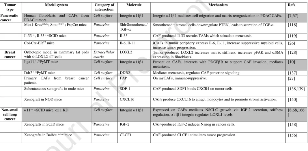
In the context of PDAC, the importance of integrins is indicated in experiments where administration of FAK inhibitors left tumor angiogenesis, apoptosis and necrosis unaffected but reduced tumor size and the number of CAFs and tumor-associated macrophages (TAMs) within tumors[112]. To further determine the relative importance of TME integrins it would require conditional deletion of FAK or specific integrin chains in CAFs. The widely expressed integrin v3 has been implied in PDAC in a mechanism where CAF-produced osteopontin interacts with v3 on PDAC cells to stimulate EMT and cancer stem cell-like properties by modulating FOXM1 expression at the tumor stroma interface (Fig. 2) [113]. These osteopontin producing CAFs most likely correspond to the myCAFs mentioned above. Separate studies of human colon cancer have similarly demonstrated crucial interactions between a distinct subset of CAFs at tumor stroma interface interacting with osteopontin, which act by contributing to the formation of microenvironmentally defined cancer stem cells [114]. These data demonstrating intense signaling activity at stroma-tumor interface fit well with the findings that CAFs at tumor stroma interface in breast cancer and pancreatic cancer are distinct from CAFs elsewhere in the tumor[26,27].
In support of a role of direct interactions of CAFs with collagen, a novel function-blocking antibody to integrin 11 can block PDAC CAF adhesion to collagen, collagen remodeling and spheroid invasion in a manner dependent on the total repertoire of integrin collagen receptors [67]. Based on immunohistochemical data with a commercial antibody to integrin 11 combined with in vitro data involving PDAC CAFs, it is suggested that 111 indeed is a major integrin, which can stimulate PDAC cell invasion in an heterospheroid system [7]. Using a novel mono-specific monoclonal antibody to integrin 11, we can confirm that 11 is expressed on CAFs in PDAC tumor stroma in vivo, but in NG2-negative CAFs. It will be important to further study the role of 111 in PDAC to determine the origin of 11-expressing CAFs using cell lineage tracing. In separate studies the role of fibronectin in the PDAC TME has been studied. Interestingly, it has been demonstrated that PDAC cells in a 3D collagen matrix migrate on elongated fibroblasts protrusions via cancer cell integrin 51 adhering to fibronectin deposited on the fibroblast cell surface[115]. In a separate study v3 integrin is suggested to be a colon cancer CAF integrin, which together with 51 is involved in FN fibrillogenesis depositing fibronectin on cell surface and directing tumor cell
Journal Pre-proof
invasion[60]. It will be interesting to determine whether v3 has this function in different types of CAFs.As already mentioned, in a detailed study of PDAC CAF interactions with the 3D ECM it is elegantly demonstrated that v5 regulates endocytosis of 51 integrin and thereby also influencing myofibroblastic activation of these cells (Fig. 1) [57].
The potential role of PDAC CAFs has been examined in relation to physiological laminin ligands where it is established that laminin-332 interacts with CAF 31 to support PDAC cell migration[55].
To summarize the role of integrins on PDAC CAFs, current data suggest that 111 and 51 are major integrins in matrix assembly and matrix reorganization involved in tumor cell growth and cell migration. v3 and v5 have both been found to assist or regulate the activity of 51 whereas little is known about cross-talk of 111 with other integrins in the PDAC context. In tissue fibrosis v1 integrin plays an orchestrating role by activating TGF- on myofibroblasts; it will be important to determine if it has a similar role on (a) particular PDAC CAF subtype(s).
3.3 Paracrine signaling in pancreatic cancer TME
Separate studies of genetic or pharmacological inhibition of Sonic hedgehog (Shh) in pancreatic CAFs revealed similar effects as Ozdemir et al. with undifferentiated tumors and decreased survival in mice as a results of the disturbed Hedgehog (Hh) signaling[116,117]. In a study by Pitaressi et al. the reason for this somewhat unexpected finding is clarified[118]. Hh signaling molecule Smoothened (Smo) in stromal cells lead to increased proliferation of PDAC cells, which could be linked to a RFN5 E3 ubiquitin ligase- mediated degradation of PTEN in Smo-null fibroblasts[118]. PTEN- deficient fibroblasts in turn were found to activate TGF-synthesis,which stimulated PDAC growth. Further studies of the mechanism suggested that hyaluronan synthesis is increased in PTEN-/- CAFs via increased activity of hyaluronan synthase 3 leading to decreased hydraulic permeability of the ECM. In support of this mechanism being relevant in PDAC disease, low stromal PTEN levels in PDAC patients correlated with poor overall survival. In conclusion, although data had suggested a role for Hh signaling as a target pathway in PDAC therapeutics, experimental data now paint a picture of complex tumor-stroma cross talk in pancreatic cancer involving Hh. Another recent example of the importance of stroma CAFs involves a pancreatic cancer model and Panc02 cells. In this model PDGFR-positive CAFs were found to produce IL-33, recruit TAMs and promote their differentiation into M2 macrophages[119] (Fig.2, Table 2). IL-33 in turn stimulated the synthesis of MMP-9 by TAMs, which has been suggested to be a major factor promoting metastasis from microvessels. This is an interesting experimental model and it would be interesting to trace these CAFs and analyze the dynamics of the changing integrin repertoire during tumorigenesis. During metastasis CAFs have been described to accompany the tumor cells[120], but so far no similar association between tumor cells and TAMs have been described; this is a possible scenario worthy of further investigation. In an interesting study, single-cell RNA sequencing of PDAC cells co-cultured with CAFs identified PDAC subpopulations with proliferative (PRO) or EMT hallmarks, which were confirmed in patient PDAC tumors [121]. In absence of CAFs, PDAC cells were mostly double-negative for these hallmarks, whereas PDAC with high CAF content were predominantly double-positive (DP), the latest being associated with poor patient survival. Mechanistically, the authors showed
Journal Pre-proof
that CAF-secreted TGF-β1 drove the DP phenotype by activating the MAPK and STAT3 signaling pathways in PDAC cells.A central question in future studies will be to try and determine which are the tumor-supportive and which are the tumor-suppressive types of CAFs in PDAC. Further studies of the paracrine signaling in myCAFs and iCAFs should also focus on integrin expression repertoire and the relative contribution of specific integrins to promoting and tumor-suppressive CAF functions. It will also be important to determine whether different CAF subpopulations hold prognostic value and have different functional properties.
3.4 TME-mediated Chemoresistance in pancreatic cancer
Using orthotopic genetic animal models such as KPC models as well as biopsy material from pancreatic cancer patients, production of insulin-like growth factors (IGFs) by TAMs and CAFs have been demonstrated to contribute to TME-mediated chemoresistance[32,122]. In a study of Ireland et al., IGF receptors on tumor cell responded to IGFs by promoting proliferation and survival. Treatment with IGF-blocking antibody in combination with gemcitabine reduced tumor growth. Since integrins have been shown to crosstalk and associate with IGF receptors[123,124], it will be interesting to determine if they also contribute to IGF1R-mediated chemoresistance. Some pancreatic cancer tumors are characterized by increased activation of IL-1 receptor associated kinase 4 (IRAK-4) in the stroma. In these tumors CAFs and PDAC cells both contribute to IL1-production and IRAK4 phosphorylation in a feedforward circuitry, resulting in fibrosis and chemoresistance[125]. When IRAK4 is silenced in such experimental tumors the ability of CAFs and PDAC to promote fibrosis is reduced and the combined administration of IL1- antibody and gemcitabine increases the effect of chemotherapy. The extensive desmoplasia in pancreatic cancer is generally recognized as being a barrier for successful immunotherapy. Following the new data indicating an ever-increasing heterogeneity of CAFs, dynamic changes from one state to another, it is clear that we are beginning to appreciate the true complexity of the pancreatic tumor stroma. As we learn more, the chances also increase that we will reach a better understanding of the diverse roles of the pancreatic TME in chemoresistance.
4. Breast
4.1 CAF heterogeneity in breast cancer
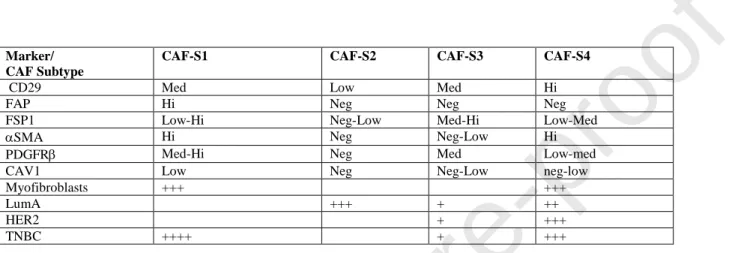
Breast is a complex organ, which undergoes hormonally regulated changes. In normal mouse breast the stroma largely determines glandular epithelium development. Two subsets of mammary gland fibroblasts have been identified in human mammary gland, lobular (CD105high/ CD26low) and interlobular (CD105low/ CD26high) fibroblasts[126]. CAF heterogeneity in breast stroma has been identified in a study of human breast cancer[27] and in mouse models [43,127]. In the human breast study the authors classify four different subsets of CAFs, called CAFS1-CAFS4 using a combination of 6 different antibodies (Table 3).
Notably, two of the subsets, CAF-S1 and CAF-S4 express high levels of SMA, but only CAF-S1 expresses fair amounts of FAP. CAF-CAF-S1 is found close to the tumor, attracts T-cells and contributes to immunosuppression. On a cellular molecular basis, the immune suppressive function of CAF-S1 partly depends on dipeptidylpeptidase 4 (DPP4, also known as CD26) and DPP4-mediated cleavage of CXCL10, leading to a reduction in T-cell recruitment to the tumor (Fig. 2.). The careful study by Costa et al. [27] also indicates that within the four CAF subclasses there is probably even more heterogeneity. It is interesting to note that the CAF-S1 display similar characteristics to the myCAF population in pancreatic cancer. However, whereas in pancreas the corresponding myCAF subset is thought to be pericyte-derived, it is unknown what the origin of these cells are in the breast.
Journal Pre-proof
Using single cell RNA sequencing, the issue of CAF heterogeneity was independently addressed in the MMTV-PyMT mouse model of breast cancer at late stages of tumor progression[127]. This study identified four transcriptionally distinct subsets of CAFs presenting different functionalities and biophysical properties. The four subsets of CAFs, termed as vCAFs (vascular CAFs), mCAFs (matrix CAFs), cCAFs (cell cycle CAFs) and dCAFs (developmental CAFs) presented distinct spatial location within the tumor parenchyma. vCAFs was shown to originate from the perivascular compartment with cCAFs being a segment of proliferative vCAFs. Conversely, mCAFs was shown to mostly derive from resident fibroblasts, while dCAFs seemed to originate from the malignant epithelial compartment via EMT. Interestingly, PDGFR was specifically expressed by mCAFs, whereas PDGFRwas expressed by all CAF subsets, with exception of dCAFs. In contrast to the previously mentioned study[27], FAP and αSMA markers were not specifically associated to a distinct subset of CAFs, but rather displayed a salt-and-pepper expression pattern in all four CAF subpopulations. These discrepancies are presumably related to breast cancer subtypes and stages, species differences and detection methodologies.As already mentioned, applying cell lineage tracing methods in the PyMT mouse model demonstrated the contribution of mesenchymal, non-hematopoietic bone marrow cells to a PDGFR-, clusterin+-breast cancer CAF subpopulation [43]. In vitro, bi-directional paracrine signaling between tumor cells and this subpopulation of CAFs had effects on both tumor cells and the CAFs. In this model clusterin, which has pleiotropic effects including stimulating endothelial cell proliferation, was suggested to promote tumor growth mainly via enhancing angiogenesis. This further highlights the complexity of fibroblast heterogeneity in breast cancer and suggests that this challenging issue requires additional investigation with regards to biomarker expression, spatial localization and functionality of these CAFs in all the subtypes and stages of breast cancer disease.
4.2 Matrix receptors and desmoplasia in breast cancer; integrin 11 on CAFs
One of the most notable features of tumor-stroma interactions in breast cancer is the desmoplastic reaction. Extensive desmoplastic reaction detected in normal breast tissue in form of mammographic density is strongly correlated to an increased risk of breast cancer development and has been proposed as a diagnostic and prognostic marker. Indeed, invasive ductal carcinomas often appear as a scirrhous mass of a stellate morphology caused by the high desmoplastic reaction observed in these tumors. The ECM composition and architecture associated to this fibrotic reaction emerge from an intimate crosstalk between fibroblasts and epithelial cells in breast tissues. Desmoplasia has also been linked to increased activation of integrins in breast cancer. In a breast cancer model, tumor secreted lysyl oxidase-like 2 (LOXL2) activates fibroblasts and promotes the expression of SMA in a FAK-dependent manner[128]. A previous landmark paper has demonstrated that increased tumor stroma stiffness promotes tumor progression by 1 integrin signaling in a FAK and Rho-signaling dependent manner [50]. Likewise, in a mouse model of breast cancer, FAK inhibition decreases tumor growth and reduces infiltration of leukocytes and macrophages[129,130]. Together, these studies support the notion that 1 integrin and FAK sustains the pro-tumor functions of CAFs. From the perspective of CAF heterogeneity, the use of CD29 as a biomarker of CAFs in the study by Costa et al. [27] is interesting, since CD29 (integrin 1), is a common integrin subunit of 12 different integrin heterodimers, and is thus widely expressed on all cells of the body[49]. Keeping this in mind, CD29 has limited use as a biomarker on its own, and even in combination with other markers, extreme care has to be taken when using high or low CD29 expression as criteria to identify a certain CAF subtype.
Journal Pre-proof
The cooperation between integrins and receptor tyrosine kinase (RTKs) in tumor and stromal cells regulates cell invasion during metastatic dissemination in breast cancer. In this context, we have recently shown that stromal integrin α11 displays a tumorigenic and pro-metastatic activity in breast cancer and strongly associates with a PDGFRβ+ CAF subset (Fig.1) [10]. Integrin α11 expression is strongly upregulated in the stromal compartment during mammary tumor progression. Histological analyses revealed a strong association between integrin α11 and PDGFRβ, both in clinical breast cancer samples and in the pre-clinical transgenic mouse MMTV-PyMT model. Among several tested stromal markers (PDGFR, PDGFR, SMA, FAP, FSP1 and NG2), this collagen-binding integrin was mostly associated with a PDGFR+ CAF subpopulation at late stages of invasive tumors. As both integrin 11 and PDGFR are well-known regulators of ECM, it is plausible that the identified CAF subset overlaps with CAF subpopulation described in the previously aforementioned study[127]. Indeed, genetic ablation of integrin α11 in the PyMT model drastically reduced not only tumor growth and metastasis, but also the desmoplastic reaction in these tumors, further highlighting the contribution of this specific11+ CAF subset to tumor progression through ECM regulation. This is further supported by the fact that mCAFs are thought to derive from resident fibroblasts, as well as integrin 11/PDGFR+ CAFs. Mechanistically, this study revealed that integrin 11/PDGFR crosstalk in CAFs endows breast cancer tumor cells with pro-invasive features through the deposition of tenascin-C (Fig.1, Table 2). Tenascin-C was strongly expressed by the same subset of CAFs expressing integrin 11 and PDGFR in the late stage PyMT tumors, as well as in clinical samples of invasive breast cancer. Overall, this study discloses an example of a collaborative crosstalk between an integrin and a growth factor receptor in CAFs, which acts as a driver of tumor invasiveness in breast cancer. Similar molecular partnerships have been previously reported, although not on CAFs. Indeed, microenvironment-induced c-Met/β1 integrin complex formation was shown to sustain breast cancer metastasis via the promotion of c-Met phosphorylation, as well as an increase of integrin affinity for fibronectin on the tumor cells[131]. Further examples of cooperation between integrins and growth factors receptors in the context of cancer are thoroughly discussed in previous reviews[132,133]. It is worth noting that 2 integrin subunit, which heterodimerizes with 1 integrin subunit to form another fibrillar collagen-binding integrin, exerts opposite functions to 111 in a related mouse breast cancer model. In contrast to α11 integrin chain, integrin α2 chain is expressed not only by CAFs, but also by tumor cells and other stromal cells. Furthermore, unlike integrin α11, the 2 subunit is downregulated in human breast cancer and acts as a metastasis suppressor in a murine model[134]. Indeed, α2-deficient MMTV-neu mice display increased metastasis, which is suggested to result from the increased capacity of tumor cells to intravasate into the bloodstream. These studies suggest opposite effects of two of the 1 integrin family members with affinity for collagen (α2β1 and α11β1) in breast cancer. Discoidin domain receptor 2 (DDR2) is a cell surface tyrosine kinase activated by collagens[5]. The function of DDR2 in both tumor cells and CAFs in a breast cancer model have been studied by performing global and tumor cell specific deletion of DDR2[135]. Global deletion of DDR2 does not affect primary tumor growth but results in reduced metastasis. Closer examination reveals that DDR2-/- stroma contains reduced amounts of fibrillar collagen, with reduced diameter and reduced organization (DDR2 CAF dependent function). DDR2 expression in tumor cells in turn appears to contribute to collective tumor invasion, suggested to occur by DDR2-dependent stabilization of EMT factor SNAIL1. Since there is a certain amount of crosstalk between 1 integrins and DDR receptors it is possible that integrins, together with DDRs, in this model also are involved in tumor cell invasion and metastasis [136]. In support of a role of DDR2 in metastasis, small molecule allosteric inhibitor WRG-28 inhibitsJournal Pre-proof
tumor-microenvironment interaction and tumor invasion[137]. It will be interesting to sort out a possible cooperation of DDR2 and integrins in tumor metastasis and whether such a link exists, identify the specific integrin(s) involved.In summary, an important cooperation between integrin and RTKs is detected in breast cancer CAFs, raising a number of interesting questions. Future studies will for example determine if one and the same integrin can cooperate with different RTKs in different tumor stroma contexts or if the cooperation is integrin-specific and limited to some kinases. This also extends to mechanism of TME-mediated chemoresistance where data is emerging on the role of integrins in mediating chemosresistance to drugs targeting RTKs[99]. These mechanisms have mainly been described in cancer cells, but similar mechanisms are likely to operate in CAFs. Finally, the role of v integrins (TGF- activating mechanisms), 51 integrin and fibronectin matrix assembly, in relation to the collagen remodeling 111 integrin will be important to study more in detail in breast cancer, just as in other desmoplastic tumor types. 4.3 Paracrine mechanisms in breast cancer TME
In a now seminal paper, an important role of stromal cell-derived factor 1 (SDF1; released from CAFs) and the corresponding receptor CXCR4 (on breast cancer cells), in tumor growth, was demonstrated [138,139] (Fig.2). In a more recent study, using a mouse model for triple negative breast cancer (TNBC), monocytes and myeloid-derived suppressor cells (MDSC) were found to negatively affect survival, which was ascribed to their immunosuppressive role and effect on invasion and angiogenesis[140]. When analyzing the effect MDSCs on stroma formation it was found that these cells stimulated stroma formation by activating CAFs and by recruiting immunosuppressive myeloid cells. Curiously, this effect was specific for TNBC cells and not seen with other breast cancer types and found to be related to their synthesis and secretion of CXCL16.
PDGF-BB-PDGFRβ signaling is one of the main pathways, which promotes the fibrotic reaction in cancer. In a paracrine manner, tumor and stroma-derived PDGFs activate PDGFRβ on CAFs and pericytes and promote ECM deposition and remodeling, increase the interstitial fluid pressure, sustain the angiogenic process and restrain the immune surveillance[141]. Stromal PDGFRs have been proposed for long-time as biomarkers for prognosis and response to RTK inhibitors (RTKIs) in cancer disease[83]. Previous works established that high PDGFRβ expression in CAFs and pericytes is associated with aggressiveness and poor prognosis in breast cancer [83]. Multivariate analyses revealed a positive correlation between high stromal and perivascular PDGFRβ expression and poor prognosis markers such as high histopathological grade, high proliferation, estrogen receptor negativity and HER2 amplification[82,142]. Furthermore, increased serum PDGF level in breast cancer patients was positively correlated with disease prognosis and recurrence in breast cancer [143]. Elevated PDGFRβ stromal levels have been also related to impaired therapeutic response. A previous study revealed that breast cancer patients with low stromal and perivascular PDGFRβ expression benefit more from chemotherapeutic agents such as tamoxifen and epirubicin than patients with high PDGFRβ expression[84].
Similarly, PDGF-CC-PDGFRα paracrine signaling has also been reported to contribute CAF-cancer cell crosstalk in breast CAF-cancer. In a recent study, by using clinical specimens and a genetically MMTV-PyMT modified mouse model for PDGF-CC, the authors demonstrated that tumor epithelium-derived PDGF-CC induces a basal-like ER-negative phenotype, rather than a luminal ER-positive molecular phenotype of BC through the activation of CAFs[144]. These PDGF-CC-activated CAFs secrete pro-tumorigenic growth factors such as hepatocyte growth factor (HGF), insulin-like growth factor binding protein 3 (IGFBP3) and the secreted glycoprotein Stanniocalcin-1 (STC1) to promote fibrotic and angiogenic responses in the
Journal Pre-proof
TME of PyMT tumors. Furthermore, genetic ablation and pharmacological inhibition of PDGF-CC resulted in a conversion of basal-like phenotype of breast cancer into a ER-positive state, which conferred sensitivity to tamoxifen therapy. This study highlights CAFs as functional mediators of the molecular subtype of breast cancer and as TME regulators of the therapeutic response to endocrine therapy.4.4 TME-mediated chemoresistance in breast cancer
Hedgehog ligand activity is detected in one third of TNBC. In an animal model of TNBC with increased Hh levels, an increased production of fibroblast growth factor 5 (FGF5) and an increased collagen remodeling activity was observed in CAFs[145]. The increased concentration of remodeled collagen at tumor stroma interface correlated with increased pFAK levels as well as increased number of CSCs at the tumor-stroma interface. In this breast cancer model, treatment with the smoothened inhibitor (SMOi) sensitized mice to chemotherapy. It will be interesting to determine what specific integrins are present and mediate the increased pFAK at the tumor stroma interface.
Studies in breast and pancreas cell lines offer additional detail as to how CSC formation via EMT may occur. Snail1, which is a central transcription factor in EMT is an unstable protein that is ubiquinated. In experiments performed by Lambies et al., deubiquitination by a specific ubiquitinase (USP27X) contributes to Snail1 stability in turn contributing to increased EMT, increased amounts of CSCs and increased chemoresistance[146]. In addition, Snail1 stabilization in CAFs contributes to increased CAF activation. It will be interesting to see whether CAF Snail1 in this context integrates with integrin-dependent mechanoregulated signaling. Such mechanoregulation in myofibroblasts involving Snail1 has recently been shown to contribute to fibrosis and depend on both YAP/TAZ and MRTF transcription factors[147].
In an impressive study of breast cancer chemoresistant patients, CAF subsets were identified[6]. These CAFs expressed the usual markers, SMA, PDGFR, FAP, FSP1, but were distinguished by the expression of two additional cell surface markers CD10 (zinc metalloprotease) and GPR77 (anaphylatoxin receptor). In this careful study the activation of GPR77 by autocrine production of C5a lead to: 1) production of IL-6 which in turn increased the abundance of CSCs (by providing a survival niche for CSCs) as well as, 2) increased expression of the multidrug transporter ABCG2, largely responsible for the observed chemoresistance. The CD10+GPR77+ CAF subset thus sustained cancer stemness and promoted tumor resistance. Targeting this subset of CAFs furthermore restored chemosensitivity. The authors suggest that targeting the CD10+GPR77+ CAF subset could be an effective strategy against CSC-driven solid tumors. It will be interesting to relate this subset of CAFs with the different breast cancer CAF subsets identified by Costa et al., as well as with CAF subsets in other cancer forms [27]. It will also be important to characterize the role of integrins in TME-mediated chemoresistance in breast cancer.
5. Lung
5.1 Fibroblast heterogeneity
The lung is also a complex organ where fibroblasts have a number of functions associated with normal lung function. Cell lineage tracing has been performed to identify and characterize the origin of fibroblast subsets[148], but in mouse lung, single-cell transcriptional analysis has been even more instrumental and has resulted in the identification of five subsets of fibroblasts in healthy lung and six subsets in fibrotic lung, in addition to a mesothelial subtype [19]. In normal lung these were grouped as myofibroblasts (Acta2+), col3a1 matrix fibroblasts (Col3a1+;Itga8+), Col4a1 matrix fibroblasts (Col4a1+;dcn+), lipofibroblasts (Lp1+) and mesenchymal progenitors (CD52+)[19]. In the fibrotic lung a distinct fibroblast