RGDS and DGEA-induced [Ca
2+] signalling in human dermal fibroblasts
P. Mineur a,1 A. Guignandon b,1 Ch.A. Lambert a, M. Amblard c, Ch.M. Lapière a, B.V. Nusgens a
a Lab. Biologie des Tissus Conjonctifs, University of Liège, Belgium
b Lab. Biologie du Tissu Osseux, INSERM E366, University of Saint-Etienne, France c Lab. des Aminoacides, Peptides et Protéines, University of Montpellier I et II, France
Abstract
A pulse of short peptides, RGDS and DGEA in the millimolar range, immediately elicits in normal human fibroblasts a transient increase of intracellular Ca2+ ([Ca2+]
i). In the present study, we show that this [Ca2+]i
occurs in an increasing number of cells as a function of peptides concentration. It is specific of each peptide and inhibited at saturating concentration of the peptide in the culture medium. The [Ca2+]
i transient depends on
signalling pathways slightly different for DGEA and RGDS involving tyrosine kinase(s) and phosphatase(s), phospholipase C, production of inositol-trisphosphate and release of Ca2+ from the cellular stores. GFOGER, the
classical collagen binding peptide of α1- α2- and α11-β1 integrins, in triple helical or denatured form, does not produce any Ca2+ signal. The [Ca2+]
i signalling induced by RGDS and DGEA is inhibited by antibodies against
β1 integrin subunit while that mediated by RGDS is also inhibited by antibodies against the α3 integrin. Delay in the acquisition of responsiveness is observed during cell adhesion and spreading on a coat of fibronectin for RGDS or collagen for DGEA or on a coat of the specific integrin-inhibiting antibodies but not by seeding cells on GFOGER or laminin-5. This delay is suppressed specifically by collagenase acting on the collagen coat or trypsin on the fibronectin coat. Our results suggest that free integrins and associated focal complexes generate a Ca2+ signal upon recognition of DGEA and RGDS by different cellular pathways.
Keywords: Integrin; Ca2+ signalling; RGDS; DGEA; Fibroblast
1. Introduction
The engagement of integrins with their immobilized ligands generates a cascade of intracellular signals including activation of kinases, release of phosphatidylinositol by phospholipases and increased concentration of
intracellular-free calcium [1,2]. The minimal recognition sequence of fibronectin and a number of other
extracellular macromolecules is RGD (Arg-Gly-Asp) [3]. This sequence recognized by different integrins is able to block cell adhesion to diverse adhesive proteins [4]. RGD-containing short peptides are also known to increase cytosolic calcium ions in various types of cells [5]. The RGD sequence is also found in the αl and α2 chains of type I collagen, although under a cryptic conformation [6] and the attachment of cells to native collagen type I is not modified by RGD peptides. Integrins α1β1, α2β1 and α11β1 are the major cell surface receptors of collagen type I [7-9] and recognize a distinct binding site containing the GFOGER sequence [10-12]. A DGEA (Asp-Gly-Glu-Ala) sequence present in the cyanogen bromide peptide αlCB3 of the type I collagen was reported to bind to α2β1 integrin and to inhibit the α2β1-mediated adhesion of platelets to collagen and that of T47D to collagen and laminin [13] and to interfere in the α2β1 signalling in many cell types [14-19]. DGEA is also able to induce Ca2+ transients in the human osteoblastic SaOS-2 line [20,21]. RGD and DGEA are
also present in cell surface proteins such as L1 CAM that can associate through these sequences in cis with the βl integrin subunit. CHL1, a murine close homolog to L1 was recently shown to modulate neuronal cell migration and other integrin-dependent functions through these sequences [22,23].
In this work, we used RGDS and DGEA in the millimolar range to trigger an immediate rise of intracellular calcium in human fibroblasts. This [Ca2+] transient of short duration resulted of the release of the ions from the
cellular stores by a phosphoinositide pathway triggered by protein kinase(s) and phospholipase(s). The goal of this study was to better understand the mechanism used by the cells to perceive the signal carried by the peptides and the implication of integrins in the process.
2. Materials and methods 2.1. Reagents
RGDS, AAAA, bradykinin, ionomycin, calyculin A, LaCl3, EGTA, BAPTA-AM, thapsigargin, herbimycin A,
genistein, neomycin, poly-L-lysine, and bovine serum albumin were purchased from Sigma (St Louis, MI), xestospongin C, okadaic acid, PP2 and U73343 from Calbiochem (San Diego, CA), U73122 from ICN (Costa Mesa, CA), PP1 from Alexis Corporation (Lausen, Switzerland), trypsin from Life Technologies Inc. (Rockville, MD) and highly purified bacterial collagenase from Advance Biofactures Corporation (Lynbrook, NY). DGEA was from Research Genetics (Huntsville, AL) and KGDS from Bachem AG (Bubendorf, Switzerland). (GPO)nGFOGER(GPO)n was a generous gift of M. Hook, Houston, Texas [11]. Function blocking monoclonal
antibodies anti-human integrin subunits α1 (clone FB12), α2 (clone P1E6), α4 (clone P4G9) and α6 (clone GoH3) were from Chemicon (Temecula, CA), α3 (clone P1B5) and rabbit anti-mouse IgG from DAKO (Carpinteria, CA), α5 (clone P1D6) and β1 (clone P4C10) from Life Technologies Inc. Anti-human αv (clone 17E6) is a generous gift from S. L. Goodman [24]. Fetal calf serum and DMEM were from Life Technologies Inc., Fluo3-AM from Molecular Probes (Eugene, OR), borosilicate coverslip Lab-Tek® from Falcon (Becton
Dickinson, Franklin Lakes, NJ). Type I collagen was purified from bovine skin as described by Delvoye et al. [25] and fibronectin was purified from human plasma as described by Hayashi and Yamada [26]. Laminin-5 was kindly offered by M. Aumailley (Dpt. Biochemistry II, University of Koeln, Germany).
2.2. Cell culture
Normal human skin fibroblasts were grown from skin biopsies of a young healthy donor by the explant
procedure described earlier [25] and used between passages 8 and 13. Human rhabdomyosarcoma RD cells were purchased from Biowhittaker (Walkersville, MD). All cells were mycoplasma-free and routinely cultured in DMEM in the presence of 10% fetal calf serum (FCS), 2 mM glutamine, penicillin-streptomycin (100 IU/ml each) and ascorbic acid (50 µg/ml) (=standard medium) at 37 °C in a humidified atmosphere containing 5% CO2.
2.3. Fluo3-AM loading and fluorescence recording
For routine experiments, cells were plated in borosilicate culture chambers (Lab-Tek®) for 18 h in DMEM-10%
FCS, rinsed with serum-free medium and incubated with the fluorophore Fluo3-AM (10 µM) in serum-free medium for 1 h. In short term adhesion experiments (up to 150 min), the trypsinized cells were plated in serum-free medium and the Fluo3-AM added for the last 20 min. After loading, cells were washed and immediately tested in serum-free medium. The observation of the fluorescence emitted by the Fluo3-labeled cells started 5 min after the last washing. Microscope fields taken at random in the culture were examined by a confocal microscope (Meridian, Akemos, MI). At a magnification of 63 x the microscope field contained 5-12 cells. The Fluo3-loaded cells were excited by an Argon LASER at 488 nm and the emitted fluorescence recorded at 530 nm in each cell of the field and in real time. Image processing and data computing were performed using the
Meridian software. The intensity of the emitted fluorescence was recorded every second during at least 100 s. As the overall fluorescence intensity varied from cell to cell, the level of fluorescence of each cell at the beginning of the recording was normalised to one arbitrary unit. The baseline of resting cells spontaneously oscillated by ± 5% around the level of the first image acquisition. A 10% rise above the baseline was considered as a significant calcium rise and the cell regarded as responsive. The results were expressed as the percentage of responding cells.
2.4. Stimulation by the peptides
After a 20 s period of baseline recording, 10 µl of a solution in water or in phosphate-buffered saline (PBS) of the various peptides (DGEA, AAAA, RGDS, KGDS) of the indicated amount (in nanomoles) was gently added on top of the cells under microscopic examination. Ten µl of an aqueous solution of bradykinin (2 nmol) was similarly injected before completion of the recording to test the ability of the examined cell population to produce a [Ca2+]
i rise. The peptide GFOGER in triple helical conformation by renaturation at 4 °C overnight or
denatured by heating at 70 °C 10 min. was similarly tested using an amount of 10 nmol. 2.5. Coating of culture surfaces
When indicated borosilicate culture chambers (Lab-Tek®) were coated with fibronectin (30 µg/ml in PBS),
native type I collagen (20 µg/ml in PBS), poly-L-lysine (100 µg/ml) or monoclonal anti-integrin subunits βl (10 µg/ml), α3 (10 µg/ml) or α5 (10 µg/ml). One ml of solutions was used per square centimeter. The fibronectin
solution was incubated for 1 h at room temperature. The unoccupied reactive sites were blocked with 0.2% heat denatured bovine serum albumin (BSA), washed with PBS at room temperature and dried. Monomeric collagen coats were prepared by drying the collagen solution at 4 °C, washing and drying at room temperature. Poly-L-lysine solution was adsorbed overnight at 4 °C. Laminin-5 was coated overnight at 4 °C, blocked with 1% heat denatured BSA for 1 h at room temperature and washed with PBS. All the coated slides were stored at 4 °C. The anti-integrin coated surface were prepared as described by Schwartz [1]. The borosilicate slides were coated first with 50 µg/ml by rabbit anti-mouse IgG for 1 h at 37 °C, washed with PBS, blocked with 1% heat denatured BSA, washed again, incubated for 1 h with 10 µg/ml of the specific monoclonal antibodies (anti-β1, anti-α3 or anti-α5 integrins) in 1% heat denatured BSA, rinsed with PBS and immediately used. Coating with the GFOGER peptides was obtained by incubating the slides overnight at 4 °C with a 1 mM solution in phosphate-buffered saline (PBS) of the triple helical peptide, washed in PBS and blocked with 1% heat-denatured BSA. All reactions and testing were performed at maximum 25 °C.
2.6. Enzymatic digestion of coated substrates
Fibroblasts were plated in serum-free medium on fibronectin and native type I collagen coated coverslips for 60 min and loaded with Fluo3-AM (10 µM) during the last 20 min. After washing with PBS, the cultures were incubated for 3 min at 25 °C with trypsin (10 µg/ml) in DMEM or with highly purified bacterial collagenase (50 U/ml) in 0.05 M Tris, 0.15 M NaCl, 2 mM CaCl2 pH 7.5 before adding the peptide agonist DGEA or RGDS and
recording the fluorescence.
2.7. Pharmacological investigations
The following pharmacological investigations were performed on fibroblasts plated on glass for 18 h before Fluo3-AM loading. Genistein (10 µM), thapsigargin (2 µM), BAPTA-AM (30 µM) neomycin (10 µM) were added 3 h before loading, PP1 and PP2 (20 µM) were added 90 min before use, herbymicin A (10 µM),
xestospongin C (20 µM), calyculin A (500 pM) and okadaic acid (lOµM) at the time of loading, EGTA (2 mM), La3+ (80 µM), U73122 and U73343 (1 µM)just before fluorescence analysis. Anti-βl and anti-α-integrins subunit
monoclonal antibodies were added at a final concentration of 10 µg/ml of culture medium 30 min before testing the effect of the peptides.
2.8. Cytotoxicity assays
The cytotoxicity of the various agents used in this study was tested by measuring the activity of the mitochondrial succinate dehydrogenase [27] as previously described [28].
Comparisons between groups were based on parameters using the one-way analysis of variance (ANOVA). When the ANOVA test was significant, the Tukey-Kramer multiple comparisons test was used to compare the groups versus control.
3. Results
3.1. DGEA and RGDS induce an intracellular free calcium [Ca +]
i transient in fibroblasts
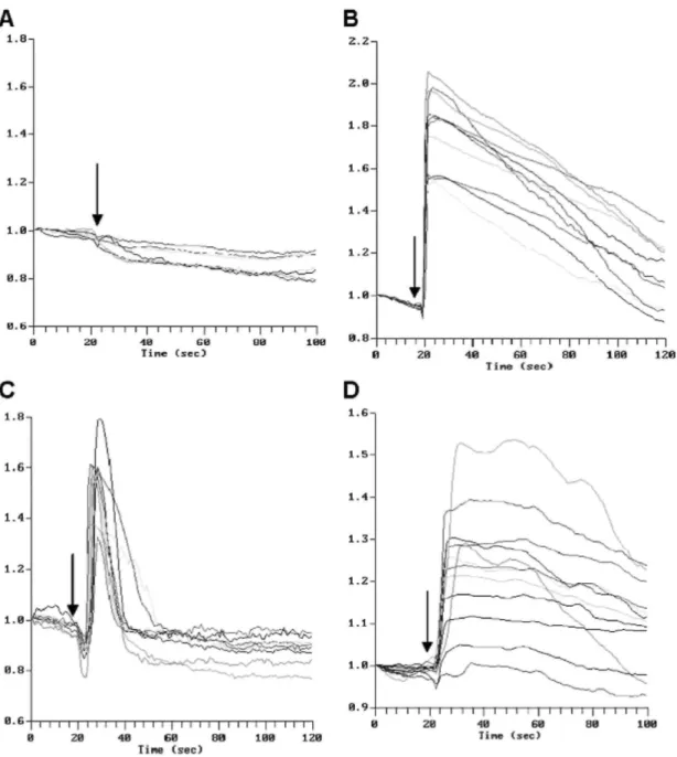
Single-cell, real-time fluorescence recording was performed by confocal microscopy on human skin fibroblasts attached for 18 h to glass coverslips in Lab-Tek® chambers and loaded with Fluo3-AM. Typically, the
fluorescence in resting fibroblasts was diffuse within the cytoplasm with scattered spots of higher intensity and accentuated in the perinuclear area (not illustrated). The variability in fluorescence intensity before normalisation to 1.0 was between 1000 and 2000 fluorescence units for a scale of 0 to 4095. The subcellular localisation of the fluorescence in resting cells remained similar for the total duration of data acquisition (at least 100 s) while the overall fluorescence intensity only slightly decreased with time (Fig. 1A), likely due to
bleaching. The compartmentalization of the dye was suppressed by ionomycin (1 µM) that produced an uniform fluorescent signal in the cell. The calculated mean intracellular free calcium concentration ([Ca2+]
i) was 120 ± 30
nM, a value in close agreement to that reported in the literature [29]. To assess the ability of the cells to respond by a rise in [Ca2+]
i, the fibroblasts were always stimulated with bradyki-nin (2 nmol) at the end of each
experiment. It induced a rise of fluorescence mainly localised in and around the nucleus. A similar burst of fluorescence was observed in fibroblasts excited by 10 nmol DGEA or RGDS in water or phosphate-buffered saline. The recording of fluorescence intensity as a function of time in the individual cells of the microscope field is illustrated in Fig. 1. No significant increase of the fluorescence was induced by AAAA (Fig. 1A) or by the solvents alone (not shown). Bradykinin induced a rise of fluorescence in every cell within a few seconds
(Fig. 1B) and its intensity peaked at 150 to 200% of the initial value before slowly and progressively returning to the baseline. A similar increased fluorescence was induced by DGEA (Fig. 1C) within ±10 s to reach a peak at ± 150% and a rapid return to the baseline value. RGDS also rapidly induced a significant rise (±130%) of
fluorescence followed by a slower decline to the baseline values (Fig. 1D) while KGDS, at the same concentration, had no effect (not shown). Prolonged acquisition of data up to 5 min failed to detect cells responding later by a significant peak. The peptide-induced Ca 2+ rise could be repeated several times. To assess
that the peak of fluorescence was actually due to an increased [Ca2+]
i, the cells were loaded with the
membrane-permeant calcium chelator BAPTA-AM for 3 h prior to addition of the peptides. This resulted in a total
suppression of the Ca2+ transients in response to the active peptides (Table 1). The peak of fluorescence induced
by both peptides was similar in the fibroblasts attached for 18 h on a glass support uncoated or coated with monomeric collagen, fibronectin, laminin-5 or poly-L-lysine.
Fig. 1. Time-course variation (in s) of the fluorescence in glass-plated fibroblasts treated with (A) AAAA (10
nmol), (B) bradykinin (2 nmol), (C) DGEA (10 nmol) and (D) RGDS (10 nmol). Fluo3-AM loaded cells were treated with the various peptides at the indicated time (arrow) during the recording of the fluorescence. The fluorescence intensity of the individual cells was normalised to 1 at the beginning of the recording.
The calcium response of the human dermal skin fibroblasts was further analyzed by varying the concentration of DGEA and RGDS. The number of responsive cells increased according to a semi-log dose-response relationship (Fig. 2) up to a maximum of 90% of the cell population upon stimulation by 10 nmol of peptide, the half-maximal response being observed at 0.1 nmol. By contrast, the size of the fluorescence peak, i.e., the increase in intracellular Ca2+ in the responding cells, was independent of the concentration of the stimulating peptide. A
pulse of 10 nmol of peptide was selected for the following experiments.
The peptide GFOGER, the known collagen I binding site of the αl-, a2- and α11-β1 integrins, in the triple helical configuration or denatured, added at the same amount (10 nmol) did not induce any Ca2+ transient. This addition
did not interfere with the production of the [Ca2+]
i transient produced by DGEA or RGDS or the [Ca2+]i signal
produced by bradykinin.
3.2. DGEA- and RGDS-induced [Ca2+]
i transients involve calcium mobilization from internal stores by a slightly
different mechanism
The origin of the calcium ions participating in the [Ca2+] transient in response to both DGEA and RGDS peptides
was investigated by using a variety of agents known to interfere with calcium movements (Table 1). Addition of EGTA to chelate calcium in the extracellular space or of La3+ ions, known to block the plasma membrane
calcium channels, 10 min prior to addition of DGEA, RGDS or bradykinin, did not affect the calcium responses. Treatment of the cells for 3 h with thapsigargin, an inhibitor of the endoplasmic reticulum ATPases-dependent calcium pump [30], suppressed the [Ca2+]
i transients induced by both peptides and by bradykinin. Xestospongin
C, known as an inhibitor of inositol-trispho-sphate (IP3) receptor [31], reduced the DGEA- and RGDS-induced signals. These data indicate that the rise in [Ca2+]
i induced by DGEA and RGDS depends upon an ΓP3-mediated
mobilization of the ion from the internal stores and not by a significant influx of the ion from the extracellular space. Xestospongin C did not inhibit the [Ca2+]
i transient induced by bradykinin after pulsing with DGEA or
RGDS. This absence of inhibition is related to the short half-life of xestospongin C after replacing the medium before pulsing with the peptides [32].
Table 1: Effect of inhibition of various signalling pathways on the [Ca 2+]
itransients induced by DGEA (10
nmol), RGDS (10 nmol) and bradykinin (2 nmol) in fibroblasts plated on glass for 18 h
Agents Target Percentage of cells responding to Number of cells/experiments
DGEA RGDS Bradykinin after DGEA RGDS
DGEA RGDS None 89 ± 7 90 ±7 97 ± 5 100 97/10 115/10 BAPTA-AM Intracellular Ca2+ 0* 0* 0 0 16/2 12/2 EGTA Extracellular Ca2+ 90 ± 9 87 ± 11 100 100 30/3 17/3 La3+ Plasma membrane Ca2+ channels 96 ± 6 72 ± 13 100 100 26/3 27/3 Thapsigargin ER Ca2+-ATPase 0* 0* 0 0 18/2 16/2
Xestospongin C IP3-mediated Ca2+ Release 22 ± 32* 15 ± 21* 100 100 17/2 18/2
Genistein Tyrosine kinases 0 ± 0* 5 ± 7* 100 90 ± 0 16/2 20/2
Herbimycin A Tyrosine kinases 70 ± 23 20 ± 13* 100 100 60/8 19/2
PP1 Src tyrosine kinases 100 ± 0 82 ± 16 92 ± 12 100 22/2 24/2
PP2 Src tyrosine kinases 100 ± 0 80 ± 28 100 96 ± 6 21/2 22/2
Okadaic acid Phosphatases 94 ± 5 15 ±19* 100 98 ± 4 35/3 56/4
Calyculin A Phosphatases 1 and 2A 100 ± 0 17 ±15* 100 100 22/3 29/3
Neomycin Phospholipase C 6 ± 7* 11 ± 10* 8 ± 1 0 36/5 27/3
U73122 Phospholipase C 6 ±10* 3 ± 5* 0 0 24/3 24/3
U73343 Inactive analog of U73122 95 ± 7 80 ± 18 100 100 21/2 25/2
ER Ca 2+-ATPase: endoplasmic reticulum Ca 2+-ATPase; IP
Fig. 2. Dose-response relationship of glass-plated fibroblasts to DGEA (♦), RGDS (▲), AAAA (x) and solvent
alone (10 µl) (O). The calcium signal was monitored as described in Materials and methods on a total of 22 to 3 8 cells in 4 separate experiments. The percentage (±SD) of responding cells is plotted against the amount of added peptide or solvent alone.
To further analyze the signalling triggered by DGEA, RGDS and bradykinin, cells were treated with a panel of agents known to inhibit different pathways originating from membrane receptors. Genistein, a broad spectrum inhibitor of tyrosine kinases, abrogated the cell response to both peptides while herbimycin A, but not PP1 and PP2, inhibitors more selective of the src-family tyrosine kinases, strongly reduced the effect of RGDS and barely affected the response to DGEA. Calyculin A and okadaic acid, phosphatases 1 and 2A inhibitors, reduced the activity of RGDS but not that of DGEA. No effect was observed with tyrphostin A23, an inhibitor of growth factor receptor tyrosine kinase activity (data not shown). The PLC inhibitors, U73122 but not its inactive analog (U73343) and neomycin, extensively reduced the Ca2+ signalling by both DGEA and RGDS as well as by
bradykinin. The toxicity of the inhibitors was tested by the MTT test as previously described [28]. All were devoid of toxicity at the concentrations and for the delay of application used here except BAPTA-AM, that reduced the mitochondrial activity of 50% after 6 h.
3.3. RGDS and DGEA target different binding molecules
To assess whether DGEA and RGDS target different receptors, DGEA- and RGDS-induced calcium responses were analyzed in cells incubated with high concentration (1 mM) of the peptides added in the medium 30 min before the assay. Cells maintained in 1 mM DGEA fully responded to RGDS and bradykinin while the response elicited by DGEA dropped to 35% (Table 2). Conversely, cells maintained in 1 mM RGDS fully responded to DGEA and bradykinin while the proportion of RGDS-responding cells decreased to 50%. These results suggest that distinct membrane receptors are used by each of the peptides.
To assess the role of integrins in the peptide-induced calcium transients, fibroblasts plated on glass in presence of 10% FCS for 18 h were treated with function blocking monoclonal antibodies against human α1, α2, α3, α4, α5, α6, av and β1 integrin subunits (10 µg/ml), alone or in combination, 30 min prior to challenging with the peptides. The anti-βl integrin antibodies blocked the Ca2+ transients induced by both DGEA and RGDS (Table
2). Irrelevant monoclonal anti-mouse IgG or heat-denatured bovine serum albumin, used at the same
concentration, did not affect the calcium signals. Blocking anti-α1, -α2, -α4, -α5, -α6 and -αv subunit integrin monoclonal antibodies, alone or in combination, were unable to inhibit the response to RGDS while it was completely suppressed by anti-α3 subunit antibodies, alone or in combination with the four other anti-α subunits. None of these seven anti-α subunit antibodies, alone or in combination, was able to block the response to DGEA. Addition of bradykinin induced a rise of [Ca2+]
i in more than 95% of the cells after treatment with each of the
antibodies, alone or combined. The inhibitory activity of anti-β1 towards RGDS and DGEA and of anti-α3
towards RGDS was similar in cells plated on coated matrices, collagen or fibronectin or poly-L-lysine (not illustrated).
Table 2: Effects of DGEA, RGDS and soluble anti-integrins antibodies on the [Ca 2+]
i transients induced by
DGEA (10 nmol), RGDS (10 nmol)and bradykinin (2 nmol )in fibroblasts plated on glass for 18 h Treatment Percentage of cells responding to Number of cells/experiments
DGEA RGDS Bradykinin after DGEA RGDS
DGEA RGDS None 84 ± 4 89 ± 2 100 100 39/3 35/3 DGEA (1 mM) 33 ± 11* 95 ± 6 100 100 24/2 25/2 RGDS (1 mM) 88 ± 6 48 ± 8* 100 96 ± 6 25/2 25/2 Anti-βl 21 ± 6* 8 ±13* 100 100 28/3 28/3 Anti-αl, -α2, -α4, -α5 74 ± 12 86 ± 9 100 100 43/3 38/3 Anti-α3 90 ± 9 0 ± 0* 98 ± 3 98 ± 3 56/4 57/4 Anti-αl, -α2, -α3, -α4, -α5 100 ± 0 13 ± 9* 100 96 ± 6 22/2 23/3 Anti-α6 74 ± 1 100 ± 0 100 100 19/2 15/2 Anti-αv 100 ± 0 94 ± 9 100 100 25/2 22/2
* P< 0.001 versus control sample.
3.4. Adhesion of fibroblasts on fibronectin or type I collagen transiently and specifically represses the [Ca2+] i
response to, respectively, RGDS and DGEA, while laminin-5 has no effect
Fibroblasts were seeded on native monomeric type I collagen, fibronectin, laminin-5 or poly-L-lysine and the peptide-induced calcium signalling was analyzed as a function of time after plating. The first observation was performed 45 min after seeding. When plated on collagen, fibronectin or laminin-5, the cells rapidly attached and progressively spread after 150 min (not shown), while on poly-L-lysine the fibroblasts were rapidly immobilized but remained rounded. On native type I collagen, the number of cells displaying a [Ca2+]
i response to RGDS was
maximal at all tested times (Fig. 3A) while the reactivity to DGEA was very low at 45 min and progressively increased to reach maximal values at 100-120 min. Conversely, on fibronectin the RGDS-induced [Ca2+]
i
signalling was low at 45 min and progressively increased with time up to 150 min (Fig. 3B). A significantly reduced reactivity to DGEA was also observed in this condition during the first 60 min. No lag period was noted for both peptides in cells plated on poly-L-lysine (Fig. 3C) or on laminin-5 (not shown). When the fibroblasts were plated, adhered and spread on GFOGER, no lag period was observed upon stimulation by DGEA, RGDS or bradykinin. The [Ca2+]
i signal in response to bradykinin was close to 100% at all time points tested on each of the
substrates (not shown). These results suggest that the recognition site specific to each peptide is temporarily unavailable during cell adhesion.
To further assess the role of the extracellular support, cells plated for 60 min, i.e., in the middle of the refractory period, on type I collagen or on fibronectin were treated either with highly purified bacterial collagenase or trypsin, washed and stimulated within 5 min by DGEA or RGDS. More than 50% of the cells seeded on collagen and treated with collagenase became responsive to DGEA (Fig. 4) while most cells in the untreated cultures or incubated with trypsin failed to respond. RGDS and bradykinin induced the expected positive response (not illustrated). Conversely, treating the cultures on a fibronectin coat by trypsin restored a significant response to RGDS (Fig. 4) while collagenase treatment was uneffective. DGEA and bradykinin induced the expected strong response (not illustrated). These observations suggest that the signalling complex for the peptides is made available by specific proteolysis of the coated proteins during the period of spreading.
To further ascertain the role of integrin-based transduction of the peptides signalling, cells were seeded on coats of anti-β1, α3 and α5 antibodies. On a coat of anti-β1 integrin bound to anti-mouse IgG, the cells attached and the [Ca2+]
i signal induced by both DGEA and RGDS was reduced at 60 min, and remained low at 150 min, i.e.,
beyond the period of non-responsiveness on ECM coats, to reach a full responsiveness only after 240 min. (Table 3). When anti-α3 integrin was used as a coat the lag period only concerned the [Ca2+] signal triggered by
RGDS. Cells seeded on a coat of anti-α5 subunit fully responded with no delay to both peptides. The [Ca2+] i
Fig. 3. Modulation of the calcium response to DGEA and RGDS by the substrate. Cells plated on (A) native type
I collagen, (B) fibronectin or (C) poly-L-lysine were loaded with Fluo3-AM during the last 20 min preceeding the stimulation by the peptides. The percentage of responding cells to DGEA (o) and RGDS (A) was recorded at increasing time after plating starting at 45 min on a total of 17 to 52 cells in 3 to 4 separate experiments for each time point. The significance of the differences versus the values at 150 min are *P<0.05, **P<0.01 and ***P<0.001 by the Tukey-Kramer test.
4. Discussion
In this study, short synthetic peptides contained in the sequence of extracellular matrix proteins were used to induce intracellular [Ca2+] transients in normal human fibroblasts. Addition of DGEA or RGDS in the millimolar
range to attached normal human fibroblasts rapidly triggered a shortlived rise in intracellular [Ca2+] shown by an
increased fluorescence of Fluo3. It was most visible in and around the nucleus as reported in other types of cells [33]. This distribution agrees with the description of the perinuclear location of agonist-stimulated elementary Ca2+ signals acting as initiator of calcium movements [34] and the release of nuclear and perinuclear Ca2+ by
[Ca2+] signals in a number of cell types among which endothelial cells [36], fibroblasts [37], osteoclasts [33,38],
epithelial cells [39], smooth muscle cells [40] and lymphocytes [41]. DGEA has been less investigated. It was shown to induce [Ca2+] transients in the osteoblast-like cell line SaOS-2 [20,21]. Here, we show that fibroblasts
respond to RGDS as well as to DGEA but not to GFOGER by calcium transients. These cells reacted to the active peptides by a concentration-dependent number of responsive cells up to a maximum of 90% and a lack of relationship between the amount of added peptide and the level of fluorescence. Such an all-or-none effect might depend on two different mechanisms, the all or none reaction and the graded response [42]. Additional work is needed to answer this question. The reason why some cells are unresponsive to the peptides while they are all responsive to bradykinin is not clear. It is not related to the basal level of fluorescence, i.e., the intracellular concentration of calcium and/or the amount of loaded fluorescent indicator, neither to the cell cycle state since mitomycin-growth arrested fibroblasts were similarly responsive (data not shown). Cell attached to poly-L-lysine that remain rounded also produced a signal of a size similar to that observed in spread cells.
While the [Ca2+] rise induced by DGEA rapidly returned to the baseline values, the signals induced by RGDS
were more persistent suggesting that the peptides target different receptors or trigger different signalling pathways. This is further supported by the positive response to one peptide in the presence of a saturating concentration of the other, the non-responsiveness to the peptides during adhesion only on a specific support, distinct effects of some inhibitors of the signalling pathways and the sensitivity to specific anti-integrin antibodies.
In our experimental model, [Ca2+]
i mobilization by RGDS or DGEA did not require calcium entry from the
extracellular medium since La3+ or EGTA in the culture medium did not affect the response. This independence
on extracellular calcium has already been observed in fibroblasts [43]. It is in sharp contrast with data published for osteoblast-like cells [21]. The difference may be explained by the low number of calcium channels in the human fibroblasts. By using a panel of pharmacological agents at non-toxic concentrations [28], we suggest that the calcium mobilized by DGEA and RGDS originates from intracellular stores and requires tyrosine-kinase(s), the release of phosphoinositides by phospholipase C and opening of the IP3-sensitive calcium channels of the intracellular stores. Furthermore, a phosphatase activity seems required for RGDS signalling as suggested by suppression of the Ca2+ signal by okadaic acid and calyculin A. Several kinases are found in vicinity of the
cytoplasmic tail of the integrins in the focal adhesions, including FAK125 and members of the src family [44].
The broad spectrum inhibitor genistein suppresses the Ca2+ signalling of both peptides. The greater sensitivity of
the RGDS-induced [Ca2+]
i signalling to herbimycin A suggests a role of one or several defined members of these
kinases family in the RGDS- but not in the DGEA-induced signalling. The src-kinases do not seem to be involved since the more specific inhibitors PP1 and PP2 are inactive. The proposed pathways are partly similar to those used in signalling by growth factor receptors and integrins [45], Interactions between cells and the extracellular matrix are regulated by small GTPases of the Rho family. These Rho GTPases are key signalling molecules regulating the architecture of the cytoskeleton [46] and the assembly of proteins into focal adhesions [47]. In preliminary experiments, RhoA, Racl and Cdc42 were knocked-down by the si-RNA technology that we previously described [48]. Si-Rac1 extensively repressed the Ca2+ signal-induced by RGDS while the signal
produced by DGEA was not affected by any of these siRNAs (personal unpublished results). As opposed to DGEA, RGDS is a recognized ligand for a large family of integrins [4], among which α3β1. Our data support a classical signalling for RGDS but do not exclude an alternate mechanism as discussed later.
Using blocking antibodies indicated that α3β1 is involved in the RGDS-induced [Ca2+]
i transient. The integrin
α3βl is very versatile and involved in adhesion, migration and signalling and may negatively cooperate with α2βl [49]. Although the preferential ligand of α3β1 integrin has been identified as laminin-5 [50], this integrin may function as a subsidiary receptor of broad specificity. It can also bind to collagens and fibronectin [7], entactin [51] and thrombospondin [52]. Recently, Akula et al. [53] demonstrated that the human herpes virus-8 (HHV-8) interacts with α3βl integrin by an RGD sequence. In our study, the absence of lag period in fibroblasts plated on laminin-5 during adhesion indicates that the signalling complex is directly available on that support as opposed to what is observed when the cells are plated on fibronectin, a more physiological support.
Activation of integrins leads to a mobilization of intracellular calcium, an ubiquitous cell signalling involved in adhesion, migration and many other functions including gene expression and apoptosis [54]. In our fibroblasts, suppression of the DGEA-induced [Ca2+] rise by monoclonal antibodies suggests the involvement of integrin(s)
of the β1 family in the process. The blocking antibodies against the α1 and α2 integrin subunit failed to suppress the response to DGEA suggesting that neither the integrin α1β1nor α2βl is the transducing receptor. A similar observation was made in the SaOS-2 cells treated with a panel of different monoclonal anti-α2 antibodies which also failed to block the DGEA-induced [Ca2+ ] transients [20]. Furthermore, the efficient induction of [Ca2+]
existence of another pathway for the reception of the message. Antibodies against the α3, a4, a5, a6 and αv integrin subunits, alone or in combination, also failed to inhibit the [Ca2+] signalling induced by DGEA. The
α11β1 integrin also recognize type I collagen [9]. The lack of function blocking antibodies for α11precluded testing its participation in the peptide-induced Ca2+ signalling at the present time. GFOGER is the recognized
ligand of the integrin α1-, a2- and α11-1 domain [56,12]. As this peptide GFOGER in triple helical or denatured form does not induce a Ca2+ transient, an alternative recognition site on the βl integrins has to be considered for
the Ca2+ signalling induced by DGEA.
Fig. 4. Effect of enzymatic digestion of the substrates on the calcium response to DGEA and RGDS during the
refractory period. Fibroblasts were plated on native type I collagen or fibronectin for 60 min and loaded with Fluo3-AM during the last 25 min. After washing, the cultures were treated with trypsin or bacterial collagenase for 5 min at room temperature and the calcium response to RGDS or DGEA was monitored on a total of 27 to 41 cells in 3 different experiments for each condition. □ no enzyme treatment; ■ collagenase; trypsin. *P<0.01, enzyme treated versus control.
Table 3: [Ca2+]
i induced by DGEA (10 nmol) and RGDS (10 nmol) in fibroblasts plated on coated subunit
integrin monoclonal antibodies
Percentage of responding cells Number of cells/ experiments DGEA RGDS Bradykinin(2 nmol) after DGEA RGDS
DGEA RGDS Anti-a.5 60 min 89 ± 6 93 ± 10 100 100 29/2 28/2 150 min 85 ± 11 100 ± 0 100 100 23/2 30/2 240 min 92 ± 11 100 ± 0 100 100 31/2 23/2 Anti-β1 60 min 32 ± 27* 31 ± 17* 100 100 26/3 51/4 150 min 17 ± 10** 32 ± 33* 95 ±6 100 38/3 26/3 240 min 96 ± 6 80 ± 2 100 100 21/2 25/2 Anti-a3 60 min 87 ± 3 34 ± 16* 95 ± 7 95 ± 6 38/4 61/5 150 min 68 ± 11 91 ± 5 100 94 ± 1 30/2 32/2 240 min 96 ± 6 75 ± 15 100 100 21/2 27/2
* P<0.05. ** P<0.01 versus the anti-α5 samples. .
The existence of a non-classical receptive site for DGEA on (a) βl integrin (s) can be postulated on the basis of the observations of Buhusi et al. [22]. Indeed, the haptotactic migration of neuronal and glial cells is potentiated by CHL1, a close homolog of the L1 cell adhesion molecules associated with αlβl and α2βl integrins. This stimulation is suppressed by mutation of the DGEA motif of CHL1 suggesting therefore the existence of a receptor site in cis on βl integrins. CHL1 has not been detected in murine fibroblasts using the antibody offered by Schachner [22] (unpublished information). Other membrane proteins might display the same role. A mechanism similar to that suggested for DGEA could also apply for RGDS. Indeed, the L1 CAM operates in neural cells a function similar to CHL1 and contains an intrinsic RGD sequence critical for promoting migration on fibronectin [23]. An operational mechanism can be hypothetized on the basis of the above mentioned
observations. Further work is planned to identify these integrins-associated proteins and their participation in the regulation of Ca2+ signalling induced by DGEA and RGDS.
The lack of responsiveness towards the peptides during adhesion and spreading on collagen for DGEA and fibronectin for RGDS may coincide with the ligand-specific recruitment of the integrins and the associated protein(s) [57]. A similar lag period during spreading was observed in fibroblasts plated on the specific anti-integrin antibodies (i.e., anti-α3 and β1 for RGDS and anti-β1 for DGEA). These observations are complemented by the recovery of responsiveness during spreading by specific proteolysis of the substrate, collagenase
suppressing the lag period of DGEA on the collagen coat while trypsin does the same to RGDS on fibronectin. Degradation of the substrate is indeed known to allow some integrins to be freed from their ligand and recycle to the cell surface [58]. The hypothesis that the integrins are not available for signalling during adhesion and spreading has been further considered. As observed by immunostaining and confocal microscopy using anti-βl antibody, the distribution of these integrins is very similar during the lag period at 45 min of attachment and at 150 min when the cells are fully spread and reactive. On fibronectin, but to some extent also on collagen, the β1 integrins are more clustered along the spreading membrane at 45 min but also present on both the ventral and dorsal faces of the cells. At 150 min, they are more evenly distributed on the total surface, ventral and dorsal. As seen by Western blotting, the sub-cellular distribution (detergent soluble and insoluble fractions) of βl integrins and vinculin are similar whatever the coat (collagen, fibronectin or poly-L-lysine) and the time after seeding (data not shown). It demonstrates that the availability of the βl integrins and their associated focal adhesions does not explain the lag period related to spreading. This observation is, however, of limited significance since all βl integrins might not behave similarly. The absence of lag period for DGEA in cells seeded on GFOGER, the known integrin recognition site of native collagen, needs further investigations. Although this peptide in triple helical configuration is a recognized ligand of α1 - α2- and α11β1 integrins, and allows attachment and spreading of the fibroblasts, it does not however represent the only recognition site of these integrins for collagen [11], RGDS and DGEA, two peptides contained in the sequence of extracellular matrix proteins and their breakdown products, trigger in fibroblasts a calcium transient by acting on two different integrins of the β1 family. The calcium is released from intracellular stores by a similar pathway using tyrosine kinase(s), PLC and IP3 for both peptides and requires phosphatase activity for RGDS alone. The present work adds support to the participation of a non classical recognition of the peptides by βl integrins.
Acknowledgements
We thank M. Aumailley for giving us laminin-5 and providing comments on the manuscript and M. Schachner for the polyclonal antibodies against CHL1. The kind gift of GFOGER by M. Hook is gratefully acknowledged. This work is supported by a grant (Prodex PEA 90099-CN1) and the European Space Agency programme, (ILSRA 2001-074).
References
[1] M.A. Schwartz, Spreading of human endothelial cells on fibronectin or vitronectin triggers elevation of intracellular free calcium, J. Cell Biol. 120 (1993) 1003-1020.
[2] K. Ahlen, A. Berg, F. Stiger, A. Tengholm, A. Siegbahn, E. Gylfe, R.K. Reed, K. Rubin, Cell interactions with collagen matrices in vivo and in vitro depend on phosphatidylinositol 3-kinase and free cytoplasmic calcium, Cell Adhes. Commun. 5 (1998) 461-473.
[3] M.D. Pierschbacher, E. Ruoslahti, Cell attachment activity of fibronectin can be duplicated by small synthetic fragments of the molecule, Nature 309 (1984) 30-33.
[5] M.D. Sjaastad, WJ. Nelson, Integrin-mediated calcium signalling and regulation of cell adhesion by intracellular calcium, BioEssays 19 (1997) 47-55.
[6] D. Gullberg, K.R. Gehlen, D.C. Turner, K. Ahlen, L.S. Zijenah, M.J. Barnes, K. Rubin, Analysis of αlβl, α2βl and α3βl integrins in cell-collagen interactions: identification of conformation dependent αlβl binding sites in cell-collagen type I, EMBO J. 11 (1992) 3865-3873. [7] EA. Wayner, W.G Carter, Identification of multiple cell adhesion receptors for collagen and fibronectin in human fibrosarcoma cells possessing unique alpha and common beta subunits, J. Cell Biol. 105 (1987) 1873-1884.
[8] R.H. Kramer, N. Marks, Identification of integrin collagen receptors on human melanoma cells, J. Biol. Chem. 264 (1989) 4684-4688. [9] T. Veiling, M. Kusche-Gulberg, T. Sejersen, D. Gullberg, cDNA cloning and chromosomal localization of human alpha(ll) integrin. A collagen-binding I domain-containing beta(l)-associated integrin alpha-chain present in muscle tissues, J. Biol. Chem. 274 (1999) 25735-25742.
[10] C.G Knight, L.F. Morton, A.R. Peachey, D.S. Tuckwell, R.W. Farndale, MJ. Barnes, The collagen-binding A-domains of integrins alpha(l)beta(l) and alpha(2)beta(l) recognize the same specific amino acid sequence, GFOGER, in native (triple-helical) collagens, J. Biol. Chem. 275 (2000) 35-40.
[11] Y. Xu, S. Gurusiddappa, R.L. Rich, R.T. Owens, D.R. Keene, R. Mayne, A. Hook, M. Hook, Multiple binding sites in collagen type I for the integrins alphal betal and alpha2 betal, J. Biol. Chem. 275 (2000) 38981-38989.
[12] W.M. Zhang, J. Kapyla, J.S. Puranen, C.G. Knight, C.F. Tiger, O.T Pentikainen, M.S. Johnson, R.W. Farndale, J. Heino, D. Gullberg, alphal 1 betal integrin recognizes the GFOGER sequence in interstitial collagens, J. Biol. Chem. 278 (2003) 7270-7277.
[13] W.D. Staatz, K.F. Fok, M.M. Zutter, S.P. Adams, B.A. Rodriguez, S.A. Santoro, Identification of a tetrapeptide recognition sequence for the alpha 2 beta 1 integrin in collagen, J. Biol. Chem. 266 (1991) 7363-7367.
[14] Y. Takeuchi, K. Nakayama, T Matsumoto, Differentiation and cell surface expression of transforming growth factor-β receptors are regulated by interaction with matrix collagen in murine osteoblastic cells, J. Biol. Chem. 271 (1996) 3938-3944.
[15] M. Sato, N. Kojima, M. Miura, K. Imai, H. Senoo, Induction of cellular processes containing collagenase and retinoid by integrin-binding to interstitial collagen in hepatic stellate cell culture, Cell Biol. Int. 22 (1998) 115-125.
[16] G Xiao, D. Wang, M.D. Benson, G. Karsenty, R.T. Franceschi, Role of the α2-integrin in osteoblast-specific gene expression and activation of the Osf2 transcription factor, J. Biol. Chem. 273 (1998) 32988-32994.
[17] M. Mizuno, R. Fujisawa, Y. Kuboki, Type I collagen-induced osteoblastic differentiation of bone-marrow cells mediated by collagen α2βl integrin interaction, J. Cell. Phys. 184 (2000) 207-213.
[18] K. Yamaguchi, H. Ura, T. Yasoshima, T. Shishido, R. Denno, K. Hirata, Establishment and characterization of a human gastric carcinoma cell line that is highly metastatic to lymph nodes, J. Exp. Clin. Cancer Res. 19 (2000) 113-120.
[19] M. Mizuno, Y. Kuboki, Osteoblast-related gene expression of bone marrow cells during the osteoblastic differentiation induced by type I collagen, J. Biochem. 129 (2001) 133-138.
[20] T.J. McCann, W.T. Mason, M.C. Meikle, F. McDonald, A collagen peptide motif activates tyrosine kinase-dependent calcium signalling pathways in human osteoblast-like cells, Matrix Biol. 16 (1997) 273-283.
[21] TJ. McCann, G. Terranova, J.W. Keyte, S.S. Papaioannou, W.T. Mason, M.C. Meikle, F. McDonald, An analysis of Ca2+ release by
DGEA: mobilization of two functionally distinct internal stores in SaOs-2 cells, Am. J. Physiol. 275 (1998) C33-C41.
[22] M. Buhusi, B.R. Midkiff, A.M. Gates, M. Richter, M. Schachner, P.F. Maness, Close homolog of L1 (CHL1) is an enhancer of integrin-mediated cell migration, J. Biol. Chem. 278 (2003) 25024-25031.
[23] K. Thelen, V. Kedar, A.K. Panicker, R.S. Schmid, B.R. Midkiff, P.F. Maness, The neural cell adhesion molecule LI potentiates integrin-dependent cell migration to extracellular matrix proteins, J. Neurosci. 22 (2002) 4918-4931.
[24] F. Mitjans, D. Sander, J. Adan, A. Sutter, J.M. Martinez, C.S. Jaggle, J.M. Moyano, H.G. Kreysch, J. Piulats, S.L. Goodman, An anti-alpha v-integrin antibody that blocks integrin function inhibits the development of a human melanoma in nude mice, J. Cell Sci. 108 (1995) 2825-2838.
[25] P. Delvoye, P. Wiliquet, J.L. Leveque, B.V. Nusgens, Ch.M. Lapiere, Measurement of mechanical forces generated by skin fibroblasts embedded in a three-dimensional collagen gel, J. Invest. Dermatol. 97 (1991) 898-902.
[26] M. Hayashi, K.M. Yamada, Domain structure of the carboxyl-terminal half of human plasma fibronectin, J. Biol. Chem. 258 (1983) 3332-3340.
[27] T. Mosmann, Rapid colorimetric assay for cellular growth and survival: application to proliferation and cytotoxicity assay, J. Immunol. Methods 65 (1983) 55-63.
[28] C. Lambert, C. Lapiere, B. Nusgens, An interleukin-1 loop is induced in human skin fibroblasts upon stress relaxation in a three-dimensional collagen gel but is not involved in the up-regulation of matrix metalloproteinase 1, J. Biol. Chem. 273 (1998) 23143-23149. [29] J.P. Kao, A.T. Harootunian, R.Y. Tsien, Photochemically generated cytosolic calcium pulses and their detection by fluo-3, J. Biol. Chem. 264 (1989) 8179-8184.
[30] O. Tharstrüp, P.J. Cullen, B.K. Drobak, M.R. Hanley, A.P. Dawson, Thapsigargin, a tumor promoter, discharges intracellular Ca2+ stores by specific inhibition of the endoplasmic reticulum Ca2(+)-ATPase, Proc. Natl. Acad. Sci. U. S. A. 87 (1990) 2466-2470. [31] J. Gafni, J.A. Munsch, T.H. Lam, M.C. Catlin, L.G. Costa, T.G. Molinski, I.N. Pessah, Xestospongins: potent membrane permeable blockers of the inositol 1,4,5-triposphate receptor, Cell 19 (1997) 723-733.
[32] L. Jiang, V. Jha, M. Dhanabal, V.P. Sukhatme, S.L. Alper, Intracellular Ca(2+) signaling in endothelial cells by the angiogenesis inhibitors endostatin and angiostatin, Am. J. Physiol.: Cell Physiol. 280 (2001) C1140-C1150.
[33] G. Shankar, I. Davison, M.H. Helfrich, W.T. Mason, M.A. Horton, Integrin receptor-mediated mobilisation of intracellular calcium in rat osteoclasts, J. Cell Sci. 105 (1993) 61-68.
[34] D. Thomas, P. Lipp, S.C. Tovey, M.J. Berridge, W. Li, R.Y. Tsien, M.D. Bootman, Microscopic properties of elementary Ca2+ release sites in non-excitable cells, Curr. Biol. 10 (2000) 8-15.
[35] N. Itano, S. Okamoto, D. Zhang, S.A. Lipton, E. Ruoslahti, Cell spreading controls endoplasmic and nuclear calcium: a physical gene regulation pathway from the cell surface to the nucleus, Proc. Natl. Acad. Sci. U. S. A. 100 (2003)5181-5186.
[36] M.A. Schwartz, K. Denninghoff, Alpha v integrins mediate the rise in intracellular calcium in endothelial cells on fibronectin even though they play a minor role in adhesion, J. Biol. Chem. 269 (1994) 11133-11137.
[37] P.W. Tsao, S.A. Mousa, Thrombospondin mediates calcium mobilization in fibroblasts via its Arg-Gly-Asp and carboxyl-terminal domains, J. Biol. Chem. 270 (1995) 23747-23753.
[38] Z. Zimolo, G. Wesolowski, H. Tanaka, J.L. Hyman, J.R. Hoyer, G.A. Rodan, Soluble alpha v beta 3-integrin ligands raise [Ca2+]i in
rat osteoclasts and mouse-derived osteoclast-like cells, Am. J. Physiol. 266 (1994) C376-C381.
[39] M.D. Sjaastad, B. Angres, R.S. Lewis, W.J. Nelson, Feedback regulation of cell-substratum adhesion by integrin-mediated intracellular Ca2+ signalling, Proc. Natl. Acad. Sci. U. S. A. 91 (1994) 8214-8218.
[40] X. Wu, J.E. Mogford, S.H. Platts, G.E. Davis, G.A. Meininger, M.J. Davis, Modulation of calcium current in arteriolar smooth muscle by αvβ3 and α5β1 integrin ligands, J. Cell Biol. 143 (1998) 241-252.
[41] M. Weismann, A.H. Guse, L. Sorokin, B. Broker, M. Frieser, R. Hallmann, G.W. Mayr, Integrin-mediated intracellular Ca2+ signalling in Jurkat T lymphocytes, J. Immunol. 158 (1997) 1618-1627.
[42] M.D. Bootman, T.R. Cheek, R.B. Moreton, D.L. Bennett, M.J. Berridge, Smoothly graded Ca2+ release from inositol 1,4,5-trisphosphate-sensitive Ca2+ stores, J. Biol. Chem. 269 (1994) 24783-24791.
[43] H.J. Visch, G.A. Rutter, W.J. Koopman, J.B. Koenderink, S. Verkaart, T. de Groot, A. Varadi, K.J. Mitchell, L.P. van den Heuvel, J.A. Smeitink, PH. Willems, Inhibition of mitochondrial Na+-Ca2+ exchange restores agonist-induced ATP production and Ca2+ handling in human complex I deficiency, J. Biol. Chem. 279 (2004) 40328-40336.
[44] W.T. Arthur, L.A. Petch, K. Burridge, Integrin engagement suppresses RhoA activity via a c-Src-dependent mechanism, Curr. Biol. 10 (2000) 719-722.
[45] K.M. Yamada, B. Geiger, Molecular interactions in cell adhesion complexes, Curr. Opin. Cell Biol. 9 (1997) 76-85. [46] A. Hall, Rho GTPases and the actin cytoskeleton, Science 279 (1998) 509-514.
[47] N.A. Hotchin, A. Hall, The assembly of integrin adhesion complexes requires both extracellular matrix and intracellular rho/rac GTPases, J. Cell Biol. 131 (1995) 1857-1865.
[48] C.F. Deroanne, D. Hamelryckx, T.T. Ho, C.A. Lambert, P. Catroux, CM. Lapiere, B.V. Nusgens, Cdc42 downregulates MMP-1 expression by inhibiting the ERK1/2 pathway, J. Cell Sci. 118 (2005) 1173-1183.
[49] R.B. Lichtner, A.R. Howlett, M. Lerch, J.A. Xuan, J. Brink, B. Langton Webster, M.R. Schneider, Negative cooperativity between α3βl and α2βl integrins in human mammary carcinoma MDA MB 231 cells, Exp. Cell Res. 240 (1998) 368-376.
[50] W.G. Carter, M.C. Ryan, P.J. Gahr, Epiligrin, a new cell adhesion ligand for integrin alpha 3 beta 1 in epithelial basement membranes, Cell 65 (1991) 599-610.
[51] S. Dedhar, K. Jewell, M. Rojiani, V. Gray, The receptor for the basement membrane glycoprotein entactin is the integrin alpha3/betal, J. Biol. Chem. 267 (1992) 18908-18914.
[52] H.C Krutzsch, B.J. Choe, J.M. Sipes, N.H. Guo, D.D. Roberts, Identification of an alpha(3)beta(1) integrin recognition sequence in thrombospondin-1, J. Biol. Chem. 274 (1999) 24080-24086.
[53] S.M. Akula, N.P. Pramod, F.Z. Wang, B. Chandran, Integrin α3βl (CD49c/29) is a cellular receptor for Kaposi's sarcoma-associated herpesvirus (KSHV/HHV-8) entry into the target cells, Cell 108 (2002) 407-419.
[54] P.A. Janmey, The cytoskeleton and cell signalling: component localization and mechanical coupling, Physiol. Rev. 78 (1998) 763-781. [55] B.M. Chan, P.D. Kassner, J.A. Schiro, H.R. Byers, T.S. Kupper, M.E. Hemler, Distinct cellular functions mediated by different VLA integrin alpha subunit cytoplasmic domains, Cell 68 (1992) 1051-1060.
[56] J. Emsley, C.G Knight, R.W. Farndale, M.J. Barnes, R.C. Liddington, Structural basis of collagen recognition by integrin alpha2betal, Cell 101 (2000)47-56.
[57] M.A. Schwartz, S.J. Shattil, Signalling networks linking integrins and Rho family GTPases, TIBS 25 (2000) 388-391.
[58] R. Visse, H. Nagase, Matrix metalloproteinases and tissue inhibitors of metalloproteinases: structure, function, and biochemistry, Circ. Res. 92 (2003) 827-839.
![Table 1: Effect of inhibition of various signalling pathways on the [Ca 2+ ] i transients induced by DGEA (10 nmol), RGDS (10 nmol) and bradykinin (2 nmol) in fibroblasts plated on glass for 18 h](https://thumb-eu.123doks.com/thumbv2/123doknet/6340425.167071/5.892.98.838.668.1031/inhibition-various-signalling-pathways-transients-induced-bradykinin-fibroblasts.webp)

![Table 2: Effects of DGEA, RGDS and soluble anti-integrins antibodies on the [Ca 2+ ] i transients induced by DGEA (10 nmol), RGDS (10 nmol)and bradykinin (2 nmol )in fibroblasts plated on glass for 18 h](https://thumb-eu.123doks.com/thumbv2/123doknet/6340425.167071/7.892.111.725.148.375/effects-soluble-integrins-antibodies-transients-induced-bradykinin-fibroblasts.webp)

![Table 3: [Ca 2+ ] i induced by DGEA (10 nmol) and RGDS (10 nmol) in fibroblasts plated on coated subunit integrin monoclonal antibodies](https://thumb-eu.123doks.com/thumbv2/123doknet/6340425.167071/10.892.101.594.681.992/table-induced-fibroblasts-plated-subunit-integrin-monoclonal-antibodies.webp)
