Université de Montréal
Novel Molecular Mechanisms of Neuronal and Vascular
Protection in Experimental Glaucoma
Par
MOHAMMADALI ALMASIEH
Département de pathologie et biologie cellulaire Faculté de Médecine
Thèse présentée à la Faculté des études supérieures en vue de l’obtention du grade de Philosophiæ Doctor (Ph.D.)
en neurocytologie moléculaire
Avril 2012
Université de Montréal
Faculté des études supérieures et postdoctorales
Cette thèse de doctorat intitulée :
«Novel Molecular Mechanisms of Neuronal and Vascular
Protection in Experimental Glaucoma»
Présentée par :
MOHAMMADALI ALMASIEH
a été évaluée par un jury composé des personnes suivantes :
Karl J.L. Fernandes, Ph.D. président-rapporteur Adriana Di Polo, Ph.D. directeur de recherche Christian Casanova, Ph.D. co-directeur Hélène Girouard, Ph.D. membre du jury William H. Baldridge, Ph.D. examinateur externe Laurent Descarries, Ph.D. représentant du doyen de la FES
RÉSUMÉ
Le glaucome est la deuxième cause de cécité irréversible dans le monde. La perte de vision qui se produit lors du glaucome s’explique par une dégénérescence du nerf optique et une mort progressive et sélective des cellules ganglionnaires de la rétine (CRG). L'hypertension oculaire est un facteur de risque majeur dans le glaucome, mais des défauts du champ visuel continuent à se développer chez un contingent de patients malgré l'administration de médicaments qui abaissent la pression intraoculaire (PIO). Par conséquent, bien que la PIO représente le seul facteur de risque modifiable dans le développement du glaucome, son contrôle ne suffit pas à protéger les CRGs et préserver la fonction visuelle chez de nombreux patients. Dans ce contexte, j'ai avancé l'hypothèse centrale voulant que les stratégies de traitement du glaucome visant à promouvoir la protection structurale et fonctionnelle des CRGs doivent agir sur les mécanismes moléculaires qui conduisent à la mort des ces neurones.
Dans la première partie de ma thèse, j'ai caractérisé l'effet neuroprotecteur de la galantamine, un inhibiteur de l'acétylcholinestérase qui est utilisé cliniquement dans le traitement de la maladie d'Alzheimer. Cette étude s’est basée sur l'hypothèse que la galantamine, en modulant l'activité du récepteur de l'acétylcholine, puisse améliorer la survie des CRGs lors du glaucome. Nous avons utilisé un modèle expérimental bien caractérisé d'hypertension oculaire induite par l’administration d'une solution saline hypertonique dans une veine épisclérale de rats Brown Norway. Les résultats de cette étude (Almasieh et al. Cell Death and Disease, 2010) ont démontré que l'administration quotidienne de galantamine améliore de manière significative la survie des corps cellulaires et des axones CRGs. La protection structurelle des CRGs s’accompagne d’une préservation remarquable de la fonction visuelle, évaluée par l'enregistrement des potentiels évoqués visuels (PEV) dans le collicule supérieur, la cible principale des CRGs chez le rongeur. Une autre constatation intéressante de cette étude est la perte substantielle de capillaires rétiniens et la réduction du débit sanguin associé à la perte des CRGs dans le glaucome expérimental. Il est très intéressant que la galantamine ait également favorisé la protection de la microvascularisation et amélioré le débit sanguin rétinien des animaux glaucomateux (Almasieh et al. en préparation). J'ai notamment démontré que les neuro-et vasoprotections médiées par la galantamine se produisent par
l'activation des récepteurs muscariniques de l'acétylcholine.
Dans la deuxième partie de ma thèse, j'ai étudié le rôle du stress oxydatif ainsi que l'utilisation de composés réducteurs pour tester l'hypothèse que le blocage d'une augmentation de superoxyde puisse retarder la mort des CRG lors du glaucome expérimental. J'ai profité d'un composé novateur, un antioxydant à base de phosphine-borane (PB1), pour tester sur son effet neuroprotecteur et examiner son mécanisme d'action dans le glaucome expérimental. Les données démontrent que l'administration intraoculaire de PB1 entraîne une protection significative des corps cellulaire et axones des CRGs. Les voies moléculaires conduisant à la survie neuronale médiée par PB1 ont été explorées en déterminant la cascade de signalisation apoptotique en cause. Les résultats démontrent que la survie des CRGs médiée par PB1 ne dépend pas d’une inhibition de signalisation de protéines kinases activées par le stress, y compris ASK1, JNK ou p38. Par contre, PB1 induit une augmentation marquée des niveaux rétiniens de BDNF et une activation en aval de la voie de survie des ERK1 / 2 (Almasieh et al.
Journal of Neurochemistry, 2011).
En conclusion, les résultats présentés dans cette thèse contribuent à une meilleure compréhension des mécanismes pathologiques qui conduisent à la perte de CRGs dans le glaucome et pourraient fournir des pistes pour la conception de nouvelles stratégies neuroprotectrices et vasoprotectrices pour le traitement et la gestion de cette maladie.
Mots-clés: glaucome, cellule ganglionnaire de la rétine, neuroprotection, inhibiteur de l'acétylcholinestérase, muscarinique, superoxyde, facteur neurotrophique dérivé du cerveau, kinases 1 et 2 régulées par des signaux extracellulaires, microvascularisation rétinienne, débit sanguin rétinien.
SUMMARY
Glaucoma is the second cause of irreversible blindness worldwide. Loss of vision in glaucoma is accompanied by progressive optic nerve degeneration and selective loss of retinal ganglion cells (RGCs). Ocular hypertension is a major risk factor in glaucoma, but visual field defects continue to progress in a large group of patients despite the use of drugs that lower intraocular pressure (IOP). Therefore, although IOP is the sole modifiable risk factor in the development of glaucoma, its regulation is not sufficient to protect RGCs and preserve visual function in many affected patients. To address this issue, I put forward the central hypothesis that effective therapeutic strategies for glaucoma must interfere with molecular mechanisms that lead to RGC death to successfully promote structural and functional protection of these neurons.
In the first part of my thesis, I characterized the neuroprotective effect of galantamine, an acetylcholinesterase inhibitor that is clinically used for the treatment of Alzheimer’s disease. The specific hypothesis of this study was that galantamine, by modulating acetylcholine receptor activity, can improve the survival of injured RGCs in glaucoma. A well characterized experimental model of ocular hypertension induced by administration of a hypertonic saline into an episcleral vein of Brown Norway rats was used. The results of this study (Almasieh et al. Cell Death and Disease, 2010) demonstrated that daily administration of galantamine significantly improved the survival of RGC soma and axons in this model. Structural protection of RGCs correlated with substantial preservation of visual function, assessed by recording visual evoked potentials (VEPs) from the superior colliculus, the primary target of RGCs in the rodent brain. An interesting finding during the course of my thesis was that there is a substantial loss of retinal capillaries and a reduction in retinal blood that correlates with RGC loss in experimental glaucoma. Interestingly, galantamine also promoted the protection of the microvasculature and improved retinal blood flow in ocular hypertensive animals (Almasieh et al. in preparation). Importantly, I demonstrated that galantamine-mediated neuro- and vasoprotection occur through activation of muscarinic acetylcholine receptors.
In the second part of my thesis, I investigated the role of oxidative stress and the use of reducing compounds to test the hypothesis that blockade of a superoxide burst may delay RGC death in experimental glaucoma. I took advantage of a novel
phosphine-borane based antioxidant compound available to us (PB1) to investigate its neuroprotective effect and mechanism of action in experimental glaucoma. The data demonstrate that intraocular administration of PB1 resulted in significant protection of RGC soma and axons. I also explored the molecular pathways leading to PB1-mediated neuronal survival by analyzing the components of survival and apoptotic signaling pathways involved in this response. My results show that PB1-mediated RGC survival did not correlate with inhibition of stress-activated protein kinase signaling, including ASK1, JNK or p38. Instead, PB1 led to a striking increase in retinal BDNF levels and downstream activation of the pro-survival ERK1/2 pathway (Almasieh et al. Journal of
Neurochemistry, 2011).
In conclusion, the findings presented in this thesis contribute to a better understanding of the pathological mechanisms underlying RGC loss in glaucoma and might provide insights into the design of novel neuroprotective and vasoprotective strategies for the treatment and management of this disease.
Key words: glaucoma, retinal ganglion cell, neuroprotection, acetylcholinesterase inhibitor, muscarinic, superoxide, brain-derived neurotrophic factor, extracellular signal-regulated kinase 1/2, retinal microvasculature, retinal blood flow.
TABLE OF CONTENTS
RÉSUMÉ ... iii
SUMMARY ... v
TABLE OF CONTENTS ... vii
LIST OF TABLES ... xiii
LIST OF FIGURES ... xiv
LIST OF ABBREVIATIONS ... xvi
ACKNOWLEDGEMENTS ... xix
CHAPTER 1 ... 1
I. GENERAL INTRODUCTION ... 1
I.1. GLAUCOMA: DEFINITION, PREVALENCE AND RISK FACTORS ... 2
I.2. PATHOLOGICAL AND CLINICAL FEATURES OF GLAUCOMA ... 3
I.2.1. Loss of RGCs, optic disc cupping and axonal damage ... 3
I.2.2. Loss of visual field ... 6
I.3. GLAUCOMA CLASSIFICATION AND EXPERIMENTAL MODELS ... 8
I.3.1. Primary angle-closure glaucoma ... 10
I.3.1.1. Experimental models of primary angle-closure glaucoma ... 11
I.3.2. Primary open-angle glaucoma ... 11
I.3.2.1. Experimental models of primary open-angle glaucoma ... 12
I.3.2.1.a. Primate model of POAG ... 12
I.3.2.1.b. Rodent models of POAG ... 12
I.3.3. Normal tension glaucoma... 14
I.4. MECHANISMS OF NEURONAL DAMAGE IN GLAUCOMA ... 15
I.4.1. The role of vascular dysfunction in the pathology of glaucoma ... 15
I.4.1.1. The retinal circulation ... 15
I.4.1.2.a. Nitric oxide ... 18
I.4.1.2.b. Endothelins ... 18
I.4.1.3. Vascular degeneration ... 20
I.4.1.4. Neurotrophic factors and vascular reactivity ... 20
I.4.2. Oxidative stress ... 22
I.4.2.1. Neuronal antioxidant systems ... 23
I.4.2.2. Oxidative stress and activation of apoptotic pathways ... 24
I.4.2.3. ROS and retinal vasculature ... 26
I.4.2.3.1. ROS and vascular tone ... 26
I.4.3. Nitrosative stress ... 27
I.4.4. The role of neurotrophic factors ... 29
I.4.4.1. The neurotrophin family and their receptors ... 29
I.4.4.2. The neurotrophic factor deprivation hypothesis ... 30
I.4.4.3. Axonal transport failure ... 32
I.4.4.4. Neurotrophin supplementation therapies ... 34
I.4.4.5. BDNF/TrkB signaling ... 34
I.4.4.6. Neurotrophins and neuronal redox homeostasis ... 35
I.4.5. Excitotoxic Damage ... 37
I.4.5.1. Glutamate levels in glaucomatous retinas ... 38
I.4.5.2. The role of glial cells: glutamate transporters... 39
I.4.5.3. The role of glial cells: cytokines ... 40
I.4.5.4. AMPAR mediated excitotoxicity ... 42
I.4.6. Common neurodegenerative pathways: glaucoma and Alzheimer’s disease43 I.4.6.1. Drug based neuroprotective strategies for treatment of glaucoma ... 45
I.4.6.2. Retinal cholinergic system and glaucoma ... 46
I.4.6.3. AChRs and neuroprotection ... 47
I.5. OBJECTIVES OF THE THESIS, HYPOTHESES AND EXPERIMENTAL APPROACHES ... 50
CHAPTER 2 ... 52 II. FIRST ARTICLE: “STRUCTURAL AND FUNCTIONAL
GALANTAMINE-MEDIATED ACTIVATION OF MUSCARINIC ACETYLCHOLINE
RECEPTORS”. ... 52
II.1. ABSTRACT ... 54
II.2. INTRODUCTION ... 55
II.3. MATERIALS AND METHODS ... 56
II.3.1. Experimental animals ... 56
II.3.2. Retrograde labeling of RGCs ... 56
II.3.3. Ocular hypertension surgery and optic nerve axotomy ... 57
II.3.4. Measurement of intraocular pressure (IOP) ... 57
II.3.5. Drug delivery ... 58
II.3.5. Quantification of RGC soma and axons... 59
II.3.6. Visual evoked potential (VEP) and electroretinogram (ERG) recordings . 59 II.3.7. Statistical analysis ... 61
II.4. RESULTS ... 61
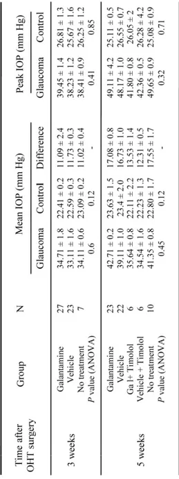
II.4.1. Galantamine protects RGC soma and axons from hypertension-induced death ... 61
II.4.2. Galantamine-mediated neuroprotection is not due to decreased IOP ... 63
II.4.3. RGC functional deficits in glaucoma are improved by galantamine ... 64
II.4.4. ACh muscarinic, but not nicotinic, receptors mediate the neuroprotective effect of galantamine in experimental glaucoma ... 65
II.5. DISCUSSION ... 66
II.6. REFERENCES... 71
II.7. TABLES ... 81
II.8. FIGURES ... 82
III. SECOND ARTICLE: “RETINAL MICROVASCULATURE PROTECTION CORRELATES WITH RETINAL GANGLION CELL SURVIVAL AND BLOOD
FLOW RESTORATION IN EXPERIMENTAL GLAUCOMA”. ... 88
III.1. ABSTRACT ... 90
III.2. INTRODUCTION ... 91
III.3. MATERIAL AND METHODS ... 92
III.3.1. Experimental animals... 92
III.3.2. Retrograde labeling of RGCs ... 92
III.3.3. Ocular hypertension surgery and measurement of intraocular pressure . 92 III.3.4. In vivo drug delivery ... 93
III.3.5. Quantification of RGC soma ... 93
III.3.6. Isolectin staining ... 94
III.3.7. Quantitative autoradiography ... 94
III.3.8. Retinal arteriole isolation and vessel diameter measurements ... 95
III.4. RESULTS ... 96
III.4.1. Loss of the retinal microvasculature occurs concomitantly with RGC death in experimental glaucoma ... 96
III.4.2. Galantamine mediates vasoprotection in experimental glaucoma ... 97
III.4.3. Retinal blood flow impairment in experimental glaucoma is partially restored by galantamine ... 97
III.4.4. Galantamine stimulates retinal arteriole relaxation ... 98
III.4.5. Muscarinic acetylcholine receptors mediate the vasodilator and vasoprotective effects of galantamine in experimental glaucoma ... 98
III.5. DISCUSSION ... 100
III.6. REFERENCES ... 102
III.8. FIGURES ... 110
CHAPTER 4 ... 114
IV. THIRD ARTICLE: “A CELL-PERMEABLE PHOSPHINE-BORANE COMPLEX DELAYS RETINAL GANGLION CELL DEATH AFTER AXONAL INJURY THROUGH ACTIVATION OF THE PRO-SURVIVAL EXTRACELLULAR SIGNAL-REGULATED KINASES 1/2 PATHWAY”. ... 114
IV.1. ABSTRACT ... 116
IV.2. INTRODUCTION ... 117
IV.3. MATERIALS AND METHODS ... 119
IV.3.1. Experimental animals ... 119
IV.3.2. RGC retrograde labeling ... 119
IV.3.3. Optic nerve injury paradigms ... 119
IV.3.3.1. Optic nerve axotomy ... 119
IV.3.3.2 Ocular hypertension (morrison model) ... 120
IV.3.4. Phosphine-borane complex synthesis ... 120
IV.3.5. In vivo drug delivery... 120
IV.3.6. Quantification of RGC soma and axons ... 121
IV.3.7. Western blot analysis ... 122
IV.3.8. Statistical analysis ... 123
IV.4. RESULTS ... 123
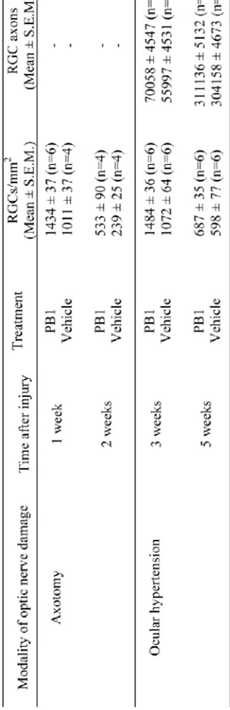
IV.4.1. Intraocular delivery of the phosphine-borane compound PB1 protects RGCs from axotomy-induced death ... 123
IV.4.2. PB1 protects RGC soma and axons in experimental glaucoma ... 124
IV.4.3. PB1-mediated RGC neuroprotection requires activation of the extracellular signal-regulated kinases 1/2 pathway ... 125
IV.5. DISCUSSION ... 127
IV.7. TABLES ... 144
IV.8. FIGURES ... 145
CHAPTER 5 ... 152
V. GENERAL DISCUSSION ... 152
V.1 STRUCTURAL PROTECTION IN GLAUCOMA ... 153
V.1.1 Galantamine, an acetylcholinesterase inhibitor, protects RGCs soma against IOP induced cell death. ... 153
V.1.2. Phenylphosphine borane reducing complex 1 (PB1), promotes RGC survival in different paradigms of optic nerve injury. ... 154
V.1.3. Protection of RGC axons in glaucoma ... 155
V.1.4. Protection of retinal microvessels in glaucoma ... 156
V.2. FUNCTIONAL PROTECTION IN GLAUCOMA ... 157
V.2.1. Galantamine treatment results in the recovery of visual evoked potentials ... 157
V.2.2. Galantamine improves retinal blood flow in glaucomatous eyes ... 159
V.3. MECHANISMS OF NEUROPROTECTION IN GLAUCOMA ... 160
V.3.1. Acetylcholine receptors and galantamine-mediated neuronal and vascular protection ... 160
V.3.2. Molecular mechanisms of PB1-mediated RGC neuroprotection... 164
V.4. GENERAL CONCLUSIONS ... 166
REFERENCES ... 167
APPENDIX A ... 205
MAINTENANCE OF AXO-OLIGODENDROGLIAL PARANODAL JUNCTIONS REQUIRES DCC AND NETRIN-1 ... 205
APPENDIX B ... 247
LIST OF TABLES
CHAPTER 2Table 1. Intraocular pressure (IOP) elevation in glaucomatous eyes……….81
CHAPTER 3
Table 1. Autoradiography values of the blood flow for each retinal isopter………109
CHAPTER 4
Table 1. PB1-induced RGC soma and axonal survival in axotomy and ocular hypertension models……….……….………144
LIST OF FIGURES
CHAPTER 1Figure 1. A schematic diagram of the retina demonstrating the principal cell types involved in retinal signaling………...4 Figure 2. Grey scale plot of visual field derived from short-wavelength automated perimetry showing large arcuate scotoma in the superior visual field……..7 Figure 3. A schematic presentation of the structures in the angle of the eye and aqueous humour circulation………..9 Figure 4. Sources of blood supply for the retina and optic nerve………17 Figure 5. The neurotrophic factor deprivation hypothesis………..33 Figure 6. Activation of ERK1/2 survival pathway via BDNF and TrkB signaling…36 Figure 7. Neuronal and glial component of retina in the excitotoxicity………..41 Figure 8. Activation of survival pathways via nAChRs signaling………...48
CHAPTER 2
Figure 1. Galantamine protects RGC soma in glaucoma…….………..…….….82 Figure 2. Comparative analysis of RGC survival in experimental glaucoma……....83 Figure 3. Galantamine protects RGC axons in glaucoma………....84 Figure 4. Galantamine-mediated neuroprotection is not due to decreased
intraocular pressure………..………85 Figure 5. RGCs functional deficits in glaucoma are improved by galantamine……86 Figure 6. The neuroprotective effect of galantamine in glaucoma is mediated by activation of muscarinic ACh receptors……….87
CHAPTER 3
Figure 1. Loss of inner retinal capillaries along with RGCs in experimental
glaucoma………..…110 Figure 2. Galantamine protects inner retinal capillaries in experimental glaucoma ………..…111
Figure 3. Galantamine-mediated improvement of the retinal blood flow in
glaucomatous retinas………..………112 Figure 4. Galantamine mediates vasodilation and vasoprotection via signaling through muscarinic acetylcholine receptors………..……..…113
CHAPTER 4
Figure 1. Chemical structure of bis (3-Propionic Acid Methyl Ester)
phenylphosphine borane reducing complex 1 (PB1)…..……...145 Figure 2. The phosphine-borane compound PB1 protects RGCs from axotomy- induced death………....…146 Figure 3. PB1 protects RGC soma in experimental glaucoma………..147 Figure 4. PB1 attenuates axonal loss in experimental glaucoma……….……...…148 Figure 5. The pro-apoptotic ASK1 signaling pathway is not regulated by PB1…..149 Figure 6. PB1 increases retinal BDNF and activates ERK1/2…………...…………150 Figure 7. PB1-mediated RGC neuroprotection requires activation of ERK1/2…..151
CHAPTER 5
LIST OF ABBREVIATIONS
AAV adeno-associated virus ACh acetylcholineAChE acetylcholinesterase AD Alzheimer’s disease
AGIS advanced glaucoma intervention study
AMPA α-amino-3-hydroxy-5-methyl-4-isoxazolepropionic acid APL allosteric potentiating ligand
APP amyloid precursor protein ARE antioxidant response element
ARRIVE animal research: reporting in vivo experiments ASK1 apoptosis signal regulating kinase 1
Aß amyloid beta
BDNF brain-derived neurotrophic factor cAMP cyclic adenosine monophosphate ChAT choline acetyltransferase
CNS central nervous system
CNTGSG collaborative normal-tension glaucoma study group CREB cAMP response element binding protein
DHß-E dihydro-ß-erythroidine hydrobromide DMSO dimethyl sulfoxide
DNA deoxyribonucleic acid
EAAT excitatory amino acid transporter ERG electroretinogram
ERK 1/2 extracellular signal-regulated kinases 1/2 ET-1 endothelin-1
GABA gamma-aminobutyric acid
GAPDH glyceraldehyde 3-phosphate dehydrogenase GFAP glial fibrillary acidic protein
GLAST glutamate-aspartate transporter GLT-1 glutamate transporter-1
GSH glutathione
HIF-1α hypoxia-inducible factor-1 alpha Hsp heat-shock protein
IOP intraocular pressure JNKs c-Jun N-terminal kinases LGN lateral geniculate nucleus L-NAME N-nitro-L-arginine methyl ester L-NMMA N-monomethyl-L-arginine
mAChR muscarinic acetylcholine receptor
MAPKKK mitogen-activated protein kinase kinase kinase MAPKs mitogen-activated protein kinases
MEK1 mitogen-activated protein kinase kinase 1 MLA methyllycaconitine citrate
mm millimeters
MMA mecamylamine hydrochloride mRNA messenger RNA
nAChR nicotinic acetylcholine receptor
NADPH nicotinamide adenine dinucleotide phosphate
NF-κB nuclear factor kappa-light-chain-enhancer of activated B cells NGF nerve growth factor
NMDA N-methyl-D-aspartic acid NO nitric oxide
NOLA N-nitro-L-arginine NOS nitric oxide synthase Nrf2 NF-E2-related factor-2 NT-3 neurotrophin-3
NT-4/5 neurotrophin-4/5
NTG normal tension glaucoma OHT ocular hypertension ONH optic nerve head
PB1 bis (3-propionic acid methyl ester) phenylphosphine borane complex 1 PBS phosphate-buffered saline
PFA paraformaldehyde
PI3K phosphatidylinositol 3-kinase PKC protein kinase C
PLC phospholipase C
POAG primary open angle glaucoma PRZ pirenzepine dihydrochloride Redox reduction-oxidation reactions RGCs retinal ganglion cells
RNA ribonucleic acid
RNFL retinal nerve fiber layer ROS reactive oxygen species
SAP standard achromatic automated perimetry SAPKs stress-activated protein kinases
SC superior colliculus
SCO scopolamine hydrobromide SOD superoxide dismutase
SWAP short-wavelength automated perimetry TM trabecular meshwork
TNFα tumor necrosis factor-alpha Trk tropomyosin related kinase TRO tropicamide
TRX thioredoxin
TRXR thioredoxin reductase
TXNIP thioredoxin -interacting protein VEP visual evoked potential
XIAP x-linked inhibitor of apoptosis protein [14C]-IMP N-isopropyl-p-14C-iodoamphetamine µm micrometers
ACKNOWLEDGEMENTS
I am heartily thankful to my supervisor, Dr. Adriana Di Polo, for the opportunity to work in her lab and for providing the guidance and encouragement that enabled me to pursue the projects. I have benefited greatly from her scientific vision and thoughtful criticism throughout the years and I am grateful for her kind support.
I would also like to express my deep appreciation to my co-supervisor Dr. Christian Casanova for his support and trust on me and for helping to expand the prospects of this work.
I would like to thank all the co-authors and collaborators of my papers in particular Drs. Melanie Kelly, Leonard Levin and Elvire Vaucher and the members of their labs for their support and contribution.
I would furthermore like to thank the members of my thesis jury for accepting to evaluate my work. I would also like to express my appreciation to members of my advisory and pre-doctoral committees Drs. Nicole Leclerc, Guy Doucet and Jean-Francois Bouchard for their valuable advice and guidance.
Many thanks to the members of the Di Polo lab past and present. It was my pleasure to work with Mike, Vincent, Yu, Johanne, Fred, Annie, Stephan and Marius. Many thanks to Philippe for his kind support. I would like to express my gratitude to Marilyne, Barbara, Ariel, Mathieu, Dara, Jorge and Sri for being awesome and for their friendship and spirit throughout the years. I would also like to thank members of the Casanova lab, Brian, Martin, Matthieu and Reza and in particular to thank Geneviève, Karine, Marouane, Nawal and Marilyse for their help and support.
Many thanks to my friend in the department: Casimir, Moogeh, Li, Jie, Greg, David, Isabel, Sarah-Jane, Mylène and Bahram. Special thanks to my friend Hamid for insightful discussions.
I would further like to thank the professors and members of the Department of Pathology and Cell Biology specially Dr. Nicole Leclerc, Dr. Mark Pelletier, Monique, Marielle, Michel Lauzon and Christine for their support. I also thank Faculté des études supérieures et postdoctorales and Groupe de Recherche sur le Système Nerveux Central for providing financial support.
“A hair divides what is false and true”
- Omar Khayyám
To my father, Mahmood, for his dedication to the family and to science To my mother, Ashraf, for her love for us and for her wisdom
To my brother, Reza, for his perseverance and support To my sister, Neda, for her kindness and talent
CHAPTER 1
I.1. GLAUCOMA: DEFINITION, PREVALENCE AND RISK FACTORS
Glaucoma is a group of chronic optic neuropathies characterized by progressive visual field defects and optic disc damage which ultimately leads to irreversible blindness due to the loss of retinal ganglion cells (RGCs). It is estimated that more than 60 million people suffer from glaucoma worldwide (Quigley and Broman, 2006) and according to a report from the World Health Organization, glaucoma is the second cause of blindness globally accounting for 12.3% of total cases (Resnikoff et al., 2004).
There are a number of risk factors that increase the incidence of glaucoma. Demographic factors including age and ethnic background are among the most recognized risk factors for glaucoma. For example people over the age of 60 have a much higher risk of developing glaucoma (Coleman and Miglior, 2008). Also, the rate of glaucoma progression is faster in individuals diagnosed with glaucoma within certain population subgroups (Sommer et al., 1991; Varma et al., 2004). Moreover, belonging to a certain ethnic background significantly lowers the age at which those individuals are at risk of developing glaucoma. For instance, individuals of African-descent 40 years and older are at higher risk of developing glaucoma and have the highest rate of blindness due to glaucoma (Congdon et al., 2004; Leske, 2007). Family history is another recognized risk factor for developing glaucoma as individuals with a family member with glaucoma have a four times higher risk of developing this disease (Wiggs, 2007).
Elevated intraocular pressure (IOP) is an important risk factor for developing glaucoma. Considering that the normal value of IOP for adult individuals is usually around 15 mmHg, people with an IOP over 21 mmHg are known as ocular hypertensive (Quigley et al., 1994). Interestingly, there is a correlation between systemic blood pressure and IOP levels as an increase or a reduction of systemic blood pressure results in higher or lower IOP, respectively (Klein et al., 2005; McLeod et al., 1990). Indeed, high systemic blood pressure is considered a risk factor in glaucoma (Deokule and Weinreb, 2008). Although IOP is regarded as an important risk factor, it is not an accurate predictor of glaucoma since more than 30% of the glaucoma patients have an IOP in the normal range (Nemesure et al., 2007).
Corneal thickness is also considered another risk factor and thinner central corneas have been correlated with visual field loss in glaucoma (Medeiros et al., 2003).
High myopia is another risk factor correlating with glaucoma occurrence (Loyo-Berros and Blustein, 2007; Mastropasqua et al., 1992). An association between diabetes and glaucoma has also been suggested (Bonovas et al., 2004). Some cases of diabetes also have higher IOP levels compared to control individuals (Klein et al., 1984; Tielsch et al., 1995).
I.2. PATHOLOGICAL AND CLINICAL FEATURES OF GLAUCOMA
I.2.1. Loss of RGCs, optic disc cupping and axonal damage
Glaucoma is characterized by damage to the neural components of the visual pathway including the retina, the optic tract and the brain. The visual information in the retina travels vertically through photoreceptors, bipolar cells and finally RGCs. RGCs play an essential role in vision because they are the only neurons responsible for relaying the visual signal from the eye to the higher centers in the brain (Figure 1). One of the highlights of glaucoma pathology is the selective loss of RGCs, which is characteristic of all glaucoma patients (Kendell et al., 1995; Quigley, 1999).
RGCs axons extend from their cell bodies over the inner surface of retina to reach the optic disc. They join to form axon bundles at the retinal nerve fiber layer supported by both the processes of Müller cells and astrocytes (Radius and Anderson, 1979). The optic disc or optic nerve head (ONH) is the region where RGC axons exit the eye to form the optic nerve. The ONH includes a prelaminar area formed by loose trabecular glial tissue and bundles of unmyelinated nerve fibers. The laminar area of the ONH is called the lamina cribrosa; it is composed of parallel collagen laminas located in a canal in the posterior part of the sclera (Jonas et al., 1991). The lamina cribrosa forms a sieve-like network and axons of RGCs exit the retina through its pores; it also allows passage of the central retinal vessels (Hernandez et al., 1986).
The area of the ONH that contains the nerve fibers, called the neuroretinal rim, surrounds the central and slightly depressed part called the cup (Jonas et al., 1988). Increased depth of the optic cup is a well known clinical feature of glaucoma (Jonas et al., 1999). ONH cupping is one of the first detectable signs of glaucoma and often precedes the loss of visual field (Pederson and Anderson, 1980). The optic disc cupping
Figure 1. A schematic diagram of the retina demonstrating the principal cell types involved in retinal signaling. The photoreceptor cells (rods and cones) are the outermost neuronal layer of the retina. Photoreceptors are light-sensitive and transduce light stimuli into electrical signals. The signal is subsequently transferred from the photoreceptors to bipolar cells. The outer plexiform layer (OPL) contains the synaptic connections between photoreceptors to bipolar cells and horizontal cells. Bipolar cells make synaptic connections with RGCs in the inner plexiform layer (IPL). RGC axons travel toward the optic disc in the nerve fiber layer and exit the eye forming the optic nerve. Cellular components of the retina are supported by Müller cells and the interconnections provided by horizontal cells and amacrine cells participate in retinal processing and modifications of visual signal. Source of image: Mohammadali Almasieh.
correlates with changes in the organization of connective tissue bundles and thinning of laminar beams in the lamina cribrosa in experimental and human glaucoma (Fukuchi et al., 1992; Miller and Quigley, 1987; Morrison et al., 1990). ONH changes also include abnormal deposition of extracellular matrix components such as collagen, laminin and elastin (Hernandez et al., 1990; Johnson et al., 1996; Morrison et al., 1990). ONH tissue remodeling occurs partly due to activation of resident glial cells since abnormal deposition of extracellular material correlates with increased expression of elastin mRNA in local astrocytes (Pena et al., 2001). Activated and proliferating astrocytes, characterized by hypertrophic soma and expression of glial fibrillary acidic protein (GFAP), are found in human and experimental glaucoma (Hernandez et al., 2008; Johnson et al., 2007a). Tissue remodeling can also result in biomechanical alterations at the ONH leading to stress-induced damage of RGC axons at this location (Burgoyne, 2010).
I.2.2. Loss of visual field
Glaucoma is not the only optic neuropathy accompanied by visual loss; however, based on the characteristic changes of the optic disc and the typical patterns of visual field loss, glaucomatous visual defects can be distinguished from other optic neuropathies. Because damage to RGC axons happens mostly at the superior and inferior parts of the ONH and considering that nerve fibers make a superior or inferior arc around the horizontal raphe before entering the optic disc, typical arcuate scotomas are the characteristic visual field loss pattern in the glaucoma (Figure 2) (Hood and Kardon, 2007; Spector, 1990). It is typical for glaucomatous vision loss to be peripherally located as occurrences of scotomas are usually paracentral; central vision will remain intact until very late in the course of the disease. Several methods are currently used for diagnosis of glaucomatous visual field abnormalities including standard achromatic automated perimetry (SAP) or selective perimetry (Sakata et al., 2007; Sample et al., 2000; Sharma et al., 2008). Due to considerable overlap in the receptive fields of different RGC types, SAP does not allow detection of visual defects unless 40-50% of RGCs are already lost. However, short-wavelength (blue-yellow color sensitive) automated perimetry (SWAP) that targets the small bistratified RGCs, enables physicians to detect functional loss years
FIGURE 2.
Figure 2. Grey scale plot of visual field derived from short-wavelength automated perimetry showing large arcuate scotoma in the superior visual field. Three panels indicate progressive loss of visual field. Different gray spectra show the level of visual loss and black indicates the blind spots. Source of image: adapted from Clement et, al., Br J Ophthalmol. 2009.
earlier because it is selective for a particular ganglion cell type (Johnson et al., 1993; Sit et al., 2004).
Pathological changes in glaucoma are also detected in visual centers in the brain and could account for visual defects. The lateral geniculate nucleus (LGN), a major central target of RGC axons in primates, shows significant atrophy and neuronal loss in primate experimental glaucoma (Ito et al., 2009; Yücel et al., 2001). Activated glial cells play important roles in the degenerative changes in the LGN as astrocytosis and microglial activation have been reported in the LGN and visual cortex in experimental glaucoma (Lam et al., 2009).
I.3. GLAUCOMA CLASSIFICATION AND EXPERIMENTAL MODELS
Glaucoma has been divided into two major categories: primary angle-closure glaucoma (PACG) and primary open-angle glaucoma (POAG). However, other sub-categories have been added to accommodate for the diversity of pathologies. The production, circulation and drainage of the aqueous humour (the clear fluid that fills the anterior and posterior chambers of the eye) are determining factors for the IOP level of the eye. Therefore, the anatomical structures of the eye involved in the production and circulation of the aqueous humour will be reviewed briefly.
The aqueous humour is produced by the ciliary epithelium of the ciliary body, provides nutrition for the lens and removes metabolic waste as it flows through the pupil into the anterior chamber of the eye (Krupin et al., 1986). The aqueous humour fills the anterior chamber providing nutrition to the cornea as well. There is an area in the anterior chamber where the cornea and iris join, the angle, where the drainage of aqueous humour takes place (Figure 3). Aqueous fluid flows toward the angle where it enters the trabecular meshwork (TM) (Tamm, 2009), a sieve-like structure that filters and directs the aqueous fluid into the Schlemm's canal (Johnstone, 2004). A portion of the aqueous humour leaves the anterior chamber through an alternative route called the uveo-scleral pathway (Gabelt and Kaufman, 2005; Goel et al., 2010). Several models of inducible and spontaneous glaucoma rely on blockade of the aqueous humour drainage and will be discussed in subsequent sections.
FIGURE 3.
Figure 3. A schematic presentation of the structures in the angle of the eye and aqueous humour circulation. The aqueous humour is produced by the ciliary body and enters the anterior chamber via the pupil. The trabecular meshwork (TM) is located in the angle between the cornea and iris and provides a circumferential outlet for drainage of aqueous. In PACG, access to the TM is limited or severed due to physical closure of the anterior chamber angle; whereas in POAG, the TM is accessible, however ultrastructural changes in the TM itself result in reduction of outflow. Aqueous humour enters Schlemm's canal and after being collected in a circumferential venous plexus, joins the blood circulation via episcleral veins. Source of image: Kwon et. al., N. Engl. J. Med. 2009.
I.3.1. Primary angle-closure glaucoma
PACG is characterized by the blockade of the aqueous humour drainage and/or its circulation. This results in increased IOP and consequent damage to the retina and optic nerve. If the IOP increase is sudden and acute, it is often accompanied by pain and redness of the eye, blurry vision, headache and nausea (Sihota, 2011). The name “angle-closure” was originally used after the observation that the aqueous humour drainage is blocked in the angle of the eye resulting in IOP build up. PACG is primarily caused by an abnormal contact between anterior segment structures of the eye, including the cornea, iris, TM, ciliary body, and the lens. This could happen due to abnormal dilation of the pupil causing congestion of the iris in the angle of the eye. There is also a condition called plateau iris, characterized by an abnormal position of ciliary processes that push and hold the iris forward placing it too close to the TM (Kumar et al., 2008). Some PACG cases also involve pupillary blockade of the aqueous fluid. Pupillary blockade happens when the back of the iris adheres to the lens and blocks the passage of the aqueous humour to the anterior chamber. This not only increases the pressure in the posterior chamber but also pushes the periphery of the iris forward resulting in angle closure (Tarongoy et al., 2009). The extent of pupillary blockade and angle closure determines the magnitude and time-course of IOP increase, therefore PACG has been divided into acute (with sudden and severe IOP increase) and chronic (with a more gradual IOP increase) subcategories. When angle-closure happens as a result of other ocular diseases, it is referred to as secondary angle-closure glaucoma. For instance, secondary angle-closure glaucoma could happen due to an anterior-shift of the lens after ocular trauma (Sankar et al., 2001).
People of Asian descent and individuals with small axial eye length, have a greater risk of developing PACG. The common procedure for the treatment of acute PACG is to lower the IOP by medications (Hoh et al., 2002). Surgical techniques such as peripheral iridotomy, the generation of small holes in the peripheral iris using a laser, might also be required (Lam et al., 2007; Tarongoy et al., 2009). Analysis of the eyes with acute PACG revealed that although IOP was lowered immediately after diagnosis, there was still a significant reduction in the thickness of the retinal nerve fiber layer (RNFL) weeks after the acute episode (Aung et al., 2004).
I.3.1.1. Experimental models of primary angle-closure glaucoma
Several breeds of dogs are prone to develop glaucoma (Reinstein et al., 2009) and the high prevalence of glaucoma in some pure-bred dogs suggests a genetic basis for this disease (Gelatt and MacKay, 2004). For instance, a canine model (Basset Hounds) of hereditary PACG shows gradual narrowing and collapse of iridocorneal angles leading to complete angle-closure by 20 months of age; angle-closure in these dogs is accompanied by IOP build up, cupping of ONH and loss of RGCs (Grozdanic et al., 2010). However, the high cost of purchasing and housing dogs, their requirements for special care and the inherent problem of handling dogs in large experimental groups, results in limited use of this model.
Laser photocoagulation has been adapted for mice to induce the closure of the anterior chamber angle (Aihara et al., 2003). A diode laser is focused on the corneal limbus to create burn spots that directly attach the iris root to the peripheral cornea; consequently, obstruction of the aqueous outflow results in elevation of IOP (Aihara et al., 2003). Despite the differences in the structure of ONH between mice and humans, such as the lack of lamina cribrosa (May and Lütjen-Drecoll, 2002), the elevation of IOP in this model is accompanied by significant loss of RGC axons (Mabuchi et al., 2003). However, the small size of the mice eye demands a high level of expertise by the experimenter in using the laser as excessive or misplace laser burns could result in abrupt IOP increase, inflammatory response and retinal damage.
I.3.2. Primary open-angle glaucoma
Primary open-angle glaucoma (POAG) is the most common form of the glaucoma worldwide. POAG is characterized by changes in the optic disc, damage to RGC axons in the optic nerve, loss of RGCs in the retina and gradual visual field defects (Quigley, 2005). POAG is not necessarily associated with IOP elevation, however, high IOP is a major risk factor for developing POAG and a significant percentage of POAG cases show a gradual increase of IOP over the years (Leske et al., 2003). The increase in IOP is not necessarily accompanied with severe changes in anterior angle structures as previously described for PACG. Since resistance to the aqueous humour outflow is a major determinant of IOP levels, age-related or pathologic changes in the TM or Schlemm's
canal might lead to the gradual increase of the outflow resistance and IOP build up (Gabelt and Kaufman, 2005; Wordinger and Clark, 1999). Although morphological changes in optic disc vary little between PACG and POAG (Boland et al., 2008; Nouri Mahdavi et al., 2011), functional evaluation demonstrated that PACG patients have a more diffuse pattern of visual field loss compared to POAG patients (Boland et al., 2008; Rhee et al., 2001).
I.3.2.1. Experimental models of primary open-angle glaucoma
Our understanding about the mechanism of aqueous humour outflow and its importance in regulating IOP has been used to develop a number of experimental glaucoma models. Several animal species are suitable for induction of experimental ocular hypertension (OHT) and have provided useful functional and structural information about glaucoma onset and progression.
I.3.2.1.a. Primate model of POAG
In this model, Rhesus or Cynomolgus monkeys are subjected to laser photocoagulation, that by creating burn spots on the circumference of the TM, results in moderate IOP increase (Wang et al., 1998). A disadvantage is that multiple sessions of laser treatment are required to induce OHT. IOP elevation in this model is accompanied with ONH cupping and thinning of RNFL (Gaasterland and Kupfer, 1974). Loss of RGCs and functional defects are well documented in this model, indicating a close similarity to human POAG (Hare et al., 2001b; Hood et al., 1999; Morgan et al., 2000). Pathological changes in the visual centers in the brain such as loss of neurons in the magno and parvocellular layers of LGN are also detected (Yücel et al., 2003; Yücel et al., 2000). The high cost of monkeys, their limited availability and difficulty to work with are major disadvantages particularly for studies on neuroprotection since they require large number of animals.
I.3.2.1.b. Rodent models of POAG
The laser photocoagulation technique has also been used for induction of OHT in rodents (Levkovitch-Verbin et al., 2002b; Schori et al., 2001; Ueda et al., 1998; Wijono et al., 1999; WoldeMussie and Feldman, 1997). A beam of diode laser is concentrated on
the circumference of the TM to create burn spots; the peak IOP reaches 35-49 mmHg and it is sustained for at least 3 weeks (Levkovitch-Verbin et al., 2002b). A similar technique has been adapted for induction of OHT in mice (Gross et al., 2003; Ji et al., 2005).
The aqueous humour flows through the TM into the Schlemm's canal and then via collector channels into the limbal venous plexus and finally drains through the episcleral veins. This pathway has been used for retrograde transfusion of solutions towards the TM (Moore et al., 1993; Morrison et al., 1995). In the Morrison model of rat OHT, hypertonic saline solution is injected into an episcleral vein of Brown Norway rat eyes. Hypertonic saline causes sclerotic damage to the TM cells, disrupts the structure of the TM and gradually reduces the aqueous outflow, resulting in IOP elevation (Morrison et al., 1997). Another method of reducing the aqueous outflow is by cauterizing the episcleral veins (Shareef et al., 1995). In this model, two to three veins are isolated and blocked using an ophthalmic cautery (Shareef et al., 1995).
Obstruction of the TM and elevation of IOP can also be achieved by injection of sterile latex microspheres (Urcola et al., 2006; Weber and Zelenak, 2001) or microbeads into the anterior chamber of rodents (Chen et al., 2011; Sappington et al., 2010). Finally, a hereditary mutation in the DBA/2J mice leads to iris pigment dispersion and anterior synechia (adhesion of the iris to the cornea) that results in significant elevation of IOP by 6 months of age (John et al., 1998).
To select a suitable rodent glaucoma model for research, the objectives of the study and the advantages/disadvantages of each model should be considered carefully. For instance, rat models are generally more appropriate for ONH studies because unlike mouse, the rat ONH has a well-defined lamina cribrosa. The structure of the lamina is similar to that in primates regarding protein content, connective tissue components and blood vessels (Morrison et al., 1995). Elevation of IOP in rat models results in cupping of ONH, loss of axons in the optic nerve and RGCs death. Mouse glaucoma models in the other hand, confer the advantage of using transgenic animals to study the role of specific genes in the development of glaucoma (Whitehouse et al., 1982).
The major disadvantage of laser photocoagulation technique is the necessity of multiple treatments to achieve a steady IOP elevation. In addition, retinal oedema and haemorrhage often occur in this model. The cauterization model has a slow rate of
progress with considerable variability in the pattern and number of RGC death (Danias et al., 2006); there is also the possibility of necrosis of the eye if several episcleral trunks are blocked (Ahmed et al., 2001; Laquis et al., 1998). The DBA/2J mouse model shows great variability in the onset and progression of glaucoma between individual animals which dramatically increases the number of animals required for each experiment. The microbeads injection model is a relatively simple and promising model but there is high variability in the levels of IOP achieved following injection of microbeads (Chen et al., 2011; Sappington et al., 2010). The Morrison model is also a simple, reliable and reproducible model of experimental glaucoma in Brown Norway rats. IOP elevation in this model results in ONH cupping and gradual loss of axons in the optic nerve (Johnson et al., 1996; Morrison et al., 1997). Progressive loss of RGC in this model is also well documented (Guo et al., 2005; Johnson et al., 2007b). General anaesthetics are often used in rodents to allow IOP measurements, however, anaesthetics result in a substantial decrease of IOP (Jia et al., 2000). An advantage of the Morrison model is that Brown Norway rats are extremely docile allowing IOP measurements in unanaesthetized animals, thus providing the most accurate documentation of IOP history. Due to its advantages, particularly in regard to neuroprotection studies, the Morrison model was selected and used throughout this thesis.
I.3.3. Normal tension glaucoma
Many glaucoma patients display typical glaucomatous visual defects and RGC loss while their IOP is within the normal range (CNTGSG, 1998). These cases are classified as normal tension glaucoma (NTG). The visual field loss in NTG patient progresses with a similar rate as POAG patients, therefore diagnosis and treatment of NTG demands particular attention (Ahrlich et al., 2010). The exact cause of NTG is unknown; however, several mechanisms have been proposed. Among these are the association of visual field loss and decreased blood flow in the optic disc of NTG patients (Ciancaglini et al., 2001; Nicolela et al., 1996; Sato et al., 2006) and the higher rate of the hemorrhages in the fundus (Ishida et al., 2000; Tezel et al., 1996); suggesting a correlation between ocular blood flow and vascular dysfunction in NTG. There is also evidence of an autoimmune response, highlighted by elevated levels of antibodies against
heat shock proteins (HSPs) (Wax et al., 1998). Since glaucoma is now considered a neurodegenerative disease, oxidative damage and excitotoxicity may play a role in the pathology of NTG.
I.4. MECHANISMS OF NEURONAL DAMAGE IN GLAUCOMA
I.4.1. The role of vascular dysfunction in the pathology of glaucoma
An increasing body of evidence suggests that retinal and ONH blood flow are among the risk factors for the development of glaucoma (Flammer et al., 2002; Osborne et al., 2001). There is a significant reduction of blood flow in the ONH of PAOG patients (Piltz-seymour et al., 2001). Elevated IOP and compression of the vessels at the level of the lamina cribrosa has been proposed to be the cause of blood flow reduction (Downs et al., 2008; Feher et al., 2005). However, loss of visual field in NTG patients is also associated with decreased optic disc blood flow (Ciancaglini et al., 2001; Nicolela et al., 1996; Sato et al., 2006) and the severity of neuronal damage in NTG and POAG patients correlates with a reduction in retinal blood flow (Bjärnhall et al., 2007; Sato et al., 2006). When the IOP of healthy volunteers was temporarily increased, it resulted in a clear vasodilatory response. However, the same level of temporary IOP increase in POAG patients resulted in a much smaller vasodilatory response (Nagel et al., 2001). These observations suggest an underlying vascular deficiency contributing to the pathology of glaucoma.
I.4.1.1. The retinal circulation
In terms of blood supply, the retina is divided into two regions: i) the outer retina, including photoreceptors, outer nuclear and outer plexiform layers, is supplied by the choroid capillaries originating from the posterior ciliary arteries (Saint Geniez and D'Amore, 2004); ii) the inner retina, including the nerve fiber layer, RGC layer, inner plexiform and inner nuclear layer, is supplied by the central retinal artery (Figure 4). The branches of the central retinal artery expand to form two inner capillary networks, one superficial, at the level of nerve fiber layer and one deeper, between the inner nuclear and outer plexiform layers (Pournaras et al., 2008). The capillaries of the inner retinal
networks are in close interaction with RGCs and other neuronal-glial elements of the retina to meet the metabolic demands of this tissue.
Retinal capillaries are composed of a single layer of endothelial cells enclosed in a basement membrane shared with surrounding pericytes (Kniesel and Wolburg, 2000; Russ et al., 1998). Pericytes are specialized mural cells found in the CNS that play an integral role in vascular tone (Bandopadhyay et al., 2001). Retinal capillaries are smaller in diameter with thinner walls compare to non-neuronal capillaries and are morphologically very similar to cerebral capillaries; however, their endothelial cells have a higher density of vesicles and higher permeability compare to their counterparts in the brain (Stewart and Tuor, 1994). Retinal capillaries are also accompanied by a considerable higher number of pericytes (up to 4.5-fold) compared to cerebral capillaries and their pericyte processes cover more than 85% of the circumference of the retinal endothelial tube (Frank et al., 1987; Stewart and Tuor, 1994). Pericytes together with the glial cell’s end-foot processes, strengthen the blood-retinal barrier and play important roles in compensating for more permeable endothelial cells compared to brain capillaries (Kim et al., 2006a; Stewart and Tuor, 1994).
I.4.1.2. Regulatory mechanisms of retinal circulation
The proper regulation of ocular blood flow is vital for normal retinal function. The ocular vessels respond to the increased neuronal activity by increasing the blood flow, a phenomena known as neurovascular coupling (Garhöfer et al., 2004; Riva et al., 2005). The choroidal vascular bed is innervated by autonomic vasoactive nerves that control the choroidal blood flow (Schrder et al., 1994). Unlike the choroid, the central retinal artery has limited sympathetic innervation that ends at the level of lamina cribrosa (Laties, 1967). Therefore, local vasodilatory and vasoconstrictor mediators play important roles in regulating the retinal blood flow (Metea and Newman, 2007). The production and release of vasoactive compounds by endothelial cells contribute to the regulation of retinal vascular tone. Nitric oxide (NO) and endothelins (ET) are among the most important endothelium-derived vasoactive factors (Chakravarthy et al., 1995; Loscalzo and Welch, 1995; Takagi et al., 1996).
FIGURE 4.
Figure 4. Sources of blood supply for the retina and optic nerve. The central retinal artery and the posterior ciliary arteries are branches of the ophthalmic artery. Branches of posterior ciliary arteries enter the eye to form the choroid. The central retinal artery enters the optic nerve at 8–12mm behind the globe and after supplying the optic nerve with a few small branches, it divides into two major trunks before leaving the optic disc. The branches of the central retinal artery expand to form two inner capillary networks, one superficial, at the level of nerve fiber layer and one deeper, between the inner nuclear and outer plexiform layers. Source of image: Mohammadali Almasieh.
I.4.1.2.a. Nitric oxide
NO is a potent neurotransmitter that regulates a wide variety of cellular functions. Three different isoforms of nitric oxide synthase (NOS) are involved in NO production: neuronal (nNOS or NOS1), inducible (iNOS or NOS2) and endothelial (eNOS or NOS3) (Alderton et al., 2001). In the retina, nNOS is expressed by RGCs, photoreceptors and amacrine cells; while eNOS is expressed by endothelial cells (Neufeld et al., 2000; Shareef et al., 1999). iNOS is activated in response to inflammation or tissue damage and is thought to contribute to cytotoxic NO signaling (Calabrese et al., 2007; Chiou, 2001). In physiological concentrations, NO signals through activation of guanylyl cyclase receptors and production of cGMP (Garthwaite, 2008). NO plays an important vasodilatory role in the retina. Experimental inhibition of retinal NOS by intravitreal injection of its inhibitors N-monomethyl-L-arginine (L-NMMA) or (L-NAME) resulted in significant vasoconstriction of retinal arterioles and venules indicating that NO production is necessary for maintaining a basal level of vascular relaxation (Donati et al., 1995; Dorner et al., 2003). Hyperemia is the activity-induced increase in blood flow in a tissue and can be easily induced in the retina by light flicker (Maelicke, 2000). Inhibition of NOS significantly reduces the retinal hyperemic response to light flicker, indicating that NO also participates in the activity dependent modulation of the retinal blood flow (Dorner et al., 2003). In normal subjects, systemic inhibition of NOS significantly reduces ONH blood flow (Luksch et al., 2000). Dysfunctional NO signaling in glaucomatous eyes became evident by a much smaller response of POAG patients to NOS inhibition (Polak et al., 2007). NO levels also effect production and drainage of the aqueous humour (Whitehouse et al., 1982). Reduction of NO levels and eNOS activity has been reported in ocular tissues of POAG patients including the TM (Nathanson and McKee, 1995).
I.4.1.2.b. Endothelins
Endothelins are a family of vasoconstrictor peptides. They are released by endothelial cells and interact with their G-protein-coupled, ETA and ETB receptors
(Davenport, 2002; Yanagisawa et al., 1988). ETA receptors are mainly expressed by the
endothelin-1 (ET-1) (Pierre and Davenport, 1998). ETB receptors have a broader pattern
of expression and can be found on neuronal and glial components of the retina (MacCumber and D'Anna, 1994; Stitt et al., 1996). It appears that ETA and ETB subtypes
can mediate opposing actions in a complex manner. Blockade of the ETB receptors
significantly increases peripheral vasoconstriction (Strachan et al., 1999). Activation of ETB receptors alone has a net constrictor effect but simultaneous activation of both
subtypes is vasodilatory (Just et al., 2004; Mickley et al., 1997). Endothelial cells express ETB and its activity results in the release of relaxing factors such as nitric oxide and
prostanoids from endothelial cells (Hirata et al., 1993; Just et al., 2005).
The level of endothelins and the expression of their receptors change in experimental and human glaucoma. For instance, levels of ET-1 in serum and aqueous humour of POAG patients is higher than in control individuals (Emre et al., 2005; Nicolela et al., 2003; Noske et al., 1997; Sugiyama et al., 1995; Tezel et al., 1997). In addition, ET-1 levels in the ONH of animals subjected to experimental glaucoma increases significantly compare to control groups (Howell et al., 2011; Prasanna et al., 2005). Receptor dysfunction has been proposed to play central role in blood flow irregularities in glaucomatous eyes. While treatment of normal subjects with an antagonist of ETA resulted in a significant vasodilatory response, a similar treatment had
a lesser effect on the vascular tone of NTG patients (Henry et al., 2006; Henry et al., 1999). Recently, ETA gene polymorphisms have been considered to be a potential risk
factor for developing NTG (Ishikawa et al., 2005; Kim et al., 2006b). Endothelins might also be involved in the induction of a glial response in the glaucomatous ONH. Reactive astrocytes in the ONH of experimental and human glaucoma are characterized by the expression of ETA and ETB (Wang et al., 2006; Wang et al., 2009). Incubation of isolated
ONH astrocytes with ET-1 results in their activation and induces marked proliferation (Murphy et al., 2010; Prasanna et al., 2002). ETA is also expressed in structures of the
anterior segment such as the iris, ciliary body, trabecular cells and endothelial lining of the Schlemm's canal (Fernández-Durango et al., 2003). Therefore endothelin signaling can also participate in the production of aqueous humour and the control of its outflow. A treatment strategy based on endothelin antagonism has been proposed to control IOP
increase and to improve retinal blood flow (Fernández-Durango et al., 2003; Rosenthal and Fromm, 2011).
I.4.1.3. Vascular degeneration
In addition to vasomodulatory abnormalities, there is also evidence of structural changes in the vasculature of glaucomatous eyes. In some POAG patients, the central retinal artery is characterized by arteriosclerotic changes such as the thickening of the basement membrane, proliferation of muscle cells into the intimae, fragmentation of the inner elastic membrane and arteriosclerotic plaques in the intimae (Gottanka et al., 2005). Narrowing of the lumen of smaller arterioles in the optic nerve and deposits of amorphous material in the basement membrane of endothelium are the other pathological findings in the POAG (Feher et al., 2005; Gottanka et al., 2005). A significant decrease in the density of capillaries has been observed in the optic nerve laminar region of POAG patients and in rat experimental glaucoma (Daz et al., 2010; Gottanka et al., 2005). These observation are in agreement with initial reports by Quigley and colleagues indicating a considerable loss of disc tissue and its capillaries (Quigley et al., 1984). Fluorescein angiograms show a high rate of capillary non-perfusion in the optic disc of NTG and POAG patients (Plange et al., 2006). Pathological changes in the vasculature also play important roles in the progression of other neurodegenerative diseases such as Alzheimer's disease (Budinger, 2003). In fact, pathological vascular changes like acellular capillaries have been found in both glaucomatous retinas and in the brains of Alzheimer's patients (Brown, 2010b).
I.4.1.4. Neurotrophic factors and vascular reactivity
In addition to neurons, neurotrophic factors also regulate survival and function of non-neuronal cells. In fact production of neurotrophic factors like BDNF and NGF is not limited to neuronal tissues as several non-neuronal tissues including the elements of cardiovascular system also express neurotrophins and their receptors (Scarisbrick et al., 1993; Yamamoto et al., 1996). Interaction of neurons and vasculature through neurotrophic factors is in particular complex and plays important role in development and physiological functions of both systems.
Neurotrophins are involved in regulation of angiogenesis, for instance, activation of neurons by BDNF and NGF increases production and secretion of VEGF by neurons which in turn results in endothelial cells proliferation (Nakamura et al., 2011). Brain-derived endothelial cells themselves shown to synthesis BDNF and express TrkB and p75NTR receptors (Kim et al., 2004b). They also produce NGF and express its receptor TrkA (Moser et al., 2004). Direct activation of TrkA receptors of endothelial cells by exogenous NGF results in proliferation of these cells and also increases the secretion of NGF by endothelial cells (Moser et al., 2004). In addition, NGF improves survival of endothelial cells under oxygen-glucose deprivation conditions (Lecht et al., 2010). Local production and release of major neurotrophin family members like BDNF and NGF by endothelial cells and the interaction of these trophic factors with TrkA, TrkB and p75NTR receptors provides an autocrine/paracrine regulatory system for cell proliferation in the vasculature (Kim et al., 2004b; Tanaka et al., 2004).
Recent studies show that neurotrophic factors produced by brain endothelial cells could be important for survival of neurons as they significantly extend the survival of isolated cortical neurons (Dugas et al., 2008). Endothelial source of BDNF also provides significant support for neurons in a number of situations like oxidative stress-hypoxia and neurotoxicity (Guo et al., 2008). In addition, secration of BDNF by endothelial cells provides important support for migrating neuroblasts that originate from sub-ventricular zone or sub-ependymal zone and helps to guide them towards their targets in the brain (Leventhal et al., 1999; Snapyan et al., 2009).
Currently, our knowledge about the effects of neurotrophins on retinal blood flow is limited; however, neurotrophins like BDNF stimulate the endothelial cells to generate more NO and shown to have vasodilatory effects in other systems (Meuchel et al., 2011). It has also been shown that using the viral vectors to overexpress BDNF in cerebral vessels results in upregulation of prostacyclin (PGl2) that leads to vascular relaxation (Santhanam et al., 2010); however, no significant change in the expression or activity of the eNOS has been detected in these experiments (Santhanam et al., 2010). It is important to mention that early in the development of brain, PGI2 is the primary mediator of endothelium-dependent relaxations in cerebral circulation; however, in the adult and aging brain, nitric oxide takes over as the primary vascular vasodilator (Charpie et al.,
1994; Willis and Leffler, 2001). Interestingly, intraocular application of BDNF results in a higher expression of vasoactive intestinal polypeptide by amacrine cells that by relaxing smooth muscle cells leads to vasodilation (Cellerino et al., 2003).
Intraocular application of BDNF significantly increases survival of RGCs in the traumatic optic nerve damage models and also increases NOS activity as it has been evaluated by NADPH-diaphorase reactivity (Klcker et al., 1998). However, it is suspected that BDNF also results in activation of iNOS that considered to be a limiting factor for the survival of RGCs due to promotion of nitrosative stress through formation of peroxynitrite (Klcker et al., 1998; Klcker et al., 1999). Further studies in line with development of new experimental protocols are needed to evaluate the role of neurotrophic factors in the improvement of retinal blood flow and protection of RGCs in pathological conditions.
I.4.2. Oxidative stress
Oxidative stress, caused by the imbalance between the production of reactive oxygen species (ROS) and their elimination by antioxidants, has been recognized as a central contributor to neuronal injury and death. ROS are continuously produced by mitochondria through the electron transport chain, but can also be generated by enzymatic degradation of neurotransmitters, neuroinflammatory mediators, and redox reactions (Halliwell, 2006). Increased levels of ROS like superoxide anion (O2•) and hydroxyl radical (OH•), a common feature of neurodegenerative diseases, can originate
from mitochondrial dysfunction, abnormal protein folding, and defective ubiquitination and proteasome degradation systems (Andersen, 2004).
There is evidence that oxidative damage occurs in experimental models of optic nerve injury and in human glaucoma. For example, the presence of DNA damage and accumulation of protein and lipid peroxidation products have been documented in the TM and retinas from experimental ocular hypertension models and POAG patients (Babizhayev and Bunin, 1989; Izzotti et al., 2003; Ko et al., 2005; Moreno et al., 2004; Sacc et al., 2005; Tezel et al., 2005). Furthermore, an intracellular ROS superoxide burst has been proposed to be a critical death signal triggered by axonal injury leading to RGC apoptosis (Geiger et al., 2002; Kanamori et al., 2010; Lieven et al., 2006; Nguyen et al.,
2003; Swanson et al., 2005). During hypoxia, the generation of mitochondrial ROS is necessary for activation of the hypoxia-inducible factor-1 alpha (HIF-1α), a transcriptional regulator required for the induction of a variety of genes (Chandel et al., 2000; Duranteau et al., 1998). Expression of HIF-1α and HIF-1α target genes, including erythropoietin, heat-shock protein 27 (Hsp-27) and vascular endothelial growth factor, increase in experimental rat glaucoma (Ergorul et al., 2010), and high levels of HIF-1α are found in the retina and optic nerve head of glaucoma patients (Tezel and Wax, 2004).
I.4.2.1. Neuronal antioxidant systems
Cells are naturally equipped with an arsenal of protective antioxidant systems such as superoxide dismutase (SOD), catalase, and glutathione peroxidase and glutathione reductase (Vendemiale et al., 1999). The SOD family of enzymes catalyze the dismutation of superoxide into molecular oxygen (O2) and H2O2, decreasing the chance
of OH• formation (Fridovich, 1995). The byproduct of this process, H2O2, is toxic and
therefore is converted to water by catalases and glutathione peroxidases. Logic follows that an insufficiency in ROS neutralizing mechanisms in RGCs might play a role in the progression of glaucoma and, in fact, circumstantial evidence exists in favor of this idea. SOD activity decreases in the trabecular meshwork of patients with glaucoma (Behndig et al., 1998; De La Paz and Epstein, 1996) and in the retina in experimental ocular hypertension (Moreno et al., 2004). RGCs are particularly vulnerable to the lack of the copper- and zinc-containing cytoplasmic form of SOD (SOD-1), as SOD1 knock-out mice are characterized by loss of RGC function, accumulation of superoxide in RGC soma and age-dependent RGC loss (Hashizume et al., 2008; Yuki et al., 2011). Exogenous supplementation or overexpression of SOD-1, however, have yielded conflicting results: while some studies showed neuroprotection of axotomized RGCs following SOD-1 administration (Kanamori et al., 2010; Schlieve et al., 2006), others reported accelerated RGC death in transgenic mice overexpressing SOD-1 (Levkovitch-Verbin et al., 2000).
The regulation of cellular redox status is provided by the glutathione and the thioredoxin (TRX) systems. Glutathione is a tripeptide molecule composed of glutamate, cysteine and glycine; in its reduced form, reduced glutathione (GSH) is a necessary