HAL Id: hal-02997894
https://hal.archives-ouvertes.fr/hal-02997894
Submitted on 10 Nov 2020
HAL is a multi-disciplinary open access
archive for the deposit and dissemination of sci-entific research documents, whether they are pub-lished or not. The documents may come from teaching and research institutions in France or abroad, or from public or private research centers.
L’archive ouverte pluridisciplinaire HAL, est destinée au dépôt et à la diffusion de documents scientifiques de niveau recherche, publiés ou non, émanant des établissements d’enseignement et de recherche français ou étrangers, des laboratoires publics ou privés.
In situ surface film evolution during Mg aqueous
corrosion in presence of selected carboxylates
A. Maltseva, S.V. Lamaka, K.A. Yasakau, Di Mei, D. Kurchavov, M.L.Zheludkevich, G. Lefèvre, P. Volovitch
To cite this version:
A. Maltseva, S.V. Lamaka, K.A. Yasakau, Di Mei, D. Kurchavov, et al.. In situ surface film evolution during Mg aqueous corrosion in presence of selected carboxylates. Corrosion Science, Elsevier, 2020, 171, pp.108484. �10.1016/j.corsci.2020.108484�. �hal-02997894�
1
In situ
surface film evolution during Mg aqueous
corrosion in presence of selected carboxylates
A. Maltseva1,*, S.V. Lamaka2, K.A. Yasakau3, Di Mei2, D. Kurchavov1, M.L. Zheludkevich2, G. Lefèvre1, P. Volovitch
1,*
1
Chimie ParisTech, PSL Research University, CNRS, Institut de Recherche de Chimie Paris, (IRCP) F-75005 Paris, France
2
Magnesium Innovations Centre (MagIC), Helmholtz-Zentrum Geesthacht, Germany
3
Department of Materials and Ceramic Engineering, CICECO - Aveiro institute of materials, University of Aveiro, 3810-193 Aveiro, Portugal
* corresponding authors: alina.maltseva@chimieparistech.psl.eu; polina.volovitch@chimieparistech.psl.eu
Abstract:
Mechanisms of inhibition of Mg aqueous corrosion in presence of chloride by sodium salicylate (Sal), 2,5-pyridinedicarboxylate (PDC) and fumarate (Fum) were studied by in situ Raman spectroscopy, ATR-FTIR, GD-OES and hydrogen collection. In
situ detected surface films were composed by Mg(OH)2 nano-crystals and included
inhibitors. All carboxylates significantly modified Mg(OH)2 growth kinetics as well as
pevented chloride incorporation in the film. Vibrational spectra of the surface films demonstrated specific interactions between the carboxylates and the surface: adsorption of Sal and Fum on the oxide/hydroxide, precipitation of coordination polymer by PDC, dissolution of iron inclusions via formation of iron-Sal soluble complexes.
Highlights:
Carboxylates modify kinetics of Mg(OH)2 film growth on Mg during aqueous
corrosion;
Mg(OH)2 nano-crystals formed in inhibitors solutions limit Cl
access to Mg surface;
Salicylate adsorbs chemically on Mg(OH)2, dissolves Fe-containing inclusions;
2,5-pyridinedicarboxylate precipitates in the form of coordination polymer;
Inhibition by fumarate seems to be due to adsorption via carboxylate group.
Key words:
A. Mg; B. in situ Raman spectroscopy; B. Glow Discharge Optical Emission Spectroscopy; B. ATR-FTIR; C. organic inhibitors; C. surface film.
2
1. Introduction
Corrosion of Mg and its alloys has been extensively studied in recent decades due to their potential application in numerous areas from engineering materials to bio-implants and batteries [1–10]. Particularly, numerous new inhibitors for Mg corrosion are reported regularly [11–18]. In a recent review [11] Mg inhibitors were classified by the mechanism of their action (anodic, cathodic or mixed type) and by the type of their interaction with the substrate (adsorption/precipitation type). Cathodic inhibition was reported for inorganic anions - phosphates and chromates [13] - as well as for anions containing IV-VI elements As, Se and Ge [16,17,19]. Reduced corrosion rate of Mg-As alloys and strong inhibition effect of soluble arsenates on corrosion rate of Mg [18] were ascribed to the ability of arsenate to poison the cathodic reaction by inhibiting hydrogen recombination [16]. Similar effect was reported for Ge [17].
Salts of carboxylic acids (carboxylates) are often regarded as efficient green corrosion inhibitors for different metals, including Mg [12,20–28]. In the literature, their inhibition effect is generally attributed to the interaction between negatively charged carboxyl groups and the positively charged sites on the metal surface. However, the inhibitions efficiency of carboxylates significantly varies for different metals and environments [12,20–28] and the exact inhibition mechanisms obviously depends on the chemical structure of carboxylate molecule.
For example, carboxylates with the long alkyl carbon chain are believed to be effective inhibitors due to specific interactions (physical or chemical adsorption [27,29]) with metallic surfaces by polar carboxylic group and formation of so-called self-assembled hydrophobic layer due to the aliphatic tail [20,24,27]. Di-(or poly-) carboxylic acids (HOOC(CH2)nCOOH) sometimes demonstrate better corrosion efficiency as compared to
their monocarboxylic analogues [22,30]. Presence of additional carboxylic group in the molecule may change the inhibition mechanism. Formation of well-organized adsorption layers on metals is less probable because of the repulsion of COO- groups for
-OOC(CH2)nCOO
chains. Thus, inhibition efficiency in this case is often attributed to formation of weakly soluble salts (or complexes) with cations of the dissolving metal [30].
Carboxylates containing electron donor / acceptor groups (benzene ring, heteroatoms and/or additional functional groups) tend to form stable complexes with metals. Nature of these complexes may play a dual role in terms of corrosion protection. On the one hand, formation of thermodynamically stable soluble complexes with metals can potentially promote metal dissolution [22,31–33]. On the other hand, formation of so-called “surface complexes”, which is basically a chemical adsorption on metal oxides or hydroxides [34], may provide corrosion protection. Very clear example which illustrates these two particular cases is the steel corrosion in presence of benzoate and salicylate anions (salts of benzoic and 2-hydroxybenzoic acids), which are known to form soluble
3 complexes with Fe. Indeed, they do not slow down the active dissolution of iron in neutral aqueous solutions, or can even accelerate it. However, both act as corrosion inhibitors for the oxidized iron surface, preventing the disruption of the passive state by aggressive species such as Cl– or SO4
[22,35–37]. Indeed, these complexing agents were reported to be chemically adsorbed on metal oxides and hydroxides [38–40]. Adsorption of organic molecules can influence the morphology of the formed corrosion products, limiting crystal growth and favoring nucleation [41]. Therefore, formation of the surface complexes can be accompanied by the reduction of the crystallite size in corrosion products [39,42] and, as a consequence, the formation of more compact surface films which potentially can ensure better corrosion protection.
Carboxylates containing several electron-donor groups (or/and hetero atoms) were reported to form polymeric complexes with the rare-earth metals [43]. Thus, precipitation of long polymeric chains containing rare earth cations and organic ligands was used to explain the synergistic inhibition mechanism in these cases [44,45]. Apparently, not only rare earth metals can form long polymeric chains, synthesis of coordination polymers with Ca [46], Cu [47], Cd, Mn, Ni [48] was reported. Therefore, this phenomenon also should be taken into account when interpreting inhibition mechanism.
Recently, a concept of metal complexing by carboxylates and a balance between stability of different metal complexes provided a basis for a new Mg corrosion inhibition approach proposed by Lamaka et al. [49]. The main idea was based on the suppression of detrimental effect of noble inclusions (mostly, Fe) which are always present in magnesium based engineering materials and can act as local cathodes. Authors assumed that the initial corrosion process leads to the detachment of iron particles from magnesium substrate, these iron particles dissolve with formation of soluble iron cations, which further can be reduced to metallic iron and re-precipitate on the substrate, creating local cathodes and accelerating corrosion [50]. Prevention of the last step due to the complexation of Fe2+/3+ is the general principle of proposed inhibition approach, which has been further extended by testing of more than 150 chemical compounds [12].
Screening of different carboxylates as potential inhibitors for Mg alloys demonstrated very different types of behavior [12,51], even though all types of carboxylates (aliphatic, aromatic (benzene ring) and heterocyclic) carboxylates can be potentially adsorbed on Mg surface and form complexes with Fe. While 2,5 – pyridine-dicarboxylic acid (PDC) and sodium fumarate (Fum) demonstrated high inhibition efficiency for all tested types of Mg (pure Mg, Al- and rare earth containing Mg alloys), the effect of sodium salicylate (Sal) varied significantly as a function of the alloy [51]. Sal inhibited corrosion of Mg alloys and commercial purity Mg (CP-Mg) containing more than 220 ppm of Fe, but it accelerated corrosion of high purity Mg (HP-Mg) containing 51 ppm of Fe. 2,5-PDC and Fum showed the effects characteristic for mixed-type inhibitors, while 3-methyl-salycilate (which is believed to act the same as Sal [12]) performed more as cathodic inhibitor, complying with the iron re-deposition model.
4 Investigation of the surface film evolution in presence of inhibiting species may clarify some aspects of inhibiting mechanism. Therefore, this article aims to study corrosion products evolution on pure Mg, namely their growth kinetics, morphology, spatial and elemental depth distribution in presence of selected carboxylates (Sal, PDC, Fum) and verify the hypotheses of specific interaction of inhibiting species with corroding Mg surface as well as Fe-complexation involved in the corrosion inhibition mechanism. Surface analysis techniques such as in situ Confocal Raman Microscopy (CRM), ex situ Attenuated Total Reflectance Fourier-Transform Infrared Spectroscopy (ATR-FTIR), Glow Discharge Optical Emission Spectroscopy (GD-OES) are used in this work.
2. Experimental details
2.1. Materials and chemicals
As-cast commercial purity magnesium (CP-Mg342) and high purity magnesium (HP-Mg51) were provided by Helmholtz Centrum Geesthacht. The average elemental composition determined by Spark OES (Spark analyser M9, Spectro Ametek, Germany) is presented in Table 1. Prior to the experiments, Mg pieces were ground with 800, 1200, 2400, 4000 SiC paper, using ethanol as a lubricant, rinsed with ethanol and dried in a stream of compressed air.
All electrolytes were prepared using analytical grade reagents (Sigma Aldrich) and Millipore water. The list of chemicals tested in this study as inhibitors is shown in Table 2
(pKa values are taken from [52–54]). Amount of the reagents, necessary to prepare 0.05 M
solution, was added to 0.1 M NaCl solution and stirred until complete dissolution. The initial pH of Sal and Fum were 5.3 and 6.7 respectively; the pH of the commercial pyridinedicarboxylic acid was adjusted by 1M NaOH solution to the value of 5.5. All the experiments were also repeated in unbuffered 0.1 M NaCl solution without inhibitors for comparison.
For the interpretation of in situ Raman and IR spectra of Mg, the reference compounds listed in Table 3 were prepared.
2.2. Hydrogen evolution measurements
Hydrogen evolution measurements were performed to calculate the inhibiting efficiency of the selected carboxylates. For this, the ground plates of CP-Mg342 and HP-Mg51 were placed in 0.1 M NaCl solution with and without the inhibitors. The surface area of each sample was measured and varied between 15.6 to 17.5 cm2 while the reaction flask of contained 500 ml of the constantly stirred electrolyte. The commercial eudiometers (Neubert-Glas, Germany) combined with an electronic balances were used for
5 a continuous automated recording of the weight of water displaced by the evolved hydrogen [55]. The values of the inhibiting efficiency (IE, %) were calculated as follows:
)/ (1)
where and are the normalized volume of hydrogen evolved in pure NaCl
solution or NaCl containing 0.05M solution of corrosion inhibitor.
2.3. Raman spectroscopy
Raman spectra were recorded on Renishaw InVia Confocal Raman Microscope (CRM), using the green laser (doubled Nd:YAG, 532 nm) with the power of 50 mW. Objectives with ×5 and ×20 magnification were used to make the optical images of the sample surface. In situ Raman measurements were performed with Leica long working distance objective (×50, numerical aperture NA = 0.5).
The surface evolution was studied in situ and the kinetics of the surface film growth was measured using the methodology described in detail in [56], Kinetic Raman Mapping. Briefly, a freshly prepared Mg sample was mounted in a flow cell with the glass optical window (active surface 3.14 cm2, electrolyte thickness ~1 mm). The H2 bubbles coming
from the water reduction reaction were continuously removed from the corroded surface by the solution flow. 25 ml of the used solution recirculated through the cell with the rate of 5 ml/min. Solution pH was continuously measured during the experiments. Initial neutral pH reached the values of 10 – 11 for 5-10 minutes of the experiment for all the solutions except PDC (it took 40 min in this case), which was used in acidic form.
Raman mapping was performed every 7 to 10 minutes during 2 hours on the areas of 100×100 μm with the step of 10 µm. Two series of Raman spectra were recorded: in range of 100 - 1800 cm-1 (typical for organic groups) and 2500 – 3800 cm-1 (organic groups but also OH-groups).Exposure time was 0.2 sec for each location on the surface, the total time of each map recording was about 2 minutes.
The reference spectra of the inhibitors solutions were collected in saturated solutions. The used Raman set up was not able to detect the peaks of diluted solutions used for in situ experiments. In situ detected Raman bands of organic molecules could be hence attributed to the adsorbed molecules.
2.4. Attenuated Total Reflectance Fourier-Transform Infrared Spectroscopy (ATR-FTIR)
Infrared spectra were measured for both, dry and wet surfaces, using a Thermo Scientific Nicolet 6700 Fourier Transform Infrared (FTIR) spectrometer equipped with a mercury cadmium telluride detector cooled at 77 K by liquid nitrogen. The ATR accessory was a horizontal ZnSe crystal coated with diamond (A = 2.54 mm2 ) with single reflection and an angle of incidence of 45° (Smart Miracle from PIKE). The spectra were recorded
6 with resolution 4 cm−1 and were averaged from 256 scans. OMNIC software was used for the data collection and treatment.
The ATR spectra were recorded on the wet Mg surface after 2 h of exposure in a flow-cell with the studied electrolyte. The humid sample was pressed against the ATR crystal and the measurement was made. The spectrum of the unreacted electrolyte containing the inhibitor was used as the background, which is a standard procedure in adsorption studies aiming to detect only adsorbed species. No other correction was applied afterwards.
2.5. Glow Discharge Optical Emission Spectroscopy (GD-OES)
GD-OES analysis was performed with a GD-Profiler 2 (HORIBA Jobin Yvon) using Quantum software. The instrument configuration included a pulsed radiofrequency (RF) generator, a standard HJY glow discharge source with an anode of 4 mm internal diameter and an automatic impedance matching system between the RF generator and the GD source. Mixture of Ar with 10000 ppm of H2 was prepared by Air Liquide and used as
the discharge gas. The optical system was purged by high purity N2 in order to detect
emission lines in the UV region. The Differential Interferometry Profiling (DiP) system integrated in the instrument allowed a direct depth measurement along with the elemental depth analysis. Elemental depth profiling was performed under Ar/H2 pressure of 750 Pa
and a power of 17 W. The used spectral wavelengths were: Mg - 285.217 nm, C - 156.149 nm, Cl - 134.730 nm and O - 130.223 nm.
GD-OES depth profiling was performed on the samples after 30 min (in case of NaCl solution) or 2h (in case of inhibitors) of the electrolyte circulation in a flow-cell.
2.6. Scanning electron microscopy (SEM)
Field emission gun scanning electron microscope (FEG SEM) Hitachi SU-70 was used with an applied accelerating voltage of 15 kV and a working distance of 9 mm in secondary electron imaging mode. All samples were sputtered with a carbon layer for enhancing surface conductivity and eliminating charge effects on non-conductive regions.
SEM analysis was performed on CP-Mg342 samples after 30 min of the reaction with studied solutions in a flow cell. The solutions were recirculated with the rate of 5 ml/min. Solution residuals were flushed off from the sample surface with deionized water and the metal surface was dried in N2 flow. The central area of the samples was chosen for
7
3. Results
3.1. Surface morphology and H2 evolution
Fig. 1(a-f) illustrates the corroded surfaces after 2 hours of exposure in the flow cell
with recirculating solution of 0.1 M NaCl, with and without the inhibitors. The high resolution optical images (right side of each figure) were taken in situ under the electrolyte flow. The general view of the surface is shown after the sample removal from the cell, in order to illustrate the difference between the corroded zones and zones which were not exposed to the electrolyte. For CP-Mg342, it is clear from Fig.1a that the surface is more attacked on the sample, which was in contact with the NaCl solution without inhibitors.
Fig. 1a reveals the filiform-like corrosion, which looks like black threads of about 10 μm width (right part of the figure, with higher magnification). The surface which was in contact with the inhibitor solutions (Fig. 1 c, e, f) has different appearance in comparison with the pristine area, however no filaments were detected after 2 hours, suggesting a partial corrosion protection.
For HP-Mg 51, no filaments were observed in any case, at least for 2 h of experiment (Fig. 1 b, d). In case of HP-Mg51, Sal acts as an etching agent revealing differently oriented grains (Fig. 1d, right part). As expected from the previous work, Sal accelerates corrosion of HP-Mg51, because the positive effect of Fe-binding (dominating in case of CP-Mg342) is negligible in comparison with the Mg2+ binding effect in Fe-deficient alloy (Log KFe(III) = 36.8 and Log KMg(II) = 4.7) [12]. In presence of Fum and PDC
the surface looked similar to the surface of CP-Mg342 (Fig. 1 e, f), so it is not shown in this figure.
Described surface appearance is in good agreement with the hydrogen evolution curves (Fig. 1 g). HP-Mg51 showed much lower corrosion rate compared to that of CP-Mg342. The inhibiting efficiency (Table 2) of three carboxylates applied to CP-Mg342 varied between 84 to 89%, while it was lower when applied to HP-Mg51 (58% for Fum and 74% for PDC). Concomitantly, fitting the visually observed performance in Fig. 1d, Sal accelerated dissolution of HPMg51 exhibiting negative inhibiting efficiency of -244%.
The micrographs of CP-Mg342 ppm surface after 30 min of exposure to different solutions are shown in Fig. 2. All the micrographs were taken on the magnesium matrix, at least 20 µm away from coarse Fe-containing inclusions. After the exposure to the non-inhibited NaCl electrolyte, Fig. 2a, the surface is covered by rosettes consisting of well-defined Mg(OH)2 platelets. After the contact with the Sal-containing solution (Fig. 2b) the
surface film has a sponge-like morphology consisting of irregularly shaped lamellas with a thickness of about 50 nm and does not display well-defined platelets. The corrosion products formed in the solutions containing PDC (Fig. 2c) and Fum (Fig. 2d) display a network consisting of nanosheets with the average thickness of about 25 nm for Fum and
8 less than 20 nm PDC respectively. In the former case, the cells of the network are much larger than in the latter case. Clearly, the morphology of corrosion product films formed in the presence of the inhibitors differs from the morphology of the film formed in the pure NaCl solution.
This difference in morphology is coherent with the expected strong effect of the synthesis conditions and precursors on the resulting morphology of Mg hydroxides [57]. By varying the synthesis conditions, sources of magnesium, surfactants, solvents, pH etc. it is possible to obtain platelet-, tube-, needle-, and lamella-like nanostructures. Interestingly, similar lamellar Mg(OH)2 structures, as the presented in Fig. 2 b, c, d, were
obtained on Mg metal in formamide / water mixture electrolytes at 80°C [58]. The observed structure has been ascribed to the binding of formamide molecules to magnesium atoms, which inhibited the growth of Mg(OH)2 perpendicular to the (0 0 0 1) plane [58].
Organic inhibitors employed in this work may have similar tendency to adsorb onto the hydroxide surface and tailoring the surface morphology forming nanosheets and irregular particles (Fig. 2b,c,d). Nanoplatelets of Mg(OH)2 crystals observed in (Fig. 2a) have very
common shapes which are typical for the products of many different Mg hydroxides produced with or without addition of various compounds [57].
3.2. Corrosion products growth detected by vibrational spectroscopy
In situ Raman spectra of all the samples demonstrated the typical for –OH vibrations
in Mg(OH)2 band at 3652 cm-1 [59]. Spectra for both types of Mg showed similar
tendencies (except HP-Mg51 in Sal solution, where Mg(OH)2 was not detected at all), so
only CP-Mg342 is presented in Fig. 3a. Additional small peaks at 3675 cm-1 and 3710 cm-1 appearing in the spectra in the presence of the inhibitors were attributed to the surface –OH groups of Mg(OH)2 in our previous work [56]. The presence of these peaks
in Raman spectra implies the well-developed surface and the smaller crystals compare to the sample exposed to the solution without inhibitors.
Mg(OH)2 (bulk brucite) growth kinetics was estimated by the evolution of the
3652 cm-1 peak area in in situ Raman spectra, after background suppression and the normalizing of the signal by the maximum of water band (3415 cm-1), see [56] for more details. The peak area was calculated as it is shown in Fig. 3a (spectrum “without inhibitors”, the shaded zone between the spectrum and the baseline passing through 2 points on the spectrum with the abscissa 3640 and 3665 cm-1). For HP Mg-51, the brucite peak was very small and its evolution did not exceed the precision of the measurement, making impossible to quantify the kinetics of the corrosion product growth. For commercial purity magnesium, Mg-342, the representative kinetics of the brucite growth on corroding Mg in different electrolytes is shown in Fig. 3b. As expected based on our previous results [56], Mg(OH)2 growth is fitted by the linear law when exposed to pure
NaCl solution. It is obvious from the comparison of the curves slopes, that Mg(OH)2
9 inhibitors. With Sal and PDC, during approximately 40 minutes Mg(OH)2 growth rate
seems not to be affected by the presence of the inhibitor but it decreases notably after 40 minutes, whereas in presence of Fum no induction period was noticed and Mg(OH)2
growth rate is low from the very beginning of the experiment.
3.3. Carboxylate interaction with corroding Mg by vibrational spectroscopy
In situ Raman spectra and IR spectra in range of 750 – 1750 cm-1 show the fingerprints of different functional groups typical for the organic inhibitors themselves (Fig. 4-6 a). In situ Raman spectra and IR spectra recorded on wet samples, just after the reaction with the studied solution were compared with the spectra of saturated inhibitors solutions and with different synthetic reference samples (Table 3) in order to conclude the specific interaction of inhibitors molecules with the sample surface or formation of soluble complexes.
Firstly, one should note that in case of Sal (CP-Mg342), the signal corresponding to organic molecules (750 – 1750 cm-1) was detected by Raman spectroscopy after around 40 minutes of the experiment. This fact is in agreement with the Mg(OH)2 kinetics trend
suggesting that the inhibiting action of Sal as well as slowing down of Mg(OH)2 growth
starts at the same time. Secondly, marked shifts in band positions were discovered for CP-Mg342 compared to Sal solution and in situ spectra on HP-Mg51, indicating the strong chemical interaction with the surface of CP-Mg342 (Fig.4a). Similar shifts in band position when compared with Sal solution spectra were noticed in IR spectra (Fig. 4b).
IR and Raman spectra of different reference compounds (wet powders of Mg(OH)2,
Fe(OH)3, precipitated in presence of Cl
and Sal from the corresponding chlorides by addition of NaOH, Fe(III) complex with Sal and carbon steel immersed in Sal solution for 2 hours) were also recorded. Comparing the observed Raman shifts with the shifts obtained on the reference samples we could attribute the most of the bands positions on CP-Mg342 to the Mg(OH)2 precipitated in presence of Sal (Fig. 4). However, some
additional shifts matched well with the positions corresponding to the most intensive peaks of Fe(III)Sal complex (Fig. 4). Detailed peaks positions and assignments are presented in
Table 4 (bands assignment was based on [38,40,60–64]). These results imply the Sal
adsorption on the precipitated hydroxide and formation of soluble complex with Fe(III) incorporated in the corrosion products.
Raman spectra recorded on both Mg samples in presence of PDC solution didn’t reveal any difference in band position as compared with the spectra of PDC solution during the first 40 min (Fig. 5a, only CP-Mg342 is shown). However, the intense fluorescent background appeared after 40 min (the time corresponding to the time of the modification of the brucite growth kinetics) so that the Raman signals were impossible to observe at the end of experiment. In contrast, IR spectra recorded after exposure to the flow-cell demonstrate some differences as compared to the spectrum of PDC solution (see
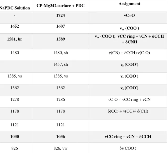
10 appears on spectra of wet Mg sample; bands in range of 1580 – 1610 cm-1 attributed to asymmetric stretching modes of COO- groups [66,67] are shifted. Nevertheless, position of symmetric modes of COO- (stretching at 1362 and 1385, bending at 826 cm-1) remains the same. Bending mode of aromatic system (1030 cm-1) is shifted towards higher wavenumbers compared to solution spectrum.
No shifts if compared to the Fum solution were noticed in Raman spectra recorded on both Mg samples in presence of Fum (Fig. 6a, only CP-Mg342 is shown). IR spectra of both Mg samples demonstrate insignificant shifts compare to spectrum of Fum solution (see Table 6 and Fig. 6b, [69,70]). Additional shoulder at 1395 cm-1, which is absent in spectrum of Fum solution and assigned as symmetric stretching vibration of COO- groups appears on spectrum of Mg samples. Additionally, a slight broadening of the band at 1569 cm-1 was observed. These two shoulders might be attributed to symmetric and asymmetric stretches of the carboxylate bonds and indicate chemisorption of Fum.
3.4. Elemental depth distribution of corroded surfaces from GD-OES
Fig. 7 illustrates the elemental depth profiles of C, O, Mg and Cl, obtained for the
samples of CP-Mg342 corroded in absence and in presence of the inhibitors (depth profiles of HP-Mg51 had the same tendency, so only Mg-342 profiles are shown).
Generally, the interface between the metal and the oxide/hydroxide layer is defined by the inflection (decrease) of the oxygen and inflection (increase) of magnesium signal between 25 and 75 % of the maximal intensity. For the reference sample exposed in 0.1 NaCl solution without inhibitors, we had to reduce the exposure time in order to avoid the formation of too porous and rough surface, which is not suitable for GD-OES; therefore, the oxide/hydroxide layer seems thinner than for the other samples. The thickness of the oxide layer formed during 2 hours of the exposure in the solutions containing inhibitors correlates well with the kinetics data obtained by in situ Raman spectroscopy: the oxide layer for Fum is the thinnest among the studied samples.
It should be noted, that the intensity of any element does not provide the information about its mass without a calibration procedure. However, for our purposes it is sufficient to compare the profiles of the similar samples and estimate the preferential presence of a particular element qualitatively. It can be seen from the figure, that the intensity of Mg as the principal element is similar within the oxide layer (and in the bulk) for all the samples, making possible to assume similar emission efficiency during erosion of different samples for other elements and to compare the relative intensity of all elements between the samples.
It is clear from Fig. 7a that without inhibitors the oxide/hydroxide layer is enriched by Cl. Since no Na was detected in the oxide/hydroxide layer (not shown), most probably, the Cl presence in the surface film corresponds to Cl- anions adsorption on positively
11 charged Mg(OH)2 [71]. Depth profiles of the samples exposed in flow cell in presence of
inhibitors demonstrate carbon enrichment in the oxide layer, indicating the presence of the organic molecules in the surface film. Interestingly, the carbon distribution in the oxide layer varies between the samples exposed in the solutions of different inhibitors. For Sal, the carbon level is much higher as compared to other inhibitors. For PDC, carbon signal reaches the maximum within the oxide/hydroxide layer while it seems that the carbon-containing layer separates two different oxidized layers: O and Mg signals are evolving in two steps (Fig. 7c).
4. Discussion
4.1. General features of Mg corrosion in presence of carboxylates
It is commonly accepted that Mg reactivity strongly depends on two factors: barrier properties of the surface film and distribution of more noble impurities in Mg matrix [72]. Considering the surface film itself, its hydrophobic properties, crystal morphology and crystal size can play an important role in barrier protection. Access of water and diffusion of aggressive species to the metal substrate through the layer consisting of the large crystals with low surface area is easier than in case of the compact layer. The low fraction of brucite compared to the inner MgO can also indicate the stabilization of the inner oxide [73].
The second factor playing an important role in Mg reactivity is the presence of Fe-rich inclusions in Mg matrix. The detrimental effect of Fe (along with other transition metals like Ni and Cu) has also been repeatedly reported by multiple authors starting from as early as 1942 [12,50,74,75]. The role of more noble than Mg inclusions is usually associated with the formation of microgalvanic cells between the inclusion and Mg matrix, where the inclusion acts as a local cathode, accelerating H2 evolution and material
degradation. Influence of Fe content is obvious from difference in the corrosion rate for HP-Mg51 and CP-Mg220 containing 51 and 220 ppm of Fe respectively reported previously [12] and evidenced in this work by the surface appearance of corroded samples and H2 evolution for HP-Mg51 and CP-Mg342 containing 51 and 342 ppm Fe (Fig. 1),
showing that after 24 hours of immersion, the corrosion rate of CP-Mg342 is twenty times higher than that of HP-Mg51 (Fig. 1g).
Thus, further discussion of inhibition mechanisms of three investigated carboxylates is based on explanation of how the inhibiting species influence both of these factors: surface film growth and interaction with Fe-riched inclusions.
Firstly, the common effect of all studied carboxylates is the formation of more compact layer of Mg(OH)2 (as compared with the reference NaCl solution), which limits
12 of the organic molecules with high electron density. Formation of the surface film with more compact morphology was evidenced by SEM images (Fig. 2). In situ Raman spectra in range of -OH vibrations (Fig. 3a, satellite peaks at 3675 and 3710 cm-1, attributed to the surface –OH groups) also supported the hypothesis of formation of smaller crystals with well-developed surface in presence of organic molecules. GD-OES profiles showed much lower content of Cl in the oxidized layer (Fig. 7 b-d) and higher content of C, supporting the competitive adsorption of organic molecules on the surface film. However, the reason of such an effect, most probably, is different for various inhibitors, according to other results (shifts of peaks position in Raman and IR spectra, Mg(OH)2 growth kinetics,
carbon depth distribution in GD-OES profiles). Therefore, each carboxylate is discussed separately.
4.2. Dual action of sodium salicylate (Sal) on Mg corrosion
First of all, the accumulation of Sal molecules on corroded surface of CP-Mg342 sample (C signal in GD-OES depth profile, Fig. 7), was significantly higher than for others inhibitors. According to Raman and IR spectra of CP-Mg342 sample exposed in Sal solution (Fig. 4), this accumulation can be assigned to a chemical adsorption of Sal. Significant shifts in bands position as compared with Sal solution spectra, suggested the modification of Sal molecule geometry caused by strong interaction between Sal and the surface. According to the shifts of COO- (in Raman spectra: 1605, 1575, 1393, 821 cm-1) and -OH (1252 cm-1) bands, both of these groups participate in the interaction [38]. Comparison with the reference samples demonstrated that the most of the shifts can be explained by the interaction with Mg(OH)2. Indeed, Sal is known as an agent stabilizing
the nano-particles of metal oxides or hydroxides due to the adsorption on their surface [39]. The fact that Sal inhibits corrosion of CP-Mg342 and accelerates HP-Mg51 is also coherent with Sal stabilization on Mg(OH)2 layer. Initial reactivity of CP-Mg342 is higher
as compared to HP-Mg51 due to the presence of significant amount of Fe inclusions [12], therefore Mg(OH)2 precipitates very fast, as confirmed by in situ growth kinetics. Hence,
Sal is able to adsorb on the freshly formed hydroxide. In contrast, HP-Mg51 corrodes slowly, so Sal interacts with metallic Mg (or the surface oxide which is every time present on Mg), promoting its fast dissolution, which is similar to the effect of Sal reported for metallic iron and oxidized iron surface [22,35–40].
Additional shifts, different from those observed on Mg(OH)2 precipitated in the
presence of Sal, were however observed in vibrational spectra of CP-Mg342 sample but not for HP Mg51 sample exposed in Sal containing solutions; namely the peak at 1605 cm
-1
, broad peak at 1317 cm-1 for Raman spectra and broad band at 1531cm-1 in IR spectra. The apparent bands position shift in vibrational spectra can be explained by assuming two distinct species with fixed absorption frequencies, which vary only their population and consequently their relative intensities, which can be caused by the effect of temperature or
13 concentration [76,77]. In our case, all the measurements were done on the same instrument and at the same temperature. The effect of concentration can eventually take place, because the surface area (area of adsorption sites) of precipitated Mg(OH)2 might be not
the same as on corroded Mg sample. However, the difference between the spectra of CP-Mg 342 sample and HP CP-Mg51 sample in the region relevant for the organic groups, leads us to an alternative explanation of the shifts in vibrational spectra. We suggest that the shifts are related to the presence of soluble Fe-Sal complex in the surface layer, given the coincidence of these shifts with the most intensive characteristic peaks of Fe(III)-Sal soluble complex for both, IR and Raman spectra (Fig. 4). This suggests that besides the stabilization of Mg(OH)2 nano-crystals, Sal inhibition efficiency can be related to the
binding of Fe ions coming from dissolution of detached from Mg substrate Fe-rich particles. This prevents iron re-deposition and inhibits cathodic reaction. This mechanism is in agreement with the data reported in the literature [12,49,51] suggesting the cathodic type of inhibition by Sal, as well as with the absence of the inhibiting effect on HP-Mg51 sample, poor in active iron-rich intermetallic particles. Apart from complexing Fe3+ (Log KFe(III) = 36.8), Sal forms stable complexes with Mg
2+
(Log KMg(II) = 4.7 [78]). Given this, the
positive effect of iron complexing, outweighing in case of CP-Mg342, is offset by accelerated Mg dissolution in case of Fe-poor HP-Mg51.
Kinetics of Mg(OH)2 growth on CP-Mg342 in presence of Sal is described by a two
steps trend (Fig. 2b): the first 40 minutes it evolves like in solution without inhibitor (step 1), and then the growth rate of Mg(OH)2 slows down (step 2). H2 evolution (Fig.1g,
CP-Mg342) demonstrates a similar two-steps tendency as Mg(OH)2 growth, even if the time
of the transition is not exactly the same as in the in situ Raman experiments. Such a big difference in the transition time can be explained by the difference in the experimental set up, impacting the ratio between the active surface and the solution volume. Step 1 (40 min in case of Raman experiments and around 6 h in case of H2 evolution measurements)
corresponds probably to the time needed to dissolve the Fe-containing particles and stabilize the formation of Mg(OH)2 particles with well-developed surface. Thus, the
inhibition effect (Step 2) of Sal starts only after this “induction period”.
To sum up, it is likely that the inhibition efficiency of Sal can be explained by both Fe-complexing and stabilization of Mg(OH)2 nano-crystals due to the strong chemical
interaction with the surface. The proposed mechanism is schematically presented in Fig. 8.
4.3. Coordination polymer film formation by 2,5-pyridinedicarboxylate (PDC)
For PDC, GD-OES profile differs significantly from those of other inhibitors (Fig. 7c). During the sputtering of the oxide/hydroxide layer, intensities of all elements evolve in two steps. This fact could be explained by formation of two distinct layers with different sputtering rate. Probably, it corresponds to the inner layer of MgO/Mg(OH)2 and
the outer layer of MgPDC. Carbon intensity reaches the maximum at the interface between these two layers, suggesting the enhanced accumulation of PDC molecules there.
14 Secondly, the high fluorescence background appears in Raman spectra of CP-Mg342 after an “induction period” of 40 min, as it was mentioned in section 3.2. PDC molecule itself does not provide fluorescence background, as evidenced by Raman spectrum of PDC solution (Fig. 5a). However, formation of a specific network of the organic molecules which contains a chain of alternating double and single bonds, called conjugation, or π-electron delocalization can cause fluorescence [79,80]. It was reported that PDC can form so-called coordination polymer with different dimensionality, consisting of the long chains of PDC molecules bonded due to the electrostatic interaction with metal ions [80–82]. Many of reported coordination polymers have fluorescent properties [46,80,83].
Analysis of IR spectra of CP-Mg342 (Fig. 5b) demonstrated that bands of carboxylates (COO-) were shifted as compared to the solution spectra (1607 - νas, 1457 - νs
[68]). Band at 1724 cm-1 corresponding to C=O stretching of COOH, is absent in the PDC solution spectrum and appears in the spectrum of Mg sample, suggesting that the delocalization of electron density of carboxylic group disappears, instead the -(C=O)-O- is taking place; which can be schematically illustrated by a mechanism shown in Fig. 9.
ν(C=C) and ν(C=N) vibrations in PDC solution at 1581 and 1030 cm-1 are shifted to higher wavenumbers on Mg sample spectra (1589 and 1036 cm-1). Therefore, most likely the coordination occurs through both, COO- group and pyridine N [83] The proposed structure of PDC complex is presented in Fig. 9.
As it was described in Section 2.1, PDC was prepared from PDCA by the addition of NaOH and pH adjustment to ≈ 5. It means that one of the COOH group can be still protonated at the very beginning of experiment (pKa2 = 4.64, which is close to
experimental value of pH). Thus, the “induction period” of 40 min in kinetics curve of Mg(OH)2 growth can be related either to the period which is necessary for increasing pH
[80] and formation of the polymer film or to the time necessary to accumulate sufficient amount of Mg2+ to precipitate coordination polymer.
To summarize, precipitation of coordination polymer consisted of long chains of PDC molecules bonded due to the electrostatic interaction with metal cations is the possible explanation of Mg corrosion inhibition by PDC, as schematically illustrated in
Fig. 9. If such a film is formed, the inhibitor affects both, cathodic and anodic reactions,
and hence acts as a mixed type inhibitor. Similarly, conclusion about the mixed inhibiting behavior (with more pronounced anodic inhibition) of related 2,5-PDC was drawn in our previous paper [51].
4.4. Adsorption of Fumarate (Fum) on corroding Mg
Despite the well-developed Mg(OH)2 surface, accumulation of Fum on corroding
15 CP-Mg342 (Fig. 7), the intensity of C within the oxide layer is lower than in the oxide layers formed in the presence of other inhibitors but it is still higher than for the bulk indicating the presence of organic molecules.
In situ Raman spectra in the range of 700 – 1700 cm-1 (Fig. 6a) do not allow to detect any significant shifts as compared with Fum solution, indicating that the interaction is weak and does not modify significantly the geometry of the adsorbed molecule [84]. In contrast, the ex situ IR spectrum of Fum on CP-Mg342 (Fig . 6b) demonstrates a shoulder at 1395 cm-1 in addition to the main band at 1372 cm-1. This shoulder could be assigned to a carboxylate group chemically interacting with the surface, as was observed for fumarate adsorbed on TiO2 [85], acetate on TiO2 [85], benzoate on quartz [86], laurate on alumina
[86], oxalate and malonate on metal (oxy)hydroxides [87]. The separation of the adsorption bands corresponding to the carboxylate antisymmetric and symmetric stretching can be used to speculate on the mode of coordination [76]. For the shoulder at 1395 cm-1 the separation is around 174 cm-1, which could be interpreted as a bridging bidentate surface complex (both oxygen atoms of one carboxylate group participate in bonding, as schematically illustrated in Fig. 10). However, our results are not sufficient to conclude if both COO- groups of Fum or only one of them participate in adsorption. Additional study is also necessary to elucidate the orientation of the adsorbed carboxylate groups of fumarate.
Conclusions
Surface analysis techniques such as in situ Raman spectroscopy in kinetic mapping mode, ATR-FTIR and GD-OES were applied to study the effect of selected carboxylates (Sal, Fum and PDC) on the surface film formation and evolution during corrosion of pure Mg in aqueous solutions containing Cl-. Namely, the growth kinetics of the surface film, its chemical composition, depth elemental distribution and morphology were studied.
Raman spectra in the range of -OH vibrations as well as SEM images demonstrated that addition of all of these inhibitors results in the decrease of the size of Mg(OH)2
crystals in the corrosion products layer. GD-OES elemental depth profiles demonstrated that Cl-accumulation and diffusion through the oxidized layer were efficiently prevented.
Three different film formation mechanisms, relevant for inhibition, were proposed: adsorption of Fum on the metallic Mg surface or MgO, stabilization of Mg(OH)2 particles
due to the chemical adsorption of Sal and precipitation of coordination polymer at the interface between more and less hydrated oxide layers in case of PDC. Characteristic peaks of soluble Fe(III)Sal complex have been found in the spectra of CP-Mg342 sample corroded in presence of Sal, thus Fe-complexing mechanism more likely takes place in
16 case of Sal inhibition. This mechanism is also consistent with the fact that Sal only inhibits corrosion of CP-Mg342 with high Fe content and was previously described as cathodic inhibitor.
Acknowledgements
KY thanks Researcher grant (IF/01284/2015) and Project CICECO-Aveiro Institute of Materials, FCT Ref. UID/CTM/50011/2019, financed by national funds through the FCT/MCTES and co-financed by FEDER under the PT2020 Partnership Agreement.
References:
[1] D. Schloffer, S. Bozorgi, P. Sherstnev, C. Lenardt, B. Gollas, Manufacturing and characterization
of magnesium alloy foils for use as anode materials in rechargeable magnesium ion batteries, J. Power Sources. 367 (2017) 138–144. doi:10.1016/j.jpowsour.2017.09.062.
[2] H. Hornberger, S. Virtanen, A.R. Boccaccini, Biomedical coatings on magnesium alloys - A
review, Acta Biomater. 8 (2012) 2442–2455. doi:10.1016/j.actbio.2012.04.012.
[3] Y. Zhang, J. Xu, Y.C. Ruan, M.K. Yu, M. O’Laughlin, H. Wise, D. Chen, L. Tian, D. Shi, J.
Wang, S. Chen, J.Q. Feng, D.H.K. Chow, X. Xie, L. Zheng, L. Huang, S. Huang, K. Leung, N. Lu, L. Zhao, H. Li, D. Zhao, X. Guo, K. Chan, F. Witte, H.C. Chan, Y. Zheng, L. Qin, Implant-derived magnesium induces local neuronal production of CGRP to improve bone-fracture healing in rats, Nat. Med. 22 (2016) 1160–1169. doi:10.1038/nm.4162.
[4] J. Gonzalez, R.Q. Hou, E.P.S. Nidadavolu, R. Willumeit-Römer, F. Feyerabend, Magnesium
degradation under physiological conditions – Best practice, Bioact. Mater. 3 (2018) 174–185. doi:10.1016/j.bioactmat.2018.01.003.
[5] M.A. Deyab, Decyl glucoside as a corrosion inhibitor for magnesium-air battery, J. Power Sources.
325 (2016) 98–103. doi:10.1016/j.jpowsour.2016.06.006.
[6] F.W. Richey, B.D. McCloskey, A.C. Luntz, Mg Anode Corrosion in Aqueous Electrolytes and
Implications for Mg-Air Batteries, J. Electrochem. Soc. 163 (2016) A958–A963. doi:10.1149/2.0781606jes.
[7] M. Deng, D. Höche, S. V. Lamaka, D. Snihirova, M.L. Zheludkevich, Mg-Ca binary alloys as
anodes for primary Mg-air batteries, J. Power Sources. 396 (2018) 109–118.
doi:10.1016/j.jpowsour.2018.05.090.
[8] D. Höche, S. V. Lamaka, B. Vaghefinazari, T. Braun, R.P. Petrauskas, M. Fichtner, M.L.
Zheludkevich, Performance boost for primary magnesium cells using iron complexing agents as electrolyte additives, Sci. Rep. 8 (2018) 1–9. doi:10.1038/s41598-018-25789-8.
[9] M. Yuasa, X. Huang, K. Suzuki, M. Mabuchi, Y. Chino, Discharge properties of Mg-Al-Mn-Ca
and Mg-Al-Mn alloys as anode materials for primary magnesium-air batteries, J. Power Sources. 297 (2015) 449–456. doi:10.1016/j.jpowsour.2015.08.042.
[10] S. V. Lamaka, J. Gonzalez, D. Mei, F. Feyerabend, R. Willumeit-Römer, M.L. Zheludkevich, Local pH and its Evolution near Mg Alloy Surfaces Exposed to Simulated Body Fluids, Adv. Mater. Interfaces. 1800169 (2018) 1–6. doi:10.1002/admi.201800169.
[11] X.-P. Guo, G.-L. Song, J.-Y. Hu, D.-B. Huang, Corrosion inhibition of magnesium (Mg) Alloys, in: Corros. Prev. Magnes. Alloy., Elsevier, 2013: pp. 61–84. doi:10.1533/9780857098962.1.61. [12] S. V. Lamaka, B. Vaghefinazari, D. Mei, R.P. Petrauskas, D. Höche, M.L. Zheludkevich,
Comprehensive screening of Mg corrosion inhibitors, Corros. Sci. 128 (2017) 224–240. doi:10.1016/j.corsci.2017.07.011.
17
[13] G. Williams, R. Grace, R.M. Woods, Inhibition of the localized corrosion of Mg alloy AZ31 in chloride containing electrolyte, Corrosion. 71 (2015) 184–198. doi:10.5006/1376.
[14] N. Dang, Y.H. Wei, L.F. Hou, Y.G. Li, C.L. Guo, Investigation of the inhibition effect of the environmentally friendly inhibitor sodium alginate on magnesium alloy in sodium chloride solution, Mater. Corros. 66 (2015) 1354–1362. doi:10.1002/maco.201408141.
[15] J. Twu, P.K. Dutta, Raman-Spectroscopic Studies of Intercalated Molybdate Ions in Layered Metal-Hydroxides, Chem. Mater. 4 (1992) 398–401. doi:10.1021/cm00020a031.
[16] N. Birbilis, G. Williams, K. Gusieva, A. Samaniego, M.A. Gibson, H.N. McMurray, Poisoning the
corrosion of magnesium, Electrochem. Commun. 34 (2013) 295–298.
doi:10.1016/j.elecom.2013.07.021.
[17] R.L. Liu, M.F. Hurley, A. Kvryan, G. Williams, J.R. Scully, N. Birbilis, Controlling the corrosion and cathodic activation of magnesium via microalloying additions of Ge, Sci. Rep. 6 (2016) 1–12. doi:10.1038/srep28747.
[18] D. Eaves, G. Williams, H.N. McMurray, Inhibition of self-corrosion in magnesium by poisoning
hydrogen recombination on iron impurities, Electrochim. Acta. 79 (2012) 1–7.
doi:10.1016/j.electacta.2012.05.148.
[19] Z. Feng, B. Hurley, M. Zhu, Z. Yang, J. Hwang, R. Buchheit, Corrosion Inhibition of AZ31 Mg
Alloy by Aqueous Selenite (SeO32-), J. Electrochem. Soc. 166 (2019) C520–C529.
doi:10.1149/2.0911914jes.
[20] G. Boisier, A. Lamure, N. Pébère, N. Portail, M. Villatte, Corrosion protection of AA2024 sealed anodic layers using the hydrophobic properties of carboxylic acids, Surf. Coatings Technol. 203 (2009) 3420–3426. doi:10.1016/j.surfcoat.2009.05.008.
[21] K. Xhanari, M. Finšgar, M. Knez Hrnčič, U. Maver, Ž. Knez, B. Seiti, Green corrosion inhibitors
for aluminium and its alloys: A review, RSC Adv. 7 (2017) 27299–27330.
doi:10.1039/c7ra03944a.
[22] Y.I. Kuznetsov, Organic corrosion inhibitors: Where are we now? A review. part III. Passivation and the role of the chemical structure of organophosphates, Int. J. Corros. Scale Inhib. 6 (2017) 209–239. doi:10.17675/2305-6894-2017-6-3-1.
[23] M. Ormellese, L. Lazzari, S. Goidanich, G. Fumagalli, A. Brenna, A study of organic substances as inhibitors for chloride-induced corrosion in concrete, Corros. Sci. 51 (2009) 2959–2968. doi:10.1016/j.corsci.2009.08.018.
[24] B. Lin, Y. Zuo, Corrosion inhibition of carboxylate inhibitors with different alkylene chain lengths on carbon steel in an alkaline solution, RSC Adv. 9 (2019) 7065–7077. doi:10.1039/C8RA10083G. [25] J. Wysocka, M. Cieslik, S. Krakowiak, J. Ryl, Carboxylic acids as efficient corrosion inhibitors of
aluminium alloys in alkaline media, Electrochim. Acta. 289 (2018) 175–192.
doi:10.1016/j.electacta.2018.08.070.
[26] G. Žerjav, I. Milošev, Carboxylic acids as corrosion inhibitors for Cu, Zn and brasses in simulated urban rain, Int. J. Electrochem. Sci. 9 (2014) 2696–2715.
[27] N. Dinodi, A.N. Shetty, Alkyl carboxylates as efficient and green inhibitors of magnesium alloy
ze41 corrosion in aqueous salt solution, Corros. Sci. 85 (2014) 411–427.
doi:10.1016/j.corsci.2014.04.052.
[28] D. Daloz, C. Rapin, P. Steinmetz, G. Michot, Corrosion Inhibition of Rapidly Solidified Mg-3% Zn-15% Al Magnesium Alloy with Sodium Carboxylates, Corrosion. 54 (1998) 444–450. doi:10.5006/1.3284872.
[29] I. V. Chernyshova, S. Ponnurangam, P. Somasundaran, Adsorption of fatty acids on iron (hydr)oxides from aqueous solutions, Langmuir. 27 (2011) 10007–10018. doi:10.1021/la2017374. [30] U. Rammelt, S. Koehler, G. Reinhard, Electrochemical characterisation of the ability of
dicarboxylic acid salts to the corrosion inhibition of mild steel in aqueous solutions, Corros. Sci. 53 (2011) 3515–3520. doi:10.1016/j.corsci.2011.06.023.
[31] F. Wormwell, A.D. Mercer, Sodium benzoate and other metal benzoates as corrosion-inhibitors in water and in aqueous solutions, J. Appl. Chem. 2 (2007) 150–160. doi:10.1002/jctb.5010020307. [32] L. Hamadi, S. Mansouri, K. Oulmi, A. Kareche, The use of amino acids as corrosion inhibitors for
18
[33] E.A. Batista, M.L.A. Temperini, An in situ SERS and FTIRAS study of salicylate interaction with copper electrode, J. Solid State Electrochem. 11 (2007) 1559–1565. doi:10.1007/s10008-007-0357-6.
[34] M.A. Blesa, A.D. Weisz, P.J. Morando, J.A. Salfity, G.E. Magaz, A.E. Regazzoni, The interaction of metal oxide surfaces with complexing agents dissolved in water, Coord. Chem. Rev. 196 (2000) 31–63. doi:10.1016/S0010-8545(99)00005-3.
[35] D. Eurof Davies, Q.J.M. Slaiman, Throughout the present investigation pure Fe supplied by the British Iron and, Corros. Sci. 11 (1971) 671–682.
[36] D. Eurof Davies, Q.J.M. Slaiman, Mechanism of the corrosion inhibition of iron by sodium benzoate-III. The role of oxygen, Corros. Sci. 13 (1973) 891–905. doi:10.1016/S0010-938X(73)80071-X.
[37] B.M. Vinoda, J. Manjanna, Dissolution of iron in salicylic acid and cation exchange between Fe(II)-salicylate and Na-montmorillonite to form Fe(II)-montmorillonite, Appl. Clay Sci. 97–98 (2014) 78–83. doi:10.1016/j.clay.2014.05.005.
[38] M. V. Blber, W. Stumm, An In-Situ ATR-FTIR Study: The Surface Coordination of Salicylic Acid on Aluminum and Iron(III) Oxides, Environ. Sci. Technol. 28 (1994) 763–768. doi:10.1021/es00054a004.
[39] I.L. Ardelean, L.B.N. Stoencea, D. Ficai, A. Ficai, R. Trusca, B.S. Vasile, G. Nechifor, E. Andronescu, Development of Stabilized Magnetite Nanoparticles for Medical Applications, J. Nanomater. 2017 (2017). doi:10.1155/2017/6514659.
[40] E.C. Yost, M.I. Tejedortejedor, M.A. Anderson, Insitu CIR-FTIR characterization of salicylate complexes at the goethite/aqueous solution interface, Environ. Sci. Technol. 24 (1990) 822–828. doi:10.1021/es00076a005.
[41] A. Alamdari, M.R. Rahimpour, N. Esfandiari, E. Nourafkan, Kinetics of magnesium hydroxide precipitation from sea bittern, Chem. Eng. Process. Process Intensif. 47 (2008) 215–221. doi:10.1016/j.cep.2007.02.012.
[42] J.A. Dirksen, T.A. Ring, Fundamentals of Crystallization: Kinetic Effects on Particle Size Distributions and Morphology, Chem. Eng. Sci. 46 (1991) 2389–2427. doi:10.1016/0009-2509(91)80035-W.
[43] T. Behrsing, G.B. Deacon, P.C. Junk, The chemistry of rare earth metals, compounds, and corrosion inhibitors, in: Rare Earth-Based Corros. Inhib., Woodhead Publishing Series in Metals and Surface Engineering, 2014: pp. 1–37. doi:10.1533/9780857093585.1.
[44] M. Forsyth, C.M. Forsyth, K. Wilson, T. Behrsing, G.B. Deacon, ATR characterisation of synergistic corrosion inhibition of mild steel surfaces by cerium salicylate, Corros. Sci. 44 (2002) 2651–2656. doi:10.1016/S0010-938X(02)00024-0.
[45] M. Forsyth, B. Hinton, Rare Earth-Based Corrosion Inhibitors, Woodhead Publishing, 2015. doi:10.1016/C2013-0-16231-9.
[46] R.K. Vakiti, B.D. Garabato, N.P. Schieber, M.J. Rucks, Y. Cao, C. Webb, J.B. Maddox, A. Celestian, W. Pan, B. Yan, Synthesis and Characterization of Two- and Three-Dimensional Calcium Coordination Polymers Built with Benzene-1,3,5-tricarboxylate and/or Pyrazine-2-carboxylate, Cryst. Growth Des. 12 (2012) 3937–3943. doi:10.1021/cg3003349.
[47] N. Singh, P. Vishnoi, G. Anantharaman, Coordination polymers based on copper carboxylates and angular 2,5-bis(imidazol-1-yl)thiophene (thim2) ligand: Sequential structural transformations, CrystEngComm. 17 (2015) 2153–2161. doi:10.1039/c4ce02428a.
[48] H. Wu, H.Y. Liu, J. Yang, B. Liu, J.F. Ma, Y.Y. Liu, Y.Y. Liu, Series of coordination polymers based on different carboxylates and a tri(4-imidazolylphenyl)amine ligand: Entangled structures and photoluminescence, Cryst. Growth Des. 11 (2011) 2317–2324. doi:10.1021/cg200005q.
[49] S. V. Lamaka, D. Höche, R.P. Petrauskas, C. Blawert, M.L. Zheludkevich, A new concept for corrosion inhibition of magnesium : Suppression of iron re-deposition, Electrochem. Commun. (2015). doi:10.1016/j.elecom.2015.10.023.
[50] D. Höche, C. Blawert, S. V. Lamaka, N. Scharnagl, C. Mendis, M.L. Zheludkevich, The effect of iron re-deposition on the corrosion of impurity-containing magnesium, Phys.Chem.Chem.Phys. 18 (2015) 1279–1291. doi:10.1039/c5cp05577f.
19
[51] J. Yang, C. Blawert, S. V. Lamaka, K.A. Yasakau, L. Wang, D. Laipple, M. Schieda, S. Di, M.L. Zheludkevich, Corrosion inhibition of pure Mg containing a high level of iron impurity in pH neutral NaCl solution, Corros. Sci. 142 (2018) 222–237. doi:10.1016/j.corsci.2018.07.027.
[52] A.E. Martell, R.M. Smith, Critical Stavility Constants, Other organic ligands, V.3, Springer, New York, 1977. doi:10.1007/978-1-4757-1568-2.
[53] A.E. Martell, R.M. Smith, Critical stability constants, Aminoacids,V. 1, Springer, New York, 1974.
[54] A.E. Martell, R.M. Smith, Critical Stability Constants: Second Supplement, Springer Science & Bisness Media, 1989. doi:10.1007/978-1-4615-6761-5.
[55] D. Mei, S. V. Lamaka, C. Feiler, M.L. Zheludkevich, The effect of small-molecule bio-relevant organic components at low concentration on the corrosion of commercially pure Mg and Mg-0.8Ca alloy: An overall perspective, Corros. Sci. 153 (2019) 258–271. doi:10.1016/j.corsci.2019.03.039.
[56] A. Maltseva, V. Shkirskiy, G. Lefèvre, P. Volovitch, Effect of pH on Mg(OH)2 film evolution on
corroding Mg by in-situ Kinetic Raman Mapping (KRM), Corros. Sci. 153 (2019) 272–282. doi:10.1016/j.corsci.2019.03.024.
[57] G. Balducci, L. Bravo Diaz, D.H. Gregory, Recent progress in the synthesis of nanostructured magnesium hydroxide, CrystEngComm. 19 (2017) 6067–6084. doi:10.1039/c7ce01570d.
[58] G. Zou, R. Liu, W. Chen, Z. Xu, Preparation and characterization of lamellar-like Mg(OH)2
nanostructures via natural oxidation of Mg metal in formamide/water mixture, Mater. Res. Bull. 42 (2007) 1153–1158. doi:10.1016/j.materresbull.2006.09.008.
[59] B. Downs, S. Robinson, H. Yang, P. Mooney, RRUFF Project, Dep. Geosci. Univ. Arizona. (2015). http://rruff.info/.
[60] B. Humbert, M. Alnot, F. Quilès, Infrared and Raman spectroscopical studies of salicylic and salicylate derivatives in aqueous solution, Spectrochim. Acta - Part A Mol. Biomol. Spectrosc. 54 (1998) 465–476. doi:10.1016/S1386-1425(97)00239-4.
[61] M.C. Alvarez-Ros, S. Sánchez-Cortés, J. V. García-Ramos, Vibrational study of the salicylate interaction with metallic ions and surfaces, Spectrochim. Acta - Part A Mol. Biomol. Spectrosc. 56 (2000) 2471–2477. doi:10.1016/S1386-1425(00)00328-0.
[62] D. Philip, A. John, C.Y. Panicker, H.T. Varghese, FT-Raman, FT-IR and surface enhanced Raman scattering spectra of sodium salicylate, Spectrochim. Acta - Part A Mol. Biomol. Spectrosc. 57 (2001) 1561–1566. doi:10.1016/S1386-1425(01)00395-X.
[63] M.A. Elbagerma, G. Azimi, H.G.M. Edwards, A.I. Alajtal, I.J. Scowen, In situ monitoring of pH titration by Raman spectroscopy, Spectrochim. Acta - Part A Mol. Biomol. Spectrosc. 75 (2010) 1403–1410. doi:10.1016/j.saa.2010.01.008.
[64] P.J.. Goulet, R.F. Aroca, Chemical adsorption of salicylate on silver - A systematic approach to the interpretation of surface-enhanced vibrational spectra, Can. J. Chem. 82 (2004) 987–997. doi:10.1139/v04-075.
[65] L. Wasylina, E. Kucharska, Z. Weglinski, A. Puszko, Chemistry of Heterocyclic Compounds, Vol.35, No. 2, 1999, Chem. Heterocycl. Compd. 35 (1999) 210–218.
[66] G. Świderski, M. Kalinowska, I. Rusinek, M. Samsonowicz, Z. Rzączyńska, W. Lewandowski, Spectroscopic (IR, Raman) and thermogravimetric studies of 3d-metal cinchomeronates and dinicotinates, J. Therm. Anal. Calorim. 126 (2016) 1521–1532. doi:10.1007/s10973-016-5818-7. [67] M. Karabacak, S. Bilgili, A. Atac, Molecular structure investigation of neutral, dimer and anion
forms of 3,4-pyridinedicarboxylic acid: A combined experimental and theoretical study,
Spectrochim. Acta - Part A Mol. Biomol. Spectrosc. 135 (2015) 270–282.
doi:10.1016/j.saa.2014.06.130.
[68] Y. Song, X. Wang, S. Zhang, J. Wang, S. Gao, S. Chen, Lanthanide-Coordination Polymers with Pyridinedicarboxylic Acids: Syntheses, Structures, and Luminescent Properties, Zeitschrift Für Anorg. Und Allg. Chemie. 642 (2016) 681–691. doi:10.1002/zaac.201600135.
[69] G. Lefèvre, T. Preočanin, J. Lützenkirchen, Attenuated total reflection - Infrared spectroscopy applied to the study of mineral - aqueous electrolyte solution interfaces: a general overview and a case study, in: T. Theophanides (Ed.), Infrared Spectrosc., Intech, 2012. doi:10.5772/36459.
20
interfaces. 2. Outer-sphere adsorption of maleate and implications for dissolution processes, Langmuir. 20 (2004) 4996–5006. doi:10.1021/la036288y.
[71] M.M. Rakhimova, N.Z. Yusupov, T.M. Nurmatov, J.A. Davlatshoeva, L. Kvyatkovskaya, A.R. Bakhtibekova, M. Bobonazarov, Salicylate Coordination Compounds of Iron, Models of Their Formation, Am. J. Chem. 3 (2013) 23–29. doi:10.5923/j.chemistry.20130302.01.
[72] M. Esmaily, J.E. Svensson, S. Fajardo, N. Birbilis, G.S. Frankel, S. Virtanen, R. Arrabal, S. Thomas, L.G. Johansson, Fundamentals and advances in magnesium alloy corrosion, Prog. Mater. Sci. 89 (2017) 92–193. doi:10.1016/j.pmatsci.2017.04.011.
[73] Y. Yan, A. Maltseva, P. Zhou, X. Li, Z. Zeng, O. Gharbi, K. Ogle, M. Vaudescal, M. Lahaye, M. Esmaily, N. Birbilis, On the in-situ aqueous stability of an Mg-Li-(Al-Y-Zr) alloy: Role of Li, Corros. Sci. In Press (2019). doi:10.1016/j.corsci.2019.108342.
[74] R.E. McNulty, J.D. Hanawalt, Some corrosion characteristics of high purity magnesium alloys, J. Electrochem. Soc. 81 (1942) 423–433. doi:10.1149/1.3071389.
[75] S. Thomas, O. Gharbi, S.H. Salleh, P. Volovitch, K. Ogle, N. Birbilis, On the effect of Fe
concentration on Mg dissolution and activation studied using atomic emission
spectroelectrochemistry and scanning electrochemical microscopy, Electrochim. Acta. (2016). doi:10.1016/j.electacta.2016.05.164.
[76] S.R. Ryu, I. Noda, Y.M. Jung, Positional Fluctuation of IR Absorption Peaks: Frequency Shift of a Single Band or Relative Intensity Changes of Overlapped Bands, Am. Lab. 43 (2011) 40–43. [77] S.R. Ryu, I. Noda, Y.M. Jung, What is the Origin of Positional Fluctuation of Spectral Features :
True Frequency Shift or Relative Intensity Changes of Two Overlapped Bands?, Appl. Spectrosc. 64 (2010) 1017–1021. doi:10.1366/000370210792434396.
[78] J.A. Dean, Lange’s Handbook of Chemistry, 15th ed., McGraw-Hill, New York, 1999.
[79] V. Masilamani, H.M. Ghaithan, M.J. Aljaafreh, A. Ahmed, R. Thagafi, S. Prasad, M.S. Alsalhi, Using a Spectrofluorometer for Resonance Raman Spectra of Organic Molecules, 2017 (2017). doi:10.1155/2017/4289830.
[80] L. Pan, T. Frydel, M.B. Sander, X. Huang, J. Li, The Effect of pH on the Dimensionality of Coordination Polymers, Inorg. Chem. 40 (2001) 1271–1283. doi:10.1021/ic001012o.
[81] M.C. Das, S.K. Ghosh, E.C. Sañudo, P.K. Bharadwaj, Coordination polymers with pyridine-2,4,6-tricarboxylic acid and alkaline-earth/lanthanide/transition metals: Synthesis and X-ray structures, J. Chem. Soc. Dalt. Trans. (2009) 1644–1658. doi:10.1039/b814066a.
[82] B. Ay, N. Dogan, E. Yildiz, I. Kani, A novel three dimensional samarium(III) coordination polymer with an unprecedented coordination mode of the 2,5-pyridinedicarboxylic acid ligand: Hydrothermal synthesis, crystal structure and luminescence property, Polyhedron. 88 (2015) 176– 181. doi:10.1016/j.poly.2014.12.035.
[83] S. Caglar, E. Dilek, S.H. Alisir, B. Caglar, New copper(II) complexes including pyridine-2,5-dicarboxylic acid: synthesis, spectroscopic, thermal properties, crystal structure and how these complexes interact with purified PON 1 enzyme, J. Coord. Chem. 69 (2016) 2482–2492. doi:10.1080/00958972.2016.1188295.
[84] R.P. Eischens, W.A. Pliskin, The Infrared Spectra of Adsorbed Molecules, Adv. Catal. 10 (1958) 1–56. doi:10.1016/S0360-0564(08)60403-4.
[85] F.P. Rotzinger, J.M. Kesselman-Truttmann, S.J. Hug, V. Shklover, M. Grätzel, Structure and
Vibrational Spectrum of Formate and Acetate Adsorbed from Aqueous Solution onto the TiO 2
Rutile (110) Surface, J. Phys. Chem. B. 108 (2004) 5004–5017. doi:10.1021/jp0360974.
[86] J.D. Kubicki, L.M. Schroeter, M.J. Itoh, B.N. Nguyen, S.E. Apitz, Attenuated total reflectance Fourier-transform infrared spectroscopy of carboxylic acids adsorbed onto mineral surfaces, Geochim. Cosmochim. Acta. 63 (1999) 2709–2725. doi:10.1016/S0016-7037(99)00194-5.
[87] K.D. Dobson, A.J. McQuillan, In situ infrared spectroscopic analysis of the adsorption of aromatic carboxylic acids to TiO2, ZrO2, Al2O3, and Ta2O5 from aqueous solutions, Spectrochim. Acta - Part
21
Table 1. Chemical composition of pure Mg and alloys as analyzed by spark optical emission
spectroscopy. The values are in ppm or in at. % when indicated.
Mg (at.%) Ag Al Ca Ce Cu Fe La Mn Ni Pb Si Sn Zn Zr CP-Mg342 99.96 <0.5 40 <1 7 4 342 <5 24 <2 <4 <1 <3 5 <5 HP-Mg 51 99.98 0.7 50 <1 <4 <1 51 <5 8 <2 <4 <1 <3 10 23
22
Table 2. Organic compounds studied in present work and respective inhibiting efficiency (IE) for
CP-Mg342 and HP-Mg51. The tests were performed with Na salts at the initial pH of the electrolyte varying in the range from 5.3 to 6.7
Name Abbreviation Structural formula of neutralized acid pKa of respective acid IE, % (CP-Mg342) IE, % (HP-Mg51) Sodium Salicylate Sal 2.8* 84 ± 1 -244 ± 29 Sodium Fumarate Fum 2.85* 4.10* 87 ± 1 58 ± 6 Sodium 2,5 – Pyridine-dicarboxylate PDC 2.35** 4.64** 89 ± 1 74±5
the value given was measured at * ionic strength of 0.1, and 25oC ** ionic strength of 0.5, and 20oC
23
Table 3. Synthetic reference samples used for the interpretation of Raman spectra.
Chemical composition
Preparation procedure
Mg(II)-Sal soluble complex 25 ml 0.1 M MgCl2 (0.508 g MgCl2·6H2O) + 0.05 M
NaSal (0.200 g of salt)
Mg(OH)2 25 ml 0.1 M MgCl2 (0.508 g MgCl2·6H2O) + 25 ml
0.1 M NaOH (1.000 g NaOH). White precipitate was
centrifuged and filtered
Mg(OH)2 in presence of Sal 25 ml 0.1 M MgCl2 (0.508 g MgCl2·6H2O) + 0.05 M
NaSal (0.200 g of salt) + 25 ml 0.1 M NaOH (1.000 g
NaOH)
White precipitate was centrifuged and filtered.
Fe(OH)3 25 ml 0.1 M FeCl3 (0.675g FeCl3·6H2O) + 25 ml 0.1
M NaOH (1.000 g NaOH)
Brown precipitate was centrifuged and filtered
Fe(III)-Sal soluble complex 25 ml 0.1 M FeCl3 (0.675g FeCl3·6H2O) + 0.05 M
NaSal (0.200 g)
Fe(OH)3 in presence of Sal 25 ml 0.1 M FeCl3 (0.675g FeCl3·6H2O) + 0.05 M
NaSal (0.200 g of salt) + 25 ml 0.1 M NaOH (1.000 g
NaOH)
Brown precipitate was centrifuged and filtered
Sal adsorbed on carbon steel Carbon steel plate grinded with 800, 1200, 2400 SiC
paper and immersed for 2 h in 25 ml solution of 0.05 M
NaSal (0.200 g of salt, pH adjusted to 10.2by 1M
24
Table 4. IR and Raman spectral data (cm-1) and band assignments for NaSal solution, CP-Mg342 exposed in NaSal solution, precipitated in presence of NaSal Mg(OH)2, soluble complex of Fe(III)Sal.
Sal Solution
CP-Mg342 surface + Sal
Mg(OH)2 + Sal Fe(III)Sal
Assignment IR Raman IR Raman IR Raman IR Raman
1624, 1592 1625, 1595 1605 1605 1603, 1589 1595, br 1602 1601 ν8b + ν(C=O)+δ(Cφ–OH) 1572 1574, br 1567 br 1575, br 1565, br 1575 1574 ν (C-C) ring or νas (COO -) - - 1536, br - 1531, br - 1544 νas (COO -) 1489 1489 1470 1470, sh 1467 1468 C-C ring, (19a) 1457 1460 1449 1450 1447, s 1453, m 1455 1455 C-C ring or δ(OH) 1387 1387 1389, sh 1393, sh 1388 1392 1359 1350, s νs (COO -) 1342 1347, br 1369, br 1375, s 1373, sh 1376, sh νs (COO -)+ δ Cph-O 1300, sh 1306 1309, br 1317, s 1317, s, br 1312, s ν (C-C) ring, 14 1254 1256 1250 1252 1254 1255 1242 1240 ,m ν (Cφ -O) - 1225 - - δCH 1159 1160, vw 1157, vw 1160, vw - ? 1147 1147 1140 1143, m 1146 1144 δCC (10) 1033 1034 1045, s 1044, s 1041 1043, br 1037, vw δCH 861 867, w 877 - 872 887 886 810 814, s 818 821 821 840 ν (Cφ–COO -)+ (Cφ–OH)+δs (COO-)
v- very; s-strong;m- medium, w- weak; sh- shoulder; br- broad. ν indicates a stretching mode, δ an in-plane bending mode (+) in-phase movement; (−) out-of-phase movement; Cφ – carbon of the aromatic ring