HAL Id: hal-03100940
https://hal.archives-ouvertes.fr/hal-03100940
Submitted on 6 Jan 2021
HAL is a multi-disciplinary open access
archive for the deposit and dissemination of
sci-entific research documents, whether they are
pub-lished or not. The documents may come from
teaching and research institutions in France or
abroad, or from public or private research centers.
L’archive ouverte pluridisciplinaire HAL, est
destinée au dépôt et à la diffusion de documents
scientifiques de niveau recherche, publiés ou non,
émanant des établissements d’enseignement et de
recherche français ou étrangers, des laboratoires
publics ou privés.
Assessing the onset of calcium phosphate nucleation by
hyperpolarized real-time NMR
Emmanuelle Weber, Thomas Kress, Daniel Abergel, Steffi Sewsurn, Thierry
Azaïs, Dennis Kurzbach
To cite this version:
Emmanuelle Weber, Thomas Kress, Daniel Abergel, Steffi Sewsurn, Thierry Azaïs, et al.. Assessing the
onset of calcium phosphate nucleation by hyperpolarized real-time NMR. Analytical Chemistry,
Amer-ican Chemical Society, 2020, 92 (11), pp.7666-7673. �10.1021/acs.analchem.0c00516�. �hal-03100940�
1
Assessing the onset of calcium phosphate nucleation by
hyperpolarized real-time NMR
Emmanuelle M. M. Weber
[a], Thomas Kress
[b], Daniel Abergel
[c], Steffi Sewsurn
[d], Thierry Azaïs
[d],*,
Dennis Kurzbach
[b],*[a] Radiological Sciences Laboratory, Department of Radiology, Stan-ford University, Richard M. Lucas Center for Imaging, 201 Welch Rd, Stanford, CA 94305, United States
[b] University Vienna, Faculty of Chemistry, Institute of Biological Chemistry, Währinger Str. 38, 1090 Vienna, Austria E-mail: dennis.kurzbach@univie.ac.at
[c] Laboratoire des Biomolécules, LBM, Département de chimie, École Normale Supérieure, PSL University, Sorbonne Université, CNRS, 24 rue Lhomond, 75005 Paris, France
[d] Sorbonne Université, CNRS, Collège de France, Laboratoire de Chimie de la Matière Condensée de Paris (LCMCP), 4, place Jussieu, F-75005, Paris, France
E-mail: thierry.azais@upmc.fr
Abstract: We report an experimental approach for high-resolution real-time monitoring of transiently formed species occurring during the onset of precipitation of ionic solids from solution. This is made possible by real-time nuclear magnetic resonance (NMR) monitoring using dissolution dynamic nuclear polarization (D-DNP) to amplify signals of functional intermediates and is supported by turbidimetry, cryo-electron microscopy and solid-state NMR measurements. D-DNP can provide drastic signal improvements in NMR signal amplitudes, permitting dramatic reductions in acquisition times and thereby enabling to probe fast interaction kinetics such as those underlying the formation of pre-nucleation species (PNS) that precede solid-liquid phase separation. This experimental strategy allows, at unprecedented detail, for investigation of the
formation of calcium phosphate (CaP)-based minerals by 31P
NMR - a process of substantial industrial, geological, and biological interest. So far, many aspects of the mechanisms of CaP nucleation remain unclear due to the absence of experimental methods capable of accessing such processes on sufficiently short time scales. The approach reported here aims to address this by an improved characterization of the initial steps of CaP precipitation, permitting the detection of PNS by NMR and determination of their formation rates, exchange dynamics and sizes. Using D-DNP monitoring, we find that under our conditions i) in the first 2 seconds after preparation of oversaturated calcium phosphate solutions, PNS with a
hydrodynamic radius of Rh ≈ 1 nm are formed; and ii) following
this rapid initial formation, the entire crystallization processes proceed on considerably longer timescales, requiring > 20 s to form the final crystal phase.
The formation of solid calcium phosphate (CaP) phases is a highly important process in many industrial, geological and biological contexts,1-2 and is of profound interest for fields of research ranging from mineralogy to biomineralization including
bone and tooth formation.3-4 However, despite their importance,
the events occurring at the onset of CaP precipitation remain to a large extent unclear and are thus the subject of increasing research efforts. Strikingly, it was only recently that the observation of (meta-) stable so-called pre-nucleation clusters
(PNC)5 has challenged the classical nucleation and growth
theory (CNT),6 in which solid crystallized nuclei were assumed to
be unstable below some critical size. Indeed, the necessity for an alternative theory of crystallisation that would account for the existence of such PNCs, described as dynamic and soluble
aggregates, and rate-limiting entities (Fig. 1)7-12, is still a matter
of debate. In such a theory of non-classical crystallization pathways (NCP), PNCs constitute the first-step of a complex pathway of numerous stages (including the formation of dense liquid phases, amorphous transient intermediates and aggregation through oriented attachment) that eventually lead to the crystallized solid. This description of crystallization is often used in the field of biomineralization, particularly in cases of
in-vivo precipitation of calcium phosphate or calcium carbonate
particles with a controlled crystalline phase, size, morphology and 3D repartition. However, some authors argue that PNCs do not contradict the CNT, thereby alleviating the need of new
theories.13-16 According to this conception, stable precursors are
often described as subcritical nuclei that possess a higher energetic state than the macroscopic crystal (>1 micron) but that
remain in thermodynamic equilibrium with the solution. 13-16
However, the important point is that, regardless of the theory, the structure and the dynamic behavior of precursor species remain poorly understood, and the factors that control the associated nucleation events remain unclear, leaving significant
gaps in the current understanding of crystallization.17
While PNC typically denotes a stable or metastable nanometric solute, we herein term pre-nucleation species or (PNS) any CaP aggregate that occurs in solution during the early onset of CaP precipitation. This will ensure sufficient generality to our conclusions by avoiding any reference to a particular nucleation or crystallization theory (CNT or NCP). In this sense, the PNS terminology introduced here should be understood as neutral. This distinction is all the more important as the concepts of nucleation theories and crystallization pathways can be used to describe different processes.
PNS are here understood as nanometeric solutes constituted of loosely coordinated ions in dynamic equilibrium with the corresponding free solvated species. Recently, PNS have been
categorized by Zahn11 into two types, which we here denote as i)
Type 1 PNS, (corresponding to PNC)5, which are stable or metastable in solution and only participate in precipitation upon receiving further stimuli; and ii) type 2 PNS, which appear
transiently during the onset of the phase separation event8 and
which are notoriously challenging to detect and characterize due to their limited life-times. This distinction is retained here, although type 1 and 2 PNS may (or may not) share similar structural and dynamic properties – a point that remains to be investigated.
2
Figure 1. Distinction between classical (CNT, top) and non-classical
crystallization pathways (NCP, bottom) as adapted from the work by Gebauer and Wolf.8 In the classical picture, materials are often
depicted to crystallize via nucleation of critical crystalline seeds and subsequent growth through ion-by-ion attachment. In contrast, NCP descriptions often assume (meta)stable pre-nucleation clusters that persist in solution, prior to their aggregation into amorphous solids or liquid transient intermediates and their subsequent precipitation. While the classical pathway is thermodynamically controlled, the NCP is thought to be dynamically controlled to a large degree.8 Among other examples, type 1 PNS have been observed to be
stable in solutions of calcium phosphate,18 calcium carbonate5 or
iron (oxyhydr)oxides.12 However, in the case of calcium
phosphates, only a small number of studies18-20 report direct
experimental evidence of PNS, despite their importance for the understanding of bone biomineralization, where anomalous mineral formation might impact some bone pathologies (e.g.,
Paget's disease).21 Regarding type 2 PNS, experimental
observations are even scarcer. Indeed, only Habraken et al.9
have reported such data. This lack of experimental data warrants deeper investigation.
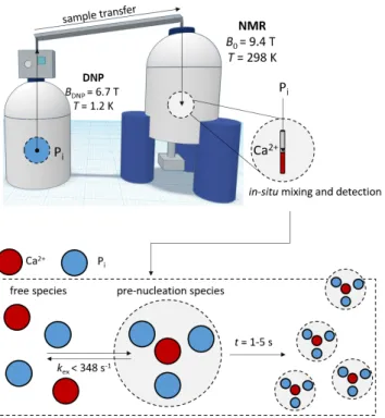
Figure 2. Schematic representation of the experimental setup for real-time
monitoring of CaP PNS formation by D-DNP. Inorganic phosphate Pi is
hyperpolarized by DNP at 1.2 K to boost the 31
P NMR magnetization. The hyperpolarized sample is then rapidly dissolved and transferred to an NMR spectrometer where it is mixed in-situ with a Ca2+
-containing solution. After mixing, free and PNS-bound Pi are in dynamic equilibrium, while the PNS
formation is monitored in real time. The process takes place on a 1-5 s time scale at 25˚C and pH 7.8.
To help answering to the current need for insights into PNS, we introduce here an experimental strategy that can provide insight into the very early stage of the CaP formation with high temporal as well as atomistic resolution. Our approach targets two different aspects of the process: a) Atomic-level details of the initial stages of CaP formation including the first detection of type 2 PNS by NMR - providing a microscopic perspective on PNS formation to confirm the existence of type 2 PNS, and if successfully observed, track their behavior; b) Characterization of the evolution of the collective solution/precipitate system, providing a complementary macroscopic perspective on the phase separation process.
The proposed methodology employs dissolution dynamic nuclear polarization (D-DNP, Fig. 2), a technique for obtaining drastically signal-enhanced nuclear magnetic resonance (NMR)
spectroscopy22-24, used to detect, monitor and characterize PNS
formation (point (a)). The D-DNP NMR results are supplemented by real-time turbidity measurements, cryo-electron microscopy, and solid-state NMR, to gain complementary access to larger CaP aggregates and solid precipitates (point (b)).
Experimental Section
D-DNP
250 µL of a Pi solution (100 mM TRIS buffer at pH 7.8)
supplemented with 50 mM of TEMPOL and mixed 1:1 v/v with
glycerol-d8 as cryo-protectant (yielding a final Pi concentration of
0.5 M) was hyperpolarized at a temperature of 1.2 K in a magnetic field of 6.7 T during 1.5 h in a prototype of a Bruker DNP polarizer. The microwave frequency was set to either 187.90 GHz (positive DNP) or 188.38 GHz (negative DNP) and modulated at 1 kHz with a saw tooth function over a bandwidth
of 100 MHz.25
The hyperpolarized sample was then dissolved by a burst of 5
mL (hence diluted 20-fold to a Pi concentration of 25 mM)
superheated D2O at 180˚C and 10.5 bar and propelled through a
0.9 T “magnetic tunnel”26 by pressurized helium at 7 bar within 4
s to a 9.4 T Bruker Avance II NMR spectrometer where it was mixed (in less than 1 s) 4:1 v/v with 500 mL TRIS buffer (yielding
20 mM Pi) containing 0, 8 or 13 mM CaCl2 (final concentration)
at 298 K. The final volume was 2.5 mL. The spectrometer was not field-frequency locked to allow the detection to start as soon as possible. After dissolution and mixing the resulting pH was determined to be 7.8±0.2.
Detection was achieved at intervals of 0.5 s by applying
non-selective 30˚ pulses with a 31P carrier frequency set to 0 ppm.
The spectral width was 40 ppm using a 10 mm BBO probe. All data were analyzed using home-written MATLAB scripts. All spectra were apodised with Gaussian window functions. Signal intensities were extracted by adapting a strategy published
before27-30 using a Lorentzian fit implemented in the ‘fit’ function
of MATLAB. The scripts employed the GNAT and EasySpin tool
boxes for data loading and apodization.31-32
To extract the kinetic rates from the D-DNP experiments, the signal intensity time traces were fitted to the following functions for mono- and bi-exponential behavior respectively:
𝑆 𝑡 = 𝑆!exp −𝑅!"#𝑡 + 𝑆!" (1)
𝑆 𝑡 = (𝑆!!exp −𝑅!"#𝑡 − 𝑆!!!exp −𝑅!"𝑡 ) +𝑆!" (2)
Where 𝑆 𝑡 is the signal amplitude at time point t after mixing, 𝑆!!
3
functions to modulate the bi-exponential behavior and 𝑆!" the
equilibrium signal amplitude for longer times.
The time traces were fitted using the bisquare method as
implemented in the MATLAB curve fitting tool. Note that 𝑅!"#
does not equal 𝑅! but includes all causes of phenomenological
signal loss.
Turbidimetry
Turbidimetry measurements were performed with a home-built turbidimeter using a SEN0189 turbidity sensor and a home-written Arduino control system. The experiments were
performed at room temperature and the sampling rate was 1 s-1.
Cryo-EM
After mixing of phosphate and CaCl2 solutions under similar
concentrations as in the D-DNP experiments (13 mM Ca2+), the
specimens were frozen as fast as possible by plunging into liquid ethane cooled by liquid nitrogen (LEICA EM CPC, Vienna, Austria). The delay between sample preparation and freezing was ca. 15-30 s. The cryofixed specimens were mounted into a Gatan cryoholder (Gatan inc., Warrendale, PA) for direct observation at 93 K (-180 °C) in a JEOL 2100HC cryo-TEM operating at 200 kV with a LaB6 filament. Images were recorded in zero-loss mode with a Gif Tridiem energy-filtered-CCD camera equipped with a chip with 2k x 2k pixels (Gatan Inc., Warrendale, PA). Acquisition was accomplished with Digital Micrograph software (versions 2.31.734.D, Gatan inc., Warrendale, PA).
SS NMR
Solid-state nuclear magnetic resonance (NMR) experiments were conducted using an Avance-III 300 Bruker spectrometer.
Precipitates were produced with a concentration of [Ca2+] = 13
mM, were packed into 4 mm (O.D.) zirconia rotors, and experiments were conducted using magic angle spinning (MAS).
The spinning frequency was νMAS = 14 kHz using a Bruker 4BL
CP/MAS 1H/BB probe. The recovery delays for 1H relaxation
were set to 10 s. A two-dimensional 1H-31P heteronuclear
correlation spectrum was recorded with 128 t1 increments with
40 transients each. The cross-polarization (CP) contact times
were set to tCP = 3 ms. Proton chemical shifts were referenced to
TMS at 0 ppm, whilst 31P chemical shifts were referenced to
H3PO4 (85% w/w aqueous solution) at 0 ppm.
Estimation of the PNS radius
Under the assumption of a simple isotropic rotation of the PNS,
the transverse relaxation rate of an isolated 31P spin depends on
its CSA ∆𝜎 according to15:
𝑅!=!"! 𝛾!𝐵!∆𝜎 ! !!𝜏!+!! !!!!!!! (3)
where 𝛾! denotes the gyromagnetic ratio of nucleus i, 𝐵! static
the magnetic field, 𝜔! the Larmor frequency and 𝜏! the rotational
correlation time. In the extreme narrowing limit where 𝜔!≫ 𝜏!,
eq. 1 reduces to:
𝑅!=!!!"! 𝛾!𝐵!∆𝜎 !𝜏! (4)
The effective hydrodynamic radius Rh can be calculated
according to the Stokes-Einstein relation: 𝑅!= !!"!!!"!
!
(5)
Where 𝑘 denotes the Boltzmann constant, 𝑇 the temperature
and 𝜂 the dynamic viscosity. It follows that
𝑅!!=!!"!!"𝐶𝑅! (6)
With the constant
𝐶 =!!!"! 𝛾!𝐵!∆𝜎!! (7)
For free Pi in the absence of Ca2+, we determined that R2(Pi)=
5.3 s-1 under our experimental conditions by conventional
spin-echo experiments. Assuming a CSA of 68 ppm (as reported for
brushite)33 for 31P in a PNS and 3 Γ
2(Pi)= Γ2(PPNS) > R2(PPNS) as explained in the main text, eqs. 3-7 yield 𝑅!< 0.94 nm for the
PNS. Note that this value is an estimate and must be interpreted with care.
All linewidths have been determined after stabilization of the sample, i.e. 2 s after completion of the mixing processes to avoid biases due to shim instabilities (see Supplementary Material Figs. S3-S5).
Results and Discussion
1. The birth of a mineral witnessed by D-DNP
By combining D-DNP with real-time NMR measurements, we have developed the capability to directly observe and analyze transient PNS that may be present immediately after preparation of oversaturated CaP solutions. In our hands, such signals remained below the detection threshold of conventional NMR (see Supplementary Material Fig. S1), in the absence of signal enhancement by D-DNP. D-DNP amplifies NMR signals of various substrates through a “hyperpolarization” procedure (Fig. 2) that consists of i). microwave irradiation of a sample that contains a paramagnetic polarization agent (PA; here 50 mM
TEMPOL) in a magnetic field (B0,DNP = 6.7 T) at cryogenic
temperatures (TDNP = 1.2 K), followed by ii) rapid heating and
dissolution of the sample, and iii) the transfer (with a transfer
time ttransfer = 4 s) of the hyperpolarized sample to a liquid-state
NMR spectrometer for detection (B0,NMR = 9.4 T at TNMR = 298 K).
Our strategy for real-time monitoring of CaP formation is
thus based on i) the production of hyperpolarized phosphate (Pi)
followed by ii) sample dissolution and rapid mixing with a Ca2+
-containing solution in-situ in an NMR spectrometer, and iii) the
time-resolved detection of NMR spectra of hyperpolarized 31P
nuclei, which reports on the presence of PNS and on the local phosphate environments (see Fig. 2). This strategy allows one to monitor the Pi interaction with Ca2+ on a time scale of a few seconds immediately after mixing of the two interacting components. For the case at hand, NMR real-time monitoring was achieved using a single pulse-acquisition scheme applied at
a rate of 2 s-1 and a flip angle of 30˚. Comparable strategies by
Katz-Brull and co-workers34 have already shown their value in
medicinal contexts.
The observation of PNS on timescales of ca. 10 s was made possible with a D-DNP prototype system, which enables complete mixing of the two components within ca. 1 s upon
arrival of the Pi solution in the NMR tube. NMR detection was
4
First, we analyzed the dependence of the hyperpolarizedphosphate spectra on the Ca2+ concentration (denoted [Ca2+])
directly after in-situ mixing. Fig. 3 displays 31P spectra obtained
within 2 s the mixing of 20 mM hyperpolarized Pi with solutions
of varying Ca2+ concentration (concentrations given after mixing).
In the absence of any Ca2+ ions, a single narrow signal is
observed at δ(31P) = 0.7 ppm with a linewidth of λ(P
i) = 35.2±6.9
Hz. This signal decays monotonically with time and can be
considered as fingerprint of free, hyperpolarized Pi in solution
under our conditions. This is in contrast to the spectra observed
in the presence of Ca2+ ([Ca2+] = 8 mM and 13 mM).
For [Ca2+] = 8 mM resonances were detected at δ(31P) = 0.6
ppm and 1.6 ppm. The signals were fitted to Lorentzian line
shapes of λ(Pi) = 129.5±4.1 and 128.3±8.0 Hz. These two lines
were assigned to free Pi (high-field signal) and to phosphate
interacting with Ca2+ ions (low-field signal). The signal of free P
i was identified by its monotonic decay after mixing, while the
signal of the Ca2+-binding species initially grows (characteristic
for the formation of CaP species) before decreasing to naught. (See Section 2 for details.)
At a calcium ion concentration of [Ca2+] = 13 mM, again two
peaks appear, although with different chemical shifts (δ(31P) =
1.3 for free and 2.7 ppm for bound Pi) and different linewidths
(λ(Pi) = 233.4±5.8 Hz and 58.1±4.4 Hz). The signal for free Pi is
again characterized by a monotonic decay, while PNS-bound phosphate shows the characteristic build-up and decay behavior
indicative of Ca2+-interaction.
1a. Line shape Analyses
The D-DNP results provide key information about two distinct aspects of PNS formation: i) the size and motional
freedom of the developing solute species (via the 31P
linewidths); and ii) on the phosphate exchange rate (via the
number of 31P signals observed):
i) linewidth-based information – determination of PNS size:
In liquid-state NMR spectroscopy, the homogeneous linewidth of a signal is proportional to the intrinsic transverse relaxation rate
of the nuclear spin R2, which in turn depends on the correlation
time τc of rotational diffusion of the spin-bearing moiety. It is
therefore likely that the observed similar linewidths at [Ca2+] = 8
mM indicate that both free and interacting species have comparable rotational diffusion properties, despite their different chemical shifts that indicate distinct chemical environments. In
contrast, the differing linewidths at [Ca2+] = 13 mM suggest that
the Ca2+-interacting species takes part in the formation of a
cluster that is significantly larger than free Pi. However, for the case at hand, we only compare relative linewidths within a single detection (to avoid biases due to shim instabilities etc.) and measure the sum Γ of homogeneous and inhomogeneous linewidths. Therefore, we can only state that R2 < Γ. Under the assumption of fast rotational diffusion of a spherical particle, where R2 ∝ τc holds, a three-fold broader line, as observed for
the broad signal at [Ca2+] = 13 mM (Fig. 3), indicates at most a
three-fold longer rotational diffusion time, corresponding to a
maximum increase by a factor 31/3 (≈ 1.4) of the hydrodynamic
radius Rh according to the Stokes-Einstein equation. This
enables an estimate of Rh < 0.9 nm for the hydrodynamic radius
of the transiently observed Ca2+-binding P
i species that we
Figure 3. Observation of PNS in supersaturated out-of-equilibrium solutions. Three CaP concentrations were evaluated by D-DNP. Injection of
a Pi solution into (a) pure buffer (concentrations: [Pi] = 20 mM; [Ca2+] = 0 mM), (b) & (c) into a solution of Ca2+ (concentrations: [Pi] = 20 mM;
[Ca2+
] = 8 & 13 mM; all concentrations are final concentrations in the NMR tube after mixing). a) Centre: a single Pi signal is seen which is
indicative of the free Pi in solution. Right: evolution of the 31P NMR signals after mixing at t = 0 at a calcium ion concentration of [Ca2+] = 0, and
T = 298 K. The signal intensity decreases exponentially towards thermal equilibrium after mixing. b) Centre: At a concentration of [Ca2+
] = 8, pH 7.8 and T = 298 K two signals appear, one corresponding to free Pi (red) and a second indicating phosphate interacting with calcium (PPNS;
blue). Right: Signal intensity of free Pi and PPNS. While the former decrease monotonically to equilibrium, the signal of PPNS builds up during the
first 2.5 s after mixing before it decays to naught as a consequence of relaxation and growth/aggregation of the PNS. c) A similar behaviour as in b) is observed at a concentration of [Ca2+
] = 13, and T = 298 K, but the line of free Pi shifts to the left of the PNS signal (see main text). pH values after mixing are indicated. The signal intensities were referenced to their equilibrium signal intensities Seq at t à∞.
5
detect here during the onset of calcium phosphate precipitation.The inequality takes all exchange processes and other inhomogeneous broadening effects into account (cf. Materials and Methods) It should be noted that concentration-dependent chemical exchange is a well-documented property of metal-ligand complexes and of many biological systems, such as the
exchange of lipids between vesicles16 or of DNA-transcription
factor complexes.17,18 A comparable concentration-dependence
could here contribute to the growing line width upon increasing
Ca2+ ion concentrations. Indeed, growing counter ion
concentrations might accelerate theexchange between free and
PNS-bound phosphate species thereby broadening the 31P
resonances.
On the short time scales of the D-DNP experiments, it is yet complicated to assess the exchange rates of the species corresponding to the individual lines, as signal averaging and mixing time incrementation are hardly feasible. A possible way to obtain exchange rates would be the use of ultrafast exchange spectroscopy (i.e. single scan approaches combined with EXSY/NOESY) as proposed in combination with D-DNP by Swisher et al.35
Nevertheless, our finding is in agreement with the estimated radius of stable type 1 PNS reported by Dey et al.18 (0.44 nm)
and of type 2 PNS reported by Habraken et al.36 (0.45 to 0.65
nm), who both found that this size would be consistent with PNS composed of three phosphate units. We therefore conclude that we directly observe nanoscopic objects that emerge during the onset of the precipitation of calcium phosphate in oversaturated solutions, i.e. type-2 PNS which are constituted of at most 3
phosphate units and (henceforth denoted PPNS).
It should be noted that the chemical shift of free Pi changes
in our hands when varying the Ca2+ concentration. This might be
a consequence of relatively low buffer concentrations and counter ion effects in solution including the variation of the ionic
strength.37 The analyses presented above however hold, as they
are not affected by this fact. Further reference data for the line shape analysis is included in the Supplementary Material (Fig. S2-S5).
ii) Chemical shift-based information – determination of dynamic exchange rates: At [Ca2+] = 8 and 13 mM, the simultaneous presence of two separate signals indicates that the
exchange rate (kex) of Pi between its free (Pi) and bound forms
(PPNS) is slow on the NMR timescale, i.e. kex < Δν, where Δν
denotes the difference in resonance frequencies of the two signals. The upper bound of the exchange rate at the two
concentrations can thus be estimated to be 249 and 348 s-1,
respectively.
1b. Real-time analyses
In addition to the information contained in static PNS spectra, real-time detection in D-DNP experiments provides key insights into the kinetics of PNS formation.
Fig. 3a to c (middle and right panels) show time traces of the NMR signal intensities of free Pi (red) as well as PPNS (blue)
when present. At all tested Ca2+-concentrations, the signal
intensity of free phosphate decays exponentially, primarily due
to the return of the hyperpolarized 31P spins to thermal
equilibrium. At 8 mM ≤ [Ca2+] ≤ 13 mM, the observed Ca2+
-interacting species PPNS though displays a build-up of signal
intensity during the first 2 s after mixing. The PPNS signal
intensity here reports on the presence of Ca2+-interacting
phosphate and thus on the formation of PNS. The build-up starts
directly after mixing of the two components so that a significant PNS signal could be detected already 0.5 s after mixing. This is then followed by a decay and eventual disappearance of the signal. In addition to the return to thermal equilibrium of the
hyperpolarized 31P spins, the P
PNS signal may also decay for t >
2 s if the growth or aggregation of the PNS generates species with molecular weights that are too high to allow detection by
solution-state NMR (i.e. post-nucleation species). No PPNS signal
could be detected in the time-series after 10 s at [Ca2+] = 8 mM,
and after 5 s at [Ca2+] =13 mM.
The time traces of the Pi and PPNS signals could be fitted respectively to mono-exponential (decay only) and bi-exponential (build-up followed by decay) functions. Both decreasing functions account for all sources of signal loss (growth, conversion, rf pulses, paramagnetic relaxation etc.) by
a single rate constant RDEC. Therefore, in case of bi-exponential
behavior, the build-up rate RBU reflects only the formation of
PNS-bound phosphate. Table 1 lists the obtained decay rates
RDEC and build-up rate constants RBU. Importantly, RBU relates to
the kinetics of PNS formation as the signal growth corresponds
to the formation rate of Ca2+ interacting phosphate species.
The Pi decay rates are largely independent of the Ca2+
concentration showing that longitudinal relaxation is not strongly
impacted by varying ionic strength. Similarly, the PPNS build-up
rates, RBU(PNS), are constant within the error margin. Thus,
under our conditions the rate of PNS formation appears to be
independent of the Ca2+ concentrations. In contrast, the variation
of the RDEC(PNS) values reflects a dependence of
post-nucleation aggregation as well relaxation processes on [Ca2+].
Indeed, the higher the Ca2+ concentration the faster the decay
rate indicating accelerated aggregation at elevated counter ion quantities (cf. turbidimetry data in section 2).
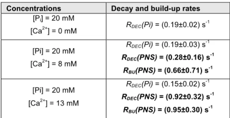
Table 1. Characteristic decay rates RDEC and build-up rates RBU observed in the D-DNP experiments. (The Methods section contains details on the fitting procedure.)
Concentrations Decay and build-up rates
[Pi] = 20 mM
[Ca2+] = 0 mM RDEC(Pi) = (0.19±0.02) s -1 [Pi] = 20 mM [Ca2+] = 8 mM RDEC(Pi) = (0.19±0.03) s-1 RDEC(PNS) = (0.28±0.16) s-1 RBU(PNS) = (0.66±0.71) s-1 [Pi] = 20 mM [Ca2+] = 13 mM RDEC(Pi) = (0.15±0.02) s-1 RDEC(PNS) = (0.92±0.32) s-1 RBU(PNS) = (0.95±0.30) s-1
It should be mentioned that the D-DNP approach employs additives such as glycerol and the polarizing agent (TEMPOL). These specific conditions might influence the precipitation process, which therefore cannot directly be compared to previously published data on CaP systems.
2. The Macroscopic Phase Transition
To compare the atomistic perspective that D-DNP provides
with a macroscopic picture that includes the entire Ca2+/P
i/buffer system, we performed real-time turbidimetry experiments to monitor the precipitation events. These experiments provided observations of the aggregation processes occurring within 10
and 20 s (depending on the Ca2+ concentrations), whereas the
6
experiments. This suggests that significant amounts of type 2PNS can only be found during the onset of the event, although the macroscopically observable phase separation process takes longer.
However, it should be considered that the NMR signal decay is in part determined by relaxation effects. Therefore, the faster decay of the NMR signals in the D-DNP experiments as compared to the turbidity curves does not necessarily prove that the corresponding species also disappear faster. Instead, they might persist for longer periods, yet remain invisible to NMR experiments as a consequence of rapid longitudinal relaxation.
The curves in Fig. 4a represent the intensity of light that
passes through the NMR sample tube after in-situ mixing of Pi
-and Ca2+-containing solutions (20 mM P
i, 0, 10 or 20 mM CaCl2).
CaP particle growth after mixing leads to visible light scattering, as seen by the decreasing sample transparency. We thereby observed a phase transition event within ~10 or 20 s, for calcium ion concentrations of 10 or 20 mM, respectively. In contrast, the
PPNS signals observed by D-DNP dropped to zero after 5 or 10 s,
respectively.
Hence, PNS, i.e. nanoscale inhomogeneities that initiate the CaP precipitation event, might be associated with the early stage of the process, while nucleation can be associated with later stages. Similar convergence between nanoscale inhomogeneities that precede macroscopic phase transitions is
well documented in the field of polymer physics.38-40 For example,
stimuli-responsiveness of polymers is based on nanoscale inhomogeneities that precede the gelation of a polymer solution
and often determine the cooperativity of such transitions.40
Possibly, a similar conjunction might be witnessed here in the context of CaP formation. This is also in agreement with the convergence of classical and non-classical crystallization
theories that has been proposed by Habraken et al.9
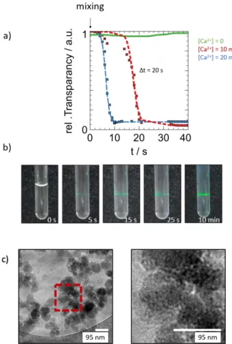
Figure 4. Macroscopic observations of CaP precipitation. a)
Turbidimetry experiments expressed as time dependence of light transmissibility (detected intensity) of CaP solutions after mixing of 20 mM Pi with different concentrations of Ca2+ ions. By this means
the phase transition was observed to take 10-20 s depending on the Ca2+ concentration (grey shade). The higher the Ca2+ concentration,
the faster the precipitation. Note that in agreement with the D-DNP data, the precipitation event proceeds faster at higher Ca2+
concentrations. b) Temporal evolution of the NMR sample tube after mixing of Pi and Ca2+. Within 10 min., macroscopic precipitates
cause light to scatter. Even within the first 5 s, i.e. during the period accessible for D-DNP, light scattering was already observed. c) Cryo-electron micrographs confirm the presence of ∼ 100 nm large CaP cluster ca. 30 seconds after mixing of the two phosphate and calcium chloride solutions. The red box indicates the area magnified on the right. The amorphous clusters feature an internal architecture due to aggregation of smaller particles of ca. 5-10 nm.
Therefore, to corroborate the turbidity data, Fig. 4b illustrates how a green laser beam is scattered only 5 s after in-situ mixing
of Pi and Ca2+-containing solutions in the NMR sample tube.
This hints towards aggregates of several hundreds of nanometers, which are too large to be detected by liquid state NMR yet accessible by turbidimetry already a few seconds after mixing. It appears therefore reasonable to assume that aggregation of PNS plays a large role in the disappearance of the NMR signals and that relaxation alone cannot account for the rapid signal disappearance. This interpretation would also explain why PNS could only be detected during the first ∼5 s after mixing by D-DNP, although the entire precipitation process takes ca. 10-20 s, judging from the turbidity experiments. We want to stress that the particle size should be interpreted with care though, as turbidity can be correlated with particle size distributions only in combination with further X-ray or light scattering data.
Cryo-electron microscopy (EM) experiments (Fig. 4c) confirmed the presence of large (∼ 100 nm) CaP amorphous particles already 30 seconds after initiation of the precipitation event. Interestingly, they display an internal structure made of smaller particles (ca. 5-10 nm). These observed structure might nevertheless stem from aggregation of secondary particles composed of PNS. Indeed, a pathway cannot be excluded that explains the disappearance of PNS (as detected by D-DNP) by aggregation of particles through secondary nucleation as it has
been described for ACC precipitation13.
Figure 5. Solid-state NMR spectrum (HETCOR) of the final
precipitate. A brushite-only structure was observed, which includes 2 water molecules per phosphate unit. The solid was recovered by centrifugation form the final mixture of the D-DNP experiments, 10 min. after mixing of the two interactants.
7
Finally, solid-state NMR measurements revealed the finalcrystallographic phase following the real-time experiments. We determined the internal architecture of crystallites formed 10 min
minutes after mixing by means of solid-state 1H-31P
heteronuclear correlation (HETCOR) spectroscopy.41-42 The
spectrum in Fig. 5 features characteristic resonances of
CaHPO4⋅2H2O, i.e. of brushite crystals. Such spectra remained
unchanged for 2 days after sample preparation.
Notably, the spectrum in Fig. 5 displays a brushite-only internal architecture with two well-defined peaks stemming from
the two types of protons within the crystalline phase (i.e. H2O
and HPO42-). Hence, starting from solute PNS that form on a
time scale of a few seconds, macroscopic brushite crystallites emerge on longer timescales to shape the final product. The system thereby undergoes a disorder-to-order transition from nanoscale inhomogeneities (observed after 0.5 - 8 s) to macroscopic crystals (observed after 10 min - 2 h) on a seconds-to-minutes timescale.
Conclusions
We present an experimental spectroscopic strategy that enables the analysis of the initial phases of mineral precipitation events and that reports on PNS size and kinetics, ion exchange rates as well as macroscopic phase behavior and the architecture of the final solid.
As PNS are currently assumed to constitute the first step in precipitation and growth of crystalline CaP, their rate of formation is possibly defining the growth-rate limiting factor that determines the kinetics of CaP precipitation from aqueous solution. Real-time monitoring of PNS formation by D-DNP allows one to obtain key information about these important nanoscopic entities. The method is here reported with an example to CaP formation in a biomineralization context, yet wide ranges of concentrations, pH and temperatures as well as other systems (e.g. carbonate, silicate or nitrate) are in principle accessible on time scales ranging from milliseconds up to several minutes.
By integrating real-time techniques with microscopy and solid-state measurements many aspects of the precipitation of CaP from aqueous solution could be captured, from the formation of initial PNS to the final precipitate.
Based on NMR, our approach capitalizes on an intrinsically high (atomistic) resolution and adds a temporal dimension to this method’s portfolio - an approach that might prove valuable for the investigation of transient intermediates, precipitation events and interaction monitoring in general and that should have a widespread potential to monitor structure and kinetics of functional intermediates in the contexts biomineralization and crystallization.
Supporting Information
D-DNP reference experiments; fitting results for real-time D-DNP experiments.
Acknowledgements
The authors thank Mrs. Ludovica M. Epasto, Mrs. Estelle Loh as well as the entire D-DNP team at LBM (and in particular to: Dr. M. Baudin, Prof. G. Bodenhausen, Mr. D. Guarin, MSc.) for its continuous support. This research was supported by the French Centre National de la Recherche Scientifique (CNRS), the
European Research Council (ERC contract ‘HYPROTIN’, grant agreement 801963). The authors thank Bruker BioSpin for providing the D-DNP equipment and the EM Service of the IBPS (CNRS FR 3631) at UPMC. The authors thank the Cluster of Excellence MATISSE led by Sorbonne Universités.
Keywords: Dissolution DNP • Calcium Phosphate
Crystallization • Prenucleation Species • NMR Spectroscopy • Biomineralization
8
References1. LeGeros, R. Z., Calcium phosphate-based osteoinductive materials. Chem Rev 2008, 108 (11), 4742-53.
2. Wang, L. J.; Nancollas, G. H., Calcium Orthophosphates: Crystallization and Dissolution. Chem Rev 2008, 108 (11), 4628-4669.
3. Habraken, W.; Habibovic, P.; Epple, M.; Bohner, M., Calcium phosphates in biomedical applications: materials for the future? materialdtoday 2015, 19, 69-87.
4. Glimcher, M. J., Bone: Nature of the calcium phosphate crystals and cellular, structural, and physical chemical mechanisms in their formation. Rev Mineral Geochem 2006,
64, 223-282.
5. Gebauer, D.; Volkel, A.; Colfen, H., Stable prenucleation calcium carbonate clusters. Science 2008, 322 (5909), 1819-22.
6. Karthika, S.; Radhakrishnan, T. K.; Kalaichelvi, P., A Review of Classical and Nonclassical Nucleation Theories. Cryst.
Growth Des. 2016, 11, 6663-6681.
7. Demichelis, R.; Raiteri, P.; Gale, J. D.; Quigley, D.; Gebauer, D., Stable prenucleation mineral clusters are liquid-like ionic polymers. Nature Communications 2011, 2.
8. Gebauer, D.; Wolf, S. E., Designing Solid Materials from Their Solute State: A Shift in Paradigms toward a Holistic Approach in Functional Materials Chemistry. J Am Chem
Soc 2019, 141 (11), 4490-4504.
9. Habraken, W. J.; Tao, J.; Brylka, L. J.; Friedrich, H.; Bertinetti, L.; Schenk, A. S.; Verch, A.; Dmitrovic, V.; Bomans, P. H.; Frederik, P. M.; Laven, J.; van der Schoot, P.; Aichmayer, B.; de With, G.; DeYoreo, J. J.; Sommerdijk, N. A., Ion-association complexes unite classical and non-classical theories for the biomimetic nucleation of calcium phosphate. Nat Commun 2013, 4, 1507.
10. Dorvee, J. R.; Veis, A., Water in the formation of biogenic minerals: peeling away the hydration layers. J Struct Biol 2013, 183 (2), 278-303.
11. Zahn, D., Thermodynamics and Kinetics of Prenucleation Clusters, Classical and Non-Classical Nucleation.
Chemphyschem 2015, 16 (10), 2069-2075.
12. Scheck, J.; Wu, B.; Drechsler, M.; Rosenberg, R.; Van Driessche, A. E.; Stawski, T. M.; Gebauer, D., The Molecular Mechanism of Iron(III) Oxide Nucleation. J Phys Chem Lett 2016, 7 (16), 3123-30.
13. Carino, A.; Testino, A.; Andalibi, M. R.; Pilger, F.; Bowen, P.; Ludwig, C., Thermodynamic-Kinetic Precipitation Modeling. A Case Study: The Amorphous Calcium Carbonate (ACC) Precipitation Pathway Unravelled. Crystal Growth & Design 2017, 17 (4), 2006-2015.
14. Carino, A.; Ludwig, C.; Cervellino, A.; Muller, E.; Testino, A., Formation and transformation of calcium phosphate phases under biologically relevant conditions: Experiments and modelling. Acta Biomater 2018, 74, 478-488.
15. Andalibi, M. R.; Wokaun, A.; Bowen, P.; Testino, A., Kinetics and Mechanism of Metal Nanoparticle Growth via Optical Extinction Spectroscopy and Computational Modeling: The Curious Case of Colloidal Gold. ACS Nano 2019, 13 (10), 11510-11521.
16. Mohammed, A. S. A.; Carino, A.; Testino, A.; Andalibi, M. R.; Cervellino, A., In Situ Liquid SAXS Studies on the Early Stage of Calcium Carbonate Formation. Part Part Syst Char 2019, 36 (6).
17. Gebauer, D.; Kellermeier, M.; Gale, J. D.; Bergstrom, L.; Colfen, H., Pre-nucleation clusters as solute precursors in crystallisation. Chem Soc Rev 2014, 43 (7), 2348-71. 18. Dey, A.; Bomans, P. H.; Muller, F. A.; Will, J.; Frederik, P.
M.; de With, G.; Sommerdijk, N. A., The role of prenucleation clusters in surface-induced calcium phosphate crystallization.
Nat Mater 2010, 9 (12), 1010-4.
19. Wang, L.; Li, S.; Ruiz-Agudo, E.; Putnis, C. V.; Putnisc, A., Posner's cluster revisited: direct imaging of nucleation and
growth of nanoscale calcium phosphate clusters at the calcite-water interface CrystEngComm 2012, 14, 6252-6256 20. Zhang, Q.; Jiang, Y.; Gou, B.-D.; Huang, J.; Gao, Y.-X.;
Zhao, J.-T.; Zheng, L.; Zhao, Y.-D.; Zhang, T.-L.; Wang, K., In Situ Detection of Calcium Phosphate Clusters in Solution and Wet Amorphous Phase by Synchrotron X ray
Absorption Near-Edge Spectroscopy at Calcium K Edge.
Crystal Growth and Design 2015, 15, 2204-2210.
21. Paul Tuck, S.; Layfield, R.; Walker, J.; Mekkayil, B.; Francis, R., Adult Paget's disease of bone: a review. Rheumatology
(Oxford) 2017, 56 (12), 2050-2059.
22. Ardenkjaer-Larsen, J. H.; Fridlund, B.; Gram, A.; Hansson, G.; Hansson, L.; Lerche, M. H.; Servin, R.; Thaning, M.; Golman, K., Increase in signal-to-noise ratio of > 10,000 times in liquid-state NMR. Proc Natl Acad Sci 2003, 100 (18), 10158-63.
23. Kovtunov, K. V.; Pokochueva, E. V.; Salnikov, O. G.; Cousin, S. F.; Kurzbach, D.; Vuichoud, B.; Jannin, S.; Chekmenev, E. Y.; Goodson, B. M.; Barskiy, D. A.; Koptyug, I. V.,
Hyperpolarized NMR Spectroscopy: d-DNP, PHIP, and SABRE Techniques. Chem Asian J 2018.
24. Jannin, S.; Dumez, J. N.; Giraudeau, P.; Kurzbach, D., Application and methodology of dissolution dynamic nuclear polarization in physical, chemical and biological contexts. J
Magn Reson 2019, 305, 41-50.
25. Weber, E. M. M.; Sicoli, G.; Vezin, H.; Frébourg, G.; Abergel, D.; Bodenhausen, G.; Kurzbach, D., Sample Ripening through Nanophase Separation Influences the Performance of Dynamic Nuclear Polarization. Angew. Chem. Int. Ed. 2018, 10.1002/anie.201800493.
26. Milani, J.; Vuichoud, B.; Bornet, A.; Mieville, P.; Mottier, R.; Jannin, S.; Bodenhausen, G., A magnetic tunnel to shelter hyperpolarized fluids. Rev Sci Instrum 2015, 86 (2). 27. Miclet, E.; Abergel, D.; Bornet, A.; Milani, J.; Jannin, S.;
Bodenhausen, G., Toward Quantitative Measurements of Enzyme Kinetics by Dissolution Dynamic Nuclear Polarization. J Phys Chem Lett 2014, 5 (19), 3290-5. 28. Sadet, A.; Emmanuelle M. M. Weber; Jhajharia, A.;
Kurzbach, D.; Bodenhausen, G.; Miclet, E.; Abergel, D., Kinetic rates of chemical metabolic processes determined by NMR boosted by dissolution dynamic nuclear polarization.
Chem. Eur. J. 2018, 24, 5456 – 5461.
29. Ivanov, K. L.; Kress, T.; Guarin, D.; Baudin, M.; Abergel, D.; Bodenhausen, G.; Kurzbach, D., Relaxation of long-lived modes in NMR of deuterated methyl groups. J. Chem. Phys. 2018, 149 (5), 054202.
30. Kress, T.; Walrant, A.; Bodenhausen, G.; Kurzbach, D., Long-Lived States in Hyperpolarized Deuterated Methyl Groups Reveal Weak Binding of Small Molecules to Proteins.
J Phys Chem Lett 2019, 10 (7), 1523-1529.
31. Stoll, S.; Schweiger, A., EasySpin, a comprehensive software package for spectral simulation and analysis in EPR. J Magn Reson 2006, 178 (1), 42-55.
32. Castanar, L.; Poggetto, G. D.; Colbourne, A. A.; Morris, G. A.; Nilsson, M., The GNAT: A new tool for processing NMR data. Magn Reson Chem 2018, 56 (6), 546-558.
33. Rothwell, W. P.; Waugh, J. S.; Yesinowski, J. P., High-resolution variable-temperature phosphorus-31 NMR of solid calcium phosphates. J. Am. Chem. Soc. 1980, 102, 2637-2673.
34. Nardi-Schreiber, A.; Gamliel, A.; Harris, T.; Sapir, G.; Sosna, J.; Gomori, J. M.; Katz-Brull, R., Biochemical phosphates observed using hyperpolarized 31P in physiological aqueous solutions. Nat Commun 2017, 8 (1), 341.
35. Leon Swisher, C.; Koelsch, B.; Sukumar, S.; Sriram, R.; Santos, R. D.; Wang, Z. J.; Kurhanewicz, J.; Vigneron, D.; Larson, P., Dynamic UltraFast 2D EXchange SpectroscopY (UF-EXSY) of hyperpolarized substrates. J Magn Reson 2015, 257, 102-9.
9
36. Habraken, W. J., The integration of ion potentiometricmeasurements with chemical, structural, and morphological analysis to follow mineralization reactions in solution.
Methods Enzymol 2013, 532, 25-44.
37. Costello, A. J. R.; Glonek, T.; Wazer, J. R. V., Phosphorus-31 Chemical Shift Variations with Countercation and Ionic Strength for the Various Ethyl Phosphates. Inorg. Chem. 1975, 15, 972-974.
38. Kurzbach, D.; Reh, M. N.; Hinderberger, D., Nanoscale inhomogeneities in thermoresponsive triblock copolymers.
Chemphyschem 2011, 12 (18), 3566-72.
39. Kurzbach, D.; Hassouneh, W.; McDaniel, J. R.; Jaumann, E. A.; Chilkoti, A.; Hinderberger, D., Hydration Layer Coupling
and Cooperativity in Phase Behavior of Stimulus Responsive Peptide Polymers. J Am Chem Soc 2013, 135 (30), 11299-11308.
40. Kurzbach, D.; Junk, M. J. N.; Hinderberger, D., Nanoscale Inhomogeneities in Thermoresponsive Polymers. Macromol
Rapid Commun 2013, 34 (2), 119-134.
41. Crutchfield, M. M.; Callis, C. V.; Irani, R. R.; Roth, G. C., Phosphorus Nuclear Magnetic Resonance Studies of Ortho and Condensed Phosphates. Inorg. Chem. 1962, 1, 813-817. 42. Wazer, J. R. V.; Callis, C. F.; Shoolery, J. N., NUCLEAR
MAGNETIC RESONANCE SPECTRA OF THE
CONDENSED PHOSPHATES. J. Am. Chem. Soc. 1955, 77, 4945-4956.