HAL Id: dumas-02071738
https://dumas.ccsd.cnrs.fr/dumas-02071738
Submitted on 18 Mar 2019
HAL is a multi-disciplinary open access
archive for the deposit and dissemination of sci-entific research documents, whether they are pub-lished or not. The documents may come from teaching and research institutions in France or abroad, or from public or private research centers.
L’archive ouverte pluridisciplinaire HAL, est destinée au dépôt et à la diffusion de documents scientifiques de niveau recherche, publiés ou non, émanant des établissements d’enseignement et de recherche français ou étrangers, des laboratoires publics ou privés.
Stratification du risque de mort subite chez les patients
présentant un syndrome de Brugada et un aspect de
repolarisation précoce
Romain Tixier
To cite this version:
Romain Tixier. Stratification du risque de mort subite chez les patients présentant un syndrome de Brugada et un aspect de repolarisation précoce. Médecine humaine et pathologie. 2018. �dumas-02071738�
HAL Id: dumas-02071738
https://dumas.ccsd.cnrs.fr/dumas-02071738
Submitted on 18 Mar 2019
HAL is a multi-disciplinary open access
archive for the deposit and dissemination of sci-entific research documents, whether they are pub-lished or not. The documents may come from teaching and research institutions in France or abroad, or from public or private research centers.
L’archive ouverte pluridisciplinaire HAL, est destinée au dépôt et à la diffusion de documents scientifiques de niveau recherche, publiés ou non, émanant des établissements d’enseignement et de recherche français ou étrangers, des laboratoires publics ou privés.
Stratification du risque de mort subite chez les patients
présentant un syndrome de Brugada et un aspect de
repolarisation précoce
Romain Tixier
To cite this version:
Romain Tixier. Stratification du risque de mort subite chez les patients présentant un syndrome de Brugada et un aspect de repolarisation précoce. Médecine humaine et pathologie. 2018. <dumas-02071738>
UNIVERSITE de BORDEAUX
U.F.R. des SCIENCES MEDICALES
Année 2018 Thèse n° 3078
Thèse pour l’obtention du
DIPLOME D’ETAT DE DOCTEUR EN MEDECINE
Présentée et soutenue publiquement le vendredi 07 septembre 2018 par
Romain TIXIER
Né le 22 avril 1990 à Fontenay-Aux-Roses (92)
STRATIFICATION DU RISQUE DE MORT SUBITE CHEZ
LES PATIENTS PRESENTANT UN SYNDROME DE
BRUGADA ET UN ASPECT DE REPOLARISATION
PRECOCE
Directeur de thèse
Monsieur le Professeur Fréderic SACHER Jury
Monsieur le Professeur Jean-Michel HAISSAGUERRE, président Monsieur le Professeur Pierre BORDACHAR
Madame le Docteur Mélèze HOCINI
Monsieur le Docteur Jean-Baptiste GOURRAUD, rapporteur Monsieur le Docteur Josselin DUCHATEAU
TABLE DES MATIERES
REMERCIEMENTS ... 3
INTRODUCTION: RISK STRATIFICATION IN BRUGADA SYNDROME ... 6
SYMPTOMS ... 6 AGE ... 7 GENDERCONSIDERATION ... 8 FAMILYBACKGROUND ... 9 GENETICASPECTS ... 9 ECGCHARACTERISTICS ... 10
BRUGADA ECG PATTERN ... 10
FRAGMENTED QRS ... 11
VENTRICULAR LATE POTENTIALS ... 12
S-WAVE IN LEAD D1 (S1 PATTERN)... 12
EARLY REPOLARIZATION PATTERN (ERP) ... 13
REPOLARIZATION DISPERSION ... 14
AVR SIGN ... 15
SINUS NODE DYSFUNCTION ... 15
PROGRAMMEDELECTRICALSTIMULATION(PES) ... 15
PROPOSEDCOMPOSITESCORES ... 17
REFERENCES ... 19
EARLY REPOLARIZATION PATTERN IS NOT RELATED WITH VENTRICULAR ARRHYTHMIAS IN BRUGADA PATIENTS ... 23 ABSTRACT... 23 INTRODUCTION ... 24 METHODS ... 25 STATISTICALANALYSIS ... 26 RESULTS... 27 DISCUSSION ... 28 REFERENCES ... 32
REMERCIEMENTS
Monsieur le Professeur Jean-Michel HAISSAGUERRE, président du jury
Je vous remercie de me faire l’honneur d’être le président du jury de cette
thèse. Je mesure la chance qui m’est donnée d’apprendre à vos côtés la
rythmologie. Votre abnégation et la recherche permanente de l’excellence
ont permis l’émergence de nombreuses avancées scientifiques ces
dernières années, aujourd’hui symbolisées par le développement du LIRYC.
Vous êtes un modèle pour nous tous.
Monsieur le Professeur Frédéric SACHER, directeur de thèse
Je te remercie de m’avoir proposé de travailler sur ce sujet passionnant et
de m’avoir accompagné tout au long de ce travail. Tes précieux conseils, ta
disponibilité et ta bonne humeur m’ont aidé à le mener de manière agréable
et efficace.
A Monsieur le Professeur Pierre BORDACHAR, juge
Je te remercie de me faire le plaisir de juger ce travail. Nous avons la chance
de bénéficier d’une formation en cardiologie d’une qualité rare et c’est en
grande partie grâce à ton implication dans l’organisation de ces
enseignements.
A Madame le Docteur Mélèze HOCINI, juge
Je vous remercie de me faire le plaisir de juger ce travail. Votre rigueur, votre
dynamisme et vos qualités humaines sont un exemple à suivre pour nous
tous.
Monsieur le Docteur Jean-Baptiste GOURRAUD, rapporteur
Je vous remercie d’avoir accepté d’être le rapporteur de cette thèse et
d’avoir été l’un des coordonnateurs de ce travail conjoint sur le syndrome de
Brugada.
Monsieur le Docteur Josselin DUCHATEAU, juge
Je te remercie de me faire le plaisir de juger cette thèse ainsi que pour tes
conseils avisés. Ta curiosité scientifique et ta compréhension de
l’électrophysiologie cardiaque sont un modèle pour moi. Travailler à tes
Messieurs les Professeurs Raymond ROUDAUT, Stéphane LAFITTE,
Jean-Michel HAISSAGUERRE, Pierre JAIS, Pierre BORDACHAR, Frédéric
SACHER, Pierre COSTE, Joël CONSTANS, ainsi que Monsieur le Docteur
Nicolas DELARCHE, je vous remercie pour votre accueil chaleureux au sein
de vos services au long de mon internat.
Monsieur le Professeur Vincent PROBST, Madame le Professeur Aurélie
THOLLET, Monsieur le Professeur Philippe MABO et Madame le Docteur
Nathalie BEHAR, merci d’avoir avec Monsieur le Docteur Jean-Baptiste
GOURRAUD et Monsieur le Professeur Frédéric SACHER dirigé
conjointement ce travail sur le syndrome de Brugada.
Messieurs les Docteurs Philippe RITTER, Sylvain PLOUX, Nicolas DERVAL,
Arnaud DENIS, Thomas PAMPRUN, Nicolas KLOTZ, Rémi CHAUVEL et
Nicolas WELTE merci pour votre bonne humeur et votre constante
disponibilité, c’est un réel plaisir de travailler et d’apprendre avec vous au
quotidien.
Merci à Mesdames et Messieurs les Docteurs Marina DIJOS, Cécile
VINCENT, Sunthareth YIEM, Marion LAINE, Georgios PAPAIOANNOU,
Grégoire MASSOULIE, Antoine MARTY, Laura CETRAN, Benjamin SEGUY,
Pierre POUSTIS, Xavier ZIRPHILE, Raphael LASERRE, Hughes BADER,
Maxime DE GUILLEBON, Jean-François RIVIERE, Romain BOULESTREAU
et Simon VITTE.
Merci à Jean BRIAND et Pauline BERTHOME pour leur indispensable aide
de recueil de données pour ce travail.
Un grand merci aux équipes paramédicales des services avec lesquelles j’ai
eu le plaisir de travailler et plus particulièrement celles du service du 3
eet de
Merci à mes parents et à ma sœur Héloïse pour tout ce qu’ils m’ont apporté,
ainsi qu’à mes grands-parents, oncles, tantes et cousin(e)s avec qui nous
formons une famille unie dont je suis très fier.
Merci à Floriane, je suis tellement heureux de t’avoir à mes côtés.
Merci à Julie M., Julie G., Bérangère et Kévin pour notre belle amitié depuis
toutes ces années et, je l’espère, pour toutes celles à venir, qu’importe les
circonstances.
Merci à Jeanne et Mathieu notamment pour leurs précieux conseils au cours
de mes études de médecine ainsi qu’à Pauline et Agathe.
Audrey, Mathieu, Max, Marianne, Adrien, Rim, Manu, Manue, Auré, Guitou,
Simon, Chloé, Jules, Fanny, Thomas, Aude, Serena et Thibault merci pour
nos embuscades mémorables et leurs souvenirs indélébiles.
Merci à Guillaume, Bénédicte, Yann, Laurène, Anaïs, Arnaud, Chloé,
Clémentine … sans oublier Roby !
Merci à Marie, Émilie, Jean et Hugo, team de co-internes de rêve, avec qui
l’on ne s’ennuie jamais à l’hôpital en encore moins en dehors.
Merci à Maud pour notre soutien mutuel pendant certaines périodes un peu
troubles de notre internat... Ainsi qu’à Antoine, Boubou [#FormationTortue],
Maëlys, Chacha, Flo, Aude, Clem et Rémi. Les moments que nous passons
ensemble, souvent autour d’une bonne bouteille et d’un bon repas, sont
tous plus agréables les uns que les autres.
Merci à Julie, Jérémie et Corentin, complices de l’inoubliable virée
sud-africaine [#orphanage]
Merci aux agenais d’adoption pour ce semestre d’exception : Loulou,
Charly, Sébastien, Marion, Adèle, Martin, Caroline, Lucie, Elsa, Solène,
Amandine, Adrian, Adrien et Florine.
Merci à Virginie, Marion et Benjamin grâce à qui les journées ont souvent
été moins longues.
INTRODUCTION: RISK STRATIFICATION IN
BRUGADA SYNDROME
Brugada syndrome (BrS) is an inherited arrhythmia syndrome affecting ionic channels mainly located in the anterior right ventricular outflow track1,2. Most patients
remain asymptomatic for life but a minority can develop palpitations, syncope or nocturnal agonal respiration due to ventricular tachycardia (VT) or fibrillation (VF), potentially leading to Sudden Cardiac Death (SCD). While therapeutic management in symptomatic BrS is well defined, consisting of Implantable Cardiac Defibrillator (ICD) implantation3, risk stratification
in asymptomatic BrS remains challenging due to a lower rate of events in this young and healthy population with long life expectancy compared to a significant ICD devices
morbidity4. Furthermore, nowadays newly diagnosed BrS patients are mainly drug induced
and/or asymptomatic patients5,6 addressed from family screening or systematic ECG
screening which need us to improve risk stratification and management of this category of patients. This is a review of known risk stratification criteria.
SYMPTOMS
Symptomatic patients either with history of aborted SCD or history of typical
arrhythmic syncope are a group at very high risk of arrhythmias recurrence7. In the first
cohort of 334 BrS patients described in by Brugada et al.1, after an average follow-up of
33±39 months, the annual rate of arrhythmias recurrences was 8.8% in patients with history
of syncope and 13.7% in patients with history of aborted SCD. The FINGER study8
analyzed 1029 BrS patients during a median follow-up of 31.9 months, the annual rate was 1.9% in patients with history of syncope and 7.7% in patients with history of aborted SCD. In the Sieira and al. study published in 2017, the annual rate of arrhythmias recurrence was 2.2% in patients with history of syncope and 11.1% in patients with history of aborted SCD.
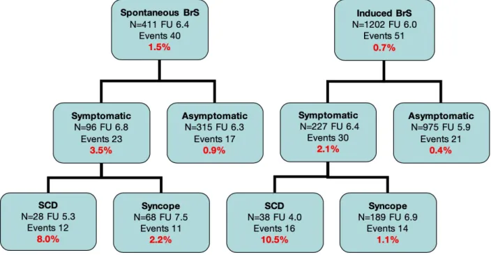
These differences could be explained by the fact that initial cohorts included patients at higher risk, by recruitment bias or by center effect. Our cohort of 1613 BrS patients followed during a median period of 5.2[1.9;9.6] years showed an annual rate of recurrence of 8% among spontaneous BrS ECG pattern patients with history of aborted SCD, 2.2% among spontaneous BrS ECG pattern patients with history of syncope, 10.5% among induced BrS ECG pattern patients with history of SCD, 1.1% among induced BrS ECG pattern patients with history of syncope. These high rates of recurrence justify the implantation of defibrillation devices in symptomatic patients despite their potential complications.
On the other hand, asymptomatic patients are at lower risk of arrhythmic event since overall annual rates of occurrence range are between 0.5% and 0.8%. However, one
should not forget this still represents more than 100 times the risk in general population6
which emphasize the necessity of a more precise risk stratification among these asymptomatic patients.
AGE
Annual rates of ventricular arrhythmias recurrences are made on assumption that events rates are constant over time. In our cohort of 1613 patients, annual rate of event was 2.0% in patients younger than 17, 1.0% in patients between 17 and 59 and 0.3% in patients older than 59.
There is only few data concerning young patients, especially under 18, so prevalence of BrS in children isn’t precisely defined, and even if BrS in children is rarer than in adults, fatal arrhythmic event can occur even at young age1,7,9, more likely during a fever episode10.
In a small cohort of 95 patients under 19, Corcia and al.11 found, after a median follow-up
of 33.8[9.0;97.7] months, an overall 1.9% annual rate of events, 6.9% in previously symptomatic patients and 0.28% in asymptomatic patients. When BrS is symptomatic in children, it seems to be a far more severe form of the disease.
In elderly patients, some data suggests a lower risk after 60 years old12, data from
our cohort supports this assumption. However, patients over 60 presenting with symptoms and newly diagnosed with BrS should undergo ICD implantation as recommended by latest expert consensus report.
Therefore, the period between 30 and 50 seems to represent the critical period for arrhythmias events. Mean age of first event is reported around 40, events are rarer before 20 or after 60 years old13. One hypothesis relies on testosterone levels14.
GENDER CONSIDERATION
Despite equal genetic transmission of affected genes, all large studies showed that BrS is 8 to 10 times more prevalent in males than in women. Furthermore, women seem to display less severe form of BrS than men.
In the largest cohort of BrS women with 494 female patients, Berthome and al.15
showed that women were less symptomatic at baseline (69% vs. 77%, p<0.001), showed less frequently spontaneous BrS pattern ECG (17% vs. 29%, p<0.001), and had a lower annual rate of arrhythmia events (0.5% in asymptomatic women with spontaneous BrS ECG pattern, 0.4% in women with induced BrS ECG pattern). Also, mean age at first event was higher in women than in men (48.6±17.8 years vs. 43±14.2 years, p<0.001). In multivariate analysis, predictors of arrhythmias recurrence were proband status (HR 10.2, p=0.01), aborted SCD (HR 69.4, p<0.0001), syncope (HR 6.8, p=0.02), QRS fragmentation (HR 20.2, p=0.02, leading to an annual rate of arrhythmias recurrence of 3.8% in asymptomatic women with fragmented QRS in this study) and QRS duration above 120ms (HR 4.7, p=0.03).
Another study from Sieira and al.16 comparing 228 women and 314 men affecting by
BrS found an annual rate of arrhythmias recurrence of 0.7% in women and 1.9% in men. Clinical presentation was also more favorable in women in this study with less symptomatic
patients and less spontaneous BrS pattern ECG in the women group. Predictors of recurrence were aborted SCD (HR 25.4, p<0.01) contrary to the presence of syncope (HR 1.4, p=0.69), and presence of SND (HR 9.1, p=0.04).
It should be noted that, in these two studies, spontaneous BrS pattern ECG does not appear to be a predictor of arrhythmias recurrence.
Possible hypothesis explaining these gender differences in BrS are a smaller Ito current in the RVOT in women17 and differences in testosterone levels14,18,19.
FAMILY BACKGROUND
Majority of large studies had failed to highlight a relation between family history of
SCD and future personal risk of VF8,9,20–24, even considering only SCD in first relative or SCD
in first relative at a young age25,26.
In a 2017 Sieira and al.27 study designed to build a risk stratification score, although
globally a history of family SCD was not statistically associated with future arrhythmic events, considering SCD in first degree relative younger than 35 years old only conferred a three-fold VF risk (HR 2.9; 95% CI [1.2;7.0], p=0.02).
However, in 2 studies from 20169 and 201711, by the same group of authors,
focalizing on risk assessment in young patients, family history of SCD was not statistically related to future rhythmic events no matter age of occurrence or level of degree-relative.
GENETIC ASPECTS
Genetic testing is positive in up to 35% of BrS patients and in most cases (75%) it
involves one of the 300 known mutations of the SCN5A gene28,29 (encoding the alpha
Few small studies reported correlation between genetic and risk of arrhythmic event in BrS30,31. Nevertheless, relationship between presence, type or precise site of SCN5A
mutations and risk of arrhythmic event has not been made in large cohorts of BrS patients8,13,21,32.
Furthermore, whereas BrS was considered as a monogenic disease with an autosomal dominant mode of transmission with incomplete penetrance, recent findings, studying genotype/phenotype correlations among individuals of BrS families, highlighted the fact that BrS inheritance could be much more complex with presence of multiple
genetic factors modulating the risk of presence of BrS ECG phenotype29.
ECG CHARACTERISTICS
BRUGADA ECG PATTERN
Showing a spontaneous BrS ECG pattern has been constantly related to a greater risk of arrhythmic events in large Brugada cohorts5,8,20.
In the FINGER study, among asymptomatic patients, showing a spontaneous BrS ECG pattern was associated with a 0.81%/year risk versus a 0.35%/year risk in induced BrS ECG pattern patients, among patients with previous history of syncope, risk varied between 2.3%/year in spontaneous BrS ECG pattern patients and 1.44% in induced BrS ECG pattern patients8.
In a study published in 2016, focusing on prognosis of drug induced BrS patients, Sieira and al.33 showed an annual arrhythmic recurrence rate of 0.4% in asymptomatic
induced BrS ECG pattern patients vs. 1.2% in asymptomatic spontaneous BrS ECG pattern patients. It should be noted that during follow-up, 13.7% of initially drug induced BrS ECG pattern showed a spontaneous BrS ECG pattern, reminding BrS ECG pattern is fluctuant over time and risk assessment should be repeated on regular basis.
Besides, evaluation of BrS ECG pattern burden by 24-hours 12-leads ECG monitoring could be useful to improve risk stratification as it was retrospectively linked to
a greater risk of ventricular arrhythmias34 but another study did not find this relationship35
and real life application could be technically difficult.
Concerning fever-induced BrS ECG pattern patients, a retrospective study from
Mizusawa36 reported an annual risk, after a 43.6±37.4 months follow-up, ranging from 3.0%
in patients with previous history of VF, 1.3% in patients with previous history of syncope, to 0.9% in asymptomatic patients.
FRAGMENTED QRS
Fragmented QRS (fQRS), defined by the presence of an abnormal QRS complex
fragmentation with ³4 spikes in 1 or ³8 spikes in all of the leads V1, V2 and V337 has been
repeatedly linked to a worst prognosis20,37–41. In order to asses properly the presence or
absence of fQRS, special attention should be made about ECG filters since it requires low pass filter with high frequency cut-off (150Hz).
In our cohort of BrS patients, the presence of fQRS was associated with a greater risk of ventricular events (HR 3.37; 95% CI [1.37;8.32], p=0.008). Fragmented QRS were found only in 1.5% (N=25) of BrS patients, but among those 25 patients, 5 had recurrences of ventricular arrhythmias.
As previously mentioned, in Berthome and al.15 study, in women, fQRS was found to
be an independent risk factor in multivariate analysis (HR 20.2, p=0.02, leading to an annual rate of arrhythmias recurrence of 3.8% in asymptomatic women with fragmented QRS in this study).
Two 2017 meta-analysis by Meng and al.42 and Rattanawong and al.43 highlighted a
three to four fold risk in fQRS patients (respectively HR 3.88; 95% CI [2.26;6.65], p<0.00001 and HR 3.36; 95% CI [2.09;5.38], p<0.001).
VENTRICULAR LATE POTENTIALS
Ventricular Late Potentials (LP) are based on the analysis, using signal average ECG, of the total filtered QRS duration, the root mean square voltage of the 40ms terminal portion
of the QRS (RMS40) and the duration of the low amplitude electric potential component
(<40μV) of the terminal portion of the QRS (LAS40). LP are considered positive when 2 of 3
criteria are met.
A Yoshioka study44 reported, in BrS patients, a prolonged total filtered QRS duration,
a prolonged LAS40 and a diminution of RMS40 at night compared with the day.
One study45 with 11 BrS patients reported differences in modifications of these
parameters compared to healthy individuals during Isoproterenol infusion. In BrS patients
decreased RMS40 and prolonged LAS40 were reported after Isoproterenol infusion,
whereas total filtered QRS duration was unchanged. This could be consistent with an intraventricular conduction disturbance of low amplitude revealed during Isoproterenol infusion.
A 43 BrS patients prospective study46 reported an increased arrhythmic risk in BrS
patients with positive LP (HR 10.9, 95% CI [1.1;104.3], p=0.038).
However, further prospective data from bigger studies is needed in order to confirm these results.
S-WAVE IN LEAD D1 (S1 PATTERN)
Conduction delay in RVOT is a suggested pathophysiological basis in BrS47–50. Calo
and al.24 hypothesized that a deep or large S1 pattern could reflect conduction delay in
RVOT and therefore constitute an attractive tool for risk assessment. They included 347 patients without previous history of SCD (78.4% male; mean age 45±13.1 years; mean follow-up was 48±38.6 months), in whom 59.1% showed a S1 pattern. Amplitude, mean
duration and mean amplitude duration area of S1 were significantly higher in patients who developed arrhythmic events compared to patients who presented a syncope than in patients who remained asymptomatic. For clinical practice proposed cut-off were
amplitude ≥1mV, duration ≥40ms and mean amplitude area ≥1mm2. Interestingly, invasive
RV mapping was performed in some patients with (8) and without (4) S1 pattern showing significantly longer endocardial activation time in S1 patients with significant delays in RVOT, more fragmented and abnormal electrograms, lower mean voltage in RVOT and bigger areas of abnormal voltage, localized exclusively in the RVOT region.
EARLY REPOLARIZATION PATTERN (ERP)
ERP is defined by elevation of J-point ³0.1mm (0.1 mV) in ³2 contiguous inferior (DII, DII and VF) and/or lateral (D1, VL, V4-V6) leads in which J-point is the peak of an end QRS
notch and/or the onset of an end QRS slur51. ERP is a common ECG finding seen in 2-5%
of the general population but BrS patients seem to display ERP more frequently than general population.
BrS and ERP are often gathered into “J-wave syndrome” appellation52 since both
syndromes shared some clinical and pathophysiological mechanisms. Both of them are associated with life threatening ventricular arrhythmias, occurring predominantly in young male patients without structural heart disease, and triggered by short-coupled premature
ventricular complex during sleep or low level of physical activity53–55. In both syndromes,
ECG patterns are dynamic and accented by vagal tone, corporal temperature. During electrical storm, administration of B-adrenergic agonists, quinidine or temporary pacing lead to an amelioration of symptoms in both cases.
Nevertheless, differences should be noticed: regions in which ECG abnormalities are seen are different since BrS pattern is found in right precordial leads corresponding to the right ventricular outflow track while ERP is found in inferior and/or lateral leads
corresponding to inferior and/or lateral left ventricle walls; sodium blocker test leads to an increase of BrS pattern and decrease of ER pattern; incidence of late potential signal on average ECG and incidence of atrial fibrillation are higher in BrS patients compared to ERP patients. Furthermore, precise pathophysiological mechanisms underlying BrS and ERP are not fully understood as it remains a matter of debate between the depolarization and the repolarization hypotheses.
Controversial data exist about value of ERP in BrS patients. Among the five main first studies exploring arrhythmic risk of ERP in BrS patients 3 of them reported higher arrhythmic risk in ERP patients, one reported higher risk in inferolateral ERP only and the other one did not. These five studies were gathered into a meta-analysis by Georgopoulos et al., aggregating a total of 1375 patients, reporting that patients with BrS displaying ERP had a statistically higher risk of arrhythmic events compared to those without ERP and that an inferolateral pattern was at higher risk but without statistical significance.
REPOLARIZATION DISPERSION
Tpeak-Tend (Tpe) interval is calculated by the difference between QT interval duration (measured between QRS onset and end of T-wave) and QT peak interval duration (measured between QRS onset and T-wave peak) on leads V1 to V4. It has been proposed has a marker of repolarization dispersion between endocardium and epicardium. Maury
and al.56 showed in a 325 BrS patient cohort that increased Tpe interval was an independent
predictor of arrhythmic occurrences (OR 9.61; 95% CI [3.13;29.41], p<0.0001) using a
100ms cut-off value. These observations were as well made in previous smaller study57,58.
Besides, in a recent meta-analysis59 studying Tpe interval for risk stratification in different
cardiac diseases in general population, Tpe interval prolongation was associated with arrhythmic or mortality outcomes in general population with highest risk in the subgroup of BrS patients constituted from 6 different studies (OR 5.68; 95% CI [1.57;20.53], p=0.001).
T-Wave Alternans (TWA), defined by beat-to-beat fluctuation in T-wave amplitude, reflects also repolarization dispersion and high TWA variation has been linked to worst
prognosis in BrS patients in some studies60,61 but confirmation is needed.
AVR SIGN
In a 2007 study62, the aVR sign, defined as R wave ≥ 0.3 mV or R/q ≥ 0.75 in lead
aVR, has been described as a risk factor for recurrence of arrhythmias in BrS patients. The R wave amplitude or R/q ratio in lead aVR was significantly greater in patients experiencing a recurrence, since 84% of BrS patients with presence of aVR sign but only 27% of BrS patients without aVR sign had events during follow-up. It should be noted that these results are based only on a 24 patients cohort.
SINUS NODE DYSFUNCTION
Sinus Node Dysfunction (SND) is clinically defined by sinus bradycardia according to age and activity, sinus pauses ≥2.5 seconds and/or chronotropic incompetence (impossibility to reach 85% of age predicted maximum heart rate during exercise). SND has sometimes been linked to rhythmic events in particular in young patients9,11 and
women16. Nonetheless, further investigation in large studies is needed to confirm this
hypothesis.
PROGRAMMED ELECTRICAL STIMULATION (PES)
Controversial data exists about the use of PES in risk stratification for BrS patients.
Although some data, especially first papers from Brugada and al.22,23, initially suggests its
prognostic significance, following studies failed to confirm a link between positive PES and
were inducible using a stimulation protocol with up to 3 extrastimuli in 2 sites (RV apex and RVOT). After a mean follow-up of 36±8 months, no statistical difference was found between induced patients and non-induced patients regarding arrhythmias recurrence (p=0.67). Using a 2 extrastimuli protocol, inducibility rate dropped to 20.5%, but in this subgroup no statistical difference was neither found (p=0.89). Consequently, the Expert Consensus Paper in 2013 dropped indication for ICD implantation after PES induced arrhythmia from
IIa to IIb3. Notably, questions were raised about patients in whom PES should be
performed, stimulation protocols used, outcomes definition, and PES reproducibility.
Sieira and al. in 2015 published a study63 with 404 patients with a mean follow up of
74.3±57.3 months. Ventricular arrhythmias were induced in 18.1% of patients using stimulation protocol with up to 3 extrastimuli in the RV apex. PES inducibility was related to arrhythmias recurrence (HR 8.3; 95% CI [3.6;19.4], p<0.01).
A meta-analysis from Sroubek and al. in 201664 pooling 1312 patients without
personal history of aborted SCD found that, after adjustments for age, sex, cohort, ECG and symptoms presentation, a positive PES with up to 2 extrastimuli was related with more
arrhythmia events in BrS patients (HR 2.66; 95% CI [1.44;4.89], p=0.002). Using a 2
extrastimuli protocol, inducibility rate was 21%. After linear regression, a third extrastimulus was not statistically associated with arrhythmias recurrence. In asymptomatic patients with spontaneous BrS ECG pattern, recurrence rate was 0.78% in the non-induced group and 1.7% in the induced group which could change patient management, leading to discuss ICD indication.
Thus, PES should only be performed to precise risk stratification in asymptomatic patients with intermediate risk according to clinical status such as asymptomatic patients with spontaneous BrS ECG pattern. An aggressive stimulation protocol increase sensitivity but could lead to a diminution of specificity, stimulation in 2 RV sites (RVOT and apex) and with up to 2 extrastimuli seems to represent a good compromise. PES positivity is defined
by induction of sustained ventricular arrhythmia (VF or VT lasting more than 30 seconds or requiring external shock). PES negativity does not correctly identify low risk patients and risk stratification should be assessed according to clinical status. Reproducibility is still a matter of concern since in the PRELUDE study ventricular arrhythmias were induced in only
34% of previously inducible patients20 contrary to another study from Gasparini and al.65
that found a reproducibility rate of 82% but with only 21 patients included.
PROPOSED COMPOSITE SCORES
In order to help risk stratification in asymptomatic patients, composite scores could be useful given the fact that single risk factors aren’t able to correctly enough discriminate low risk and high risk patients26,66.
In 2014 a score has been proposed by Kawazoe and al.67 with clinical data and
mainly ECG criteria. History of syncope, QRS duration in V6, r-J interval in V1 and Tpe dispersion were included. Nevertheless, even if this score has a good performance (AUC 0.845, sensibility 97.1% and specificity 63%), it was retrospectively built on data from only 143 patients whom 140 were males and provides a future VF probability without precise time range. Furthermore, rely on electrical data such as QRS duration or Tpe dispersion, whose precise measurement are known to be tough in real life68–70, in order to decide
whether or not an ICD should be implanted, sometimes in young patients, might be tricky. Another score has been as well proposed by Sieira and al.27 to assessed risk of
arrhythmias recurrences in BrS patients (Table 1A). A score ³2 conferred an arrhythmic risk greater than 6% at 5 years (9.2%) and has been proposed as a cut off for ICD implantation by the authors since it’s the cut off used in hypertrophic cardiomyopathy (Table 2B). Despite such a score is needed to simplify assessment of arrhythmic risk and management in BrS patients, it is not certain that it allows a correct discrimination of intermediate risk patients representing the group in need for a more accurate risk assessment.
Table 1A: Proposed score model for risk
stratification Table 1B: Event free survival rate according to the score model group
Risk factor Points Score 1 year 5 years 10 years p
Spontaneous Type 1 ECG 1 0 100% 98.4% 97.2%
Early familial SCD (<35yo) 1 1 100% 96.4% 96.4% 0.79
Inducible PES 2 2 97.4% 90.8% 90.8% 0.02
Syncope 2 3 88.7% 83.4% 83.4% <0.01
Sinus Node Dysfunction 3 4 91.4% 75.2% 70.1% <0.01
Aborted SCD 4 ³5 79.3% 68.2% 61.4% <0.01
Finally, a score for risk assessment in patients under 19 years old has as well been
elaborated11 using presence of symptoms (4 points), spontaneous BrS ECG pattern (3
points), SND and/or atrial tachycardia (2 points) and conduction abnormalities (1 points). However, a cut-off of 4 points is considered to discriminate low risk and high risk patients but due to the construction score the results are mainly driven by symptoms which lowers its clinical utility.
REFERENCES
1. Brugada P, Brugada J. Right bundle branch block, persistent ST segment elevation and sudden cardiac death: A distinct clinical and electrocardiographic syndrome. A multicenter report. J Am Coll
Cardiol. 1992;20(6):1391-1396. doi:10.1016/0735-1097(92)90253-J.
2. Dumaine R, Towbin J a, Brugada P, et al. Ionic mechanisms responsible for the electrocardiographic phenotype of the Brugada syndrome are temperature dependent. Circ Res. 1999;85(9):803-809. doi:10.1161/01.RES.85.9.803.
3. Priori SG, Wilde AA, Horie M, et al. Executive summary: HRS/EHRA/APHRS expert consensus statement on the diagnosis and management of patients with inherited primary arrhythmia syndromes.
J Arrhythmia. 2014;30(1):29-47. doi:10.1016/j.joa.2013.08.001.
4. Sacher F, Probst V, Maury P, et al. Outcome after implantation of a cardioverter-defibrillator in patients with Brugada syndrome: A multicenter study-part 2. Circulation. 2013;128(16):1739-1747. doi:10.1161/CIRCULATIONAHA.113.001941.
5. Gourraud J, Barc J, Thollet A, Le H, Probst V. Brugada syndrome : Diagnosis , risk. Arch Cardiovasc
Dis. 2017;110(3):188-195. doi:10.1016/j.acvd.2016.09.009.
6. Sieira J, Brugada P. Brugada Syndrome: Defining the Risk in Asymptomatic Patients. Arrhythmia
Electrophysiol Rev. 2016;5(3):164. doi:10.15420/aer.2016:22:3.
7. Sacher F, Arsac F, Wilton SB, et al. Syncope in Brugada syndrome patients: Prevalence,
characteristics, and outcome. Hear Rhythm. 2012;9(8):1272-1279.
doi:https://doi.org/10.1016/j.hrthm.2012.04.013.
8. Probst V, Veltmann C, Eckardt L, et al. Long-Term Prognosis of Patients Diagnosed With Brugada Syndrome: Results From the FINGER Brugada Syndrome Registry. Circulation. 2010;121(5):635-643. doi:10.1161/CIRCULATIONAHA.109.887026.
9. Gonzalez Corcia MC, Sieira J, Sarkozy A, et al. Brugada syndrome in the young: an assessment of risk factors predicting future events. Europace. 2016:euw206. doi:10.1093/europace/euw206. 10. Probst V, Denjoy I, Meregalli PG, et al. Clinical aspects and prognosis of Brugada syndrome in
children. Circulation. 2007;115(15):2042-2048. doi:10.1161/CIRCULATIONAHA.106.664219.
11. Gonzalez Corcia MC, Sieira J, Pappaert G, et al. A Clinical Score Model to Predict Lethal Events in Young Patients (≤19 Years) With the Brugada Syndrome. Am J Cardiol. 2017;120(5):797-802. doi:10.1016/j.amjcard.2017.05.056.
12. Conte G, Asmundis CDE, Ph D, et al. Clinical Characteristics , Management , and Prognosis of Elderly Patients with Brugada Syndrome. J Cardiovasc Electrophysiol. 2014;25:514-519. doi:10.1111/jce.12359.
13. Adler A, Rosso R, Chorin E, Havakuk O, Antzelevitch C, Viskin S. Risk stratification in Brugada syndrome: Clinical characteristics, electrocardiographic parameters, and auxiliary testing. Hear
Rhythm. 2015. doi:10.1016/j.hrthm.2015.08.038.
14. Ezaki K, Nakagawa M, Taniguchi Y, et al. Gender Differences in the ST Segment. Circ J. 2010;74(11):2448-2454. doi:10.1253/circj.CJ-10-0221.
15. Berthome P, Tixier R, Briand J, et al. Clinical presentation and follow-up of women affected by Brugada syndrome. Manuscript submitted for publication. 2018.
16. Sieira J, Conte G, Ciconte G, et al. Clinical characterisation and long-term prognosis of women with Brugada syndrome. 2016:452-458. doi:10.1136/heartjnl-2015-308556.
17. Di Diego JM, Cordeiro JM, Goodrow RJ, et al. Ionic and cellular basis for the predominance of the Brugada syndrome phenotype in males. Circulation. 2002;106(15):2004-2011. doi:10.1161/01.CIR.0000032002.22105.7A.
18. Matsuo K, Akahoshi M, Seto S, Yano K. Disappearance of the Brugada-type electrocardiogram after surgical castration: a role for testosterone and an explanation for the male preponderance. Pacing Clin
Electrophysiol. 2003;26(July):1551-1553. doi:10.1046/j.1460-9592.2003.t01-1-00227.x.
19. Shimizu W, Matsuo K, Kokubo Y, et al. Sex hormone and gender difference - Role of testosterone on male predominance in Brugada syndrome. J Cardiovasc Electrophysiol. 2007;18(4):415-421. doi:10.1111/j.1540-8167.2006.00743.x.
20. Priori SG, Gasparini M, Napolitano C, et al. Risk stratification in brugada syndrome: Results of the PRELUDE (PRogrammed ELectrical stimUlation preDictive valuE) registry. J Am Coll Cardiol. 2012;59(1):37-45. doi:10.1016/j.jacc.2011.08.064.
21. Priori SG, Napolitano C, Gasparini M, et al. Natural history of Brugada syndrome: Insights for risk stratification and management. Circulation. 2002;105(11):1342-1347. doi:10.1161/hc1102.105288. 22. Brugada J, Brugada R, Antzelevitch C, Towbin J, Nademanee K, Brugada P. Long-term follow-up of
individuals with the electrocardiographic pattern of right bundle-branch block and ST-segment elevation in precordial leads V1 to V3. Circulation. 2002;105(1):73-78. doi:10.1161/hc0102.101354. 23. Brugada J, Brugada R, Brugada P. Determinants of Sudden Cardiac Death in Individuals with the
2003;108(25):3092-3096. doi:10.1161/01.CIR.0000104568.13957.4F.
24. Calò L, Giustetto C, Martino A, et al. A New Electrocardiographic Marker of Sudden Death in Brugada Syndrome: The S-Wave in Lead i. J Am Coll Cardiol. 2016;67(12):1427-1440. doi:10.1016/j.jacc.2016.01.024.
25. Sarkozy A, Sorgente A, Boussy T, et al. The value of a family history of sudden death in patients with diagnostic type i Brugada ECG pattern. Eur Heart J. 2011;32(17):2153-2160. doi:10.1093/eurheartj/ehr129.
26. Delise P, Allocca G, Marras E, et al. Risk stratification in individuals with the Brugada type 1 ECG pattern without previous cardiac arrest: Usefulness of a combined clinical and electrophysiologic approach. Eur Heart J. 2011;32(2):169-176. doi:10.1093/eurheartj/ehq381.
27. Sieira J, Conte G, Ciconte G, et al. A score model to predict risk of events in patients with Brugada Syndrome. Eur Heart J. 2017;38(22):1756-1763. doi:10.1093/eurheartj/ehx119.
28. Lieve KV V, Wilde AAM. Inherited ion channel diseases: A brief review. Europace. 2015;17:ii1-ii6. doi:10.1093/europace/euv105.
29. Gourraud J-B, Barc J, Thollet A, et al. The Brugada Syndrome: A Rare Arrhythmia Disorder with Complex Inheritance. Front Cardiovasc Med. 2016;3(April):1-11. doi:10.3389/fcvm.2016.00009. 30. Meregalli PG, Tan HL, Probst V, et al. Type of SCN5A mutation determines clinical severity and degree
of conduction slowing in loss-of-function sodium channelopathies. Hear Rhythm. 2009;6(3):341-348. doi:10.1016/j.hrthm.2008.11.009.
31. Nakano Y, Ochi H, Onohara Y, et al. Common variant near HEY2 has a protective effect on ventricular fibrillation occurrence in Brugada syndrome by regulating the repolarization current. Circ Arrhythmia
Electrophysiol. 2016;9(1):1-2. doi:10.1161/CIRCEP.115.003436.
32. Maury P, Rollin A, Sacher F, et al. Prevalence and Prognostic Role of Various Conduction Disturbances in Patients With the Brugada Syndrome. Am J Cardiol. 2013;112(9):1384-1389. doi:10.1016/j.amjcard.2013.06.033.
33. Sieira J, Ciconte G, Conte G, et al. Long-term prognosis of drug-induced Brugada syndrome. Hear
Rhythm. 2017;133(0):622-630. doi:10.1016/j.hrthm.2017.04.044.
34. Extramiana F, Maison-Blanche P, Badilini F, Messali A, Denjoy I, Leenhardt A. Type 1 electrocardiographic burden is increased in symptomatic patients with Brugada syndrome. J
Electrocardiol. 2010;43(5):408-414. doi:10.1016/j.jelectrocard.2010.06.011.
35. Behar N, Petit B, Probst V, et al. Heart rate variability and repolarization characteristics in symptomatic and asymptomatic Brugada syndrome. 2016. doi:10.1093/europace/euw224.
36. Mizusawa Y, Morita H, Adler A, et al. Prognostic significance of fever-induced Brugada syndrome.
Hear Rhythm. 2016;13(7):1515-1520. doi:10.1016/j.hrthm.2016.03.044.
37. Morita H, Kusano KF, Miura D, et al. Fragmented QRS as a marker of conduction abnormality and a predictor of prognosis of Brugada syndrome. Circulation. 2008;118(17):1697-1704. doi:10.1161/CIRCULATIONAHA.108.770917.
38. Kamakura S, Ohe T, Nakazawa K, et al. Long-term prognosis of probands with brugada-pattern ST-elevation in leads V 1-V 3. Circ Arrhythmia Electrophysiol. 2009;2(5):495-503. doi:10.1161/CIRCEP.108.816892.
39. Tokioka K, Kusano KF, Morita H, et al. Electrocardiographic parameters and fatal arrhythmic events in patients with brugada syndrome: Combination of depolarization and repolarization abnormalities. J
Am Coll Cardiol. 2014;63(20):2131-2138. doi:10.1016/j.jacc.2014.01.072.
40. Morita H, Watanabe A, Morimoto Y, et al. Distribution and prognostic significance of fragmented QRS in patients with brugada syndrome. Circ Arrhythmia Electrophysiol. 2017;10(3):2-5. doi:10.1161/CIRCEP.116.004765.
41. de Asmundis C, Mugnai G, Chierchia GB, et al. Long-Term Follow-Up of Probands With Brugada Syndrome. Am J Cardiol. 2017;119(9):1392-1400. doi:10.1016/j.amjcard.2017.01.039.
42. Meng L, Letsas KP, Baranchuk A, et al. Meta-analysis of fragmented QRS as an electrocardiographic predictor for arrhythmic events in patients with Brugada syndrome. Front Physiol. 2017;8(SEP):1-7. doi:10.3389/fphys.2017.00678.
43. Rattanawong P, Riangwiwat T, Prasitlumkum N, et al. Baseline fragmented QRS increases the risk of major arrhythmic events in Brugada syndrome: Systematic review and meta-analysis. Ann
Noninvasive Electrocardiol. 2017;(September):1-8. doi:10.1111/anec.12507.
44. Yoshioka K, Amino M, Zareba W, et al. Identification of High-Risk Brugada Syndrome Patients by Combined Analysis of Late Potential and T-Wave Amplitude Variability on Ambulatory Electrocardiograms. Circ J. 2013;77(3):610-618. doi:10.1253/circj.CJ-12-0932.
45. Takagi A, Nakazawa K, Sakurai T. Prolongation of LAS40 (Duration of the Low Amplitude Electric Potential Component (<40μV) of the Terminal Portion of the QRS) Induced by Isoproterenol in 11 Patients With Brugada Syndrome. Circ J. 2002;66(December):1101-1104.
stratification of patients with Brugada syndrome: A prospective study. Hear Rhythm. 2009;6(8):1156-1162. doi:10.1016/j.hrthm.2009.05.007.
47. Wilde AAM, Postema PG, Di Diego JM, et al. The pathophysiological mechanism underlying Brugada syndrome: depolarization versus repolarization. J Mol Cell Cardiol. 2010. doi:10.1016/j.yjmcc.2010.07.012.
48. Lambiase PD, Ahmed AK, Ciaccio EJ, et al. High-density substrate mapping in brugada syndrome: Combined role of conduction and repolarization heterogeneities in arrhythmogenesis. Circulation. 2009;120(2):106-117. doi:10.1161/CIRCULATIONAHA.108.771401.
49. Hoogendijk MG, Potse M, Coronel R. Critical appraisal of the mechanism underlying J waves. J
Electrocardiol. 2013. doi:10.1016/j.jelectrocard.2013.06.017.
50. Sacher F, Jesel L, Jais P, Haïssaguerre M. Insight into the mechanism of Brugada syndrome: Epicardial substrate and modification during ajmaline testing. Hear Rhythm. 2014;11(4):732-734. doi:10.1016/j.hrthm.2013.05.023.
51. MacFarlane PW, Antzelevitch C, Haissaguerre M, et al. The early repolarization pattern: A consensus paper. J Am Coll Cardiol. 2015;66(4):470-477. doi:10.1016/j.jacc.2015.05.033.
52. Antzelevitch C, Yan G-X, Ackerman MJ, et al. J-Wave syndromes expert consensus conference report: Emerging concepts and gaps in knowledge Electrophysiology (Sociedad Latinoamericana de Estimulació n Cardíaca y Electrofisiología [SOLAECE]) Europace Advance Access. Finnish Acad Sci
Sigrid Juselius Found. 2016;32(5):26-40. doi:10.1093/europace/euw235.
53. Nam G-B, Kim Y-H, Antzelevitch C. Augmentation of J waves and electrical storms in patients with early repolarization. N Engl J Med. 2008;358:2078-2079. doi:10.1056/NEJMc0708182.
54. Haïssaguerre M, Derval N, Sacher F, et al. Sudden cardiac arrest associated with early repolarization.
N Engl J Med. 2008;358(19):2016-2023. doi:358/19/2016 [pii] 10.1056/NEJMoa071968.
55. Rosso R, Kogan E, Belhassen B, et al. J-Point Elevation in Survivors of Primary Ventricular Fibrillation and Matched Control Subjects. Incidence and Clinical Significance. J Am Coll Cardiol. 2008;52(15):1231-1238. doi:10.1016/j.jacc.2008.07.010.
56. Maury P, Sacher F, Gourraud JB, et al. Increased Tpeak-Tend interval is highly and independently
related to arrhythmic events in Brugada syndrome. Hear Rhythm. 2015.
doi:10.1016/j.hrthm.2015.07.029.
57. Letsas KP, Weber R, Astheimer K, Kalusche D, Arentz T. Tpeak-Tend interval and Tpeak-T end/QT ratio as markers of ventricular tachycardia inducibility in subjects with Brugada ECG phenotype.
Europace. 2010;12(2):271-274. doi:10.1093/europace/eup357.
58. Hevia JC, Antzelevitch C, Bárzaga FT, et al. Tpeak-Tend and Tpeak-Tend Dispersion as Risk Factors for Ventricular Tachycardia / Ventricular Fibrillation in Patients With the Brugada Syndrome. 2006;47(9). doi:10.1016/j.jacc.2005.12.049.
59. Tse G, Gong M, Wong WT, et al. The T peak − T end interval as an electrocardiographic risk marker of arrhythmic and mortality outcomes: A systematic review and meta-analysis. Hear Rhythm. 2017. doi:10.1016/j.hrthm.2017.05.031.
60. Uchimura-Makita Y, Nakano Y, Tokuyama T, et al. Time-domain T-wave alternans is strongly associated with a history of ventricular fibrillation in patients with brugada syndrome. J Cardiovasc
Electrophysiol. 2014. doi:10.1111/jce.12441.
61. Sakamoto S, Takagi M, Kakihara J, et al. The utility of T-wave alternans during the morning in the summer for the risk stratification of patients with Brugada syndrome. Heart Vessels. 2016. doi:10.1007/s00380-016-0882-2.
62. Babai Bigi MA, Aslani A, Shahrzad S. aVR sign as a risk factor for life-threatening arrhythmic events
in patients with Brugada syndrome. Hear Rhythm. 2007;4(8):1009-1012.
doi:10.1016/j.hrthm.2007.04.017.
63. Sieira J, Conte G, Ciconte G, et al. Prognostic Value of Programmed Electrical Stimulation in Brugada Syndrome. Circ Arrhythmia Electrophysiol. 2015;8(4):777-784. doi:10.1161/CIRCEP.114.002647. 64. Sroubek J, Probst V, Mazzanti A, et al. Programmed ventricular stimulation for risk stratification in the
Brugada syndrome: A pooled analysis. Circulation. 2016;133(7):622-630.
doi:10.1161/CIRCULATIONAHA.115.017885.
65. Gasparini M, Priori SG, Mantica M, Coltorti F, Napolitano C, Galimberti P, Bloise R CC. Programmed electrical stimulation in Brugada syndrome: how reproducible are the results? J Cardiovasc
Electrophysiol. 2002;13(9):880-887.
66. Delise P, Allocca G, Sitta N, Di Stefano P. Event rates and risk factors in patients with Brugada syndrome and no prior cardiac arrest: A cumulative analysis of the largest available studies distinguishing ICD-recorded fast ventricular arrhythmias and sudden death. Hear Rhythm. 2014;11(2):252-258. doi:10.1016/j.hrthm.2013.10.039.
67. Kawazoe H, Nakano Y, Tokuyama T, et al. Risk Stratification of Ventricular Fibrillation in Brugada Syndrome Using Non-invasive Scoring Methods. Circulation. 2014;130(Suppl 2):A13486-A13486.
doi:10.1016/j.hrthm.2016.07.009.
68. Jain R, Tandri H, Daly A, et al. Reader- and instrument-dependent variability in the electrocardiographic assessment of arrhythmogenic right ventricular dysplasia/cardiomyopathy. J
Cardiovasc Electrophysiol. 2011;22(5):561-568. doi:10.1111/j.1540-8167.2010.01961.x.
69. De Guillebon M, Thambo JB, Ploux S, et al. Reliability and reproducibility of QRS duration in the selection of candidates for cardiac resynchronization therapy. J Cardiovasc Electrophysiol. 2010;21(8):890-892. doi:10.1111/j.1540-8167.2010.01743.x.
70. Sarubbi B, Li W, Somerville J. QRS width in right bundle branch block. Accuracy and reproducibility of manual measurement. Int J Cardiol. 2000;75(1):71-74. doi:10.1016/S0167-5273(00)00299-0.
EARLY REPOLARIZATION PATTERN IS NOT
RELATED WITH VENTRICULAR ARRHYTHMIAS IN
BRUGADA PATIENTS
ABSTRACT
Introduction: Brugada syndrome (BrS) is an inherited arrhythmia syndrome with an
increased risk of syncope and sudden death. The risk stratification of BrS patients remains challenging. An Early Repolarization pattern (ERP) has been suggested to increase the risk of ventricular arrhythmias. The aim of this study was to investigate the association between BrS and ERP with arrhythmic events in the largest cohort of BrS patients ever described.
Methods: Consecutive patients affected with BrS were recruited from 15 tertiary centers in
France between 1994 and 2015. Data were prospectively collected with an average follow-up of 6.1± 4.6 years. ECGs were reviewed by 2 physicians blinded to clinical status. ERP was defined as a 0.1 mV J wave elevation in at least 2 inferior and/or lateral leads.
Results: A total of 1613 patients (mean age 45±15 years; 1119 males, 69%) were enrolled
in this study. Three hundred and twenty-three patients (20%) were symptomatic at baseline (66 (4%) aborted sudden cardiac death (SCD) or documented VT, 257 (16%) syncope). Four hundred and eleven patients (25%) presented a spontaneous type 1 ECG. Implantable cardiac defibrillator (ICD) was implanted in 477 patients (30%). At baseline, ERP has been found in 117 patients (7%): 76 patients (5%) presented QRS slurring, 33 (2%) QRS notching and 8 (0.5%) both. ERP were more frequent in male than in female (81% male, p=0.004). No significant difference was found in the age, family history of SCD or symptoms or spontaneous type 1 ECG. During follow-up of 6.1± 4.6 years, 84 of 1496 patients without ERP (6%) and 7 of the 117 patients with ERP (6%) underwent arrhythmic events (1 SCD, 5 appropriate ICD therapy and 1 ventricular arrhythmia). Patients with ERP had not a
significantly higher rate of arrhythmic events than patients without ERP (HR: 1.16; 95% CI: 0.54-2.51; p= 0,7).
Conclusion: In the largest cohort of BrS patients ever described, ERP does not seem to be
a marker of arrhythmic event.
INTRODUCTION
Brugada Syndrome (BrS) is an inherited arrhythmia syndrome first described in 1992
by Pedro and Josep Brugada1 affecting ionic channels mainly located in the anterior right
ventricular outflow track. Most patients remain asymptomatic for life but a minority can develop palpitations, syncope or nocturnal agonal respiration due to ventricular tachycardia (VT) or fibrillation (VF), potentially leading to Sudden Cardiac Death (SCD). While therapeutic management in symptomatic BrS is well defined, consisting of Implantable Cardiac Defibrillator (ICD) implantation2,3, risk stratification in asymptomatic
BrS remains challenging due to a lower rate of events in this young population4 compared
to a significant ICD devices morbidity5,6,7. The Early Repolarization Pattern (ERP) is a
common electrocardiographic (ECG) finding seen in 2-5% of the general population which
was considered benign until it was linked to idiopathic VF in 20088,9,10. These 2 syndromes
are often gathered into the same J-waves syndromes entity since they share some cellular and ionic similarities11,12. ERP has been suggested to increase the risk of ventricular
arrhythmias in BrS and its prognosis significance have already been discussed in several
studies but existing data on the risk linked to ERP in BrS remain controversial6,13–18. The aim
of this study was to investigate the association of the ERP with arrhythmic events in the largest cohort of BrS patients ever described.
METHODS
Consecutive patients affected with BrS were prospectively recruited from 15 tertiary centers in France between February 1994 and July 2015, gathered into the same database held by Nantes University Hospital Center, FRANCE. The data collected were: gender, age at diagnosis, proband status, personal history of symptoms (syncope, documented sustained ventricular arrhythmia or aborted SCD), ECG characteristics, family history of asymptomatic or symptomatic BrS, family history of SCD, presence of ICD in primary or secondary prevention, presence of programmed electrical stimulation at diagnosis and its result, genetic analysis of SCN5A results. All subjects underwent trans-thoracic-echocardiography and if necessary exercise treadmill test or coronary angiography used at treating physician discretion in order to rule out any structural heart disease.
All 12-leads ECG were recorded, according to international standards, at a paper speed of 25 mm/s, amplitude of 10 mm/mV and with 0.16-150Hz filters. ECG were analyzed by two different physicians blinded to clinical status, in case of disagreement, a third lecture by another electrophysiologist physician was performed. BrS was confirmed in presence of spontaneous or induced (Flecainide 2mg/kg I.V. or Ajmaline 1mg/kg I.V.) type 1 ECG which was defined using latest international recommendations by ST segment elevation ³2mm (0.2 mV) in ³1 right precordial leads (V1-V3) positioned in the 4th, 3rd, or 2nd
intercostal space2. ERP was defined, using latest expert consensus report, by elevation of
J-point ³0.1mm (0.1 mV) in ³2 contiguous inferior (DII, DII and VF) and/or lateral (D1, VL,
V4-V6) leads in which J-point is the peak of an end QRS notch and/or the onset of an end
QRS slur19. All ECG were analyzed using ImageJ software (National Institutes of Health,
Bethesda, Maryland, USA). The following variables were measured: heart rate, P wave
duration in D2, PR interval duration in lead D220, QRS duration in all derivations21, QT and
QTc using Bazett formula in lead D2, S wave duration and depth in leads D1 and D222,
elevation in case of BrS in leads V1 to V3, amplitude of J-point elevation and characterization of ST-segment (horizontal or downward sloping) in case ERP in inferior
and/or lateral leads25. When 2 or more ECG were available for the same patient, ECG with
the most pronounced BrS and/or ERP features was selected.
During follow-up, an arrhythmic event was defined by occurrence of syncope, documented sustained VT, appropriate ICD shock, SCD or aborted SCD.
Ethics committees from every center have approved the study, data storage and use was approved by the French Data Protection Authority and consents were obtained for all subjects.
STATISTICAL ANALYSIS
Continuous data are presented as mean (± standard deviation) or median [25th,75th] based on the distribution. Categorical variables are presented as counts (proportions). The t-test and Mann-Whitney-U tests were performed to test for statistical differences in continuous parameters between the two groups. The chi-square or Fisher's exact test (based on expected frequency) were used to compare categorical variables between groups. The Kaplan-Meier method estimator was used to assess the time to a first cardiac event. Cox proportional-hazards regression analysis [with hazard ratios (HR) and confidence intervals (CI)] were used to evaluate the independent risk of clinical and genetic factors of interest for first cardiac events. The mean event rate per year was evaluated by the number of events occurring during the follow-up divided by the number of patients multiplied by the average duration of follow-up. A P-value <0.05 was considered statistically significant. Data analyses were performed using SAS (SAS version 9.4; SAS Institute Inc., Cary, NC).
RESULTS
A total of 1613 patients (45±15 years, 68% male), members of 1260 different families, were enrolled in the study. Demographic data and clinical status at inclusion are shown in table 1. 323 (20%) were symptomatic at baseline. Spontaneous type 1 ECG was found in 411 (25%) patients. ERP was seen in 117 patients (7%), mean J-point elevation was 0.18±0.078mV, 14 patients (12%) had a >0.2mV J-point elevation, 14 (12%) had inferolateral ERP (Figure 1). Their ECG characteristics are shown in table 2. Clinical and
ECG differences between BrS patients with and without ERP are shown in table 3. There
was no difference between the two groups regarding age, probands status, symptoms, family history of SCD, presence of ICD, presence of SCN5A mutation, positive SVP, sinus node dysfunction, P wave >120ms, PR interval >200ms, QRS duration >90ms, QTC>460ms, maximum Tpeak-Tend>100ms, spontaneous type 1 ECG, presence of BrS in peripheral lead, maximum V1 and V2 ST elevation or fragmented QRS. There were statistical differences regarding sex gender (81% male in the ERP group, p=0,004), heart rate (p=0,001), maximum V3 ST elevation (p=0.04) and presence of AvR sign (14% in the non-ERP group versus only 6% in the ERP group, p=0,01).
During a median follow-up of 5.2[1.9;9.6] years, 7 (7%) arrhythmic events occurred in the ERP group (5 appropriate ICD therapies, 1 SCD and 1 documented VT) vs. 84 (6%) in the non-ERP group. Data about BrS patients showing ERP with symptoms during follow-up are shown in table 4. None of these patients were displaying inferolateral ERP nor a >2mm J-point elevation and 5 of them were already symptomatic at baseline.
Using univariate Cox proportional hazard regression analysis, and as shown in figure 2, ERP had not a significantly higher rate of arrhythmic events than patients without ERP (HR: 1.16; 95% CI: 0.54-2.51; p= 0,7).
Figure 3 shows event rates in BrS patients according to ECG presentation and symptoms at baseline.
DISCUSSION
In our study, which is so far the largest cohort of Brugada patients with 1613 individuals followed for a median period of 5.1 years, ERP was seen in 7% of BrS patients, which is slightly more than the incidence observed in the general population, but was not related to a significantly higher rate of arrhythmic events in BrS patients. Interestingly, in BrS patients with ERP who became symptomatic during follow-up, none of them were displaying inferolateral ERP or >2mm J-point elevation which are yet characteristics considered at higher risk.
Among the five main previous studies exploring arrhythmic risk of ERP in BrS patients, results were controversial: 3 of them reported higher arrhythmic risk in ERP patients, one reported higher risk in inferolateral ERP only and the other one did not.
Definition of ERP in these five studies was the same as the definition we used in our protocol and as recommended in the latest consensus report.
These five studies were gathered into a meta-analysis by Georgopoulos et al.26
reporting that patients with BrS displaying ERP had a statistically higher risk of arrhythmic events compared to those without ERP and that an inferolateral pattern was at higher risk but without statistical significance. These studies had fewer patients than ours since their total of patients was 1375 and the largest one, by Kamakura et al. had 460 patients.
In the Letsas et al.15 study, which did not find a statistical relation between ERP and
arrhythmic events in BrS patients, patient’s characteristics were globally similar as the one included in our cohort with 30% of symptomatic BrS including 2.8% of personal aborted SCD or VF history, a male proportion of 70%, a spontaneous type 1 ECG proportion of 45%; ERP proportion was 12% with 31% of inferior ERP, 51% of lateral ERP and 17% of inferolateral ERP; the rate of arrhythmic event was 7% during a smaller follow-up of 3.7±2.3 years.
The Takagi et al.14 study reported a higher arrhythmic risk in BrS patients showing
inferolateral ERP (95% CI:1.4-10.2; p=0.003), ERP was also found in 12% of the BrS patients which were more symptomatic at baseline (42%) with 18% with personal aborted SCD history, were mainly male (94%) and showed a higher proportion of spontaneous type 1 ECG (63%).
In the Kamakura et al.17 study, only probands were included, 17% had personal
aborted SCD history, male proportion was 95%, spontaneous type 1 ECG proportion was 74%, ERP was only found in 10% of BrS patients but all of them showed inferolateral ERP; despite that, event rate remained similar as our study (7%) during a smaller follow up of 4.1±1.3 years.
In the Kawata et al.18 study the 49 patients included were only symptomatic patients
with personal history of aborted SCD, male proportion was 94%, spontaneous type 1 ECG proportion 74%, ERP proportion 63% with 29% of inferolateral ERP, 29% of descending or horizontal ST-segment and a mean J-point amplitude of 0.228±0.122 mV. During a follow-up of 7.6±3.8 years, 71% of patients had another arrhythmic event.
In the Tokioka et al.16 study, patients were also mainly male (95%), 21,5% were
symptomatic at baseline, 5.3% with personal history of aborted SCD, 63% had a spontaneous type 1 ECG, 25% showed an ERP, all of them in the inferolateral leads, there was a 10% rate of arrhythmic events during a follow-up of 3.8±3.7 years.
Differences between these previous results and our study could thus be explained by the fact that majority of patients included in our cohort are lower risk patients at baseline. Particularly, as shown in table 5, in the Kamakura, Kawata and Tokioka studies, which reported higher risk in ERP patients, patients included seem to were at very high risk regarding ERP and/or BrS characteristics with a high proportion of male, spontaneous BrS type 1 ECG, symptomatic BrS patients especially with high proportion of previous aborted
SCD, ERP in inferolateral leads and/or with a >2mV J-point elevation, characteristics which are known for worsening patients’ prognostic4,7,13,25,27–29.
Furthermore, with only 7 patients becoming symptomatic during follow-up among ERP patients, we could have lacked statistical power.
Likewise, studies reporting a higher arrhythmic risk in ERP patients are all Japanese studies. The other one, which don’t report a higher arrhythmic risk in ERP patients is about European patients, like this study. Maybe Asiatic and European patients aren’t part of a homogeneous population considering BrS and/or ERP characteristics. In this case, risk stratification strategy could differ.
BrS and ERP are often gathered into “J-wave syndrome” appellation12 since both
syndromes share some clinical and pathophysiological mechanisms. Both of them are associated with life threatening ventricular arrhythmias, occurring predominantly in young male patients without structural heart disease, and triggered by short-coupled premature ventricular complex during sleep or low level of physical activity. In both syndromes, ECG patterns are dynamic and accented by vagal tone, fever and hypothermia. During electrical storm, administration of B-adrenergic agonists, quinidine or temporary pacing lead to an amelioration of symptoms in both cases. Nevertheless, differences should be noticed: regions in which ECG abnormalities are seen are different since BrS pattern is found in right precordial leads corresponding to the right ventricular outflow track while ER pattern is found in inferior and/or lateral leads corresponding to inferior and/or lateral left ventricle walls; sodium blocker test leads to an increase of BrS pattern and decrease of ER pattern; incidence of late potential signal on average ECG and incidence of atrial fibrillation are higher in BrS patients compared to ERP patients. Furthermore, precise physiopathological mechanisms underlying BrS and ERP are not fully understood as it remains a matter of
Some limitation concerning this work should be noticed, BrS and ERP are dynamic ECG pattern, while all BrS patients in our cohort were confirmed with either spontaneous or drug induced type 1 ECG, some patients showing intermittent ERP pattern could have been misclassified.