UNIVERSITÉ DE SHERBROOKE
AUTOTAXIN PROMOTES CANCER CELL INVASION VIA THE LYSOPHOSPHATIDIC ACID RECEPTOR 4
Par Kelly Harper
Département de pédiatrie, Service d'immunologie
Mémoire présenté à la Faculté de médecine et des sciences de la santé en vue de l'obtention du grade de
maître ès sciences (M.Sc.) en immunologie
15 mai 2010
EVALUATEURS:
Pre. Claire Dubois (programme d'immunologie) Pr. Gilles Dupuis (programme d'immunologie) Pre. Nathalie Rivard (programme de biologie cellulaire)
l+I
NOTICE:
Library and Archives Canada
Published Heritage Branch
395 Wellington Street Ottawa ON K1A ON4 Canada
The author has granted a
non-exclusive license allowing Library and Archives Canada to reproduce, publish, archive, preserve, conserve, communicate to the public by
telecommunication or on the Internet, loan, distribute and sell theses worldwide, for commercial or non-commercial purposes, in microform, paper, electronic and/or any other formats.
The author retains copyright ownership and moral rights in this thesis. Neither the thesis nor substantial extracts from it may be printed or otherwise reproduced without the author's permission.
ln compliance with the Canadian Privacy Act some supporting forms may have been removed from this thesis.
While these forms may be included in the document page count, their removal does not represent any loss of content from the thesis.
l+I
Canada
Bibliothèque et Archives Canada Direction du Patrimoine de l'édition 395, rue Wellington Ottawa ON K1A ON4 CanadaAVIS:
Your file Votre référence
ISBN: 978-0-494-70733-3
Our file Notre référence
ISBN: 978-0-494-70733-3
L'auteur a accordé une licence non exclusive permettant à la Bibliothèque et Archives Canada de reproduire, publier, archiver, sauvegarder, conserver, transmettre au public par télécommunication ou par l'Internet, prêter, distribuer et vendre des thèses partout dans le monde, à des fins commerciales ou autres, sur support microforme, papier, électronique et/ou autres formats.
L'auteur conserve la propriété du droit d'auteur et des droits moraux qui protège cette thèse. Ni la thèse ni des extraits substantiels de celle-ci ne doivent être imprimés ou autrement
reproduits sans son autorisation.
Conformément à la loi canadienne sur la protection de la vie privée, quelques
formulaires secondaires ont été enlevés de cette thèse.
Bien que ces formulaires aient inclus dans la pagination, il n'y aura aucun contenu manquant.
UNIVERSITÉ DE SHERBROOKE
AUTOTAXIN PROMOTES CANCER CELL INVASION VIA THE LYSOPHOSPHATIDIC ACID RECEPTOR 4
Par Kelly Harper
Département de pédiatrie, service d'immunologie
Mémoire présenté à la Faculté de médecine et des sciences de la santé en vue de l'obtention du grade de
maître ès sciences (M.Sc.) en immunologie 15 mai 2010
SUMMARY
Tumor metastasis is a fundamental property of malignant cancer cells and the major cause of death in cancer patients. Recent studies indicate that tumor cell invasion and
metastasis may be initiated by the formation of the actin-rich cell protrusions with ECM degradation activity, invadopodia. However, despite extensive research on the biology of invadopodia, very little is known about their specific inducers during tumor progression. Autotaxin (ATX) is a secreted lysophospholipase whose expression levels within tumors correlates strongly with their aggressiveness and invasiveness. ATX produces
lyosophosphatidic acid (LPA), a phospholipid with known tumor promoting functions that acts through the G-protein coupled receptors, LPA1-6. Recently, overexpression of ATX
vivo, however, the role of other LPA receptors (LPA4-6) as well as the exact mechanisms by which ATX induces tumor metastasis remain poorly characterized.
In order to determine the involvement of ATX and LPA in invadopodia production, we used the fibrosarcoma HT-1080 cells stably transfected with ATX or shRNA targeting ATX in fluorescent matrix degradation assays. Our results demonstrate that ATX is implicated in the production of invadopodia resulting in an increase in bath their
formation and function. Using LPC or LPA, the substrate and product of ATX, we further show that invadopodia production is dependent on the production of LPA from LPC. Among the LPA receptors, LPA4 has the highest expression in HT1080 cells. Using LPA4 shRNA as well as agonists and inhibitors of the cAMP pathway, we provide evidence that LPA4 signaling through the cAMP-EPAC-Rapl axis, regulates invadopodia formation downstream of ATX. Furthermore, inhibition of Rac 1, a known effector of Rap 1 and invadopodia formation, abolished EPAC-induced invadopodia production, suggesting downstream participation of Racl. Finally, results using LPA4 shRNA support the requirement of this receptor for in vitro cell invasion and in vivo metastasis formation.
Our results suggest that ATX through LPA4 is a strong inducer of invadopodia formation that correlates with the ability of the cells to invade and metastasize. This study also revealed an unexpected signaling pathway for cell invasion involving LPA4-driven cAMP production and subsequent activation of the EPAC-Rapl-Racl axis.
L' AUTOTAXIN INDIDT L'INVASION DES CELLULES CANCÉREUSES VIA LE RÉCEPTEUR DE TYPE 4 DEL' ACIDE LYSOPHOSPHATIDIQUE
RÉSUMÉ
La formation des métastases est une propriété fondamentale des cellules cancéreuses malignes ainsi que cause principale de décès chez les patients atteints de cancer. Des etudes recent indique que l'invasion tumorale et la formation des metastases peut etre initié par la formation des protrusions riches en actine et capables de dégrader la matrice extracellulaire, appeles des invadopodes. Cependant, malgré les recherches importantes sur la biologie des invadopodes, les informations concernant les initiateurs spécifiques de ces structures lors de la progression tumorale demeurent limitées. L'autotaxin (ATX) est une lysophospholipase sécrétée dont les niveaux d'expression corrèlent avec l'agressivité et le potentiel invasif des tumeurs. L'ATX produit l'acide lysophosphatidique (LPA), un phospholipide impliqué dans la progression tumorale qui agit par l'intermédiaire de récepteurs couplés aux protéines G, LPAt-6. Il a été récemment démontré que la surexpression del' ATX et des récepteurs LPAt-3 cause une augmentation de l'invasion tumorale et de la formation de métastases in vivo, cependant, le rôle d'autres récepteurs, soit les LPA4-6, ainsi que les mécanismes exacts par lesquels l'ATX induit la formation de métastases demeurent peu connus.
Afin d'étudier l'influence del' ATX sur la production d'invadopodes, nous avons transfecté des cellules de fibrosarcome, les HT-1080, avec des gène codant soit pour l 'ATX ou des ARN m interférant. Ces cellules ont été testées dans des essais de
production d'invadopodes utilisant de la matrice fluorescente et des techniques
d'immunofluorescence afm de visualiser de façon simultanée la dégradation de la matrice et les composantes caractéristiques de ces structures. Nos résultats indiquent que l 'ATX est impliquée dans la formation et les fonctions des invadopodes. Par l'ajout du LPC ou du LPA, le substrat et le produit de l' ATX, nous avons montré que la production
d'invadopodes est dépendante de la production de LPA du LPC. Parmi les récepteurs du LPA, le LPA4 possède l'expression la plus élevée chez les cellules HT-1080. Par le biais de shARNs spécifiques au LPA4 ainsi que d'agonistes et d'inhibiteurs de la voie de l'AMPc, nos résultats indiquent que la voie de signalisationAMPc-EPAC-Rapl, induite par l'activation du LPA4, régule la formation d'invadopodes en aval de l'ATX. De plus, l'inhibition de Racl, un effecteur connu de Rapl et de la formation d'invadopodes, abolit la production d'invadopodes induite par l'activation d' EPAC, suggérant la participation de Racl en aval de EPAC. Enfm, les résultats d'expériences utilisant des shARNs du LPA4 confrrment l'implication de ce récepteur dans l'invasion des cellules in vitro et la formation de métastases.
En conclusion, nos résultats suggèrent quel' ATX via le LPA4 est un initiateur puissant de la formation d'invadopodes par les cellules tumorales, ce qui corrèle avec leur habileté à former des métastases. Cette étude a également révélée l'existence d'une voie inattendue de signalisation cellulaire dans l'invasion, impliquant la production d 'AMPc dépendante de LPA4 et l'activation subséquente de l'axe EPAC-Rapl-Racl.
TABLE OF CONTENTS
TABLE OF CONTENTS ... .!
LIST OF FIGURES ... ID LIST OF ABBREVIATIONS USED ... V SUMMARY
RÉSUMÉ
1- INTRODUCTION ... 1
1.1 Autotaxin ... 1
1.1.1 Identification and structural characteristics ... .1
1.1.2 Enzymatic activity ... 5
1.1.3 Expression and role in normal cells and tissues ... 7
1.1.4 Expression and role in pathologies ... 8
1.1.5 Regulation of expression ... 9
1.2 Lysophosphatidic acid ... 11
1.2.1 Identification ... 11
1.2.2 Production and degradation of LPA ... .12
1.2.3 LPA receptors ... 15
1.2.4 Signaling pathways ... 18
1.2.5 Implication in physiological and pathological processes ... 20
1.3 LPA receptor 4 ... 24
1.3 .2 Implications in cancer ... 25
1.3 .3 Major signaling pathway ... 26
1.3 .3 .1 Production of cAMP ... 26
1.3 .3 .2 Spatial regulation of effectors ... 27
1.3 .3 .3 cAMP eff ectors and roles in cellular functions ... 30
1.3.3.4 EPAC and Rapl ... 35
lA Invadopodia ... 38
1.4.1 Structure ... 38
1.4.2 Actin remodeling ... 39
1.4.3 ECM degradation ... 43
1.4.4 Signaling ... 45
1.4.5 Implication of invadopodia in metastatic process ... .47
1.5 Objectives and Pertinence ... 50
2 - ARTICLE ... 52 2.1 Preface ... 52 2.2 Résumé ... 53 2.3 Manuscript ... 55 3 - DISCUSSION ... 88 4 - ACKNOWLEDGMENTS ... 98 5 -REFERENCES ... 100
LIST OF FIGURES MEMOIRE
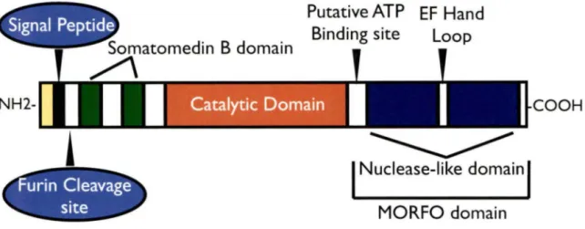
Figure 1: The major structural demains of ATX ... .4
Figure 2: Representation of the production ofLPA from LPC by ATX ... 6
Figure 3: Production and degradation pathways for LPA ... 14
Figure 4: Signaling pathways of the LPA receptors ... .19
Figure 5: Production and de gradation of cAMP ... 28
Figure 6: Overview of cAMP production and major effectors ... 32
Figure 7: Activators and effectors of Rapl ... 34
Figure 8: Invadopodium ... 49
ARTICLE Figure 1: Autotaxin induces invadopodia formation in HT1080 cells ... 65
Figure 2: ATX-induced invadopodia production is dependent on LPA production fromLPC ... 68
Figure 3: LPA-4 is implicated in invadopodia production by HT1080 cells ... 69
Figure 4: LPA-4 signais through Gs-cAMP-EPAC for invadopodia production ... 71
Figure 5: Racl activation downstream of EPAC is required for invadopodia production ... 74
Supplementary Figure 1: Involvement of Gs and Gï in ATX-induced invadopodia
production ... 76 Supplementary Figure 2: PKA inhibitors have no effect on ATX-induced invadopodia
production ... 76 Supplementary Figure 3: Rapl inhibition suppresses EPAC-induced invadopodia
AC: ADAM: ADF: AGK: AKAP: AMP: Arap3: ARF: Arp2/3: ATFl: ATP: ATX: BAD: bFGF: Bmp-2: CalDAG-GEFl: cAMP: CBP: CDC25-HD:
LIST OF ABBREVIATIONS USED adenylyl cylase
a disintegrin and metalloproteinase actin depolymerizing factor
acyl-glycerol kinase
a-kinase anchoring proteins adenosine monophosphate
Arf-GAP with Rho-GAP domain, ANK repeat and PH domain-containing protein 3
ADP Ribosylation Factors actin-related proteins
activating transcription factor 1 adenosine Triphosphate
autotaxin
bcl-2-associated death promoter basic fibroblast growth factor bone morphogenic protein 2
calcium- and diacylglycerol-binding guanine nucleotide exchange factor 1
cyclic adenosine 3 ',5 '-monophosphate CREB binding protein
CNG: cPA: CREB: CREM: CRIB: CTX: DA: DAG: DDW: DEP: DOCK4: DPP4: EBV: ECM: Edg: EGF: EPAC: E.R: ERKl/2: F-actin: FAK: FGF:
cyclic-nucleotide gated ion channel cyclic phosphatidic acid
cAMP response element-binding cAMP response element modulator Cdc42/Rac1-interactive binding choiera toxin
dominant active diacylglycerol
aspartic acid-aspartic acid-tryptophan motif dishevelled, Egl-10, pleckstrin
dedicator of cytokinesis 4 dipeptidyl dipeptidase IV Epstein-Barr virus
extracellular matrix
endothelial differentiation gene epidermal growth factor
exchange protein activated by cAMP endoplasmic reticulum
extracellular signal-regulated kinases filamentous actin
focal adhesion kinase fibroblast growth factor
GAP: GDI: GDP: GEF: GPCR: GPI: GPR: GSK3alpha: GTP: GTPase:
HKD:
HUVECs: IFN-y: IL-1~: IL-4: IP3: JNK: KDELR: LDL: LDV: LIMK:GTPase activating protein
guanosine nucleotide dissociation inhibitors guanosine diphosphate
guanine exchange factor
G-protein coupled receptor
glycosyl-phosphatidylinositol-anchored protien G-protein receptor
glycogen synthase kinase 3 guanosine triphosphate
guanosine triphosphate hydrolysing enzyme HxKxxxxD/E
human umbilical vascular endothelial cells interferon gamma
interleukin 1-beta interleukin 4
inositol triphosphate
c-Jun N-terminal kinases
endoplasmic reticulum protein retention receptor low density lipoprotein
leucine-aspartic acid-valine
LPA4: LPAAT: LPC: LPS: lysoPLD: MAG: MAPK: MEF: MMP-2 (7, 9): MORPHO: mRNA: MTl-MMP: NFAT: NFkB: N-myc: NPP2: NTA: N-WASP: PA: lysophosphatidic receptor 4
lysophosphatidic acid acyl transferases
lysophosphatidyl choline lipopolysaccharide lysophospholipase D monoacylglycerol
mitogen activated protein kinase mouse embryonic fibroblasts matrix metalloproteinase 2 (7, 9)
modulator of oligodendrocyte remodeling and focal adhesion organization
messenger ribonucleic acid membrane-type 1 MMP
nuclear factor of activated T-cells nuclear factor kappa ~
neuroblastoma derived V-mye myelocytomatosis viral related
oncogene
nucleotide pyrophosphatase/phosphodiesterase 2 N-terminal acidic domain
neuronal Wiskott-Aldrich syndrome protein
PAK: PDE: PH: PI3K: PKA: PKC: PLAl or 2: PLC: PLD: PRD: PTX: RA: Rapl (2): RAPGEF3: RAPL: RAS: REM: RGD: Riam:
Rims:
Rit:p21-activated protein kinase phosphodiesterase pleckstrin homology phosphoinositide 3-kinases protein kinase A protein kinase C phospholipase Al or A2 phospholipase C phospholipase D proline rich domain Pertussis toxin ras-association ras-proximate-1 (2)
rap guanine nucleotide exchange factor 3
regulator of adhesion and polarization enriched in lymphocytes ratsarcoma
ras-exchange motif
arginine-glycine-aspartic acid
rap 1-GTP-interacting adapter molecule rab3-interacting molecules
ROCK: RT-PCR: SlP: sAC: SEM: SFK: SH3: shRNA: SRE: Tbx2: TGF-p: Tiaml: trnAC: tyr: uPA: uPAR: VASP: VCA: VEGF: vzg-1: WASP: WAVE:
rho-associated coiled-coil-forming protein kinase reverse transcriptase polymerase chain reaction sphingosine-1-phosphate
soluble AC standard error src family kinase src-homology 3 domain
short hairpin RNA serum response element T-box transcription factor 2 transforming growth factor beta
T-cell lymphoma invasion and metastasis-inducing protein 1 transmembrane AC
tyrosine
urokinase-type plasminogen activator
urokinase receptor
vasodilator-stimulated phosphoprotein verprolin-cofilin-acidic
vascular endothelial growth factor ventricular zone gene 1
Wiskott-Aldrich syndrome protein
1- INTRODUCTION
1.1 Autotaxin
1.1.1 Identification and structural characteristics
Autotaxin (ATX), also known as Nucleotide pyrophosphatase/phosphodiesterase 2 (NPP2), was originally isolated from the culture medium of human melanoma cells (A2058) in 1991 (STRACKE et al., 1992). It was identified as a novel 125-kDa autocrine motility stimulating factor with a basic pl of 7.7 +/- 0.2 (STRACKE et al., 1992). Following its initial discovery, ATX was identified as one of the seven members of the nucleotide pyrophosphatase/phosphodiesterase (NPP) enzyme family due to its homology with phosphodiesterases (MURATA et al., 1994). All members of the NPP family have structurally related catalytic domains and nucleotide pyrophosphatase/phosphodiesterase activity whereby they hydrolyze pyrophosphate or phosphodiester bonds in nucleotides and other extracellular molecules. However, the NPP family members have very different substrate specificity (JANSEN et al., 2005) and are therefore implicated in diverse pathological processes (STEFAN et al., 2005).
ATX was originally thought to be an integral membrane protein like NPP 1 because of their overall structural similarity (MOOLENAAR, 2002;STRACKE et al., 1997). However, researchers have demonstrated that the N-terminal 27-residue hydrophobie domain of ATX is in fact a signal peptide that is removed by a signal peptidase during translation (JANSEN et al., 2005). ATX has also been shown to have a more prominent
cytoplasmic distribution than NPPl, consistent with its N-terminal hydrophobie domain being a signal peptide and nota signal anchor as in NPPl (JANSEN et al., 2005;KOIKE et al., 2006). Following the removal of this pre-peptide, the ATX pro-protein follows the classical secretory pathway, where proteins are transported outside the cell from the E.R via the Golgi apparatus. This was verified by the fact that ATX secretion was arrested by brefeldin A, an inhibitor of such transport (JANSEN et al., 2005). These observations were consistent with the finding that the majority of ATX is present in the culture medium of several cell types such as Glioblastoma Multiforme cells (JANSEN et al., 2005;KISHI et al., 2006). ATX is therefore synthesized as a pre-pro-protein and functions as a secreted protein (JANSEN et al., 2005;KOIKE et al., 2006). C-terminal to the peptidase cleavage site is a consensus sequence for furin, or related pro-protein convertases such as PACE4. lt is not known whether cleavage by furin occurs before or after secretion, however, this cleavage is not required for ATX secretion (JANSEN et al., 2005;KOIKE et al., 2006). The removal of an N-terminal octapeptide by furin, however, is associated with enhanced motility stimulating activity of ATX in certain reports (JANSEN et al., 2005). ATX cleaved by furin was shown to have a 30% increase in activity over the non-furin cleaved form in HEK293 cells (KOIKE et al., 2006). Three ATX isoforms have been found in the medium of cells and have been characterized by Giganti et al (2008) as ATX a, ~. and y. ATX a, originally discovered in A2058 cells, laclcs exon 21; ATX~, a
splice variant reported in human teratocarcinoma (LEE et al., 1996) laclcs exons 12 and 21; andATXy (PD-la), isolated from brain lacks exon 12. The mRNA transcripts ofthese
high expression in peripheral tissues while ATX y mRNA has highest expression in the brain. The ATXa mRNA isoform had the lowest expression levels in both the brain and peripheral tissues. The measured activity of ATXa protein was also very low probably due to its cleavage by an unknown factor that results in a protein of 55-66kDa with little
or no enzymatic activity (GIGANTI et al., 2008).
Besides its pre and pro protein domains, ATX contains a Modulator of Oligodendrocyte Remodeling and Focal adhes_ion Organization (MORPHO) domain implicated in
oligodendroglial process network formation and focal adhesion organization (DENNIS et
al., 2008). It also contains an EF-hand-like motif that contributes to the function of the MORFO domain, an inactive nuclease-like domain that is essential for the catalytic activity of ATX, and two cysteine-rich somatomedin B domains (YUELLING and FUSS, 2008). The somatomedin B domain, which is derived from the amino terminus of vitronectin, forms a presumed binding site for type 1 plasminogen activator inhibitor (PAI-1), and urokinase plasminogen activator receptor (uPAR) (SEIFFERT and
LOSKUTOFF, 1991;SEIFFERT et al., 1994). This suggests a relationship of ATX with
extracellular matrix proteins. In fact, ATX has recently been shown to contain an integrin binding domain (RGD) within its somatomedin B domains, that may be implicated in
lymphocyte trafficking (KANDA et al., 2008). Finally, ATX has a catalytic domain,
structurally similar to that of the NPP family, that functions as a lysophospholipase D
Putative ATP EF Hand Sematemedin B demain Binding site Leep
~
NH2- COOH
~
1
Nuclease-like demain1
MORFO demainFigure 1: The major structural demains of ATX. ATX contains an N-terminal signal peptide that is removed during translation. Following this is a consensus sequence for furin cleavage that may result in enhance activity of the protein. ATX also has two sornatomedin B demains, an inactive nuclease-like domain that contains an EF hand loop motif, and an ATP binding site. Finally there is a catalytic domain which functions as a lysophospholipase D and a MORPHO dornain implicated in oligodendrocyte remodeling.
1.1.2 Enzymatic activity
ATX was originally thought to hydrolyze pyrophosphate or phosphodiester bonds in
nucleotides due to its inclusion in the NPP family (BOLLEN et al., 2000). However,
autotaxin, has recently been shown to be molecularly identical to extracellular plasma lysophospholipase D (lysoPLD), whose activity was first discovered in human plasma in
1983 (YAMASHITA et al., 1983). LysoPLD is responsible for catalyzing the production
of Lysophosphatidic acid (LPA) from Lysophosphatidylcholine (LPC) by hydrolysis
(UMEZU-GOTO et al., 2002), see figure 2. cPA (cyclic phosphatidic acid), an analog of
LPA and intermediate in LPA formation, can also be produced from LPC by ATX
(TSUDA et al., 2006). LPC is the main physiological substrate for ATX/lysoPLD. In
fact, ATX has a 25-fold lower Km and thus higher affmity for LPC than for nucleoside
substrates, which are the natural substrates for other members of the NPP family (XIE and Meier, 2004). ATX/LysoPLD also has a higher affmity for unsaturated acyl-LPCs as
compared to saturated or ether-linked species (TOKUMURA et al., 1999). ATX,
interestingly, does not contain the HKD motifs critical for the catalytic activity of the phospholipase D (PLD) superfamily (XIE and MEIER, 2004). The hydrolysis of lysophospholipids by ATX is instead a metal-assisted reaction that occurs via a nucleotidylated threonine at the same catalytic site used for the hydrolysis of nucleotides
(GIJSBERS et al., 2003). ATX requires a metal ion, such as Co2+, for optimal lysoPLD
activity. It can also be enhanced by Ca2+ and Mg2+, which may act by stabilizing the
Lysophosphatidic Lysophosphatidytcholine acid (LPA) (LPC)
p-
o-0 0 P - 0 0'
r
_J Sn-1r
Sn-2_J c--.:>i .-3 0 OH 0 OH 0 0 - Phosphate group - Glycerol backbone - Fatty acyt chains(CH3>3
Nature Reviews 1 Cancer
Figure 2: Representation of the production of LPA from LPC by ATX. The lysoPLD activity of ATX is responsible for hydrolyzing the bond between choline and the phosphate group of LPC. This results in release of choline and LPA. (Modified figure from: MILLS and MOOLENAAR, 2003).
constitutively active (YUELLING and FUSS, 2008), however, its catalytic activity depends on an essential disulfide bridge between the catalytic and nuclease-like domains (JANSEN et al., 2009) as well as glycosylation of Asn-524 (JANSEN et al., 2007). Human ATX activity can be inhibited by EDTA, phenanthroline and ATP as well as by its products LPA, cPA and SlP (BAKER et al., 2006;VAN MEETEREN et al., 2005). LPA, generated from LPC, is considered to be responsible for the majority of ATX biological effects and will be discussed in the LPA section.
1.1.3 Expression and role in normal cells and tissues
ATX is ubiquitously expressed and is, therefore, synthesized by a variety of normal cells and tissues. Particularly high expression of ATX has been found in the brain (MILLS and MOOLENAAR, 2003), kidneys and lymph nodes (KANDA et al., 2008). ATX is implicated in many processes during normal development such as adipogenesis (SIMON et al., 2005), and central nervous system development that includes neurite morphology (DENNIS et al., 2005). It is also implicated in intestinal cell motility through activation of PLC-gamma and phosphorylation/recruitment ofvillin (KHURANA et al., 2008). ATX has been shown to regulate myelination by controlling cytoskeletal organization and FAK phosphorylation in oligodendrocytes (FOX et al., 2004). More recently, ATX has been shown to be implicated in immune functions due to the fmdings that mast cells in submucosal connective tissue secrete ATX (MORI et al., 2007) and that ATX was shown to promote the entry of lymphocytes into secondary lymphoid organs (KANDA et al.,
2008). Besicles these advancements, the most well known role of ATX is in vascular development (KHURANA et al., 2008;SATO et al., 2005).
ATX has been shown to be essential for blood vessel formation during development. ATX deficiency using knockout technology in mice leads to embryonic lethality due to impaired vessel formation in the yolk sac and embryo (VAN MEETEREN et al., 2006). The vascular defects in ATX deficient mice also resemble those in mice lacking genes involved in cell migration and adhesion such as the fibronectin and focal adhesion kinase genes. Results of this study indicated that the loss of LPA production and downstream GPCR signaling is responsible for the phenotype observed in ATX knockout mice (VAN MEETEREN et al., 2006).
1.lA Expression and role in pathologies
ATX has been implicated in numerous pathologies including Alzheimer's disease, chronic hepatitis C, multiple sclerosis, neuropathie pain, obesity and rheumatoid arthritis, but its most investigated and presumably most important role is in tumorigenesis (FERRY et al., 2003;HAMMACK et al., 2004;INOUE et al., 2008a;INOUE et al., 2008b;UMEMURA et al., 2006;WATANABE et al., 2007;ZHAO et al., 2008). ATX has been shown to be up-regulated in malignancies including breast, lung, colon, ovarian, stomach, thyroid and brain cancer, correlating with the invasiveness of these cancer cells (KEHLEN et al., 2004;KISHI et al., 2006;YANG et al., 2002;YANG et al., 1999). ATX has been shown to
proliferation, cell survival, cell motility, invasion and angiogenesis. ATX acts extracellularly and stimulates the metastatic cascade at multiple levels by acting as a
tumor cell motility factor as well as a strong inductor of the angiogenic response (NAM et
al., 2000;NAM et al., 2001). Specifically, ATX-transfected Ras-transformed NIH3T3 cells were shown to be more invasive, tumorigenic, angiogenic and metastatic than
mock-transfected contrais (NAM et al., 2000;NAM et al., 2001). ATX, especially in the
presence of LPC, has been shown to increase chemotaxis and proliferation in multiple cell
lines. 1t has also been demonstrated that several cancer cell lines release significant
amounts of LPC into the culture medium (UMEZU-GOTO et al., 2002). ATX promotes
proliferation of A2058, MDA-MB231, CHO-Kl and Edg2-RH7777 cancer cells, but not RH7777 cells that lack LPA receptors. Research, therefore, suggests that autocrine or paracrine production of LPA via ATX contributes to tumor cell motility, survival and
proliferation (BRINDLEY, 2004;UMEZU-GOTO et al., 2002). Recently over-expression
of ATX (or LPA1-3) has been shown to increase tumor invasion and metastasis of breast
cancer cells (LIU et al., 2009) while pharmacological inhibition of ATX and LPA
receptors was shown to decrease cell migration in vitro and cause tumor regression in
mice (ZHANG et al., 2009) further supporting the role of ATX in cancer progression.
1.1.S Regulation of expression
Although ATX has important roles in tumor progression, mostly due to its aberrant expression in various malignant cells ( described above ), little is known about the factors
that regulate its expression in cells. This section will detail the few stimulators and inhibitors currently known.
ATX expression can be induced by retinoic acid in a neuroblastoma cell line with N-myc amplification that is responsive to the diff erentiation inducing effects of retinoic acid (DUFNER-BEATTIE et al., 2001). ATX is also one of many genes up-regulated during Bmp-2 mediated mesenchymal development (BACHNER et al., 1998) indicating that cell differentiation might trigger ATX expression in certain cell lines. Lipopolysaccharide (LPS) induces ATX expression in the monocytic THP-1 cells via JNK and p38MAPK, resulting in enhanced immune cell migration (LI and ZHANG, 2009). Fibroblast-like synoviocytes from patients with rheumatoid arthritis have increased expression of ATX that can be down regulated by anti-inflammatory cytokines including IL-1 p, IL-4 and IFN-y (SANTOS et al., 1996). In thyroid carcinoma cell lines, the growth factors EGF and bFGF have been shown to stimulate ATX activity while the anti-inflammatory cytokines IL-4, IL-lP and TGF-P reduced its expression (KEHLEN et al., 2004). Therefore, pro-inflammatory stimuli seem to increase ATX expression while anti-inflammatory cytokines have the opposite effect. Expression of a6p4 integrin, which correlates with an invasive and migratory phenotype in advanced breast carcinomas, leads to increased expression of ATX mediated by up-regulation and activation of NFATl that binds to the ATX promotor (CHEN and O'CONNOR, 2005). ATX expression is increased by more than 100-fold in cells transformed by the viral oncoprotein v-Jun (BLACK et al.,
cells with EBV also results in induction of ATX (BAUMFORTH et al., 2005). In contrast, a candidate tumor suppressor gene for breast cancer, CST6, when expressed in breast cancer cells down-regulated the expression of ATX (SONG et al., 2006). Therefore it seems that molecular eues associated with cancer progression can induce ATX expression while tumor suppressors seem to reduce its expression. Each of the above mentioned stimuli have been investigated in very few cell types. Therefore, the precise signaling pathways and transcription factors responsible for ATX regulation remain mostly unknown so many more studies are needed to define exactly how ATX expression is regulated.
1.2 Lysophosphatidic acid 1.2.1 Identification
Lysophospholipids have been known for decades as components in the biosynthesis of cells membranes with short half lives that range from seconds to minutes (CHOI et al., 2010;SHIMIZU, 2009). They were originally thought to act as intracellular messengers (GERRARD and ROBINSON, 1984) or to mediate effects due to intrinsic chemical properties, such as calcium binding activity (SIMON et al., 1984). However, they were later found to have cell signaling roles (VOGT, 1963) and it is now known that most bioactive lipids act on cell surface GPCRs to mediate intracellular signaling (SHIMIZU, 2009). Many lysophospholipids, including LPA, have similar effects on cellular functions as polypeptide growth factors (HILL and TREISMAN, 1995). LPA (1 or 2-acyl-sn-glycerol-3-phosphate), a glycerolysophospholipid, was the first LP to be recognized as a
concentrations (TOKUMURA et al., 1978), including mitogenic and morphological effects on many cell types (ISHII et al., 2009).
LPA has a glycerol backbone, single carbon chain, and a polar headgroup (see figure 2) (MEYER ZU HERINGDORF and JAKOBS, 2007). There are multiple molecular species of LPA, consisting of acyl- or ether-linked chains with varions numbers of carbons and degrees of unsaturations. The acyl chain can be esterified at either the sn-1 or sn-2 position of the glycerol backbone(l-acyl-LPA or 2-acyl-LPA) while ether-linked LPAs carry an alkyl or alkenyl linkage at the sn-1 position (1-alkyl-LPA or 1-alkenyl-LPA). The biological activities of LPA depend on the carbon chain length and degree of unsaturation as well as the position and linkage type of the carbon chain attached to the glycerol backbone (MEYER ZU HERINGDORF and JAKOBS, 2007).
1.2.2 Production and degradation ofLPA
LPA is detected in serum, plasma and many other biological fiuids and tissues such as saliva (SUGIURA et al., 2002), follicular fiuid (TOKUMURA et al., 1999), seminal fiuid (HAMA et al., 2002), and malignant effusions (WESTERMANN et al., 1998). Major cellular sources of LPA include platelets and adipocytes (EICHHOLTZ et al., 1993;VALET et al., 1998), while postmitotic neurons, lymphoid cells, endometrial cells, erythrocytes and cancer cells are also able to produce LPA (AOKI et al., 2008;SMYTH et al., 2008;YE, 2008). Therefore, LPA may act as a circulating as well as a locally produced paracrine mediator (TAKUWA et al., 2002). LPA is found at a concentration of approximately 154pmol in cells, 0.1-6.3 µM in blood and 80-lOOnM in plasma
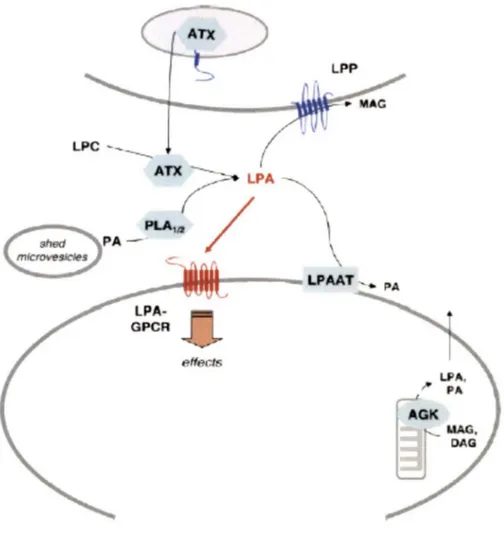
(HOSOGAYA et al., 2008;KISHIMOTO et al., 2003). As mentioned previously, the lysophospholipase D activity of autotaxin is responsible for the majority of LPA produced in vivo from the substrate LPC (UMEZU-GOTO et al., 2002). However, other enzymes and pathways remain responsible for some LPA production. These other routes of LPA synthesis include de nova LPA biosynthesis in cells through intermediate lipid metabolism, resulting in intracellular LPA, or liberation and subsequent enzymatic conversions of precursor glycerophospholipids, resulting in extracellular LPA (GOETZL and An, 1998). Extracellular LPA can be produced by the action of many different enzymes including phospholipase Al or A2 (PLAl or 2), monoacylglycerol kinase or glycerol-3-phosphate acyltransferase (PEBAY et al., 2007). PLAl/2 for example produce LPA by deacylating phosphatidic acid (PA) that is first generated intracellularly from phospholipids or diacylglycerol (AOKI et al., 2008). Figure 3 summarizes the pathways of LPA production and degradation.
LPA has short half-life attributed toits rapid degradation by lipid phosphate phosphatases, integral membrane proteins that dephosphorylate LPA to monoacylglycerol (MAG) (BRINDLEY et al., 2002), or by acylation of LPA to PA by the action of acyl transferases (LPAAT) (YAMASHITA et al., 2001). To counteract these effects, extracellular LPA is normally bound to proteins such as albumin, fatty acid binding protein, or gelsolin which act to increase the stability and facilitate transport of LPA (AOKI, 2004;GAITS et al., 1997;MILLS and MOOLENAAR, 2003;PAGES et al., 2001).
Figure 3: Production and degradation pathways for LPA. The major producer of LPA in vivo is ATX which converts LPC to LPA. Other enzymes that can also produce LPA include acylglycerol kinase (AGK) which produces LPA from MAG and PLAl/2 which can produce LPA from PA. The two major degradation pathways for LPA are acylation by LPAAT resulting in PA and phosphorylation by LPPs resulting in MAG . (Modified figure from: MEYER ZU HERINGDORF and JAKOBS, 2007).
1.2.3 LPA receptors
The first evidence of the possible existence of G-protein-coupled receptors (GPCRs) specific for LPA was the observation, by van Corven et al. in 1989, that the proliferative effects of LPA on fibroblasts were pertussis toxin sensitive and therefore mediated by G-proteins (VAN CORVEN et al., 1989). GPCRs are 7 transmembrane receptors that couple to various trimers of G-proteins to mediate intracellular signaling. Further studies also supported the existence of LPA-specific receptors (THOMSON et al., 1994;VAN DER BEND et al., 1992). Thomson et al. (1994) found LPA-specific high affinity binding sites on cell membranes and suggested that GPCRs could be responsible for mediating the LPA binding to membranes and induce a transient increase in intracellular Ca2+ levels. Van der Bend et al. (1992) found that a 32P-labeled LPA analog binds to a membrane protein of 38-40 kDa in varions cells types that was proposed to be a specific cell-surface LPA receptor (VAN DER BEND et al., 1992). However, it was not until 1996 (HECHT et al., 1996) that the first LPA receptor gene was identified in mice during the course of studies designed to identify navel GPCR genes associated with the production of neurons in mice (CHUN et al., 1999;CHUN, 1999;MASANA et al., 1995).
LPA1, originally named ventricular zone gene l(vzg-1) due toits increased localization in this area of the brain, was also known as endothelial differentiation gene 2 (Edg-2) due to its sequence similarity to an orphan receptor called Edg-1 (endothelial differentiation gene 1) cloned from endothelial cells (HLA and Maciag, 1990). Edg-2/LPA1 was subsequently cloned and identified as an LPA receptor in humans in 1997 (AN et al., 1997). LPA1 is a 41 kDA (364 a.a) protein with seven transmembrane domains, in
concordance with the structure for GPCRs (CHUN et al., 1999;CONTOS and CHUN, 1998). In human tissues LPA1 is widely expressed in almost ail tissues with high expression in brain, heart, placenta and digestive tract and lowest expression in liver and peripheral blood leucocytes (AN et al., 1998a). Interestingly, LPA1 is also expressed in several cancers (FURUI et al., 1999) such as HeLa carcinoma, SW480 colorectal adenocarcinoma, A549 lung carcinoma, and G361 melanoma, but undetectable in many leukemia lines and Burkitt's lymphoma (AN et al., 1998a).
A second LPA receptor, LPA2, originally known as Edg-4, was subsequently identi:fied due to sequence similarity with Edg-2/LPAr (CHUN, 1999;CONTOS and CHUN, 1998). In humans, LPA2 is detected in testis, pancreas, prostate, thymus, spleen, and peripheral blood leukocytes and is almost undetectable in brain, heart, placenta, and digestive tract contrary to LPA1 (AN et al., 1998a). Therefore, LPA2 is less widely distributed than LPA1. LPA2 has also been found to be expressed in various cancer cell types (AN et al., 1998a). A third related gene, Edg-7 now known as LPA3 has 60% amino acid similarity to mouse LPA1 and 2. LPA3 has a more restricted expression pattern than LPA1 and 2, having abundant expression only in human testis, prostate, heart and frontal regions of the cerebral cortex (IM et al., 2000) as well as pancreas, lung and ovary (BANDOH et al., 1999;IM et al., 2000). The Edg family has some common structural features such as lack of a cysteine residue in the :first extracellular loop found in most GPCRs and they share 50-57% amino acid identity in humans (AN et al., 1998a;ISHII et al., 2009). However, signaling induced by these receptors was unable to account for ail the cellular eff ects of LPA. The existence of additional receptors was later implied by several reports
(CONTOS et al., 2002;HOOKS et al., 2001) due to the fact that cell lines not expressing the Edg receptors were shown to have mitogenic responses to LPA.
Recently, a distinct group of LPA receptors unrelated to the Edg family receptors have been identified. In 2003 a fourth LPA receptor (LPAJp2y9/GPR23) was identified that was structurally distant from the Edg receptors (NOGUCHI et al., 2003) (LPA.i will be discussed in detail in the LPA.i section). This stimulated the identification of two additional LPA receptors, LPAs and LPA6. These three receptors are more closely related to the purinergic receptors (purino-receptor cluster), indicating that they arose from different ancestor genes than the Edg family receptors (ISHll et al., 2009).
LPA5 (GPR92-93) (KOTARSKY et al., 2006;LEE et al., 2006) was an orphan GPCR that was identified as an LPA receptor due to its close relation to LPA4 (LEE et al., 2006). Low levels of LPAs mRNA are expressed in embryonic brain, heart, placenta (KOTARSKY et al., 2006) and platelets (AMISTEN et al., 2008), while high levels were found in small intestine (specifically in the lymphocyte compartment) and moderate levels in skin, spleen, stomach, thymus, lung, liver, dorsal root ganglion cells (OH et al., 2008) and embryonic stem cells (LEE et al., 2006).
LPA6 (p2y5), was originally reported in 1996 as an orphan GPCR encoded in an intron of the retinoblastoma gene (HERZOG et al., 1996). LPA6 has ubiquitous expression including in hair follicle cells, epidermis (PASTERNACK et al., 2008), intestine (LEE et
al., 2009) and a leukemia cell line (YOON et al., 2006) with high expression in human umbilical vascular endothelial cells (HUVECs) (YANAGIDA et al., 2009).
1.2.4 Signaling pathways
The major cellular effects mediated by LPA are either growth related or cytoskeletal-dependent effects (GOETZL and An, 1998). Upon receptor activation, G-alpha subunits are separated from G-beta-gamma subunits, which remain together, now in their active state bound to GTP (WATTS and NEVE, 2005). The Edg family LPA GPCRs mediates effects by coupling to Gï, Gq or G12113 depending on cell type, receptor expression levels, or amounts of available G-proteins (AN et al., 1998b;BANDOH et al., 1999;IM et al., 2000). LPA1 and LPA2 can signal through ail three of these G-proteins while LPA3 only couples to Gq and Gï (FUKUSHIMA et al., 1998;ISHII et al., 2000)(see figure 4 for an overview of the signaling pathways). Signaling by LPA4 will be discussed in further detail later and signaling of the newest LPA receptors, LPAs and LPA6, is less well characterized and will not be discussed here.
Gï, which is pertussis toxin sensitive, mediates inhibition of cAMP production as well as stimulation of protein kinases that recroit the ras-raf cascade and activate MAP kinases, resulting in proliferation and differentiation. Gï induces FAK (focal adhesion kinase) activation leading to focal adhesion formation. PI3K is also activated downstream of Gi leading to Akt activation and cell survival or Rac activation and cell migration. Finally, tyrosine kinase-dependent induction of tyrosine phosphatases can also be promoted by Gi
!
(RAC)
Migration
! (
cAMP) Stress Aber Migrationformation
(cAMP)
src+-è:E!Ac)
Cytoskjetal
À
remodellingy
Depolymerization Integrin activationFigure 4: Signaling pathways of the LPA receptors. The major LPA signaling pathways are illustrated here and include signaling through four different G-protein families, G12m, Gi, Gq, an Gs. Signaling through these G-proteins can activate diverse proteins including RhoA, PI3K, Ras, PLC and AC. The major downstream effects of LPA signaling include cytoskeletal remodeling, cell migration, stress fiber formation, focal adhesion formation and integrin activation.
Gq mediates phospholipase C (PLC) activation, liberating inositol triphosphate (IP3) that causes mobilization of intracellular Ca2+ that can result in actin depolymerization. PLC activation downstream of Gq can also stimulate MAP kinases directly or through induction of diacylglycerol (DAG), which activates some PKC isozymes, which in tum can activate MAP kinases by activating Raf-1, (GHOSH et al., 1997). Furthermore, PKC-ô can activate src kinase activity indirectly through protein tyrosine phosphatase alpha (BRANDT et al., 2003). Finally, G12113 stimulates RhoGTPase pathways that contribute to SRE-mediated transcription as well as mediating cytoskeletal dependent functions such as stress fi.ber formation and actin polymerization. G12m also promotes activation of PLD, PI3K, RhoA and Cdc42 (FROMM et al., 1997). G~y dimers may also
participate in signaling by recruitment of PLC (BARR et al., 2000;SANKARAN et al., 1998) and association with PI3K (KUROSU et al., 1997;MAIER et al., 1999;STOYANOV et al., 1995).
1.2.5 Implication in physiological and pathological processes
LPA signaling is implicated in diverse biological processes that include tissue remodeling, wound healing (WATTERSON et al., 2007), angiogenesis, platelet aggregation, cardiovascular function, (SMYTH et al., 2008) neurogenesis, myelination, olfaction, neuropathie pain, reproduction, adipogenesis, (YE, 2008), and immunomodulation (MOOLENAAR et al., 2004;NOGUCHI et al., 2009). For instance, LPA has been identified as a main platelet-activating lipid of mildly oxidized LDL and human atherosclerotic lesions (SIESS et al., 1999). In platelets, LPA induces human platelet shape change and platelet aggregation. LPA signaling is also implicated in neuropathie
pain responses and induces neuropathie pain and demyelination of the dorsal root similar to what is observed after nerve injury (FUilTA et al., 2007).
LPA is highly implicated in embryonic development of the nervous system . It is an important mediator of physiological and pathological processes in the central nervous system and influences ail neuronal cell types. Both apoptotic and survival effects of LPA have been reported and these opposing effects may be due to concentration differences or differential expression of LPA receptors as well as cell maturation and cell density (TAKUWA et al., 2002). For example, LPA bas been demonstrated to mediate proliferative effects and morphological changes in Ventricular Zone neuroblasts (CONTOS et al., 2000a;FUKUSHIMA et al., 2000), and to influence neuronal differentiation (DOTTORI et al., 2008;SPOHR et al., 2008), including neurite formation by differentiating neurons (FUKUSHIMA et al., 2000). LPA was also shown to protect Schwann cells from apoptosis (WEINER and CHUN, 1999) and to promote cell survival of early postmitotic cortical neurons (KINGSBURY et al., 2003). However, LPA also induces growth cone collapse and neurite retraction as well as apoptosis in hippocampal neurons (FUKUSHIMA, 2004). Apoptotic effects of LPA are observed in neurons cultured for over one week while survival effects are seen in neurons cultured for 1-2 days (FUJIWARA et al., 2003).
Another important role of LPA is in reproduction. LPA is found in normal follicular fluid suggesting its involvement in normal physiology of avaries (TOKUMURA et al., 1999). LPA may also be involved in male and female reproductive physiology and pathology
(TOKUMURA et al., 1999). LPA receptor mediated signaling has been implicated in many processes involved in reproduction such as ovarian functions (CHEN et al., 2008;TOKUMURA et al., 1999), with LPA4 being highly expressed in human and mouse ovary (NOGUCHI et al., 2003), spermatogenesis, with degradation of LPA being associated with reduced spermatogenesis and LPA1-4 expression in human testis (CROI et al., 2010;NOGUCHI et al., 2003;YE, 2008), fertilization (GARBI et al., 2000), early embryo spacing (HAMA et al., 2007;YE et al., 2005), decidualization (SHIOKAWA et al., 2000) and pregnancy maintenance (ZIECIK et al., 2008). LPA signaling has also been implicated in pathologies of the reproductive system including ovarian, prostate and endometrial cancers (LPA2 and MMP-7 implicated) (GUO et al., 2006;HOPE et al., 2009;SUTPHEN et al., 2004) and endometriosis (WEI et al., 2009;WOCLAWEK-POTOCKA et al., 2009).
These physiological effects of LPA are mediated by signaling through various LPA receptors whose effects can be elucidated by use of receptor knockout mice. Knockout studies have shown that LPA1 is implicated in the initiation of neuropathie pain and is important for proliferation of astrocytes. LPA1-null mice display 50% lethality, and survivors have abnormal phenotypes such as reduced body size, craniofacial dysmorphism and reduced brain mass as well as a suckling defect (CONTOS et al., 2000a). LPA1, therefore, seems to play an important role in the central nervous system. LPA2-null mice show no obvious phenotypic abnormalities and, therefore, might have redundant functions with LPA1 as they bath couple to the same G-proteins (CROI et al., 2008). Female LPA3-null mice display delayed embryo implantation, altered embryo
spacmg, and reduced litter size (YE et al., 2005), suggesting that this receptor is implicated in reproductive functioning.
Finally, the most important role of LPA for this study is in tumor progression. LPA is known to be a patent tumor promoting molecule and influences many cellular processes implicated in tumorigenesis. LPA has an effect on the cellular motility of cancer cells by mediating cytoskeletal rearrangements via the Rho GTPases Rho and Rac (IMAMURA et al., 1993;STAM et al., 1998). This leads to stimulation or inhibition of cell migration or invasion depending on the cell type. LPA can induce proliferation and mitogenic signaling of prostate cancer cells (BUDNIK and MUKHOPADHYAY, 2002). LPA also stimulates migration and proliferation of human carcinoma cells (DLDl) as well as their adhesion to collagen type I and secretion of endothelial growth factor and IL-8, all of which can lead to an increased metastasizing potential of DLD 1 carcinoma cells (SHIDA et al., 2003). LPA is also present in the ascites from ovarian cancer patients (WESTERMANN et al., 1998;XU et al., 1998), and a significant increase in blood LPA has been found in patients with ovarian carcinoma at the first stage of this disease, suggesting an important contribution of LPA to this pathology (FANG et al., 2000). Malignant progression has also been shown to correlate with differential expression of various LPA receptor subtypes (CONTOS et al., 2000b). LPA1 over-expression in breast carcinoma cells leads to metastatic spread to bone (BOUCHARABA et al., 2004), while LPA1 signaling has been shown to mediate stimulation of motility of human pancreatic cancer cells (YAMADA et al., 2004) and induction of metastasis by human colon carcinoma cells (SHIDA et al., 2003). LPA2 is over-expressed in invasive ductal
carcinoma (KITAYAMA et al., 2004) as well as ovarian cancer (ERICKSON et al., 2001) and promotes mitogenic signaling in human colon cancer cells (YUN et al., 2005). Finally, LPA3 expression increases the aggressiveness of ovarian carcinoma (YU et al., 2008). Also, as mentioned in the previous ATX section, over-expression of LPA1-3 receptors or their pharmacological inhibition results in promotion or inhibition of cancer cell invasion, tumor progression, and metastasis (LIU et al., 2009;ZHANG et al., 2009).
1.3 LPA receptor 4
1.3.l Expression and role in physiological processes
LPA4/p2y9/GPR23 is widely expressed in embryonic tissues including brain and stem cells (LEE et al., 2007). In adults, it is abundant in ovary and is weakly expressed in many tissues including pancreas, prostate, spleen, small intestine, colon, skeletal muscle, brain, placenta, lung, liver, skin, heart, thymus and bone marrow (NOGUCHI et al., 2003). Increased mRNA expression has also been documented at implantation sites in the uterus (ISHII et al., 2009). The roles of LPA4 in physiology and disease have only started ta be uncovered. Ta date, LPA4 has been shown ta induce Rho-mediated neurite retraction and stress fi.ber formation as well as cell aggregation and rounding (LEE et al., 2007;YANAGIDA et al., 2007), AC stimulation leading ta increased cAMP levels (LEE et al., 2007;NOGUCHI et al., 2003), and Gq and Gï-mediated calcium mobilization (LEE et al., 2007;NOGUCHI et al., 2003;YANAGIDA et al., 2007). LPA4 may play arole in neuronal development including neurogenesis and neuronal migration as it increases cAMP and the transcription factor CREB, which is essential ta neuronal differentiation (RHEE et al., 2006;YANAGIDA et al., 2007). In a recent study, LPA4, coupled ta cAMP,
has been found to inhibit osteogenic differentiation reducing bone volume and trabecular thickness (LIU et al.,) signifying arole in bone homeostasis.
1.3.2 Implications in cancer
There is little information to date on the implications of LPA4 in cancer. One study has found that LPA4 expression inhibits motility and invasion of B 103 neuroblastoma cells, which do not endogenously express LPA4. In this study, knockdown of LPA4 in MEFs increased cell migration, and LPA4 expression decreased PI3K reducing Akt and Rac activation levels while increasing Rho activation. It is important to note that in the cell type studied, LPA4 couples only to Gq and G12m (LEE et al., 2008). However, another recent study has found that expression of the LPA4 receptor can induce transformation and anchorage independent growth in Myc/tbx2 -transformed cells. Mye and tbx2 are cooperating partners in cell transformation. The authors found that expression of LPA4 (or LPA1 or LPA2) in Myc/tbx2-MEFs induced a transformed phenotype observed by the increased ability of the cells to grow in soft agar (anchorage-independent growth) as well as inducing tumor formation in vivo when these cells were subcutaneously injected in mice. These effects were found to necessitate Gi signaling and activation of PI3K and ERKl/2 pathways, particularly with prolonged activation of ERK (TAGHAVI et al., 2008). Therefore the role of LPA4 in tumorigenesis is currently unclear and probably depends on cell type and G-protein coupling.
1.3.3 Major signaling pathway
LPA4 has been shown to be capable of signaling through Gï, activating PI3K and ERKl/2 (TAGHAVI et al., 2008), G12113 resulting in Rho activation (LEE et al., 2007) and Gq inducing increases in calcium (TAGHAVI et al., 2008). However, the major signaling pathway of LPA4, different from those previously discussed for the other receptors, is signaling through Gsresulting in the production of cAMP (NOGUCHI et al., 2003).
1.3.3.1 Production of cAMP
cAMP (cyclic adenosine 3' .5'-monophosphate) was first identified as a second messenger nucleotide found to have a fundamental role in the cellular response to extracellular stimuli and, therefore, control a diverse range of cellular processes (ROBISON et al., 1968). GPCRs appear to be the main receptors responsible for causing an accumulation of intracellular cAMP in response to ligand binding. The heterotrimeric G proteins coupled to GPCRs regulate ACs (adenylyl cyclases) in response to various cellular stimuli. The Gs G-protein activates adenylyl cylclases (as does Gq in some instances) while the Gï family of proteins inactivates ACs (WATTS and NEVE, 2005). These G-proteins, upon receptor activation, separate from G~y and are converted to their
GTP-bound state in which they can exert their distinctive regulatory functions on ACs (WATTS and NEVE, 2005). AC are 12 transmembrane domain proteins which are generally bound to the inside of cell membranes. Class III ACs are responsible for cAMP production (WATTS and NEVE, 2005). In humans there are 9 transmembrane AC enzymes (trnAC) and one soluble AC (sAC). However, the soluble form occurs primarily in mature spermatozoa and will not be further discussed here (JAISWAL and CONTI, 2003;WATTS
and NEVE, 2005). Gsa activates ACs by inducing a conformational change in the catalytic site upon interaction (SKIBA and HAMM, 1998). Activated AC converts ATP (adenosine triphosphate) to cAMP by creation of a cyclic phosphodiester bond with the alpha-phosphate group of ATP resulting in increased intracellular cAMP concentrations. cAMP can then be converted to AMP by cAMP-specific phosphodiesterases (PDE), see figure 5. Growth factors and PI3K can also down-regulate cAMP signaling by activating Alet and subsequently PDEs, facilitating the conversion of cAMP to AMP (ROBISON et al., 1968; DEGERMAN et al., 1997).
1.3.3.2 Spatial regulation of effectors
GPCRs are confined to specific domains of the cell membrane in association with intracellular organelles or the cytoskeleton. The ACs that they activate are found anchored nearby (JARNAESS and TASKEN, 2007) resulting in targeted cAMP production depending on the extracellular ligand and receptor activated. cAMP is further regulated through its degradation by cAMP-specific phosphodiesterases (PDEs), the only known mechanism of cAMP inactivation (JARNAESS and TASKEN, 2007). The cellular localization of PDEs is controlled by anchoring to specific subcellular compartments and recruitment into multi-protein signaling complexes, therefore, targeting them to specific subcellular locations. This allows increased cAMP concentrations in certain areas of the cell and not in others, where it will be degraded instead (JARNAESS and TASKEN, 2007). The level of intracellular cAMP is, therefore, spatially and temporally regulated by the balance between the activities of ACs and cyclic nucleotide PDEs (JARNAESS and TAS KEN, 2007).
NH2
~'C~N
o · 0 -a-
HC n ' 1 1 1 'w'C'N~H ·o- P-O-P-O-P-0-C~ 1 1 1 2 0 0 0 0 HATP
H H HOOHAdeo~yl
oycia'8!
Mg" NI-li~N,C~
HC 11 1 s· 'w_.c,N:::CH PP, + 0 - Ci:).OHcAMP
H H ·o-P - 0 OH 1 0 Ph°"""'<fiest<waœ!
Mg" ,H,0 NH2JJ'-C~N
o· HC 11 1HO-t-o-1;,~î__c:~~~p
H~
HO OHFigure 5: Production and degradation of cAMP. Activated AC converts ATP (adenosine triphosphate) to cAMP by creation of a cyclic phosphodiester bond with the alpha-phosphate group of ATP. cAMP can then be degraded to AMP by the action of cAMP-specific PDEs. From: DUMAN and NESTLER, 1999)
cAMP effectors are also spatially regulated. PK.A (Protein kinase A/cAMP-dependent protein kinase), a major effector of cAMP (to be elaborated in the cAMP effector section) is tethered to specific intracellular locations by AKAPs (A-kinase anchoring proteins), which anchor the regulatory subunits (see figure 6A). AKAPs are scaffolding proteins that form multi-protein complexes to integrate cAMP signaling (SCOTT and MCCARTNEY, 1994). There are over 50 members of the AKAP family (WONG and SCOTT, 2004) which all have similar functions while being structurally diverse (JARNAESS and TASKEN, 2007). These proteins all contain a PK.A binding domain and a unique targeting domain, with several containing additional interaction sites for formation of multivalent signaling complexes (JARNAESS and TASKEN, 2007). Sorne AKAPs are known to interact with both PD Es and PK.A for example. Targeting of PK.A isozymes is important for many physiological processes such as cAMP regulation of ion channels in the nervous system, regulation of the cell cycle involving microtubule dynamics, steroidogenesis, reproductive function, immune response and numerous intracellular transport mechanisms (TASKEN and AANDAHL, 2004).
EPAC (exchange protein activated by cAMP/cAMP-regulated guanine exchange factor) proteins, other effectors of cAMP (see cAMP effector section), are also spatially regulated. First these proteins have been shown to also interact with AKAP signaling complexes which may be responsible in part for their localization, that requires specific anchoring, to various cell compartments such as the cytosol, nucleus, nuclear envelope, and plasma membrane, resulting in different cellular functions (PONSIOEN et al., 2009). EPAC localization has also recently been found to be directly regulated by cAMP. In a
2009 study by Ponsioen et al. the authors found that cAMP binding to EPAC induced translocation of EPAC to the plasma membrane due to a conformational change that also reveals its catalytic site for Rap activation. The translocation of EPAC was found to be due to passive diffusion and depended on its DEP domain and is a dynamic and reversible event (PONSIOEN et al., 2009).
Therefore, cAMP is produced at a specific location, due to receptor and AC localization, and its diffusion in the cell is controlled by PDE. This results in targeting of increased cAMP to specific effectors, for example, PK.A or EPAC, and their associated substrates. PK.A and EPAC are also anchored close to specific effectors resulting in a controlled and speci:fic response to cAMP increases, depending on the receptor activated, that will mediate a distinct biological effect.
1.3.3.3 cAMP effectors and roles in cellular functions
cAMP has been found to be implicated in virtually a11 cellular responses such as proliferation, differentiation, apoptosis, gene transcription, metabolism, secretion, cell division and neurotransmission (CHENG et al., 2008). Therefore, it is also implicated in many pathologies including diabetes, heart failure and cancer to name a few (CHENG et al., 2008). The response to cAMP is cell type and cell context speci:fic, and in different situations can mediate opposing effects. For example, cAMP has been shown to either inhibit or stimulate cell prolif eration depending on the cell type studied or the stimuli used (BEAVO and BRUNTON, 2002;STORK and SCHMITT, 2002). The cAMP effector EPAC was shown to induce Akt phosphorylation in WRT cells and macrophages increasing gene expression and proliferation (CASS et al., 1999), while in adipocytes
EPAC was shown to inhibit Akt phosphorylation thereby reducing proliferation (ZMUDA-TRZEBIATOWSKA et al., 2007). cAMP has also been shown to inhibit keratinocyte migration (MCCAWLEY et al., 2000) or to enhance it (IWASAKI et al., 1994), depending on the concentration of cAMP used. The role of cAMP in cancer is vague, while it has been shown to inhibit tumorigenesis (O'CONNOR et al., 1998), these effects are likely to be cell type and context dependent and it is likely that cAMP can mediate opposing effects on tumorigenesis as it does on other cellular processes. For example one study has shown that cAMP suppresses MMP-2 activation (LEE et al., 2006), while another study showed that elevation of cAMP increased the expression and activity of MMP-2 (TSURUDA et al., 2004), a molecule known to be implicated in tumor cell invasion. The responses to cAMP are mediated by its three main effectors, CNGs (cyclic-nucleotide gated ion channel) whose activation by cAMP allows calcium influx, as well as PKA and EPAC two intracellular cAMP receptors whose diverse cellular functions will be discussed in the following paragraphs.
PKA is the best known cAMP effector and before the discovery of EPAC it was thought to mediate nearly a11 of the effects of cAMP. PKA is a heterotetramer composed of two regulatory subunits that when bound to cAMP dissociate from two catalytic subunits releasing the inhibition of PKA activity (KIM et al., 2007). PKA is a broad speci:ficity serine/threonine kinase that phosphorylates many different substrates including cytoplasmic or nuclear substrates, enzymes and transcription factors (KIM et al., 2007). Sorne of the processes that PKA regulates include metabolism, leaming and memory (ABEL and Nguyen, 2008), exocytosis (SZASZAK et al., 2008), transcription, cell cycle
cAMP-gated ion channel s
A
ATP cAMP Substrates
cAMP GEFs cAMP phosphodiesterases
DE P cAMP-B REM RA CDC25HD
B Epac-1 N c
Epac-2 N c
Regu latory region Catalytic region
c
EpaccAMP GE Fs
8-pCPT-2'-0 -Me-cAMP
Effectors
Figure 6: Overview of cAMP production and major effectors. A) cAMP is produced from ATP by AC that is activated by the Gs G-protein of a GPCR. Degradation of cAMP is performed by POE anchored to specific subcellular compartments by AKAPs. The three major effectors of cAMP, CNG-ion channels, PKA and EPAC are illustrated here. PKA is anchored close to the AC producing cAMP due to the action of AKAPs. B) The structural domains of EPACI and EPAC2 are shown . C) The cAMP analog 8-pCPT can activate EPACs GEF activity. (Figure from: ROSCIONI et al., 2008)
progression and apoptosis (LORENOWICZ et al., 2008). For example, PKA inhibits the interaction of 14-3-3 proteins with BAD and NFAT to promote cell survival (SASTRY et al., 2007). It activates KDELR (endoplasmic reticulum protein retention receptor), which promotes retrieval of proteins from Golgi to ER therefore maintaining the steady state of the cell (CABRERA et al., 2003). Increased cAMP levels promote survival of neuronal cells by inactivating GSK3alpha and beta via PKA-dependent mechanisms and thus prevents oncogenesis and neurodegeneration (TANil et al., 2002). PKA also mediates ERK activation controlling cell proliferation, and enhances release of stored energy, (lipolysis) (CALIPEL et al., 2006). Finally, PKA phosphorylates many transcription factors such as CREB, CREM and ATFl, allowing them to interact with transcriptional co-activators CBP and p300 to activate transcription (DANIEL et al., 1998).
EPAC is the newest member of cAMP regulated proteins. EPAC proteins were originally identified in 1998 (DE ROOIJ et al., 1998) as having cAMP binding and GEF (guanine exchange factor) domains. To date there exist 2 isoforms of the protein, EPAC-1 and EPAC-2 also known as RAPGEF3 and RAPGEF4. EPACl protein is widely expressed in tissues such as blood vessels, kidney, adipose tissue, central nervous system, ovary and uterus but not peripheral leukocytes, while EPAC2 has limited expression mainly in the central nervous system (DE ROOIJ et al., 1998;KAWASAKI et al., 1998;KILPINEN et al., 2008). Like PKA, EPAC has cAMP regulatory binding sites that, when bound to cAMP, allow the protein to be active and therefore mediate its GEF activity. EPACl contains a DEP (dishevelled, Egl-10, pleckstrin) domain responsible for membrane anchoring, a cAMP binding domain, and a Ras-association domain (RA). For catalytic
-
-ëfITT~@+s~
0
~@
I \
t
1DAG 1 lca2+ 1 1cAMP1
\ I (
Dock4 ) ( E;ac )t
frk) (
RapGRP) ·~
--- "" + / /
(
RapGAP)
~S--Sc-a~m ~Ç--- (
Spa1 ) PDZ-GEF ...,~---...:...
(E6-TP1)(RapLj - - /
~
[ Riam
J / \~
PIP3 [ ] [ Others J\ [ AF6 J Arap3
T
~
@\ /
[vav2r•m l
l
~
S S SS
t
t
/ /
~
(~
__ A_c_ti_n -'dy_n_a_m_ic_s-~)~
(1ntegrin ) ( Cadherin )Figure 7: Activators and effectors of Rap l. Rapl can be activated by E-cadherin, receptor tyrosine kinase and GPCR signaling that induce various RapGEFs such as RapGRP (CalDAG-GEF) and EPAC. Various RapGAPs, including Spa, are able to inhibit Rapl activation. The many effectors of Rapl are implicated mainly in actin dynamics or integrin activation. (Figure from: BOS, 2005)