Original Article
Effect of Treatment with Octreotide on the
Morphology of Growth Hormone-secreting
Pituitary Adenomas: Study of 24 Cases
Albert Beckers, M.D., Ph.D., Kalman Kovacs, M.D., Ph.D.,
Eva Horvath, Ph.D., Roger Abs, M.D.,
Michel Reznik, M.D., Ph.D., and Achille Stevenaert, M.D., Ph.D.
g { .
Abstract r
Twenty-four acromegalic patients were treated with octreotide subcutaneously for periods of
3 to 6 weeks (group I, 12 cases) or 6 months (group II, 12 cases) before transsphenoidal sur- gery. Radiological studies performed in 19 patients before and at the end of this treatment period revealed no changes in 8 cases. In 8 other cases, a slight reduction in tumor size was observed, and in 3 cases an important shrinkage was documented. At surgery, the adenoma- .: tous tissue appeared softer than in nonpretreated patients, facilitating the operation. Patho- logical examination revealed widening of perivascular spaces with accumulation of fibrous
tissue and more crinophagy than in nonpretreated patients but failed to reveal morphologi- :; tally pronounced cell involution as observed in prolactin-producing adenomas treated with dopamine agonists. No significant difference in frequency or extent of cellular changes was 9 noted between the t w o groups. These morphological findings seem to be more consistent with a functional inhibition of growth hormone release than with cellular alterations induced by octreotide. Endocr Pathol 2" 1 2 3 - 1 3 1 , 1991,
9 L
Departments of Endocrinol- ogy (AB), Pathology (MR), and Neurosurgery (AS), CHU, University of Liege, Belgium; Department of Pathology, St. Michael's Hospital, University of To- ronto, Canada (KK, EH);
and Department of Endocri- nology, University of Ant- werp, Belgium (RA). Address correspondence to Dr. Beckers, University of Liege, CHU-B35, Depart- ment of Endocrinology, B-4000 Sart-Tilman Liege, Belgium.
Although natural somatostatin strongly inhib- its growth hormone (GH) secretion, it could not be used successfully in the treatment of acromegaly due to its short half-life and because o f the rebound secretion o f G H after cessation of the infusion [6]. Recently, a new somatostatin analog, octreotide (Sandostatin, Sandoz Ltd., Basel, Switzerland), with a marked and prolonged inhibitory effect on G H secretion, became available for clinical use [5, 13, 15]. In this article, we report data recorded from 24 acromegalic patients treated with octreotide before transsphenoidal sur- gery. Radiological evolution and appearance of the adenoma at surgery are reviewed. Special attention is given to possible morpho- logical changes induced by octreotide.
M a t e r i a l s a n d M e t h o d s
This series includes 24 acromegalic patients (13 men and 11 women, aged 28-63 years). Diagnosis of acromegaly was made clinically and was confirmed biochemically by elevated serum G H and plasma insulinlike growth factor I (IGF-I) concentrations and radiologi- cally by demonstration of a pituitary tumor by computed tomography (CT) scanning or magnetic resonance imaging (MRI). Perti- nent clinical data are reported in Table 1.
Octreotide was given at a constant dose of 3 x t00 Ixg/day subcutaneously for 3 to 6 weeks before transsphenoidal surgery in 12 patients (group I, cases 1-12). Twelve pa- tients (group II, cases 13-24) were treated for 6 months before surgery. In this group, the
124 Endocrine Pathology Volume 2 Number 3 September 1991
Table 1. Clinical, radiological, and surgical data (groups I and 11) Previous
Treatment Case Age (yr)/ (Duration or
No. Sex Year of Treatment)
Octreotide-Induced Grading Size Reduction
Tumor Tissue D i a m e t e r (mm) Aspect at Surgery at Surgery 1 63/M B (2 yr) 2 63/F 0 3 56/F 0 4 41/F 0 5 56/F B (6 mo) 6 59/F 0 7 35/F 0 8 43/M B (2 too) 9 591M 10 56/M 11 31/M 0 R (1976) [3 (10 yr) 0 12 39/M S (1977) B (8 yr) 13 54/F 0 14 37/M 0 15 58/M 0 16 43/M 0 17 28/M 0 18 34/M 0 19 35/M 0 20 51/F 0 21 59/F 0 22 29/F 0 23 47/F 0 24 3S/M 0 I None I Slight I None I Slight II None II None II None III Slight 1II Slight III Pronounced III-A Slight W-A None I Slight I None I Pronounced II Pronounced II Slight II N C II N C II Slight III N C III None III N C III-A N C B = bromocriptine; R = radiotherapy; S = surgery; NC = no control.
9 Soft 10 Firm 10 Soft 10 Firm 13 Soft 13 Soft 15 Soft 20 Soft, necrotic, hemorrhagic 25 Soft, hemorrhagic 25 Soft, hemorrhagic 25 Soft, necrotic, hemorrhagic 50 Fibrotic 8 Soft 10 Liquid 10 Soft 12 Soft 14 Soft 14 Soft
15 Soft with fibrous septa 16 Soft
14 Soft 16 Soft
t6 Soft with hard peel 23 Firm
dose ofoctreotide was augmented to 3 x 500 lag/day w h e n a decrease in therapeutic effect was experienced, as expressed by a loss o f inhibition o f G H and I G F - I secretion. T h e last injection was given in each case 4 hours before surgery.
T h e study was approved by the local ethical committee. T h e purpose o f the study was explained to the patients, w h o gave their oral consent.
Radiological
StudyT h e pituitary region was evaluated by C T scanning using a Siemens S o m a t o m 2 N or by
M R I using a Siemens M a g n e t o m before octreotide treatment and after 3 weeks in group I and after 6 m o n t h s in group II. Grading and suprasellar extension were as- sessed as previously described [11, 22]. T h e largest diameter o f the cavity was also measured after a d e n o m e c t o m y .
Morphological S t u d y
For light microscopy, small pieces o f t u m o r tissue were fixed in Bouin's solution, dehy- drated in a series o f graded ethanols, and embedded in paraffin. Sections were stained
Acromegaly and Octreotide 1 25
Table 2, Pathological data {group t)
Light Microscopical Electron Microscopical Case No. Findings Findings
1 Acidophitic adenoma Densely granulated mam-
Immunopositivity: GH, P I L L mosomatotroph
(few cells) Lysosomal accumulation:
Fibrosis: no mild Acidophilic adenoma hnmunopositivity: GH (strong), PRL (10%), LH (occasional cells) Fibrosis: no Diffuse adenoma Immunopositivity: GH Fibrosis: no
Strongly acidophilic ade- noma tmmunopositivity: GH Fibrosis: no Diffuse adenoma [mmunopositiviw: GH, PRL (few cells) Fibrosis: no Diffuse adenoma lmmunopositivity,: GH Fibrosis: minimal, focal
Chromophobic adenoma Immunopositivity: GH (fo-
cal), PRL (few ceils) Fibrosis: minimal, mainly
perivascular
Pituitary adenoma and neu- ronal choristoma Immunopositivity: GH in
adenoma
Immunonegativity: GH in choristoma
Fibrosis: focal, partly perivascular
Chromophobic-acidophilic adenoma, with focal hem- orrhage
Immunopositivity: GH, PRL (few cells)
Fibrosis: focal, slightly perivaseular
Perivascutar fibrosis: mild Endocrine amyloid Densely granulated GH cell
adenoma
Lysosomal accumulation: moderate
Perivascular fibrosis: mild to moderate
Very densely granulated GH cell adenoma, consisting of small cells
Lysosomal accumtflation: no Widening of perivascular
space
Very densely granulated GH cell adenoma
Lysosomal accumulation: no Widening of perivascular
space
Densely granulated GH cell adenoma
Widening of perivascular space
Densely granulated GH cell adenoma
Lysosomal accumulation: mild
Perivascular fibrosis: mild to moderate
Sparsely granulated GH cell adenoma
Lysosomal accumulation: mild
Endocrine amyloid Sparsely granulated GH cell
adenoma
Lysosomal accumulation: no Perivascular fibrosis: mild to
moderate
Sparsely granulated GH cell adenoma
Lysosomal accumulation: no Perivascular fibrosis: mild to
moderate Endocrine amyloid
with hematoxylin-eosin to establish histolog- ical diagnosis.
For immnnocytochemistry, the PAP tech- nique was used [20]. Paraffin sections of 4- to 6-1~m thickness were immunostained for G H , prolactin (PP, L), luteinizing hormone (LH), follicle-stimulating hormone (FSH), thyroid-stimulating hormone (TSH), and adrenocorticotropic hormone (ACTH). Dilu- tion o f primary antibodies varied from 100 to 640; duration o f exposure to the primary antibody was 30 minutes. Negative controls were performed by incubating the sections with the same dilutions of normal rabbit serum as were employed for the correspond- ing specific antiserum.
For electron microscopy, small parts o f tumor tissue were fixed in 4% glutaraldehyde, osmicated, dehydrated, and embedded in Epon. Ultrathin sections were stained with uranyl acetate and lead citrate and investigated with a Philips 410-LS electron microscope.
Results
In group I, octreotide treatment resulted in clinical improvement and a marked reduction of serum G H and plasma IGF-I concentra- tions, except ill 1 case (case 12). In group II, tile initial inhibition of G H and IGF-I secretion with 3 x 100 lxg octreotide was only retained in 3 cases (13, 15, 16), whereas in 4 cases (18, 20, 22, 23) the same therapeutic effect was retrieved by increasing the dose. In 5 cases (14, 17, 19, 21, 24), the increase o f the octreotide dose to 3 • 500 Ixg could not correct the progressive hormonal deteriora- tion but still maintained G H and IGF-I levels below" pretreatment values.
Radiological Data
In group I, no change in tumor size was observed in 6 cases after 3 weeks of treatment (see Table 1). In 5 cases, a slight reduction could be seen, and in 1 case marked shrinkage was observed. In group II, no changes were observed after 6 months of treatment in 2 cases, whereas 3 tumors showed a slight regression and 2 others were markedly smaller.
Surgical Data
In group I, the adenomatous tissue appeared soft or hemorrhagic in 9 cases (see Table 1).
126 Endocrine Pathology Volume 2 Number 3 September 1991
Table 2. (continued)
10 Acidophilic adenoma, with nuclear pleomorphism
and cyst formation Immunopositivity: GH (strong) Fibrosis: no 11 Adenoma Immunopositivity: GH (fo- cal)
Fibrosis: focal, perivascular 12 Fibrous tissue trapping a few
small adenoma cells Immunoposidvity: GH Ob-
cal)
Fibrosis: massive
Densely granulated GH cell adenoma
Lysosomal accumulation: mild
PerivascuJar fibrosis: moder- ate
Sparsely granulated GH cell adenoma
Lysosomal accumulation: mild
Densely and sparsely granu- lated adenoma cells Lysosomal accumulation: no Endocrine amyloid
GH = growth hormone; PRL = prolactin; LH = luteinizing hormone.
In 2 cases it was firm and in 1 case fibrotic. In the latter case (12) surgical excision could not be achieved completely. In group II, the adenoma had a soft consistency in 10 cases, was liquid in 1 case, and was firm in another case.
Morphological Data
The data on cases 9 and 12 should be interpreted with care, since these patients formerly underwent pituitary radiotherapy or surgery, which could modify the morpholog- ical features o f the adenoma. Five patients received bromocriptine, but it is improbable that the morphological changes were induced by this treatment, since it was stopped at least 2 months prior to the octreotide administra- tion.
By light microscopy on hematoxylin- eosin-stained slides, the tumors o f group I were found to be partly acidophilie and partly chromophobic adenomas, exhibiting a diffuse pattern with some variation in cellularity, slight to moderate cellular and nuclear pleomorphism, and occasional mitotic figures Table 2). With the exception o f I case (9) in which acum hemorrhagic infarction was evident, the adenoma cells showed neither necrosis nor damage; the architecture was preserved. The capillaries showed no abnor- malities; no capillary occlusion, thrombosis, or endothelial cell injury was identified. In 5 cases, minimal or slight perivascular fibrosis was noted with widening o f perivascular spaces, and in 1 case (12) the fibrosis was
massive. N o inflammatory infiltration was evident. In I case (8), the adenoma cells were interspersed with nerve fibers and large cells possessing long cytoplasmic processes and a granular cytoplasm resembt}ng nerve cells. Neural tissue was revealed in one area adjacent m the adenoma. This lesion associ- ated with the somatotroph adenoma was regarded as representing an adenohypophy- seal neuronal choristoma similar to those described elsewhere [1, 18]. N o striking differences were found in group II (Table 3). One adenoma showed signs of necrosis (case 22). Five adenomas showed mild, mainly perivascular fibrosis.
By immunocytochemistry, the adenoma cells showed varying degrees ofpositivity for GH. Immunoreactivity varied from case to case and even from area to area within the same tumor. The number o f adenoma cells showing G H immunoreactivity was not counted. Based on nonquantitative evalua- tion, adenoma cells with G H immunostain- ing were regarded as being between 10 and 90% o f the total population in the various tumors. In 5 tumors from group I and 1 tumor from group II, scattered cells (tess than 10% of the cell population) yielded PRL immunoreactivity. The PRL immunoreactiv- it,/was more pronounced in one adenoma o f group II. Immunostainings for LH, FSH, TSH, and A C T H were negative in all adenomas.
By electron microscopy, the tumors o f group I showed the characteristic features o f somatotroph adenomas (see Table 2). In 7 cases the adenomas were densely granulated, in 4 adenomas the secretory granules were sparse, and in 1 case (12) the tumor was composed o f both densely and sparsely granulated cells. Eleven tumors consisted o f cells exhibiting the ultrastructural features o f somatotrophs, whereas 1 tumor (case 1) was composed ofmammosomatotrophs. In I case (8) in which the adenoma was interspersed with nerve cells, as seen by histology, the neural components were not recognized in the available electron microscopical speci- mens. In the case with acute infarction (case 9), the adenoma cells showed signs o f advanced necrosis with nuclear pyknosis, clumping ofchromatin, chromatotysis, multi- focal disruption o f the nuclear membrane, vesiculation o f the endoplasmic reticulum, and marked dilation o f the Golgi complex. The mitochondria were swollen and had lost
Acromegaly and Octreotide 127
Table 3. Pathological data {group 11)
Light Microscopical Electron Microscopical Case No. Findings Findings
13 Strongly acidophilic ade- Very densely granulated GH noma with nuclear and cell adenoma, with large cellular pleomorphism secretory granules Immunopositivity: GH Perivascular fibrosis: no Fibrosis: no
14 Acidophilic adenoma, with moderate pleomorphism Immunopositivit'y: GH Fibrosis: no 15 16 17 18 19 20 21 Acidophilic adenoma Immunopositivity: GH Fibrosis: no Chromophobic-acidophilic adenoma Immunopositiviw: GH, PRL (occasional cells)
Fibrosis: slight, focal
Cellular, partly acidophilic adenoma
hnmunopositivity: GH Fibrosis: no
Very cellular, strongly acido- philic adenoma
hnmuuopositivity: GH Fibrosis: slight, focal
Acidophilic adenoma Immunopositivity: GH Fibrosis: no
Moderately cellular, acido- philic adenoma Immunopositivity: GH Fibrosis: no
Cellular, chiefly acidophilic adenoma
Immunopositivity: GH Fibrosis: mild
Densely granulated ade- noma, with features (nu- clear morphology', fibrous bodies) of sparsely granu- lated GH cell adenoma Lysosomal accumulation: no Densely granulated GH cell
adenoma
Sparsely granulated GH cell adenoma consisting of small cells with fibrous bodies
Perivascular fibrosis: moder- ate
Sparsely granulated GH cell adenoma, with fibrous bodies
Lysosomal accumulation: no Very densely granulated Girl cell adenoma, consisting ofve W small cells with large secretory granules Widening of perivascular
space
Perivascular fibrosis: moder- ate
Partly densely, partly sparsely granulated GH celt adenoma with numer- ous fibrous bodies Lysosomal accumulation: no Endocrine amyloid
Sparsely granulated GH cell adenoma, with minor densely granulated com- ponent
Endocrine amyloid Very densely granulated GH
cell adenoma, with large secretory granules Lysosomal accumulation: strong Widening of perivascular space Endocrine amyloid
their internal compartments and, in some places, cell membranes were discontinuous. N o cell damage was identified in the remain- ing 11 tumors. The nuclei and cell m e m -
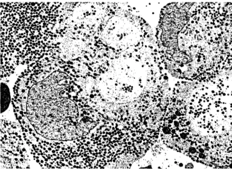
branes appeared to be normal. In 10 tumors, cell size and nuclear size did not seem to differ considerably from control cases, whereas in 2 cases (3, 12), cell and nuclear sizes appeared to be decreased. The nuclei had normal features. T h e endoptasmic reticulum membranes were conspicuous and well- preserved. T h e Golgi complexes were promi- nent and consisted of sacculi and sacs containing immature secretory granules. The fibrous bodies, a characteristic feature o f sparsely granulated G H cell adenomas [12] were noticeable without differing in size, number, and ultrastructural appearance from those seen in untreated somatotroph ade- nomas. T h e mitochondria were ovoid or rod-shaped and had tubular cristae and a moderately electron-dense matrix~ The size and number o f secretory granules seemed to be slightly larger and more numerous in 2 cases of densely granulated tumors (3, 4), whereas they were within the normal range in the rest o f the adenomas. As considerable variation existed from one tumor to the other and even between different areas of the same tumor, and because no morphometric analy- sis has been performed, definite conclusions could not be drawn as to whether the size and number o f secretory granules had changed during treatment with octreotide. The lyso- somes in 6 cases seemed to be unusually large and coalesced, containing lipid droplets and secretory granules. Crinophagy (i.e., the uptake of secretory granules into lysosomes) was more pronounced in the adenomas o f patients treated with octreotide compared to those with no octreotide medication before surgery (Fig. 1). In 11 somatotroph ade- nomas, no exowtosis was observed, whereas in the m a m m o s o m a t o t r o p h adenoma, extru- sion o f secretory granules was common. The capillaries, in general, appeared to be normal and patent; no platelet aggregation, thrombo- sis, or vascular occlusions were seen. T h e endothelial cells showed no major alterations. In 3 cases, the perivascular space was wider than in the untreated cases; accumulation o f fibroblasts and collagen fiber deposits were observed. This perivascutar fibrosis was slight to moderate in 8 tumors and absent in 3 tumors. N o interstitial fibrosis was found. It is noteworthy that 4 adenomas contained endocrine amyloid.
The morphological features by electron microscopy o f the adenomas from group 1I
128 Endocrine Pathology Volume 2 Number 3 September 1991
Table 3, Icontinued)
22 Cellular, chromophobic- Sparsely granulated GH cell acidophilic adenoma, with adenoma
signs of early necrosis Lysosomal accumulation: no Immunopositivity: GH, PRL Widening ofperivascular
(many cells) space
Fibrosis: minimal, perivas- cutar
23 Very cellular, slightly acido- Sparsely granulated GH cell
philic adenoma adenoma, consisting of
Immunopositivity: GH small cells
Fibrosis: no Perivascular fibrosis: no
24 Acidophilic adenoma Densely granulated GH cell
Immunopositivity: GH, adenoma
FSH (occasional cells) Lysosomal accumulation: no Fibrosis: mild, focal, perivas-
cular
G H = g r o w t h h o r m o n e ; PRL = protactin; F S H = tk)tlicle-stimutating h o r m o n e .
(see Table 3) were not significantly different from those noted in group I. Six densely granulated adenomas were observed. In 5 adenomas, the secretory granules were sparse and, in 1 case (19), the tumor was composed of both densely and sparsely granulated cells. One densely granulated G H cell adenoma (case 14) consisted of cells that were often spherule-shaped and possessed eccentric, crescent-shaped nucleus and fibrous bodies (Fig. 2). These findings suggest that this tumor represented an originally sparsely granulated G H cell adenoma and accumula- tion of secretory granules was the result o f
Figure 1. Mammosomatotroph adenoma os a patient (case 1} treated with oc- treotide for 3 weeks. The adenoma cells contain large, irregular secretory gran- ules, some of which have a mottled appearance (arrowhead). Note crinophagy (arrowsJ. {x 10,500.)
somatostatin analog treatment. Cell size seemed to be decreased in 2 cases (16, 18). In 3 o f the densely granulated adenomas (cases 13, 18, 21), unusually numerous and large secretory granules were observed. Striking accumulation o f tysosomes was noted in 1 case (21). Endocrine amyloid was present in 3 adenomas (Fig. 3).
Discussion
The suppressive activity ofoctreotide on G H secretion and its therapeutic value in the management o f acromegaly are now well established [7, 8, 14]. This study was initiated to assess the effect of octreotide on volume, appearance, and morphological features of G H cell adenomas. Shrinkage of G H cell adenomas would make octreotide a useful drug to prepare patients for surgery [17]. In our series, involution was obtained in t 1 of 19 cases, but it was only significant in 3 cases. Modest reduction in tumor size in 3 of 6 patients has been reported by Barnard and colleagues [4], whereas Ducasse and associ- ates [9] described major regression in 1 patient treated with an infusion ofoctreotide. Barkan and co-workers [2] observed signifi- cant reduction o f the tumor volume in 9 of 15 acromegalic patients.
The soft consistency of the adenomatous tissue found at surgery makes the operation easier. This softening effect of octreotide was recorded in 19 of our cases and was also observed by Spinas and colleagues [19] in 5 cases. However, larger series are required to reach definitive conclusions regarding the influence of octreotide on tissue softening, shrinkage of the adenoma, and potential benefits o f these effects.
The morphological response o f somato- troph adenomas to octreotide was not uni- form and not marked, despite the fact that the drug caused clinical improvement and sub- stantial decrease in serum G H levels in acromegalic patients. Lysosomes were large and numerous in several cells, and crino- phagy was observed, suggesting that hormone release was suppressed and the retained secretory products ofadenomatous cells were intracellularly degraded by lysosomes. Some adenomatous cells appeared to be smaller than those of untreated somatotroph ade- nomas and, in several tumors, varying degrees
Acromegaly and Octreotide 1 2 9
Figure 2. Growth hormone (GH) cell adenoma of a patient (case 14) treated with octreotide for 6 months. The adenoma cells are densely granulated, but oth- erwise they have ultrastructural characteristics of a sparsely granulated GH cell adenoma. (• 6,000.)
ofperivascular fibrosis was noticeable. Some tumors removed from octreotide-treated pa- tients were, however, morphologically indis- tinguishable from those not exposed to octreotide. Furthermore, no morphological difference could be evidenced between ade-
Figure 3. Foci of endocrine amyloid (asterisk) in the densely granulated growth hormone cell adenoma of a patient (case 21 ) treated with octreotide for 6 months. (x 11,800,)
nomas treated ~br 3 weeks and those treated for 6 months. Also, no important difference could be detected between adenomas o f patients presenting with an escape to the long-term treatment and those from patients who remained responsive. In the present series, with the exception of 2 adenomas with large areas o f hemorrhagic necrosis, no cell necrosis, endothelial injury', platelet aggrega- tion, vascular damage, or thrombosis was noted, indicating that octreotide had no direct cytotoxic or vasotoxic effects. The vascular change in the 2 exceptional cases was interpreted as an incidental finding, since hemorrhages and loci of necrosis also occur in tumors not exposed to octreotide [12].
Our morphological results are in agree- ment with the literature. Landolt and Oster- walder [16] found amyloid deposits and perivascular fibrosis but no consistent ultra- structural alterations in the secretory appara- tus of adenoma cells. George and colleagues [10] investigated the somatotroph adenoma o f a 36-year-old acromegalic woman treated for 10 days with octreotide. These authors noted an abundance of lysosomes with crinophagy and broadening o f the perivascu- tar space with slight fibrosis around the capillaries. Compared to 10 somatotroph adenomas not exposed to octreotide, the adenoma cells in the octreotide-treated pa- tient appeared to be smaller and contained smaller nuclei as well as cytoplasm, and lysosomes occupied a larger area of the cytoplasmic volume. The nuclear-cytoplas- mic ratio, cytoplasmic volume densities of endoplasmic reticulum, Golgi apparatus, mi- tochondria, and secretory granule diameters did not differ significantly from the control values. Barkan and co-workers [3] observed perivascular fibrosis and a reduction of both cytoplasmic and nuclear areas. These authors also reported 2 densely granulated adenomas with features o f initially sparsely granulated adenomas, resembling the morphological appearance o f i case in our series.
All these results are consistent with functional inhibition of G H release. Dopa- mine agonists, such as bromocriptine, cause reversible shrinkage o f PRL-secreting pitu- itary adenomas associated with morphologi- cally pronounced cell involution [21]. The changes are striking and are characterized by a substantial decrease in cell size and cytoplas- mic and nuclear area, an increase in the
130 Endocrine Pathology Volume 2 Number 3 September 1991
nuclear-cytoplasmic ratio, and a reduction in cytoplasmic volume densities ofendoplasmic reticulum membranes and Golgi apparatus. These changes are not apparent in G H - secreting pituitary adenomas exposed to octreotide. Thus, in tumors removed from octreotide-treated patients, a discrepancy ex- ists between functional inhibition and m o r - phological findings.
References
1. Asa SL, Scheithauer BW, Bilbao JM, Horvath E, Ryan N, Kovacs K, Randall RV, Laws ER, Singer W, Linfoot JA, Thorner MO, Vale W. A case for hypothalamic acromegaly: a clinical study of six patients with hypothalamic gangtio- cytomas producing growth hormone-releas- ing factor. J Clin Endocrinol Metab 58:796- 803, 1984.
2. Barkan AL, Kelch RP, Hopwood N J, Beitins IZ. Treatment of acromegaly with the long- acting somatostatin analog SMS 201-995. J Clin Endocrinol Metab 66:16-23, 1988. 3. Barkan AL, Lloyd RV, Chandler WF, Hatfield
MK, Gebarski SS, Kelch RP, Beitins IZ. Preoperative treatment of acromegaty with long-acting somamstatin analog SMS 201- 995: shrinkage of invasive pituitary macroade- nomas and improved surgical remission rate. J Clin Endocrinot Metab 67:1040-1048, 1988. 4. Barnard LB, Grantham WG, Lamberton P, O'Dorisio TM, Jackson IM. Treatment of resistant acromegaly with long-acting so- matostatin analogue (SMS 201-995). Ann Intern Med 105:856-861, 1986.
5. Bauer W, Briner U, Doepfner W, Haller R, Huguenin P~ Marbach P, Petcher TJ, Press J. SMS 201-995: a very potent and selective octapeptide analogue of somatostatin with prolonged action. Life Sci 31:1133-1140, 1983.
6. Besser GM, Mortimer CH, Carr D, Schally AV, Coy DH, Evered D, Kastin AJ, Turn- bridge WMG, Thorner MO, Hall R. Growth hormone release inhibiting hormone in ac- romegaly. Br MedJ 1:352-355, 1974. 7. Chiodini PG, Cozzi R, Dallabonzana D,
Oppizzi G, Verde G, Petroncini M, Liuzzi A, del Pozo E. Medical treatment of acromegaly with SMS 201-995, a somatostatin analog: a comparison with bromocriptine. J Clin Endo- crinol Metab 64:447-453, 1987.
8. Comi RJ, Gorden P. The response of serum growth hormone levels to the long-acting somatostatin analog SMS 201-995 in acro- megaly. J Clin Endocrinol Metab 64:37-42, 1987.
9. Ducasse MCR, Tauber JP, Tourre A, Bonafe
A, Babin TH, Tauber MT, Harris AG, Bayard F. Shrinking of a growth hormone-producing pituitary tumor by continuous subcutaneous infusion of the somatostatin analog SMS 201-995. J Clin Endocrinol Metab 65:1042- 1046, 1987.
10. George SR, Kovacs K, Asa SL, Horvath E, Cross EG, Burrow GN. Effect of SMS 201-995, a long acting somatostatin analogue, on the secretion and morphology of a pituitary growth hormone cell adenoma. Clin Endo- crinol (Oxf)26:395-405, 1987.
11. Guiot G, Oproiu A, Hertzog E, Fredy D. Ad~nomes hypophysaires. EMC Paris t7:340,
1969.
12. Kovacs K, Horvath E. Tumors of the pituitary gland. In: Atlas of tumor pathology, second series, fascicle 21. Washington, DC: Armed Forces Institute of Pathology, 1986, pp 1-269. 13. Lamberts SWJ, Oosterom R, Neufeld M, del Pozo E. The somatostatin analog SMS 201- 995 induces long-acting inhibition of growth hormone secretion without rebound hyperse- cretion in acromegalic patients, J Clin Endo- crinol Metab 60:1161-1165, 1985.
14. Lamberts SWJ, Uitterlinden P, Verschoor L, Van Dongeu KJ, dcl Pozo E. Long-term treatment ofacromegaly with the somatostatiu analogue SMS 202-995. N Engl J Med 313:1576-1580, 1985.
15. 1Lxmberts SWJ, Zweens M, Verschoor L, del Pozo E. A comparison among the g-rowth hormone-lowering effects in acromegaly of the somatostatin analog SMS 201-995, bro- mocriptine and the combination of both drugs. J Clin Endocrinol Metab 63:16-19, 1986.
16. Landolt AM, Osterwalder V. Perivascular fibrosis in prolactinomas: is it increased by bromocriptine? J Clin Endocrinol Metab 58:1179-1183, 1984.
17. Landolt AM, Osterwatder V, Stuckmann G. Preoperative treatment of acromegaly with SMS 201-995: surgical and pathological obser- vation. In: Ludecks DK, Tolls G, eds. Growth hormone, growth factors and acromegaly. NewYork: Raven Press, 1987, pp 229-244. 18. Scheithauer BW, Kovacs K, Randall RV,
Horvath E, Okazaki H, Laws ERJr. Hypotha- lamic neuronal hamartoma and adenohypophy- seal neuronal choristoma: their association with growth hormone adenoma of the pitu- itary. J Neuropathol Exp Neurol 42:648-663, 1983.
19. Spinas GA, ZapfJ, Landoh AM, Stuckmann G, Froesch ER. Preoperative treatment of 5 acromegalics with somatostatin analogue: en- docrine and clinical observations. Acta Endo- crinot (Copenh) 114:249-256, 1987.
Acromegaly and Octreotide 131
HG. The unlabeled antibody enzyme method ofimmunochemistry. Preparation and proper- ties of soluble antigen-antibody complex (i~orseradish peroxydase anti-horseradish per- oxidase) and its use in the identification of spirochetes. J Histochem Cytochem 18:315- 333, t970,
21. Tindatl GT, Kovacs K, Horvath E, Thorner
MO. Human protactin-producing adenomas and bromocriptine: a histological, immunocy- tochemical, ultrastrucmral, and morphometric study. J Clin Endocrinol Metab 55:1178-
1183, 1982.
22. Vezina JL, Maltais R. La selle tmcique dans t'acromdgalie. Etude radiologique. Neurochir- urgie Ig(suppl 2):35, 1973.