HAL Id: dumas-02434257
https://dumas.ccsd.cnrs.fr/dumas-02434257
Submitted on 9 Jan 2020
HAL is a multi-disciplinary open access
archive for the deposit and dissemination of sci-entific research documents, whether they are pub-lished or not. The documents may come from teaching and research institutions in France or
L’archive ouverte pluridisciplinaire HAL, est destinée au dépôt et à la diffusion de documents scientifiques de niveau recherche, publiés ou non, émanant des établissements d’enseignement et de recherche français ou étrangers, des laboratoires
Afrik : Analyse familiale du risque de kératocone
Gabrielle Lapeyre
To cite this version:
Gabrielle Lapeyre. Afrik : Analyse familiale du risque de kératocone. Sciences du Vivant [q-bio]. 2019. �dumas-02434257�
Université de Bordeaux U.F.R. DES SCIENCES MEDICALES Année 2019 N° 3082 Thèse pour l’obtention du DIPLOME D’ETAT DE DOCTEUR EN MEDECINE DISCIPLINE : OPHTALMOLOGIE Présentée et soutenue publiquement le : 10 septembre 2019 Par GABRIELLE LAPEYRE Née le 16 janvier 1991 à Toulouse
AFRIK: ANALYSE FAMILIALE DU RISQUE DE KERATOCONE
Directeur de thèse : Monsieur le Professeur David Touboul Rapporteur de thèse : Monsieur le Professeur Eric Gabison Membres du Jury : Monsieur le Professeur Jean-François Korobelnik Président Madame le Professeur Marie-Noëlle Delyfer Jury Monsieur le Professeur Pierre Fournié Jury Madame le Docteur Valentine Coste Jury Monsieur le Professeur David Touboul Directeur de thèseTable des matières
TABLE DES MATIERES 2 INDEX DES TABLEAUX ET FIGURES 3 LISTE DES ABREVIATIONS 4 INTRODUCTION 5 ARTICLE 9 ABSTRACT 11 MANUSCRIPT 12MATERIAL AND METHODS 13
RESULTS 17
DISCUSSION 19
BIBLIOGRAPHY 23
TABLES ET FIGURES 27
CONCLUSION ET PERSPECTIVES 35 ANNEXES 36 Annexe 1 Le kératocône chez l'enfant 36 Annexe 2 classifications Amsler-Krumeich 42 Annexe 3 classifications de Rabinowitz 43 BIBLIOGRAPHIE 44 SERMENT D’HIPPOCRATE 47
Index des tableaux et figures
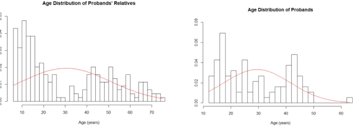
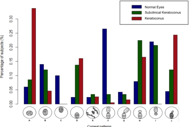
Figure 1: Distribution of age in proposers (a) and in relatives (b), in red the theoretical normal distribution. ………..……… 27 Table 1: Number of probands and close relatives according to keratoconus (KC) status. The assignment of patients in a group is conditioned by the status of the most severely affected eye..……….………...………... 28 Table 2: Eye characteristics according to keratoconus (KC) status..……….. 29 Table 3: Individual characteristics according to keratoconus (KC) status …………...……. 30 Table 4: Maximum likelihood estimates of familial correlations for the corneal thinnest point, maximal keratometry, I-S index and AAI using the class D regressive model……. 31 Table 5: Previous study on the same topic : prevalence of KC in relatives, with videokeratography... 32 Figure 2 : Description of corneal patterns according to Rabinowitz classification and KC status……….……….…….… 33 Figure 3: Anterior corneal shape on galilei topographer for one of our sibling family.. 34Liste des abréviations
CNRK : Centre National de Référence du Kératocône CHU : Centre Hospitalier Universitaire UNADEV : Union Nationale des Aveugles et des Déficients Visuels INSERM : Institut National de la Santé et de la Recherche Médicale IOP : Intraocular Pressure MD : Mean Deviation KLAP : Kératoplastie lamellaire antérieure profonde mmHg : Millimètre de Mercure PIO : Pression Intra-Oculaire SD : Standard Deviation KC : keratoconus OCT : optical coherence tomographyIntroduction
Le kératocône est défini par une ectasie postérieure de la cornée, associée à un amincissement non inflammatoire. La topographie cornéenne est le meilleur examen pour diagnostiquer cette pathologie, notamment les kératocônes à un stade peu avancé, ou suspect. La classification d’Amsler-Krumeich (annexe 1) bien que historique a toujours été utilisée pour classer les kératocônes. Cette classification n’intègre pas les données des technologies récentes de pointe, mais elle aide et sert de référence pour de nombreuses études. A ce jour aucune explication physiopathologique complète ne permet d’expliquer l’apparition d’un kératocône chez un individu. Les chercheurs évoquent le rôle de l’environnement, de la biomécanique cornéenne, de la génétique et des désordres biochimiques inflammatoires.
C’est une pathologie progressive : l’ectasie cornéenne va se majorer. Celle-ci est définie par une augmentation des indices de kératométrie : Kératométrie maximale, minimale et moyenne ; une augmentation du bombement postérieur, et un amincissement de la cornée. La progression du kératocône est le plus souvent associée à une baisse d’acuité visuelle. Afin de dépister les kératocônes évolutifs il est important de réaliser un suivi répété chez ces patients, de préférence en utilisant les mêmes machines, le même topographe à chaque consultation.
Les facteurs de risque de développement d’un kératocône évoqués dans la littérature sont : l’âge jeune, la trisomie 21, l’allergie oculaire, l’ethnie, la génétique, les stimulations mécaniques répétées de la cornée (frottement oculaire), floppy eyelid
syndrome, l’atopie, le syndrome de Marfan, le syndrome d’Ehlers-Danlos, l’amaurose congénitale de Leber.
La prise en charge du kératocône a pour but la réhabilitation de l’acuité visuelle et la stabilisation de la maladie/l’absence de progression. Elle peut se faire par deux versants, un non chirurgical et un chirurgical. Bien sûr la prise en charge non chirurgicale, quand elle est possible, doit être la première envisagée.
Il est primordial d’insister sur la compréhension de la maladie par le patient, pour que celui ci l’accepte, soit motivé pour la prise en charge, qu’il comprenne les enjeux. Mais aussi qu’il sensibilise les membres de sa famille pour le dépistage et qu’il comprenne le rôle essentiel du frottement oculaire dans la progression de la maladie. Le rôle des questionnaires de frottement oculaire est primordial dans cette prise en charge, car ils permettent une prise de conscience par les patients de ces gestes inconscients, répétitifs et traumatisants pour leurs cornées. Dans le service du CNRK de Bordeaux, il est courant que l’on filme les patients en même temps que l’on s’entretient avec eux, et que l’on regarde ensuite la vidéo afin de leur montrer les frottements oculaires inconscients qu’ils réalisent de manière répétée au cours d’une simple conversation.
La réhabilitation visuelle peut se faire à travers les verres de lunettes et les lentilles de contact rigides. Les lentilles de contact rigides aujourd’hui très développées dans le kératocône permettent un gain d’acuité visuelle supérieur aux lunettes dans de nombreux cas ; grâce à leurs capacités à corriger les astigmatismes irréguliers et à diminuer les aberrations optiques de hauts degrés. Cependant certains patients, une minorité, sont limités par la manipulation des lentilles qui peut être vécue comme non naturelle, voire angoissante, ou un environnement/contexte non adapté (travail dans la
Dans les cas où une adaptation en lentilles rigides n’est pas réalisable, ou pas satisfaisante, la pose d’anneau intra cornéen doit être envisagée si la cornée le permet (pachymétrie suffisante). La pose d’anneau intra cornéen, plus ou moins couplée à une photokératectomie thérapeutique topoguidée secondairement, permet la diminution la régularisation des astigmatismes de ces cornées, et la diminution des indices kératometriques. Ils permettent dans de nombreux cas d’obtenir une acuité visuelle satisfaisante associée à une adaptation en lunettes ou en lentilles rigides. La limite de la pose des anneaux intra cornéens est le lien étroit entre les résultats et l’expérience du chirurgien.
Les kératocônes réfractaires à ces dernières thérapeutiques et ceux présentant des opacités cornéennes centrales, voire des hydrops ont comme unique recours les greffes de cornée. Celles ci sont largement mieux maitrisées ces dernières années grâce à l’évolution de nos connaissances dans le domaine et à l’utilisation adaptée des thérapeutiques pour éviter les rejets de greffe. Mais elles restent un geste chirurgical irréversible qui signe un tournant de la maladie, à un stade très évolué. Les greffes de cornées, quand cela est possible, sont des greffes lamellaires antérieures profondes (KLAP). Elles ont l’avantage de ne pas mettre en contact la cornée greffée avec l’humeur aqueuse et de diminuer le risque de rejet de greffe. Quand une KLAP n’est pas réalisable, le chirurgien réalisera une kératoplastie transfixiante. La gestion post opératoire des greffes doit être rigoureuse. Elle nécessite une prise en charge minutieuse de l’ablation des sutures, une bonne gestion des anti-inflammatoires et du risque de rejet, et une surveillance des pathologies secondaires comme le glaucome. Le patient doit être compliant, et patient pour obtenir un résultat optique satisfaisant grâce à l’adaptation optique, secondairement en lunettes ou en lentilles rigides.
A tous les stades de la prise en charge, une thérapeutique primordiale dans le kératocône doit toujours être envisagée : le cross-linking. Celui-ci a prouvé son efficacité dans la diminution de la progression des kératocônes. Son mécanisme physiopathologique est encore discuté, entre durcissement de la cornée, baisse de la sensibilité cornéenne diminuant les frottements oculaires, et influence sur le comportement et acceptation de la maladie.
Tous les traitements chirurgicaux et les lentilles de contact présentent un risque infectieux, bien que minime, non négligeable. Ils ont un coût pour la société et le patient, et se font au travers d’une prise en charge longue et chronophage pour le patient et son médecin. Ils peuvent être source de stress, et malheureusement dans certain cas dépassés : d’échec répétitif de greffe ou de complications irréversibles ; une perte définitive de la vision peut avoir lieu.
Ces implications justifient une prise en charge précoce du kératocône et l’identification de ses facteurs de risque et de progression, afin d’optimiser le dépistage et la sensibilisation des patients.
Cette étude découle du besoin d’identifier les personnes à risque de kératocône, pour mieux orienter le dépistage ; et ce afin de réduire l’incidence de la pathologie, et sa morbidité. Elle est née sous l’impulsion et la dynamique du CNRK du Pr Colin et du Pr Touboul, grâce au financement de l’UNADEV, au travail des attachés de recherche, et grâce à la collaboration de qualité avec l’équipe de l’INSERM du Dr Bouzigon spécialisée dans la variabilité génétique et maladies humaines à Paris. Elle a pour objectif principal, en tant que cohorte prospective descriptive et épidémiologique bien menée de servir de référence dans la prévalence du kératocône chez les apparentés au premier degrés.
Article
Le manuscrit ici présenté fait l’objet d’une soumission dans une revue internationale à comité de lecture : Cornea.AFRiK: Analysis of Familial Risk for Keratoconus
Authors: Gabrielle Lapeyre1, Pierre Fournie2, Raphaël Vernet3, Stéphanie Roseng1, François Malecaze2, Emmanuelle Bouzigon3, David Touboul1
Setting: CHU Bordeaux, CRNK
1National center of keratoconus, CHU Bordeaux, France 2National center of keratoconus, CHU Toulouse, France
3Inserm, UMRS 1124, Genetic Epidemiology and Functional Genomics of Multifactorial Diseases Team, University Paris Descartes, Paris, France Acknowledgments for financial support: UNADEV Conflict of interests: none
AFRiK: Analysis of Familial Risk for Keratoconus
Abstract: Purpose: To assess the keratoconus (KC) prevalence among close relatives of affected subjects. Design: Large epidemiological, prospective and observational cohort. Methods: Setting: The study took place in the French Keratoconus National Reference Centre, in Bordeaux. Study Population: Ninety-four unrelated patients with KC and at least 2 close relatives assessable; 221 close relatives. Observation Procedures: Each patient had a thorough ocular examination including visual acuity, refraction, slit lamp biomicroscopy, indirect ophthalmoscopy, keratometry, corneal topography (Topographic Modelling System (Tomey Corporation, Japan) and Galilei V6 (Ziemer, Switzerland)), pachymetry and biomechanical properties of cornea (Ocular Response Analyzer (Reicherts, USA)). Additional data have been collected about atopy, smoking, and eye-rubbing habits. Main Outcome Measures: Keratoconus prevalence, multivariate analysis, and familial correlation analysis have been studied. Results: Of the 221 relatives, 9.05% (n=20) had a clinical manifest KC and 15.4% (n=31) had a subclinical KC. The prevalence of KC was estimated equal to 0.14 (0.07; 0.22 (CI 95%)) among parents, 0.03 (0; 0.10 (CI 95%)) among offspring, and 0.10 (0.04; 0.20 (CI 95%)) among siblings. In multivariable analysis, age and eye-rubbing were associated with an increased risk of KC. The familial correlation of KC among probands’ close relatives was estimated at 0.55 among parents, 0.29 among offspring and 0.49 among siblings. Conclusion: Being a close relative of a KC subject remains a risk factor of developing KC. This should be taken into account for KC screening in paediatric population, in family members of KC patients and for refractive surgery candidates.Introduction Keratoconus (KC) is a complex multifactorial corneal disorder characterized by corneal thinning, bulging, and progressive deformation.1 This disease is well known and has been subject to a renewing interest due to the recent development of refractive surgery which itself stems from the development of technology in corneal topography and tomography. The treatment of this condition has been subject to innovation and now relies on a wide range of possibilities such as gas permeable contact lens fitting, corneal cross-linking, intra-stromal corneal ring segment, therapeutic excimer treatment and corneal transplantation.2 Nevertheless, there is a need to account properly for the contribution of certain factors to the development and progression of KC. At this time twenty-four genes were identified as potential contributors to KC and 49 KC-related comorbidities/syndromes were found.3 A behavioural theory based on eye-rubbing offers an interesting approach for management of this corneal ectatic condition.4 Moreover, in our medical practice we are used to seeing a lot of familial history of KC and some authors found a higher prevalence of KC in relatives of patient with KC in their population.5, 6, 7 As a matter of fact, we still do not know the relative contribution of genetics, of the environment, and of behaviour in the development of KC. In order to investigate this topic, we conducted a large prospective cohort study at the French Keratoconus National Reference Centre (CRNK) to assess the KC prevalence in close relatives of KC subjects.
Methods: Consecutive unrelated KC patients (probands) with two or more family members were enrolled prospectively from January 2015 to September 2017, when they came for an ophthalmic examination at the CRNK, at the Bordeaux University and Hospital. The study, called AFRiK “Analysis of Family Risk for Keratoconus” was funded by the “Union Nationale des Aveugles et Déficients Visuels (UNADEV)” charity, a French humanitarian non-profit association. Ethical approval was obtained for this epidemiological, prospective, and observational cohort study. All patients gave their consent to participate in the study. The study was performed in accordance with the tenets of the Declaration of Helsinki. The AFRiK study includes 7 years old or older patients who were previously diagnosed as having a clinically significant KC in one or both eyes based on clinical features, slit lamp findings, keratometry, pachymetry. At least two close relatives were needed to constitute a family, including first-degree relatives (parents and offspring) and siblings. Patients were excluded if they had other ocular diseases in addition to KC. If they wore contact lenses, they had to remove them at least fifteen days prior to the examination. Each patient (KC and relatives) had a thorough ocular examination including corrected distance visual acuity (CDVA), refraction, slit lamp biomicroscopy, indirect ophthalmoscopy, keratometry (Km), corneal topography, pachymetry and biomechanical properties of cornea (when possible). We used Topographic Modelling System (TMS- Tomey Corporation, Nagoya, Japan) and Galilei V6 for topography and tomography (Ziemer ophthalmic Systems AG, Port, Switzerland) and Ocular Response Analyzer (ORA- Reicherts, Depew, USA) for biomechanics and tonometry. Additional
data have been collected by a physician assistant from standardized questions and responses about: smoking, eye-rubbing habits, asthma, urticaria, dermatitis, allergic conjunctivitis, allergic rhinitis, and allergy to drug, contact or food allergy. For simplicity reasons we group together asthma, urticaria, dermatitis, allergic conjunctivitis, allergic rhinitis, and allergy to drug, contact or food allergy condition under the name of atopy in this article. Two corneal experts examined the data to classify subjects as KC, subclinical KC (subKC) including forme fruste KC and KC suspect, or normal eyes. The stage of the disease was determined using the Amsler-Krumeich classification. We arbitrarily chose four criteria to standardize our classification between KC and subKC: pachymetry less than 510 μm, I-S index positive, AAI (asymmetric, aspheric index) positive, and abnormal corneal enantiomorphism.8 Abnormal corneal enantiomorphism was defined by one of three different measures between the right and left eye: either a difference superior to 0,3D between the two means SimK, a difference superior to 10 μm between the two corneal thinnest points, or a difference superior to 10 μm between the two AAI. At least three of the four criteria were requested to satisfy a KC diagnostic, while only two were needed for subKC. The familial pattern was studied using the Galilei topography (anterior instantaneous curvature) and classified with the Rabinowitz pattern classification. Every topography of family member was categorized according to its similarity with the topography of the family proband, using a score between zero and three (0= no similarity, 3= strong similarity).
Socio-demographic and clinical (ophthalmologic) characteristics were compared according to affection status or between probands and affected relatives using the Student’s t-test and Wilcoxon-Mann-Whitney test for quantitative variables and the Chi-square test and Fisher exact test for qualitative variables. Univariate and multivariate analyses using logistic or linear mixed models were performed to study the relationship between individual or environmental factors and the risk of KC on the one hand, and several indices of KC severity (maximal keratometry, AAI, thinnest-point corneal thickness (TPCT), I-S index, and Krumeich severity score) on the other hand. A random effect was included in all models to account for familial dependencies. To analyze the indices of KC severity related to both right and left eyes of a given individual, a second random effect was included to account for intra-individual correlations. Before analysis, AAI was log-transformed due to its skewed distribution. Analyses were done using R statistical software (R Core Team 2018). Familial correlations of KC were estimated in probands’ relatives using the Falconer’s method9 which relies on a liability threshold model. This method assumes that liability to KC is normally distributed and that the prevalence of the disease in the general population determines the threshold of liability above which a person expresses the disease. This method allows approximation of familial correlations of KC using the prevalence of the disease in general population and the observed prevalence in the probands’ relatives. For that purpose, we used a prevalence of 265/100,000 as reported by Godefrooij et al10. A familial correlation analysis was performed for four quantitative ophthalmologic indices: maximal keratometry, AAI, corneal thinnest point and I-S index. For a given individual, out of the pairs of indices relating to the right and left eyes, the one with the
highest value for maximal keratometry, AAI and I-S index, or with the lowest value for the TPCT was assigned to that individual. Prior to familial analysis, each phenotype was adjusted for age and gender using multiple regressions. As all probands were affected by KC, which is known to be associated with the four ophthalmologic indices, an indicator variable specifying the position of individuals in the family (probands, probands blood relatives (parents, offspring or sibs) or unrelated spouses) was included in the regression models. This strategy was shown to correct adequately for the ascertainment of families through affected subjects11. The regression residuals of each phenotype were then inverse-Gaussian-transformed to standard normal distributions. A familial correlation analysis was performed for each adjusted and transformed ophthalmologic index using the Class D regressive model12 which allows to construct various patterns of familial correlations of any origin (genetic and/or environmental). Briefly, the vector of observed quantitative phenotypes for a family is assumed to follow a multivariate normal distribution with an overall mean and covariance matrix expressed in terms of the phenotypic variance (assumed equal for all family members, the phenotypes being adjusted and standardized prior to analysis) and four phenotypic correlations: ρFM
(between spouse), ρFO (between father and offspring), ρMO (between mother and
offspring) and ρSS (between siblings). Parameter estimations and tests of models were carried out using maximum likelihood methods as implemented in S.A.G.E. 6.4 program (http://darwin.cwru.edu)13. Each familial correlation was tested by comparing a model including all correlations except for the one tested being set to zero to a general model including all of them. Specific patterns of correlations were then tested including a model specifying equal father-offspring and mother-offspring correlations (ρFO = ρMO = ρPO) and a model specifying equal parent-offspring (ρPO) and sib-sib correlations
Results We included 315 subjects (probands and relatives) belonging to 94 families ascertained through one KC subject. Among these families, 56.5% included the proband and his/her parents, 35% the proband and his/her offspring, 2% the proband his/her parents and his/her offspring, and 6.5% the proband and his/her siblings. The average age at examination was 29.5 years and 30.5 years for probands and relatives respectively. The distribution of age is described in figure 1. Among the 221 relatives, 20 subjects (9.05%) were diagnosed with KC in one or both eyes, and 31 (14.03%) with subKC (Table 1). The prevalence of KC was estimated equal to 0.14 (0.07; 0.22 (CI 95%)) among parents, 0.03 (0; 0.10 (CI 95%)) among offspring, and 0.10 (0.04; 0.20 (CI 95%)) among siblings. KC was bilateral in 73% of probands (n=69), and in 50% of affected relatives (n=10). In total, we had 79 bilateral KC, 20 left KC, and 15 right KC. KC patients were more often males than unaffected subjects (62.3% vs 45.8%, p=0.005). The familial correlation of KC among probands’ relatives was estimated at 0.55 among parents, 0.29 among offspring, and 0.49 among siblings. Table 2 describes clinical and ophthalmologic features according to affection status (KC, subKC, normal eyes). KC patients presented more often eye-rubbing (p=0.007), dry eye syndrome (p=0.002), and atopy (p=0.002) than patients with normal eyes. In multivariable analysis, age and eye rubbing were associated with an increased risk of KC (OR=1.02 [1.00; 1.04] and OR=2.50 [1.39; 4.50] respectively) and being a woman with a decreased risk of KC (OR=0.51 [0.29;0. 90]). Table 3 reports eye and topographic characteristics looking at each eye separately.
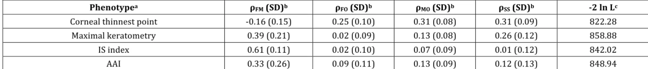
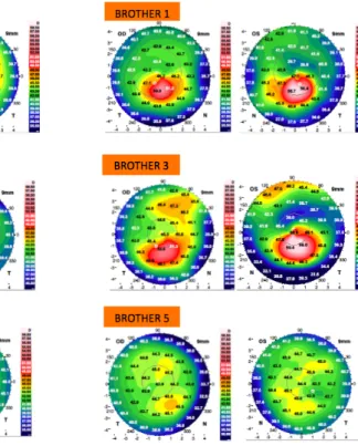
Topographic pattern in relatives and probands were collected and classified with Rabinowitz pattern classification in figure 2. One example of familial similarity for anterior corneal aspect is shown in figure 3. In multivariable analysis, age was associated with higher values of keratometry (p=0.05), AAI (p=5x10⁻¹⁰), and Krumeich severity score (p=0.03) and with lower values of thinnest-point corneal thickness (TPCT) (p=0.004). In this analysis, eye-rubbing was associated with higher values of keratometry (p=0.001), AAI (p=5x10⁻⁶), IS index (p=0.01) and Krumeich severity score (p=4x10⁻⁴); and allergy-related traits with higher values of Krumeich severity score (allergic conjunctivitis, p=0.03) and AAI or I-S index (contact allergy, with p=0.006 and p=2.5x10⁻⁴, respectively). Estimates of familial correlations for thinnest-point corneal thickness, maximal keratometry, I-S index and AAI, adjusted for family position and significant covariates are shown in table 4. For the thinnest-point corneal thickness the spouse correlation did not differ significantly from zero (P=0.32) and each of the three other correlations was significant (PρFO=0.03, PρMO=2.8x10⁻⁴, PρSS=4.7x10⁻⁴). Tests of equality of these
correlations indicated a good fit of a model with equal parent-offspring and sib-sib correlations, estimated at 0.31. For maximal keratometry the three correlations ρFM, ρMO
and ρFO did not differ from zero (p≥0.11) while the sib-sib correlation was significant
(P=0.03) and estimated at 0.25. For I-S index, only the spouse correlation was significant (P=0.04) and estimated at 0.62. For AAI, none of the four familial correlations was significant (P>0.15).
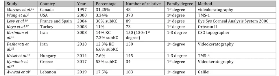
Discussion The prevalence of KC varies around the world ranging from 0.0004% in Russia to 3.3% in Iran14, while recent prevalence in Caucasian population was estimated at 265/100000.10 Regardless of ethnic considerations, the prevalence of KC in relatives is commonly considered more important than in general population, and this all the more if we take into account subKC. Our team decided in 2014 to lead this familial prospective cohort study on this topic, in which each patient and his family could have a topography and thorough ocular examination. This data collection process was completed in 2017. We are aware that having a control group could have been useful to identify subKC over general population, but we benefited from several quantitative measures provided by recent topographers and ORA, as well as environmental and topographical patterns. Only few studies used topography on relatives, and KC prevalence in those individuals varied from 2.92%5 to 31.2%.15 The prevalence differed a lot from one study to another as
described in table 5. In a recent study Awwad et al. focus on pediatric first degree relatives of KC patients and found 17,5% of KC.6 The largest study was published by
Wang et al.5 in 2000. They analysed 373 first-degree relatives of KC individuals and
found that the prevalence of KC was 3.78% in siblings and 2.92% in parents and offspring. All studies conducted thereafter reported a prevalence of KC in relatives higher than 7.6%.16 For example, Kaya et al.17 found 11% of KC in 72 relatives and
Karimian et al.18 found a prevalence of 14% in 150 relatives. In those studies, suspected
KC prevalence in relatives reached 21% to 33.3%, 18,19,20 and abnormal corneal patterns
KC, but most of the time it is detected only by asking patients about their family history using a questionnaire. Thus, 4.3% to 23.5% of KC patients reported multiple KC cases in their familly22,23. Our study population included children, men, and women of all ages. As KC is a progressive condition that appears in the teenage and young adulthood, we can suppose that some of the younger family members which did not yet develop a KC might develop a KC later on. This could have led to an underestimation of the prevalence of KC in relatives. The AFRiK study was performed in a KC national centre. This implies that our centre tends to receive more severe pathologies. Thus, almost half of our probands were diagnosed with a Krumeich stage 2 or more. A cohort multicentric follow-up would be interesting as consolidated findings might have a significant impact on KC screening. In AFRiK study the KC subjects were more often males, but the severity of KC was more important among women. When looking at the literature on prevalence and incidence, we came across discordant results. Indeed, Godefrooij et al10 found more male with KC
than women while Hwans et al24 found no sex predisposition. Fink et al25 found no
impact of gender on severity and progression of KC. However, it is difficult to compare these studies because these populations were of different ethnic groups with different range of ages. The analysis of topographic patterns allows to identify shared traits within KC families, which go beyond the spectrum of KC topographical features. However, there was a lot of heterogeneity within families. As illustrated in figure 3, we were able to identify in a
sibling pairs that were very heterogeneous. One must keep in mind that figure 3 accounts for a particularly thorough example. Most of recruited families only included three members and as such similarity can be more limited. Nevertheless, we believe that topographical patterns might be as complex and informative as fingerprinting patterns. Very few information is available in the literature on that topic. It could be of interest to extend this research to a broader population and to more extended families. This would allow us to investigate whether familial aggregation, as demonstrated by Wang et al.5, is only present in KC family or could be present in every family. Familial corneal pattern could appear as a new important datum, which calls for further research. We find a positive familial correlation and develop, four quantitative indices to characterize it further. The familial correlation estimates for these indices help us identify what could be determined by genetics. For example, corneal thickness is uncorrelated between spouses while it is correlated between parents and offspring, and between siblings. The best fitting pattern of familial correlations for corneal thickness could be accounted by an additive polygenic model. The other indices showed only significant spouse correlation (IS index) or excess of correlation between siblings (maximal keratometry) so they could be explained by other factors than genetic such as age or shared environmental factors. These help us focus on the complexity of the origin of this association between KC and close relative KC. Some authors5 put forward genetic transmission as the most probable explanation and mitigating the contribution of sporadic and environmental factors. This is supported by genetic studies3 that found several genes as potential contributors to KC, and it seems to be complex non-mendelian disease16. Moreover, biomechanical evaluation now plays a major role in KC screening26. Indeed, several studies among KC relatives suggested that
their biomechanical properties differed from those of subject from general population. 27,28 If we compare corneal hysteresis (CH) and corneal resistance factor (CHF) in relatives without KC or subKC to previous study with a control group from the same centre29, we do not observe different biomechanical properties. Actually, we do not know if biomechanical properties are influenced by genetic factors. On this topic further research is needed. More particularly, atopy is often associated with KC and it is well known that atopy is influenced by both genetic and environmental factors, but its role in the development of KC remains to be confirmed. As Bawazeer et al 30, we believe that atopy might contribute to KC through the eye-rubbing tendency of patients31. One issue with eye rubbing is that it is difficult to quantify. One could use a questionnaire with score to account for the tendency of eye rubbing. Atopy and eye rubbing are both affected by allergens, pollution, mimicry, habits and these could fit into the environmental and behaviour theory. KC patients probably evolve in the same environment and do the same things as their family, so this could explain why familial history of KC was considered as a risk factor to develop KC. As well, it should be an item to be screened in the frame of refractive surgery pretenders. KC incidence in families of KC is higher than in general population. Nevertheless, similar behaviors, exposure to eye rubbing risks, social mimicry, genetic factors related to atopy, and corneal pattern could represent confusing elements converging against the genetic affiliation theory, extremely guilt-creating and harrowing for families. Being a close relative of a person affected by KC remains a risk factor of developing a KC. Early screening is of great importance in KC relatives, and as such we should sensitize
Bibliography 1. Rabinowitz YS. Keratoconus. Surv Ophthalmol. 1998; 42(4):297-319. 2. Gomes JA, Tan D, Rapuano CJ, et al. Global consensus on keratoconus and ectatic diseases. Cornea. 2015; 34(4):359–69. 3. Loukovitis E, Sfakianakis K, Syrmakesi P, et al. Genetic Aspects of Keratoconus: A Literature Review Exploring Potential Genetic Contributions and Possible Genetic Relationships with Comorbidities. Ophthalmol Ther. 2018; 7(2):263-292. 4. Gatinel D. Eye rubbing, a sine qua non for keratoconus. Int J Kerat Ect Cor Dis. 2016; 5(1):6-12. 5. Wang Y, Rabinowitz YS, Rotter JI, Yang H. Genetic epidemiological study of keratoconus: evidence for major gene determination. Am J Med Genet. 2000; 93(5):403– 409. 6. Awwad ST, YehiaM, Mehanna C-J, Fattah MA, Saad A, Hatoum A, Al-Haddad C. Tomographic and Refractive Characteristics of Pediatric First-Degree Relatives of Keratoconus Patients. Am J Ophthalmol. 2019; 207: 71-76 7. Francois J. Afflictions of the cornea. In: Francois J, ed. Heredity in Ophthalmology. St. Louis: CV Mosby, 1961: 297–298. 8. Smadja D, Touboul D, Cohen A, et al. Detection of subclinical keratoconus using an automated decision tree classification. Am J Ophthalmol. 2013; 156:237–246. 9. Falconer DS. The inheritance of liability to diseases with variable age of onset, with particular reference to diabetes mellitus. Ann Hum Genet. 1967; 31 :1-20. 10. Godefrooij DA, de Wit GA, Uiterwaal CS, et al. Age-specific Incidence and Prevalence of Keratoconus: A Nationwide Registration Study. Am J Ophthalmol. 2017; 175:169–172.
11. Bouzigon E, Dizier MH, Krähenbühl C, et al. Clustering patterns of LOD scores for asthma-related phenotypes revealed by a genome-wide screen in 295 French EGEA families. Hum Mol Genet 2004 ; 13(24): 3103-3113. 12. Bonney GE, Opitz JM, Reynolds JF. On the statistical determination of major gene mechanisms in continuous human traits: regressive models. Am J Med Genet. 1984; 18(4):731-749. 13. S.A.G.E. Statistical Analysis for Genetic Epidemiology, Release 6.4: http://darwin.cwru.edu ; 2016. 14. Gordon-Shaag A, Millodot M, Shneor E, Liu Y. The genetic and environmental factors for keratoconus. Biomed Res Int. 2015; 2015:795738. 15. Morrow GL, Stein RM, Racine JS, et al. Computerized videokeratography of keratoconus kindreds. Can J Ophthalmol. 1997; 32(4):233-243 16. Kriszt A, Losonczy G, Berta A, et al. Segregation analysis suggests that keratoconus is a complex non-mendelian disease. Acta Ophthalmol. 2014; 92(7):e562-8 17. Kaya V, Utine CA, Altunsoy M, et al. Evaluation of corneal topography with Orbscan II in first-degree relatives of patients with keratoconus. Cornea. 2008; 27(5):531–534. 18. Karimian F, Aramesh S, Rabei HM, et al. Topographic evaluation of relatives of patients with keratoconus. Cornea. 2008; 27(8):874–878. 19. Levy D, Hutchings H, Rouland JF, et al. Videokeratographic anomalies in familial keratoconus. Ophthalmology. 2004; 111(5): 867–874. 20. Steele TM, Fabinyi DC, Couper TA, Loughnan MS. Prevalence of Orbscan II corneal abnormalities in relatives of patients with keratoconus. Clin Exp Ophthalmol. 2008; 36(9):824–830. 21. Kymionis GD, Blazaki SV, Tsoulnaras KI et al. Corneal Imaging Abnormalities in
22. Owens H, Gamble G. A profile of keratoconus in New Zealand. Cornea. 2003; 22(2):122-125. 23. Khor WB, Wei RH, Lim L, et al. Keratoconus in Asians: demographics, clinical characteristics and visual function in a hospital-based population. Clin Exp Ophthalmol. 2011; 39(4):299-307. 24. Hwang S, Lim DH, Chung TY. Prevalence and Incidence of Keratoconus in South Korea: A Nationwide Population-based Study. Am J Ophthalmol. 2018; 192:56-64. 25. Fink BA, Sinnott LT, Wagner H, et al. The influence of gender and hormone status on the severity and progression of keratoconus. Cornea. 2010; 29(1):65-72. 26. Vinciguerra R, Ambrosio R Jr, Elsheikh A, et al. Detection of keratoconus with a new biomechanical index. J Refract Surg. 2016; 32(12): 803-810. 27. Ionescu IC, Corbu CG, Nicula C, et al. The importance of corneal biomechanics in assessing first degree family members of keratoconus patients. Rom J Ophthalmol. 2018; 62(2):149-154. 28. Kara N, Altinkaynak H, Baz O, Goker Y. Biomechanical evaluation of cornea in topographically normal relatives of patients with keratoconus. Cornea 2013; 32(3):262-266. 29. Touboul D, Roberts C, Kérautret J, et al. Correlations between corneal hysteresis, intraocular pressure, and corneal central pachymetry. J Cataract Refract Surg. 2008; 34(4) :616-622. 30. Bawazeer AM, Hodge WG, Lorimer B. Atopy and keratoconus: a multivariate analysis. Br JOphthalmol. 2000; 84(8):834– 836. 31. Galvis V, Tello A, Carreno NI, et al. Risk factors for keratoconus: atopy and eye rubbing. Cornea. 2017 ; 36(1): e1.
32. Besharati MR, Shoja MR, Manaviat MR, et al. Corneal topographic changes in healthy siblings of patients with keratoconus. Int J Ophthalmol. 2010; 3(1): 73-75.
Figure 1: distribution of age in probands (a) and in relatives (b), in red the theoretical
normal distribution.
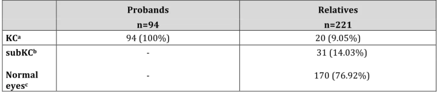
Table 1: Number of probands and close relatives according to keratoconus (KC) status. The assignment of patients in a group is conditioned by the status of the most severely affected eye. Probands Relatives n=94 n=221 KCa 94 (100%) 20 (9.05%) subKCb - 31 (14.03%) Normal eyesc - 170 (76.92%) a Patients with KC in one or both eyes b Patients with one or both eyes with subclinical KC (subKC) c Patients without KC or subKC
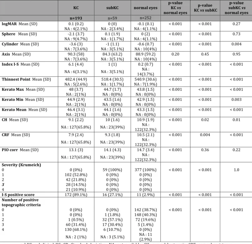
Table 2: Eye characteristics according to keratoconus (KC) status
subKC: subclinical KC; SD: Standard deviation; NA: not available; CH: corneal hysteresis; CRF: corneal resistance factor; PIO corr: corrected intraocular pressure
KC subKC normal eyes p-value KC vs
normal eyes p-value KC vs subKC p-value subKC vs normal eyes n=193 n=59 n=252 logMAR Mean (SD) 0.1 (0.2) 0 (0) -0.1 (0.1) < 0.001 < 0.001 0.27
NA : 4(2.1%) NA : 2(3.4%) NA : 4(1.1%)
Sphere Mean (SD) -2.1 (3.7) 0.1 (1.9) 0 (2) < 0.001 < 0.001 0.73
NA : 9(4.7%) NA : 1(1.7%) NA : 4(1.1%)
Cylinder Mean (SD) -3.6 (3) -1 (1.1) -0.6 (0.7) < 0.001 < 0.001 0.004
NA : 7(3.6%) NA : 3(5.1%) NA : 10(4%)
Axis Mean (SD) 90.3 (58) 84.3 (63.2) 88.9 (59.2) 0.20 0.45 0.95
NA : 7(3.6%) NA : 3(5.1%) NA : 10(4%)
Index I-S Mean (SD) 6.1 (4.4) 1 (1) 0.2 (0.7) < 0.001 < 0.001 < 0.001
NA : 6(3.1%) NA : 3(5.1%) 14(3.7%) NA :
Thinnest Point Mean (SD) 482.4 (44.9) 518.4 (30.5) 540.9 (30.6) < 0.001 < 0.001 < 0.001
NA : 5(2.6%) NA : 1(1.7%) NA : 7(1.9%)
Kerato Max Mean (SD) 48 (3.7) 44.7 (1.7) 43.8 (1.5) < 0.001 < 0.001 < 0.001
NA : 2(1%) NA : 0(0%) NA : 0(0%)
Kerato Min Mean (SD) 44.9 (2.9) 43.5 (1.6) 42.9 (1.5) < 0.001 < 0.001 0.003
NA : 2(1%) NA : 0(0%) NA : 0(0%)
Kerato Mean Mean (SD) 46.4 (3.1) 44.1 (1.6) 43.3 (1.5) < 0.001 < 0.001 < 0.001
NA : 2(1%) NA : 0(0%) NA : 0(0%)
CH Mean (SD) 9.1 (2.2) 10 (1.6) 10.9 (1.9) < 0.001 0.02 0.01
NA : 127(65.8%) NA : 23(39%) 122(32.3%) NA :
CRF Mean (SD) 7.9 (2.4) 9.3 (1.8) 10.5 (2.1) < 0.001 0.004 < 0.001
NA : 127(65.8%) NA : 23(39%) 122(32.3%) NA :
PIO corr Mean (SD) 13.1 (3) 14.1 (4.3) 14.7 (3.4) < 0.001 0.36 0.22
NA : 127(65.8%) NA : 23(39%) 122(32.3%) NA :
Severity (Krumeich) 0 0 (0%) 59 (100%) 377 (100%) < 0.001 < 0.001 1.0 1 102 (52.8%) 0 (0%) 0 (0%) 2 42 (21.8%) 0 (0%) 0 (0%) 3 28 (14.5%) 0 (0%) 0 (0%) 4 21 (10.9%) 0 (0%) 0 (0%) I-S positive score 172 (89.1%) 16 (27.1%) 11 (2.9%) < 0.001 < 0.001 < 0.001 Number of positive topographic criteria 0 0 (0%) 0 (0%) 142 (38.7%) < 0.001 < 0.001 < 0.001 1 0 (0%) 1 (1.8%) 148 (40.3%) 2 1 (0.5%) 32 (57.1%) 72 (19.6%) 3 60 (31.4%) 17 (30.4%) 5 (1.4%) 4 130 (68.1%) 6 (10.7%) 0 (0%)
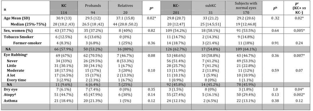
Table 3: Individual characteristics according to keratoconus (KC) status
n KC Probands Relatives pa KC- subKC
Subjects with normal eyes pb pc (KC+ vs KC-) 114 94 20 201 31 170 Age Mean (SD) 30.9 (13) 29.5 (12) 37.1 (15.8) 0.02* 29.8 (20.7) 33 (21.2) 29.2 (20.6) 0 .32 0.02* Median (25%-75%) 28 (18.2 ;43) 26.5 (18 ;42) 44 (20.8 ;50.2) 20 [12;47] 25 [14.5;51] 19 [12;46.8] Sex, women (%) 43 (37.7%) 35 (37.2%) 8 (40%) 0.82 109 (54.2%) 18 (58.1%) 91 (53.5%) 0.64 0,005* Tobacco Smoker 6 (12.5%) 6 (13.6%) 0 (0%) 11 (14.7%) 2 (14.3%) 9 (14.8%) Former-smoker 4 (8.3%) 3 (6.8%) 1 (25%) 0.36 14 (18.7%) 3 (21.4%) 11 (18%) 0.91 0.24 NA 66 (57.9%) 50 (53.2%) 16 (80%) 126 (62.7%) 17 (54.8%) 109 (64.1%) Eye Rubbinge 69 (67%) 62 (70.5%) 7 (46.7%) 0.08 53 (48.6%) 10 (58.8%) 43 (46.7%) 0.36 0.007* Never 34 (33%) 26 (29.5%) 8 (53.3%) 0.10 56 (51.4%) 7 (41.2%) 49 (53.3%) 0.59 0.07 Little 31 (30.1%) 30 (34.1%) 1 (6.7%) 28 (25.7%) 7 (41.2%) 21 (22.8%) Moderate 18 (17.5%) 15 (17%) 3 (20%) 13 (11.9%) 2 (11.8%) 11 (12%) A lot 17 (16.5%) 15 (17%) 2 (13.3%) 11 (10.1%) 1 (5.9%) 10 (10.9%) Every time 3 (2.9%) 2 (2.3%) 1 (6.7%) 1 (0.9%) 0 (0%) 1 (1.1%) NA 11 (9.6%) 6 (6.4%) 5 (25%) 92 (45.8%) 14 (45.2%) 78 (45.9%) Dry eye 7 (6.1%) 7 (7.4%) 0 (0%) 0.35 3 (1.5%) 0 (0%) 3 (1.8%) 1.0 0.04* Atopyd 51 (44.7%) 45 (47.9%) 6 (30%) 0.14 55 (27.4%) 5 (16.1%) 50 (29.4%) 0.13 0.002* Asthma 21 (18.4%) 20 (21.3%) 1 (5%) 0.12 24 (12.1%) 2 (6.5%) 22 (13.1%) 0.38 0.12 a p-value of intra-class test proposers VS relatives among KC+ subjects b p-value of intra-class test subKC VS subjects with normal eyes c Wilcoxon-Mann-Whitney test for age; Chi2 test for sex d Atopy: at least one allergy among asthma, dermatitis, conjunctivitis, allergic rhinitis, drug contact or food allergy, urticaria e The first line shows the number of subjects who answered a little, moderately a lot constantly SD: standard deviation
Table 4: Maximum likelihood estimates of familial correlations for the corneal thinnest point, maximal keratometry, I-S index and AAI using the class D regressive model Phenotypea ρFM (SD)b ρFO (SD)b ρMO (SD)b ρSS (SD)b -2 ln Lc Corneal thinnest point -0.16 (0.15) 0.25 (0.10) 0.31 (0.08) 0.31 (0.09) 822.28 Maximal keratometry 0.39 (0.21) 0.02 (0.09) 0.13 (0.08) 0.26 (0.12) 858.88 IS index 0.61 (0.11) 0.02 (0.10) 0.07 (0.09) 0.01 (0.12) 842.02 AAI 0.33 (0.26) 0.09 (0.11) 0.13 (0.09) 0.12 (0.13) 848.94 a phenotypes were adjusted for age, gender and the indicator variable specifying the position of individuals in the family with respect to the probands. The regression residuals were inverse-Gaussian-transformed to standard normal distributions.
b ρFM: spouse correlation, ρFO: father-offspring correlation, ρMO: mother-offspring correlation, ρSS: sib-sib correlation with standard deviation (SD) in parenthesis c ln L: natural logarithm of the maximum likelihood SD: standard deviation; AAI: asymmetric aspheric index
Table 5: Previous study on the same topic: prevalence of KC in relatives, with videokeratography
Study Country Year Percentage Number of relative Family degree Method
Morrow et al.15 Canada 1997 31.25% 48 1st degree videokeratography
Wang et al.5 USA 2000 3.34% 373 1st degree TMS-1
Levy et al.19 France and Spain 2004 30% subKC 89 1st degree Eye Sys Corneal Analysis System 2000
Kaya et al.17 Turkey 2008 11% 73 1st degree Orbscan II
Karimian et al.18
Iran 2008 14% KC
7.3% subKC 150 (130=1
st
degree) 1-3 degree CSO topographer
Besharati et al.32
Iran 2010 12.3% KC
6.6% subKC 150 1
st degree Videokeratography
Kriszt et al.16 Hungary 2014 7.6% 145 1-3 degree TMS-4
Kymionis et al.21
Greece 2017 53% subKC 34 1st degree Videokeratography
Awwad et al6 Lebanon 2019 17.5% 183 1st degree Galilei
Figure 3: Anterior corneal shape on galilei topographer for one of our sibling family
Conclusion et perspectives
Cette étude a révélé comme d’autres études avant elle (synthétisée dans le tableau 5 de l’article) une augmentation du risque de kératocône chez les apparentés au premier degrés. Mais elle nous donne grâce à sa puissance, et son design une prévalence actualisée du kératocône chez les apparentés au premier degrés.
Nous avons été surpris par l’importance des kératocônes subcliniques dans notre étude : près d’un quart des apparentés au premier degré ont des traits topographiques pathologiques. Ce chiffre est très nettement supérieur aux estimations d’il y a 15-20ans ce qui montre du doigt les avancées dans l’imagerie cornéenne. Les détails fournis aujourd’hui par les topographes d’élévation et les topographes utilisant les technologies OCT sont tels qu’ils nous permettent une analyse ultra précise des cornées. Cependant une des difficultés est de trouver la limite nette entre une cornée « normale » et une cornée subclinique ou suspecte, car il y a une évolution lente qui rend difficile la stadification du kératocône. Et il parait artificiel de pouvoir classer les cornées comme normales/subclinique/ou atteintes d’un kératocône. Cependant c’est une nécessité pour les études et cela en fait aussi une de leurs limites. L’étude condensée de l’ensemble de ces topographies cornéennes nous a permis d’avoir une vue d’ensemble de ces cornées regroupées par familles, et de nous rendre compte des traits similaires qui existaient au sein des familles. Ces données sont très difficiles à mettre en évidence de manière chiffrée, mais elles nous semblent être une source infinie d’information et une voie de recherche qui impliquerait la génétique et l’épigénétique. Elles pourraient aussi servir pour cartographier, identifier les personnes, comme les empreintes génétiques, et intéresser bien plus que le domaine de la recherche sur le kératocône.
Annexe 1
LE KERATOCONE CHEZ L’ENFANT
Une faible acuité visuelle chez un enfant peut altérer son développement, ses apprentissages, sa vie sociale, et sa qualité de vie. Le kératocône peut être responsable d’une baisse d’acuité visuelle et doit être pris en charge chez les enfants. Il peut être plus agressif que chez l’adulte (1,2), et nécessiter une approche thérapeutique différente. Epidémiologie : Classiquement le kératocône se manifeste entre 10 et 20 ans, la plus part des papiers dans la littérature décrivent le début de la maladie à l’adolescence, mais un cas clinique chez un enfant de 4ans a été rapporté. (3) Le kératocône chez l’enfant est initialement unilatéral, mais la majorité des enfants développeront secondairement un kératocône bilatéral dans les 16 ans à venir (4-5). Le plus souvent le kératocône est une pathologie isolée, cependant il a été associé à des pathologies telle que : la kérato-conjonctivite vernale, l’atopie, la trisomie 21, la rétinite pigmentaire, l’amaurose congénitale de Leber, les prolapsus de la valve mitrale et les pathologies du collagène telles que la maladie de Marfan et le syndrome d’Ehlers-Danlos (6, 4, 7). Une précédente étude au sein du service (8) avait montré que la sévérité du kératocône chez l’enfant était plus souvent associée au garçon, à l’allergie, au frottement oculaire et à une histoire familiale de kératocône.Dans les études, il revient régulièrement que 10% des enfants ayant un kératocône ont une histoire familiale de kératocône (9, 4, 10, 11). Une étude récente au Liban, assez comparable à la notre, a montré une prévalence du kératocône de 17,5% à 19,1% chez les enfants entre 6 et 18 ans ayant un membre de leur famille atteint au premier degré d’un kératocône (12). Bien que la prévalence du kératocône soit plus élevée au Moyen Orient, 3,3% (13), comparée aux populations européennes, 0,265% (14), cette étude confirme la présence d’antécédents familiaux comme étant un facteur de risque de développement de la maladie. La progression du kératocône : Le kératocône sur ces jeunes cornées a tendance à être plus agressif que sur celles de l’adulte (6). Le remodelage cornéen est plus important chez les enfants : leurs cornées sont moins dures, et plus viscoélastiques que chez l’adulte (15). La CLEK study a prouvé que la kératométrie, qui signe la progression du kératocône, avait une progression maximale avant 20 ans (16). Et certaines pathologies sont associées à une progression plus rapide du kératocône comme l’atopie et la kératoconjonctivite vernale. La nécessité de réaliser une greffe de cornée est utilisée dans les études comme un marqueur de stade avancé de la maladie, cependant la proportion de greffes de cornée est très variable chez l’enfant dans les études. Mais Tuf et al.(2) rapportent un pourcentage plus important de greffe de cornée chez les patients ayant présenté un kératocône dans l’enfance.
Les différences entre kératocône adulte et pédiatrique : Le kératocône chez l’enfant est plus sévère que chez l’adulte et présente une évolution rapide qui demande un suivi plus rapproché. Les cornées de l’enfant et de l’adulte sont structurellement différentes. Il existe un cross-linking naturel au court des années, qui pourrait expliquer la stabilisation de la pathologie vers l’âge de 30 ans. A l’âge du diagnostic, les kératocônes sont plus avancés chez les enfants que chez les adultes. (17) Les mêmes algorithmes de traitements sont disponibles chez l’adulte et chez l’enfant (lunettes, lentilles rigides, cross linking, anneau intra cornéen et greffe de cornée). Cependant la rapidité de l’évolution de certain kératocône chez l’enfant pousse de nombreuse équipes à réaliser des cross-linking avant même d’avoir des signes de progression kératometrique. Soeters et al. (18) ont rapporté de meilleurs résultats, basés sur l’acuité visuelle, après cross linking, chez l’enfant que chez l’adulte, alors que Vinciguerra et al. (19, 20) rapportent des meilleurs résultats chez l’adulte. Les enfants auraient tendance à poursuivre l’évolution kératométrique de leur kératocône même après cross-linking, mais il est difficile de dire si cela est dû à l’histoire naturelle de la maladie. Le cross-linking La sécurité et l’efficacité du cross linking chez l’enfant ont été démontrées dans plusieurs études utilisant un protocole standard epi-OFF ou non. Certains auteurs ont trouvé une amélioration de l’acuité visuelle après cross linking. (21, 22, 23) Les cônes
progression malgré le cross-linking. (24) Et l’on sait que la persistance d’un frottement oculaire peut aussi continuer à faire évoluer un kératocône même après un cross linking. Les risques du cross linking épithélium-OFF sont comme chez l’adulte : un haze cornéen, des cicatrices, des blépharites, une photophobie, une douleur, une baise temporaire de l’acuité visuelle et les infections. Le cross linking trans-épithélial présente moins de risque dû à l’absence d’abrasion de celui-ci, et est plus facile de réalisation chez l’enfant. Certaines études ont montré l’efficacité ce cette technique (25, 26). Mais l’épithélium serait une barrière pour la riboflavine et les UVA. Eraslan et al. (27) ont prouvé que le trans-epith était 0,7 fois moins efficace dans la stabilisation des cornées comparé au protocole standard (p=0,038). Dans tous les cas le cross linking, n’est pas efficace à 100% pour stopper l’évolution du kératocône. Les anneaux intra cornéens Les anneaux intra cornéens sont utilisés dans 20% des kératocônes intolérants aux lentilles rigides. Ils sont envisageables pour des cornées supérieures à 400microns, sans cicatrice centrale. La pose d’anneaux intra cornéen peut être couplée à un cross linking. Les rares complications sont principalement : l’extrusion de l’anneau, la néo vascularisation, l’infection, des dépôts autour de l’anneau. Chez l’enfant ce n’est pas un traitement de premier choix, bien que réversible, car leur kératocône est encore évolutif. De plus la tendance au frottement oculaire et une compliance parfois imparfaite peuvent rendre dangereuse l’utilisation d’anneau intra cornéen chez l’enfant. (1)
Cependant cette option doit être envisagée chez les adolescents, à un stade avancé de la maladie, avant une greffe de cornée. Les greffes de cornée Dans les kératocônes très avancés il y a une indication à réaliser une greffe de cornée. Une kératoplastie lamellaire antérieure profonde est préférable si cela est possible, car celle ci présente moins de risque de rejet, chez des patients jeunes qui auront besoin de plusieurs greffes au cours de leur vie. Les greffes de cornées ont un moins bon pronostic chez les enfants que chez les adultes (1, 28). Elles nécessitent des anesthésies générales répétées chez les plus jeunes, pour surveiller la pression intra oculaire et l’ablation des sutures. La gestion des sutures est plus complexe, avec des sutures qui ont tendance à plus se détendre. L’évaluation de la réfraction est difficile, et se surajoute le risque et la gestion de l’amblyopie si la maturation visuelle cérébrale n’est pas terminée. Les rejets de greffe sont plus fréquents avec le jeune âge (29, 30). La balance bénéfice risque doit être bien pesée avant de poser l’indication chirurgicale, en fonction de la clinique mais aussi du contexte de l’enfant : poly-pathologie, motivation des parents. Conclusion D’après la littérature le kératocône chez l’enfant serait plus agressif, nécessitant un suivi plus fréquent. Le cross-linking est le traitement de référence du kératocône chez l’enfant, même s’il paraît moins efficace que chez l’adulte. Les greffes de cornée et les
délicats que chez l’adulte. Le dépistage précoce et la lutte contre les frottements oculaires restent le meilleur moyen pour éviter des amblyopies profondes, et
Annexe 2
LA CLASSIFICATION D’AMSLER ET KRUMEICH
(32)Annexe 3
CLASSIFICATION DE RABINOWITZ
(33)Bibliographie
1. Kankyriya VP, Kymionis GD, Diaknois VF, et al. Management of pediatric keratoconus-evolving role of corneal collagen cross-linking : an update. Ind J Ophthalmol. 2013 ;61(8) :435-440.
2. Tuf SJ, Moodaley LC, Gregory WM, et al. Prognostic factors for the progression of keratoconus. Ophtalmology. 1994 ;101 :439-447.
3. Sabti S, Tappeiner C, Frueh BE, Corneal Cross-linking in a 4-year old child with keratoconus and down syndrome. Cornea. 2015 ;34(9) :1157-1160. 4. Gordon Shaag A, Millodot M, Shneor E, et al. The genetic and environnemental factors for keratoconus. BioMed Res Int. 2015 :19. 5. Li X, Rabinowitz YS, Rasheed K, et al. Longitudinal study of the normal eyes in unilateral keratoconus patients. Ophthalmology. 2004 ;111(3)440-446. 6. Ys R. Keratoconus. Surv Ophthalmol. 1998 ;42 :297-319.
7. Davidson AE, Hayes S, Hardcastle AJ, et al. The pathogenesis of keratoconus. Eye. 2014 ;28(2) :189-195.
8. Leoni-Mesplie S, Mortemousque B, Mesplie N, et al. Epidemiological aspects of keratoconus in children. Fr J Ophthalmol. 2012 ;35(10) :776-785.
9. El Rami H, Chelala D, Dirani A, et al. An update on the safety and efficacy of corneal collagen cross-linking in pediatric keratoconus. BioMed Res Int. 2015
10. Gonzales V, McDonnell PJ. Computer-assisted corneal topography in parents of patients with keratoconus. Arch Ophthalmol. 1992;110(10):1412–1414.
11. Rabinowitz YS, Garbus J, McDonnell PJ. Computer-assisted corneal topography in family members of patients with keratoconus. Arch Ophthalmol. 1990;108(3):365–371.
12. Awwad S-T., Yehia M, Mehanna C-J. et al. Tomographic and Refractive Charactéristics of Pediactric First-Degree Relatives of Keratoconus Patients. Am J Ophthalmlo. 2019 : 13. Waked N, Fayad AM, Fadlallah A, El Rami H. Depistage du kératocone dans une
population universitaire au Liban. J Fr Ophtalmol. 2012 ;35(1) :23-29 14. Godefrooij DA, de Wit GA, Uiterwaal CS, et al. Age-specific Incidence and Prevalence of Keratoconus: A Nationwide Registration Study. Am J Ophthalmol. 2017; 175:169–172. 15. Kotecha A, Elsheikh A, Robertsr C, et al. Corneal thickness and age-related biomechanical properties of the cornea measured with the ocular response analyzer. Invest Ophthalmol Vis Sci. 2006;47:5337–5347.
16. McMahon TT, Edrington TB, Szczotka-Flynn L, et al. Longitudinal changes in corneal curvature in keratoconus. Cornea. 2006;25:296–305. 17. Léoni-Mesplié SMB, Touboul D, et al. Scalability and severity of kertoconus in children. Am J Ophthalmol. 2012;154(1):56–62. 18. Soeters N, van der Valk R, Tahzib NG. Corneal cross-linking for treatment of progressive keratoconus in various age groups. J Refract Surg. 2014;30(7):454–460. 19. Vinciguerra P, Albe E, Frueh BE, et al. Two year corneal cross-linking results in patients younger than 18 years with documented progressive keratoconus. Am J Ophthalmol. 2012;154(3):520–526. 20. Vinciguerra R, Romano M, Carnesasca F, et al. Corneal cross-linking as a treatment for keratoconus: four-year morphologic and clinical outcomes with respect to patient age. Ophthalmology. 2013;120:908–918. 21. Toprak I, Yaylali V, Yildrim C. Visual, topographic, and pacchymetric effects of pediatric corneal collagen cross-linking. J Pediatr Ophthalmol Strabismus. 2016;54:84–89.
22. Padmanabhan P, Rachapalle R, Rajagopal R, et al. Corneal Collagen cross-linking for keratoconus in pediatric patients—long-term results. Cornea. 2017;36:138–142.
23. Ucakhan OO, Bayraktutar BN, Saglik A. Pediatric corneal collagen cross-linking: long term follow up of visual, refractive and topographic outcomes. Cornea. 2016;35:162– 168.
24. Sarac O, Caglayan M, Cakmak HB. Factors influencing progresison of keratoconus 2 years after corneal collagen cross-linking in pediatric patients. Cornea. 2016 (EPub ahead of print)
25. Caporossi A, Mazzotta C, Paradiso A, et al. Transepithelial corneal collagen crosslinking for progressive keratoconus: 24 month clinical results. J Cataract Refract Surg. 2013;39:1157–1163.
26. Ag S. Corneal Biomechanical and anterior chamber parameteres variations after 1-year of transepithelial corneal cross-linking in eyes of children with keratoconus. Middle East Afr J Ophthlamol. 2016;23:129–134.
27. Erasalan M, Toker E, Cerman E, et al. Efficacy of epithelium-off and epithelium-on corneal cross linking in pediatric keratoconus. Eye Contact Lens. 2016;43:155–161. 28. Low JR, Anshu A, Tan AC, et al. The outcomes of primary pediatric keratoplasty in
Singapore. Am J Ophthalmol. 2014;158(3):496–502.
29. Asuri MK, Garg P, Gokhle N, et al. Penetrating keratoplasty n children. Cornea. 2000;19(2):140–144.
30. Lowe MT, Keane MC, Coster DJ, et al. The outcome of corneal transplantation in infants, children, and adolescents. Ophthalmology. 2011;118(3):492–497.
31. Mukhtar S, Ambati BK, Pediatric keratoconus: a review of the literature. Int Ophthalmol. 2017:1-10
32. Amsler M. Keratocône classique et kératcône frustre, arguments unitaires. Ophthalmologica. 1946 ;104 :96-111
Serment d’Hippocrate
Au moment d’être admise à exercer la médecine, je promets et je jure d’être fidèle aux lois de l’honneur et de la probité. Mon premier souci sera de rétablir, de préserver ou de promouvoir la santé dans tous ses éléments, physiques et mentaux, individuels et sociaux.Je respecterai toutes les personnes, leur autonomie et leur volonté, sans aucune discrimination selon leur état ou leurs convictions. J’interviendrai pour les protéger si elles sont affaiblies, vulnérables ou menacées dans leur intégrité ou leur dignité. Même sous la contrainte, je ne ferai pas usage de mes connaissances contre les lois de l’humanité.
J’informerai les patients des décisions envisagées, de leurs raisons et de leurs conséquences.
Je ne tromperai jamais leur confiance et n’exploiterai pas le pouvoir hérité des circonstances pour forcer les consciences. Je donnerai mes soins à l’indigent et à quiconque me les demandera. Je ne me laisserai pas influencer par la soif du gain ou la recherche de la gloire. Admise dans l’intimité des personnes, je tairai les secrets qui me seront confiés. Reçue à l’intérieur des maisons, je respecterai les secrets des foyers et ma conduite ne servira pas à corrompre les moeurs.
Je ferai tout pour soulager les souffrances. Je ne prolongerai pas abusivement les agonies. Je ne provoquerai jamais la mort délibérément.
Je préserverai l’indépendance nécessaire à l’accomplissement de ma mission. Je n’entreprendrai rien qui dépasse mes compétences. Je les entretiendrai et les perfectionnerai pour assurer au mieux les services qui me seront demandés.
J’apporterai mon aide à mes confrères ainsi qu’à leurs familles dans l’adversité.
Que les hommes et mes confrères m’accordent leur estime si je suis fidèle à mes promesses ; que je sois déshonorée et méprisée si j’y manque.