Open Access
Methodology
Proteome alteration induced by hTERT transfection of human
fibroblast cellsk
Gabriel D Mazzucchelli*
1, Valérie Gabelica
1, Nicolas Smargiasso
1,
Maximilien Fléron
1, Wilson Ashimwe
1, Frédéric Rosu
1, Marie-Claire De
Pauw-Gillet
2, Jean-François Riou
3and Edwin De Pauw
1Address: 1Laboratory of Mass Spectrometry; CART, GIGA, University of Liège, BAT. B6C, allée de la Chimie, 3, 4000 Liège 1, Belgium, 2Histology
and Cytology Laboratory, GIGA, University of Liège, BAT. B6C, allée de la Chimie, 3, 4000 Liège 1, Belgium and 3Regulation et Dynamique des
Genomes, Museum National d'Histoire Naturelle, USM 503, INSERM U565, CNRS UMR 5153, Paris, France Email: Gabriel D Mazzucchelli* - [email protected]; Valérie Gabelica - [email protected];
Nicolas Smargiasso - [email protected]; Maximilien Fléron - [email protected]; Wilson Ashimwe - [email protected];
Frédéric Rosu - [email protected]; Marie-Claire De Pauw-Gillet - [email protected]; Jean-François Riou - [email protected]; Edwin De Pauw - [email protected]
* Corresponding author
Abstract
Background: Telomerase confers cellular immortality by elongating telomeres, thereby circumventing the Hayflick
limit. Extended-life-span cells have been generated by transfection with the human telomerase reverse transcriptase (hTERT) gene. hTERT transfected cell lines may be of outstanding interest to monitor the effect of drugs targeting the telomerase activity. The incidence of hTERT gene transfection at the proteome level is a prerequisite to that purpose. The effect of the transfection has been studied on the proteome of human fibroblast (WI38). Cytosolic and nuclear fractions of WI38 cells, empty vector transfected WI38 (WI38-HPV) and hTERT WI38 cells were submitted to a 2D-DIGE (Two-Dimensional Differential In-Gel Electrophoresis) analysis. Only spots that had a similar abundance in WI38 and WI38-HPV, but were differentially expressed in WI38 hTERT were selected for MS identification. This method directly points to the proteins linked with the hTERT expression. Number of false positive differentially expressed proteins has been excluded by using control WI38-HPV cells. The proteome alteration induced by hTERT WI38 transfection should be taken into account in subsequent use of the cell line for anti-telomerase drugs evaluation.
Results: 2D-DIGE experiment shows that 57 spots out of 2246 are significantly differentially expressed in the cytosolic
fraction due to hTERT transfection, and 38 were confidently identified. In the nuclear fraction, 44 spots out of 2172 were selected in the differential proteome analysis, and 14 were identified. The results show that, in addition to elongating telomeres, hTERT gene transfection has other physiological roles, among which an enhanced ER capacity and a potent cell protection against apoptosis.
Conclusion: We show that the methodology reduces the complexity of the proteome analysis and highlights proteins
implicated in other processes than telomere elongation. hTERT induced proteome changes suggest that telomerase expression enhances natural cell repair mechanisms and stress resistance probably required for long term resistance of immortalized cells. Thus, hTERT transfected cells can not be only consider as an immortal equivalent to parental cells but also as cells which are over-resistant to stresses. These findings are the prerequisite for any larger proteomics aiming to evaluate anti-telomerase drugs proteome alteration and thus therapeutics induced cell reactions.
Published: 17 April 2008
Proteome Science 2008, 6:12 doi:10.1186/1477-5956-6-12
Received: 30 November 2007 Accepted: 17 April 2008 This article is available from: http://www.proteomesci.com/content/6/1/12
© 2008 Mazzucchelli et al; licensee BioMed Central Ltd.
This is an Open Access article distributed under the terms of the Creative Commons Attribution License (http://creativecommons.org/licenses/by/2.0), which permits unrestricted use, distribution, and reproduction in any medium, provided the original work is properly cited.
Background
Telomeres are specialized functional DNA-protein com-plexes that cap the end of linear chromosomes. Their role is to protect chromosomes from degradation, recombina-tion, or fusion, and to prevent the chromosome ends from being detected as strand breaks. At each cell division, tel-omeres shorten until they reach a critical size that drives eukaryotic cells into replicative senescence. Telomere length therefore acts as a biological life clock. Telomerase, a ribonucleoprotein complex, is involved in telomere length maintenance in eukaryotic cells by adding telom-eric repeats to the 3' end of chromosomes. Telomerase activity is downregulated in most human cells during embryogenesis, thereby limiting their proliferative capac-ity. However, the reactivation of telomerase activity is observed in 90% of all human tumor cells, making this enzyme an attractive target for selective cancer therapy. Reconstitution of telomerase activity by ectopic expres-sion of the catalytic telomerase subunit (hTERT) cDNA stabilizes the telomere length of fibroblasts and other cell types that therefore acquire immortality [1]. Such hTERT-transfected cells have been proposed as immortal versions of normal human cells model with the advantage of indef-inite proliferation. This strategy is applied for biochemical and physiological studies of normal cell growth, differen-tiation, genetic manipulation, etc [1-4]. More recently, experiments using transplanted telomerase-immortalized cells have been conducted in immunodeficient mice [5]. Adenocortical hTERT immortalized cells were able to replace cells with deficient functions. However such cell-based therapies raise important interrogations about medium and long term incidence of hTERT-immortalized cell autotransplantation cells. Indeed, several studies have evidenced that hTERT has other physiological roles beyond maintaining telomere such as potent incidence on malignant transformation of human fibroblasts by a tel-omere length-independent mechanisms [6-8]. Further-more, the key role of telomerase in cancer cell immortalization led to the development of therapeutic strategies based on telomerase inhibition. A detailed char-acterization of hTERT-related transfection processes is therefore of outstanding interest to monitor anti-telomer-ase drug therapy.
We have studied here the sub-proteome modifications induced by hTERT transfection in the normal human fibroblast WI38 cell line. Cytoplasmic and nuclear frac-tions were analyzed by 2D DIGE. Comparison with con-trols, WI38 transfected with the empty vector (HPV) or parental untransfected WI38 cells has revealed the altered expression of proteins involved in apoptosis, cell cycle and endoplasmic reticulum homeostasis. The subfrac-tionation method used allows telomerase detection by western blots, TRAP assay (Telomere repeat amplification
protocol) experiment and reproducible extraction and isolation of the cytosolic and nuclear sub-proteomes, the latter one being enriched in low abundant proteins such as transcription factors and telomeric proteins. The inter-est of this work is to characterize the effect of hTERT expression at the proteome level of WI 38 cells for further investigating the effect of anti-telomerase drugs therapies in an integrated proteomic study.
Results and Discussion
To characterize the protein expression changes resulting from hTERT transfection, we performed a differential pro-teomic analysis in order to compare wild type WI 38 cells (WI38), hTERT transfected WI 38 cells (WI38-hTERT) and control HPV transfected WI 38 cells (WI38-HPV) har-vested at the same number of population doublings (PDL) after transfection and FACS sorting (PDL = 10). The use of this relatively low passage number in vitro main-tains in hTERT transfected cells normal cell characters, such as the capacity of contact inhibition and the karyo-type [9]. It has been shown that hTERT expression protects the WI38 transfected cells from stress-induced apoptosis and necrosis [10]. Prolonged culturing of WI38-hTERT cells leads to a loss of density-dependent growth inhibi-tion and to an onset of contact-induced, p53 dependent cell death [11]. Our cell lines presented density-depend-ent growth inhibition. For the proteomics analysis, our three cell lines were maintained at 90% confluence, and in exponential growth phase. In these conditions, the population doubling levels were similar.
2D DIGE was performed on both nuclear and cytosolic cell fractions. The subfractionation method used relies on a complementary and very different 2D gel images (Figure 1) which is necessary for low abundant nuclear proteins differential analysis. Nuclear fraction corresponds to 5.89% ± 0.41 (mean value over 6 independent experi-ments) of the total protein content. Protein spots that showed either an increase or a decrease in intensity supe-rior to 30%, together with a statistically significant Stu-dent's t-test (p < 0.05) were considered as being differentially expressed. Interestingly, numbers (Table 1) of differentially expressed proteins were found between parental cells and control HPV WI 38 cells. These modifi-cations are thus a consequence of the general transfection and are not due to hTERT gene transfection. In this study only proteins which are differentially expressed between parental cells and hTERT cells and have in addition the same expression in parental cell and control HPV WI 38 cells were selected for the study and submitted to mass spectrometry based identification. In the cytosolic frac-tion, 57 spots out of 2246 are significantly differentially expressed (Table 1), and 38 were confidently identified (Table 2). In the nuclear fraction, 44 spots out of 2172
were selected in the differential proteome analysis (Table 1), and 14 were identified (Table 3).
Functional protein association network of the proteins differentially expressed due to hTERT transfection [12]
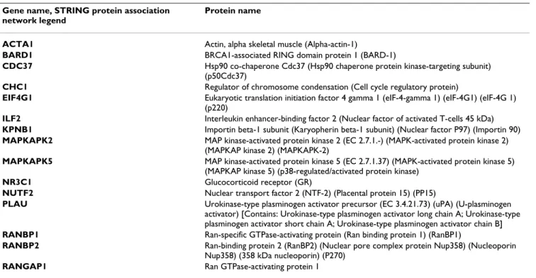
Based on STRING protein-protein interactions predictions an association network of the proteins which have their expression modified due to hTERT transfection was cre-ated (Figure 2). The Table 4 describes the 15 proteins that were automatically selected by STRING to enlarge the pro-tein association network of the Figure 2. This bioinformat-ics analysis evidenced protein groups which have their expression modified due to hTERT transfection in which proteins involved in ubiquitin-proteasome protein degra-dation pathway, nuclear-cytosol transport, endoplasmic reticulum functions, pre-mRNAs processing and cell sev-eral chaperone functions. This network will be further compared to those of anti telomerase drugs experiments.
Up regulation of the heat shock protein 90-alpha
The heat shock protein 90-alpha (hsp90α) is upregulated by a factor 1.42 in the nuclear fraction of hTERT WI38 cells. Hsp90α exerts its chaperon function to ensure the correct conformation, activity, intracellular localization and proteolytic turnover of a range of proteins that are involved in cell growth, differentiation and survival [13]. More specifically, hsp90α interacts with telomerase com-plex and occupy a central place in our protein association network (Figure 2). Hsp90α is involved in allowing RNA template of the telomerase (hTR) to bind hTERT and also in fine tuning and stabilizing the structure of the telomer-ase complex [14,15]. Its central role in the telomertelomer-ase activity modulation and in multiple signaling pathways and biological processes makes it a relevant target for anti cancer therapy. Number of study are currently evaluating clinical activity of hsp90 inhibitors in cancer therapeutics [16-22]. Our DIGE study links the overexpression of the hsp90α with the ectopic expression of hTERT and suggests a key role of this chaperone in the subsequent cell adapta-tion to immortalizaadapta-tion. In addiadapta-tion, Hiyama and cow-orkers have shown in a differential gene expression profiles study that the hsp90α is overexpressed in tumor with high telomerase activity comparing tumor with low telomerase activity [23]. Theses findings emphasize the
interest of anti hsp90 targeting drugs therapy and shows the interest of such proteomics methodology for potential biomarkers discovery and further drugs candidates charac-terization and preclinical validation.
Down-regulation of apoptotic effectors Galectin-1 and Annexin 5
Galectin-1 (Gal-1) is downregulated by a factor of 2.1 in the nuclear proteome of hTERT-transfected cells. Gal-1 is a highly conserved protein with a carbohydrate-recogni-tion domain that binds beta-galactoside. Recently Gal-1 was shown to be implicated in cell growth, apoptosis and cell differentiation and survival of effector T cells [24-27]. Exogenously added recombinant Gal-1 induces apoptosis by the mitochondrial and death receptor pathways of MA-10 tumor Leydig cells. In contrast, low concentrations of Gal-1 significantly promote cell proliferation, without inducing cell death [26]. Walzel and al. have shown that Gal-1 triggers through binding to N-linked glycans a Ca
2+-sensitive apoptotic pathway [28].
Annexin 5 is downregulated by a factor of 2.64 in the cytosolic proteome of hTERT-transfected cells. Annexins are abundant Ca2+- dependant phospholipids-binding
proteins. Depletion of endogenous annexin 5 with siRNA inhibits delta protein kinase C functions in which cellular processes such as growth, differentiation and apoptosis [29,30]. In addition Hawkins and co-workers have proven that annexin 5 lack expression results in reduced suscepti-bility to a range of apoptotic stimuli, and that annexin 5 (-/-) cells are more resistant to apoptosis [31].
The downregulation of these two proteins in hTERT-trans-fected cells suggests that telomerase expression, in addi-tion to its immortalizaaddi-tion effect, induces a protecaddi-tion against apoptosis, compared to WI 38 cells. In agreement, modulation of apoptosis have been reported [32-34].
Upregulation of Cajal bodies associated factors
Cleavage stimulator factor 50 kDa subunit (CstF-50) is upregulated by a factor of 1.57 (T-test: 1.5E-03), and cleavage and polyadenylation specificity factor 5 (CPSF-25) by a factor of 1.35 (T-test: 1.6E-02) in the nuclear pro-teome of hTERT transfected cells. CstF-50 is a subunit of Table 1: 2D DIGE spots selection.
Nuclear fraction Cytosolic fraction Total number of spots differentially expressed in WI38/WI38-hTERT 190 210
Number of spots differentially expressed due to transfection (WI38/WI38-HPV) 146 153 By difference, number of spots differentially expressed due only to hTERT overexpression (number of
identified spots are in parentheses)
44 (14) 57 (38)
Number of spots differentially expressed in 2D-DIGE experiment (Student's t-test p < 0.05 and increase or decrease intensity superior at 30%). Selection based on spots differentially expressed in WI38/hTERT WI 38, but similarly expressed in WI38 and control WI38-HPV.
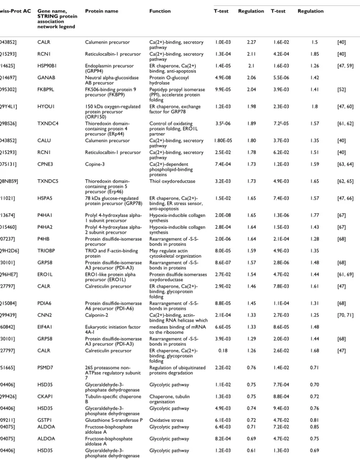
Table 2: Cytosolic differentially expressed proteins identification and regulation
hTERTt-WI38/WI38 hTERT-WI38/HPV-WI38 Reference Swiss-Prot AC Gene name,
STRING protein association network legend
Protein name Function T-test Regulation T-test Regulation
[O43852] CALR Calumenin precursor Ca(2+)-binding, secretory
pathway 1.0E-03 2.27 1.6E-02 1.5 [40] [Q15293] RCN1 Reticulocalbin-1 precursor Ca(2+)-binding, secretory
pathway 1.3E-04 2.11 4.2E-04 1.85 [40] [P14625] HSP90B1 Endoplasmin precursor
(GRP94) ER chaperone, Ca(2+) binding, anti-apoptosis 1.4E-05 2.1 1.6E-03 1.26 [47, 59] [Q14697] GANAB Neutral alpha-glucosidase
AB precursor Protein O-glucosyl hydrolase 4.9E-08 2.06 5.5E-06 1.42 [O95302] FKBP9L FK506-binding protein 9
precursor (FKBP9) Peptidyp propyl isomerase (PPI), accelerate protein folding
9.9E-05 2.04 3.9E-03 1.41 [52]
[Q9Y4L1] HYOU1 150 kDa oxygen-regulated protein precursor (ORP150) ER chaperone, exchange factor for GRP78 1.2E-03 1.98 2.3E-03 1.8 [47, 60] [Q9BS26] TXNDC4 Thioredoxin domain-containing protein 4 precursor (ERp44) Control of oxidating protein folding, ERO1L partner
3.5E-06 1.89 7.2E-05 1.57 [61, 62]
[O43852] CALU Calumenin precursor Ca(2+)-binding, secretory pathway
1.80E-05 1.80 3.7E-03 1.35 [40] [Q15293] RCN1 Reticulocalbin-1 precursor Ca(2+)-binding, secretory
pathway
2.5E-02 1.78 6.2E-02 1.51 [40] [O75131] CPNE3 Copine-3 Ca(2+)-dependent
phospholipid-binding proteins 7.4E-04 1.73 1.2E-03 1.59 [63, 64] [Q8NBS9] TXNDC5 Thioredoxin domain-containing protein 5 precursor (Erp46)
Thiol oxydoreductase 3.2E-03 1.73 4.9E-03 1.65 [62, 65]
[P11021] HSPA5 78 kDa glucose-regulated protein precursor (GRP78)
ER chaperone, Ca(2+)-binding, ER stress sensor, anti-apoptosis
1.5E-02 1.65 7.4E-03 1.57 [47, 66]
[P13674] P4HA1 Prolyl 4-hydroxylase
alpha-1 subunit precursor Hypoxia-inducible collagen synthesis 2.0E-08 1.65 1.3E-06 1.77 [67] [O15460] P4HA2 Prolyl 4-hydroxylase
alpha-2 subunit precursor Hypoxia-inducible collagen synthesis 2.8E-04 1.64 1.5E-03 1.43 [67] [P07237] P4HB Protein disulfide-isomerase
precursor Rearrangement of -S-S-bonds in proteins 2.0E-06 1.64 2.1E-04 1.28 [68] [Q9H2D6] TRIOBP TRIO and F-actin-binding
protein May regulate actin cytoskeletal organization 8.0E-05 1.59 4.9E-03 1.35 [P30101] GRP58 Protein disulfide-isomerase
A3 precursor (PDI-A3) Rearrangement of -S-S-bonds in proteins 8.6E-07 1.57 2.8E-06 1.48 [68] [Q96HE7] ERO1L ERO1-like protein alpha
precursor (ERO1L) Protein disulfide isomerases oxydoreductase 2.7E-02 1.54 4.7E-02 1.44 [61, 69] [P27797] CALR Calreticulin precursor ER chaperone,
Ca(2+)-binding, glycoprotein folding
2.9E-02 1.46 7.8E-03 1.61 [47]
[Q15084] PDIA6 Protein disulfide-isomerase A6 precursor (PDI-A6)
Rearrangement of -S-S-bonds in proteins
8.8E-05 1.45 1.1E-04 1.31 [68] [Q99439] CNN2 Calponin-2 Ca(2+)-binding,
actin-binding RNA helicase which
2.1E-04 1.33 2.7E-03 1.25 [70, 71] [P60842] EIF4A1 Eukaryotic initiation factor
4A-I
mediates binding of mRNA to the ribosome 6.6E-05 1.33 8.6E-05 1.48 [P30101] GRP58 Protein disulfide-isomerase A3 precursor (PDI-A3) Rearrangement of -S-S-bonds in proteins 3.9E-03 1.29 2.0E-03 1.44 [68] [P27797] CALR Calreticulin precursor ER chaperone,
Ca(2+)-binding, glycoprotein folding
0.18 1.26 2.6E-02 1.68 [47]
[P51665] PSMD7 26S proteasome non-ATPase regulatory subunit 7
Regulation of ubiquitinated
proteins degradation 2.2E-02 0.76 1.4E-02 0.71 [P04406] HSD35
Glyceraldehyde-3-phosphate dehydrogenase
Glycolytic pathway 1.1E-02 0.75 7.7E-04 0.70 [Q99426] CKAP1 Tubulin-specific chaperone
B Chaperone, tubulin organisation 1.3E-03 0.75 8.8E-04 0.72 [P04406] HSD35 Glyceraldehyde-3-phosphate dehydrogenase
Glycolytic pathway 4.9E-03 0.74 9.4E-03 0.76 [P09211] GSTP1 Glutathione S-transferase P Oxidative stress 6.1E-03 0.72 4.7E-02 0.81 [P04075] ALDOA Fructose-bisphosphate
aldolase A Glycolytic pathway 6.4E-03 0.71 7.2E-02 0.85 [P04075] ALDOA Fructose-bisphosphate
aldolase A Glycolytic pathway 8.2E-04 0.69 4.7E-02 0.75 [P04406] HSD35
the heterotrimer CstF. CstF and CPSF-25 are required for polyadenylation and 3'-end cleavage of mammalian pre-mRNAs. Schul and co-workers have shown that "cleavage bodies" (compact spherical fibrous structures containing CstF et CPSF factors) are intimately associated with Cajal bodies (CBs) [35]. hTERT and the telomerase RNA com-ponent, hTR, are also found in CBs. Ectopic expression of hTERT results in accumulation of hTR in CBs in primary fibroblasts or smooth muscle cells, but not in normal cells or in telomerase-negative (ALT) tumor cells [36]. It has
been proposed that CBs could act as storage sites and deliver components of the telomerase complex when needed [37]. hTR localization in CBs is an important reg-ulatory mechanism for telomere length homeostasis in human cells: mutant hTR failing to accumulate in CBs results in a functional deficiency and a decreasing associ-ation of telomerase with telomere [38]. The RNA compo-nent of human telomerase (hTR) includes H/ACA and CR7 domains required for 3' end processing, localization and accumulation. 3' end processing is a prerequisite for Table 3: Nuclear differentially expressed proteins identification and regulation
hTERTt-WI38/WI38 hTERT-WI38/HPV-WI38 Swiss-Prot AC Gene name,
STRING protein association network legend
Protein name Function T-test Regulation T-test Regulation
[P31943] HNRPH1 Heterogeneous nuclear ribonucleoprotein H
Pre-mRNAs processing 4.4E-03 1.73 3.0E-02 1.58 [P08235] NR3C2 Mineralocorticoid receptor Binds to mineralocorticoid response
elements (MRE) and transactivates target genes
6.1E-03 1.65 1.6E-02 1.53
[O14579] COPE Coatomer subunit epsilon Implicated in vesicles trafficking 4.2E-02 1.62 0.12 1.33 [Q05048] CSTF1 Cleavage stimulation factor
50 kDa subunit
Polyadenylation and 3'-end cleavage of mammalian pre-mRNAs
1.5E-03 1.57 5.6E-03 1.64 [P07900] HSPCA Heat shock protein HSP
90-alpha
Chaperone, 680kDa human telomerase complex partner [72]
1.5E-02 1.42 2.2E-04 2.05 [P62826] ARA24 GTP-binding nuclear
protein Ran
nucleocytoplasmic transport 2.0E-02 1.37 6.7E-03 1.24 [O43809] ENSP0000030 0291 Cleavage and
polyadenylation specificity factor 5
Polyadenylation and 3'-end cleavage of mammalian pre-mRNAs
1.6E-02 1.35 - 1.35
[Q13838] ATP6V1G2 splicesome RNA helicase
BAT1 Splice factor, required for mRNA export 6.3E-03 1.34 1.7E-03 1.5 [P25788] PSMA3 Proteasome subunit alpha
type 3 Protein degradation 5.6E-03 0.68 4.7E-02 0.75 [P28072] PSMB6 Proteasome subunit beta
type 6 precursor Protein degradation 3.8E-05 0.65 3.0E-04 0.66 [P08865] RPSA 40S ribosomal protein SA Protein synthesis 1.3E-04 0.58 5.4E-04 0.60 [P60842] EIF4A1 Eukaryotic initiation factor
4A-I
RNA helicase which mediates binding of mRNA to the ribosome
3.1E-02 0.54 1.2E-01 0.52 [P09382] LGALS1 Galectin-1 Cell growth, apoptosis and cell
differentiation
8.8E-07 0.50 1.2E-06 0.51 [Q12906] ILF3 Interleukin
enhancer-binding factor 3
Translation inhibitory protein of acid beta-glucosidase and other mRNAs, IL2 transcription regulator, promote the formation of stable DNA-dependent protein kinase holoenzyme complexes on DNA
1.1E-05 0.47 9.5E-04 0.66
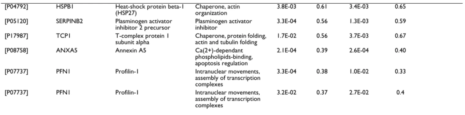
List of the proteins differentially expressed due to hTERT transfection in the nuclear fraction of human WI38 cells. Listed by decreasing regulation. [P04792] HSPB1 Heat-shock protein beta-1
(HSP27)
Chaperone, actin organization
3.8E-03 0.61 3.4E-03 0.65 [P05120] SERPINB2 Plasminogen activator
inhibitor 2 precursor Plasminogen activator inhibitor 3.3E-04 0.56 1.3E-03 0.59 [P17987] TCP1 T-complex protein 1
subunit alpha Chaperone, protein folding, actin and tubulin folding 1.7E-02 0.56 3.7E-03 0.67 [P08758] ANXA5 Annexin A5 Ca(2+)-dependant
phospholipids-binding, apoptosis regulation
2.1E-04 0.39 2.6E-04 0.40
[P07737] PFN1 Profilin-1 Intranuclear movements, assembly of transcription complexes
3.3E-04 0.38 1.0E-02 0.33
[P07737] PFN1 Profilin-1 Intranuclear movements, assembly of transcription complexes
3.2E-02 0.37 2.7E-02 0.4
List of the proteins differentially expressed due to hTERT transfection in the cytosolic fraction of human WI38 cells. Listed by decreasing regulation.
translocation of hTR to CBs [38]. A probable role of CstF-50 and CPSF-25 in hTR maturation may be an important regulation process and requires further investigations. Profilin 1 is a known component of Cajal bodies sup-posed involved in both ATP-dependant intranuclear movements and in the assembly of transcription com-plexes [39]. Our results show that this protein is downreg-ulated in cytosol hTERT transfected cells. Modulation of
this protein may also implicate CBs functions in hTERT-transfected cells.
Enhanced protective ER functions in hTERT transfected cells
22 out of the 38 proteins identified in the cytosolic frac-tion are involved in endoplasmic reticulum funcfrac-tions and are up-regulated in the hTERT transfected cells. Our results are consistent with this recent report and underline the
Nuclear and cytosolic 2D DIGE gels Figure 1
Nuclear and cytosolic 2D DIGE gels. Two dimensional gel electrophoresis images of nuclear (A, B) and cytosolic fractions
(C, D) of WI38 cells. The subfractionation method used allows reproducible extraction and isolation of the nuclear and cytosolic sub-proteomes.
implication of the CERC protein family as an important signaling pathway in hTERT transfected cells. Reticulocal-bin and calumenin belong to the CREC (Cab45, reticulo-calbin, ERC-45, calumenin) proteins, which constitute a family of EF-hand (helix-turn-helix structural motif) cal-cium binding proteins localized to the secretory pathway
[40]. It has been proposed that CREC family is essential for cell survival because homozygous deletion of a region containing the reticulocalbin gene is lethal [41]. Several reports described their important role in pathophysiolog-ical processes especially in connection with malignant transformation [42-44]. Another recent study indicates
Protein association network of the proteins differentially expressed due to hTERT transfection Figure 2
Protein association network of the proteins differentially expressed due to hTERT transfection. Protein-protein
interactions predictions from the identified proteins which have their expression modified due to hTERT transfection. Based on STRING [12]. Network Display – Nodes are either coloured (differentially expressed proteins in this study except TERT that was manually introduced) or white (nodes of a higher iteration/depth). Additional white nodes are automatically generated by STRING to maximize existing interactions. Stronger associations are represented by thicker lines. Parameters used to cre-ate the protein association network: medium confidence and 15 additional (white) nodes. Protein descriptions of the coloured nodes are described in Table 2 and 3 those of white nodes in Table 4.
that calumenin may have an autocrine or a paracrine effect on the cells in its vicinity modulating cell cycle and organization of the actin cytoskeleton [45]. Additionally, calumenin and reticunocalbin were also differentially expressed in a study comparing early passage, senescent, and hTERT-transfected endothelial cells [46]. In this study, mRNA level of these proteins were upregulated in hTERT transfected cells compared to early passage subcon-fluent cells.
ORP150, GRP78, calreticulin, PDIs, ERp44, ERp46, FKBP9 are implicated in essential ER functions and Ca2+
homeostasis [47]. Interestingly, gene knockout experi-ments have proven that ER chaperon function is required for early mammalian development. Depletion of GRP78 leads to lethality in 3.5-day-old embryos (E3.5) due to failure of embryo peri-implantation [48]. This study sug-gests that physiological ER stress may exist in early devel-opment due to increased activity of cell proliferation and protein secretion. Calreticulin deficiency is also lethal in mouse embryos at E14.5, resulting from a lesion in car-diac development [49]. Interestingly GRP78 and calreticu-lin are downregulated after birth in the healthy mature heart [50]. ER chaperones overexpression are also pro-posed to promote cancer and tumor immunity [47]. It is interesting to note that telomerase is generally active in early embryonic stage and in 90% of cancers like these chaperones.
Our results indicate that overexpression of ER chaperones is a consequence of telomerase reactivation. The fact that these proteins are selectively upregulated in hTERT WI38 cells compared to WI38 and WI38-HPV control cells dem-onstrates that hTERT induces an important modulation of the ER functions. The ER is one of the most important folding compartments within the cell, as well as an intra-cellular Ca2+ storage organelle and it contains a number of
Ca2+ regulated molecular chaperones responsible for the
proper folding of glycosylated as well as non-glycosylated proteins. ER is also capable, through Ca2+ homeostasis
modulation, to determine cellular sensitivity to ER stress and apoptosis [51]. We propose that upregulations of ER chaperones in hTERT transfected cells are responsible of a protective cell effect essential for the proliferation of hTERT immortalized cells. This protective effect is proba-bly mediated by upregulation of GRP78 and calreticulin chaperones. In addition, FKBP9 may play an important role in the control of the timing of this biological process due to the recent discovery of its molecular timer function [52]. We propose also that intracellular and potent inter-cellular anti-apoptotic signaling are mediated by proteins belonging to the CREC family in hTERT positive cells trough a Ca2+ signaling modulation. Such proteome
mod-ulation makes evidence that telomerase reactivation, in addition of elongating telomere, has several indirect effects that augment ER capacity of protein folding and degradation [53-55].
Table 4: Protein descriptions of the white nodes of the Protein association network (Figure 2)
Gene name, STRING protein association network legend
Protein name
ACTA1 Actin, alpha skeletal muscle (Alpha-actin-1)
BARD1 BRCA1-associated RING domain protein 1 (BARD-1)
CDC37 Hsp90 co-chaperone Cdc37 (Hsp90 chaperone protein kinase-targeting subunit) (p50Cdc37)
CHC1 Regulator of chromosome condensation (Cell cycle regulatory protein)
EIF4G1 Eukaryotic translation initiation factor 4 gamma 1 (eIF-4-gamma 1) (eIF-4G1) (eIF-4G 1) (p220)
ILF2 Interleukin enhancer-binding factor 2 (Nuclear factor of activated T-cells 45 kDa)
KPNB1 Importin beta-1 subunit (Karyopherin beta-1 subunit) (Nuclear factor P97) (Importin 90)
MAPKAPK2 MAP kinase-activated protein kinase 2 (EC 2.7.1.-) (MAPK-activated protein kinase 2) (MAPKAP kinase 2) (MAPKAPK-2)
MAPKAPK5 MAP kinase-activated protein kinase 5 (EC 2.7.1.37) (MAPK-activated protein kinase 5) (MAPKAP kinase 5) (p38-regulated/activated protein kinase)
NR3C1 Glucocorticoid receptor (GR)
NUTF2 Nuclear transport factor 2 (NTF-2) (Placental protein 15) (PP15)
PLAU Urokinase-type plasminogen activator precursor (EC 3.4.21.73) (uPA) (U-plasminogen activator) [Contains: Urokinase-type plasminogen activator long chain A; Urokinase-type plasminogen activator short chain A; Urokinase-type plasminogen activator chain B]
RANBP1 Ran-specific GTPase-activating protein (Ran binding protein 1) (RanBP1)
RANBP2 Ran-binding protein 2 (RanBP2) (Nuclear pore complex protein Nup358) (Nucleoporin Nup358) (358 kDa nucleoporin) (P270)
Conclusion
We show that the methodology reduces the complexity of the proteome analysis and highlights proteins implicated in other processes than telomere elongation. hTERT trans-fection enhances natural ER capacity and modulates Ca2+
cell signaling pathways potentially resulting in overpro-tection mechanisms against endogeneous and exogene-ous disorder. This hypothesis is in accordance with the identified down-regulation of apoptotic effectors Galec-tin-1 and Annexin 5. Other effects like an enhanced DNA excision repair pathway have also been reported in cell with long telomere [56]. Altogether, these observations suggest that telomerase expression enhances natural cell repair mechanisms and stress resistance probably required for long term resistance of immortalized cells. Therefore, hTERT transfected cells cannot be only consid-ered as an immortal equivalent to parental cells but also as cells which are over-resistant to stresses. The introduc-tion of hTERT gene in WI38 cells modulates the chaper-one hsp90α expression which seems to be, by its central position in the protein association network (Figure 2), an important regulator of subsequent cell adaptation mecha-nism to immortalization. This finding increases the inter-est of current anti cancer studies based on hsp90 inhibition. In addition to these observations, orthogonal analysis of our results with several cancerous cells pro-teomic studies reveals that some of the current high-lighted proteins such as hsp90α, GRP78 and calreticulin are also implicated in oncogenese and cell resistance [53-55]. Finally the model "parental WI38/hTERT WI38/HPV WI38" characterization (Table 2, 3) is the prerequisite for any larger proteomics aiming to evaluate anti-telomerase drugs proteome alteration and thus therapeutics induced cell reactions.
Methods
Cell culture and infection of WI38 cells
hTERT WI38, HPV WI38 and parental WI38 cells (human embryonic lung fibroblasts) were grown in Eagle's mini-mal essential medium with Glutamax (Invitrogen), sup-plemented with 10% fetal calf serum and penicillin-streptomycin 1% (Gibco).
Lentiviral supernatants containing hTERT or control HPV vector were a generous gift from Dr. Annelise Bennaceur-Griscelli (Institut Gustave Roussy, Villejuif, France). Briefly, WI 38 cells at 1.5 × 10⁵ cells/mL were infected at a
multiplicity of infection equal to 50 in the presence of 4 μg/mL Polybrene in complete culture medium. Enhanced green fluorescent protein-positive cells were sorted 5 days later by flow cytometry according to a high or low inten-sity of fluorescence. Populations that expressed a high intensity of fluorescence were seeded [57].
Non denaturing subfractionation method
The non denaturing method of extraction and fractiona-tion is based on the method of Gorski and co-workers [58] and has been adapted for WI38 cells. A cytosolic frac-tion and a nuclear histone-depleted proteins fracfrac-tion were obtained. We verified the presence or not of telomerase activity by Western blots and TRAP (telomeric repeat amplification protocol) assay on each fraction. Then, a 2D LC-MS/MS analysis was performed and allowed the iden-tification of more than 100 proteins per fraction. The two fractions shared a few number of common structural pro-teins and the nuclear fraction was enriched in low abun-dant proteins such as transcription factors and telomeric proteins (results not shown). For each experiment, 25 T 175 flasks 90% confluent are rinsed three times with PBS, then collected, using a scraper in a cold room at 4°C. The collected cells are centrifuged 5 minutes at 400 g and recovered. 10 mL of buffer 1 (Hepes-KOH 10 mM, pH 7.6; KCl 10 mM; Spermine/HCl 0.15 mM; Spermidine 0.5 mM; DTT 0.5 mM) (10 times a volume equivalent to the fresh weight) is added to the cell pellet, homogenized and incubated 10 minutes on ice. The tube is then centrifuged 5 minutes at 800 g and the cells are recovered. One vol-ume equivalent to the fresh weight of buffer 1 is added. The solution is homogenized with 10 strokes with a motorized potter Elvehjem homogenizer (Teflon/glass) at 4000 rpm and the disruption of the cell membrane is con-trolled by Trypan Blue staining of nuclei with an inverted phase contrast microscope. 10% in volume of buffer 2 (Hepes-KOH 10 mM, pH 7.6; KCl 1 M; Spermine/HCl 0.15 mM; Spermidine 0.5 mM; DTT 0.5 mM) is added to restore isotonicity of the solution, the solution is homog-enized with 10 strokes at 4000 rpm and centrifuged 10 minutes at 1100 g. The pellet which contains nuclei is recovered, the supernatant is collected and centrifuged 30 minutes at 24000 g. The supernatant is recovered and cor-responds to the cytosolic fraction. The pellet (which con-tains nuclei) is homogenized in buffer 4 (Hepes-KOH 10 mM, pH 7.6; KCl 100 mM; Spermine/HCl 0.15 mM; Sper-midine 0.5 mM; DTT 0.5 mM) with 5 strokes at 800 rpm and centrifuged 5 minutes at 1100 g. This step is repeated 3 times. The cleaned nuclei are homogenized in 2.25 mL of buffer (Hepes-KOH 10 mM, pH 7.6; KCl 100 mM; DTT 0.5 mM) with 5 strokes at 800 rpm and transferred to a 3 mL ultra-centrifuge tube. 250 μL of ammonium (10% V/ V) sulfate is gently added and slowly agitated for 30 utes. Then, a centrifugation is done at 90000 g for 40 min-utes at 4°C. The supernatant which contains nuclear proteins is transferred to another tube. 0.3 g/mL of ammonium sulfate are added to the solution and let under smooth agitation for 40 minutes. The solution is centrifuged at 90000 g for 30 minutes at 4°C. 1 mL of buffer 7 (Hepes-KOH pH 7.6 25 mM; KCl 150 mM; DTT 1 mM) is added to the supernatant after the centrifugation and shaken for 15 minutes. The solution is dialyzed
over-night and 2 hours again in buffer 7. The solution is centri-fuged at 24000 g for 5 minutes. The supernatant is collected and constitutes the nuclear fraction.
CyDye Labeling and Two-Dimensional Differential In-Gel Electrophoresis (2D-DIGE)
DIGE technology was used with at least triplicate experi-ment for each nuclear and cytosol subproteomes from WI38, hTERT-WI38 and HPV-WI38. The DIGE technology
allows 3 different protein fluorescent labelling by the use of Cy dyes. Two different samples are labelled by Cy3 and Cy5, whereas a pool of all samples is labelled by Cy2. A total of 12 2D-DIGE gels were realised corresponding to 24 different samples applied. The Cy2 internal standard sample reduces inter-gel experimental variations resulting in statistically improved results. Protein samples (12.5 μg each at 5 mg/mL) were labelled in 7 M urea, 2 M thiourea, 1.5% (w/v) ASB-14, 1.5% (w/v) CHAPS, 20 mM
TRIS-Experimental design Figure 3
Experimental design. Figure 3 shows an example of the experimental design applied for differential 2D-DIGE analysis and
mass spectrometry based identification. Nuclear sub-proteomes from WI38, hTERT WI38 and HPV-WI38. Each spot is selected from the standardized abundance, then excised and digested for MS/MS analysis.
HCl, pH 8.5, according to the manufacturer procedure (Amersham Biosciences part of GE Healthcare). Samples were applied at least in triplicate with inversing Cy3/Cy5 labelling. An internal standard constituted by a mix of all samples was Cy2 labelled. Differentially labelled samples (12.5 μg of each Cy2-, Cy3-, and Cy5-labeled sample) were pooled and resolved isoelectrically on 24-cm IPG strips, pH 3–10, NL on a Protean IEF cell (Bio-Rad). After active rehydration for 9 h with the sample, the isoelectric focalisation is carried out up to 65000 Vh over night. IPG strips were seeded in reduction buffer for 15 minutes (DTT 130 mM, urea 6 M, Tris-HCl 0.373 M, pH 8.8, glyc-erol 20% v/v, SDS 2% w/v) followed by an alkylation (iodoacetamide 135 mM, urea 6 M, Tris-HCl pH 8.8 0.373M, glycerol 20% v/v, SDS 2% w/v) for additional 15 minutes. The second dimension electrophoresis was per-formed overnight at 20°C in an Ettan Dalt II system (G.E. Healthcare) at 1 W per gel. Each gel was finally scanned with the Typhoon 9400 scanner (G.E. Healthcare) at the wavelengths corresponding to each CyDye. Images were analyzed with the DeCyder software 6.5 (G.E. Healthcare) according to the manufacturer. Protein spots that showed a statistically significant Student's t-test (p < 0.05) for an increased or decreased in intensity superior at 30% were accepted as being differentially expressed. Only spots that had similar abundances in WI38 and WI38-HPV, and were differentially expressed in WI38-hTERT were selected for MS identification.
Protein identification
Spots of interest were automatically excised from the gel with the Ettan Spot Picker and submitted to tryptic diges-tion. The resulting peptides were extracted and rehydrated in 10 μL of formic acid (1%). One microliter of sample was deposited on an Ancorchip MALDI plate prespotted with HCCA matrix (Bruker-Daltonics). Samples were ana-lyzed with an UltraFlex II MALDI-TOF-TOF (Bruker Dal-tonics) by MS fingerprint (spectra acquisition mass range:70–4000 m/z). Peaks with the highest intensities obtained in TOF-MS mode were subsequently analyzed by LIFT MS/MS for confirmation (mass range 40–4000). Protein identifications were carried out using the Biotools software (version: 3.0, build 2.9, Bruker Daltonics), the Mascot search engine (Version: 2.1.0) and SwissProt data-base (Sprot 50.8). All spots discussed here had a score cor-responding to a p-value < 0.001 (where P is the probability that the observed match is a random event). Identification with a p-value between 0.001 and 0.05 were all manually confirmed. Figure 3 shows an example of the general procedure applied for differential 2D analysis and mass spectrometry based identification.
Competing interests
The authors declare that they have no competing interests.
Authors' contributions
GDM, EDP, MC DPG, JFR, VG designed the study and interpreted part of the results. GDM performed most of the experiments, analyzed most of the results and drafted the manuscript. FR performed part of bioinformatics setup for mass spectrometry based identification. NS, MF and WA performed part of sample preparation and 2D DIGE experiments. All authors read and approved the final manuscript.
Acknowledgements
This work was supported by the FRS-FNRS (FRFC project 2.4.623.05). VG is a Research Associate and FR is a post doctoral fellow of the FNRS. NS and MF benefited from a FRIA PhD fellowship.
References
1. Bodnar AG, Ouellette M, Frolkis M, Holt SE, Chiu CP, Morin GB, Har-ley CB, Shay JW, Lichtsteiner S, WE Wright: Extension of life-span
by introduction of telomerase into normal human cells.
Sci-ence 1998, 279:349-52.
2. Jiang XR, Jimenez G, Chang E, Frolkis M, Kusler B, Sage M, Beeche M, Bodnar AG, Wahl GM, Tlsty TD, Chiu CP: Telomerase
expres-sion in human somatic cells does not induce changes associ-ated with a transformed phenotype. Nat Genet 1999, 21:111-4.
3. Morales CP, Holt SE, Ouellette M, Kaur KJ, Yan Y, Wilson KS, White MA, Wright WE, Shay JW: Absence of cancer-associated
changes in human fibroblasts immortalized with telomerase.
Nat Genet 1999, 21:115-8.
4. Kogan I, Goldfinger N, Milyavsky M, Cohen M, Shats I, Dobler G, Klocker H, Wasylyk B, Voller M, Aalders T, Schalken JA, Oren M, Rotter V: hTERT-immortalized prostate epithelial and
stro-mal-derived cells: an authentic in vitro model for differenti-ation and carcinogenesis. Cancer Res 2006, 66:3531-40.
5. Huang Q, Chen M, Liang S, Acha V, Liu D, Yuan F, Hawks CL, Hornsby PJ: Improving cell therapy – experiments using transplanted
telomerase-immortalized cells in immunodeficient mice.
Mech Ageing Dev 2007, 128:25-30.
6. Armstrong L, Saretzki G, Peters H, Wappler I, Evans J, Hole N, Zglin-icki von T, Lako M: Overexpression of telomerase confers
growth advantage, stress resistance, and enhanced differen-tiation of ESCs toward the hematopoietic lineage. Stem Cells
2005, 23:516-29.
7. Gonzalez-Suarez E, Samper E, Ramirez A, Flores JM, Martin-Caballero J, Jorcano JL, Blasco MA: Increased epidermal tumors and
increased skin wound healing in transgenic mice overex-pressing the catalytic subunit of telomerase, mTERT, in basal keratinocytes. Embo J 2001, 20:2619-30.
8. Serakinci N, Guldberg P, Burns JS, Abdallah B, Schrodder H, Jensen T, Kassem M: Adult human mesenchymal stem cell as a target
for neoplastic transformation. Oncogene 2004, 23:5095-8.
9. Bates SE, Zhou NY, Federico LE, Xia L, TR O'Connor: Repair of
cyclobutane pyrimidine dimers or dimethylsulfate damage in DNA is identical in normal or telomerase-immortalized human skin fibroblasts. Nucleic Acids Res 2005, 33:2475-85.
10. Gorbunova V, Seluanov A, Pereira-Smith OM: Expression of
human telomerase (hTERT) does not prevent stress-induced senescence in normal human fibroblasts but pro-tects the cells from stress-induced apoptosis and necrosis. J
Biol Chem 2002, 277:38540-9.
11. Meerson A, Milyavsky M, Rotter V: p53 mediates
density-depend-ent growth arrest. FEBS Lett 2004, 559:152-8.
12. Search Tool for the Retrieval of Interacting Genes/Proteins.
.
13. Maloney A, Workman P: HSP90 as a new therapeutic target for
cancer therapy: the story unfolds. Expert Opin Biol Ther 2002, 2:3-24.
14. Keppler BR, Grady AT, Jarstfer MB: The biochemical role of the
heat shock protein 90 chaperone complex in establishing human telomerase activity. J Biol Chem 2006, 281:19840-8.
15. Toogun OA, Dezwaan DC, Freeman BC: The hsp90 molecular
chaperone modulates multiple telomerase activities. Mol Cell
Biol 2008, 28:457-67.
16. Maloney A, Clarke PA, Naaby-Hansen S, Stein R, Koopman JO, Akpan A, Yang A, Zvelebil M, Cramer R, Stimson L, Aherne W, Banerji U, Judson I, Sharp S, Powers M, deBilly E, Salmons J, Walton M, Burlin-game A, Waterfield M, Workman P: Gene and protein expression
profiling of human ovarian cancer cells treated with the heat shock protein 90 inhibitor 17-allylamino-17-demethoxy-geldanamycin. Cancer Res 2007, 67:3239-53.
17. Sharp SY, Boxall K, Rowlands M, Prodromou C, Roe SM, Maloney A, Powers M, Clarke PA, Box G, Sanderson S, Patterson L, Matthews TP, Cheung KM, Ball K, Hayes A, Raynaud F, Marais R, Pearl L, Eccles S, Aherne W, McDonald E, Workman P: In vitro biological
charac-terization of a novel, synthetic diaryl pyrazole resorcinol class of heat shock protein 90 inhibitors. Cancer Res 2007, 67:2206-16.
18. Powers MV, Workman P: Targeting of multiple signalling
path-ways by heat shock protein 90 molecular chaperone inhibi-tors. Endocr Relat Cancer 2006, 13(Suppl 1):S125-35.
19. Powers MV, Workman P: Inhibitors of the heat shock response:
biology and pharmacology. FEBS Lett 2007, 581:3758-69.
20. Hadden MK, Galam L, Gestwicki JE, Matts RL, Blagg BS: Derrubone,
an Inhibitor of the Hsp90 Protein Folding Machinery. J Nat
Prod 2007.
21. Rodina A, Vilenchik M, Moulick K, Aguirre J, Kim J, Chiang A, Litz J, Clement CC, Kang Y, She Y, Wu N, Felts S, Wipf P, Massague J, Jiang X, Brodsky JL, Krystal GW, Chiosis G: Selective compounds
define Hsp90 as a major inhibitor of apoptosis in small-cell lung cancer. Nat Chem Biol 2007, 3:498-507.
22. Williams CR, Tabios R, Linehan WM, Neckers L: Intratumor
injec-tion of the Hsp90 inhibitor 17AAG decreases tumor growth and induces apoptosis in a prostate cancer xenograft model.
J Urol 2007, 178:1528-32.
23. Hiyama E, Hiyama K, Nishiyama M, Reynolds CP, Shay JW, Yokoyama T: Differential gene expression profiles between
neuroblast-omas with high telomerase activity and low telomerase activity. J Pediatr Surg 2003, 38:1730-4.
24. Yang RY, Liu FT: Galectins in cell growth and apoptosis. Cell Mol Life Sci 2003, 60:267-76.
25. Fluck Zacarias MF, Rico MJ, Gervasoni SI, Ilarregui JM, Toscano MA, Rabinovich GA, Scharovsky OG: Low-dose cyclophosphamide
modulates galectin-1 expression and function in an experi-mental rat lymphoma model. Cancer Immunol Immunother 2007, 56:237-48.
26. Biron VA, Iglesias MM, Troncoso MF, Besio-Moreno M, Patrignani ZJ, Pignataro OP, Wolfenstein-Todel C: Galectin-1: biphasic growth
regulation of Leydig tumor cells. Glycobiology 2006, 16:810-21.
27. Rabinovich GA, Alonso CR, Sotomayor CE, Durand S, Bocco JL, Riera CM: Molecular mechanisms implicated in galectin-1-induced
apoptosis: activation of the AP-1 transcription factor and downregulation of Bcl-2. Cell Death Differ 2000, 7:747-53.
28. Walzel H, Fahmi AA, Eldesouky MA, Abou-Eladab EF, Waitz G, Brock J, Tiedge M: Effects of N-glycan processing inhibitors on
sign-aling events and induction of apoptosis in galectin-1-stimu-lated Jurkat T lymphocytes. Glycobiology 2006, 16:1262-71.
29. Brodie C, Blumberg PM: Regulation of cell apoptosis by protein
kinase c delta. Apoptosis 2003, 8:19-27.
30. Kheifets V, Bright R, Inagaki K, Schechtman D, D Mochly-Rosen:
Pro-tein kinase C delta (deltaPKC)-annexin V interaction: a required step in deltaPKC translocation and function. J Biol
Chem 2006, 281:23218-26.
31. Hawkins TE, Das D, Young B, Moss SE: DT40 cells lacking the
binding protein annexin 5 are resistant to Ca2+-dependent apoptosis. Proc Natl Acad Sci USA 2002, 99:8054-9.
32. Xi L, Chen G, Zhou J, Xu G, Wang S, Wu P, Zhu T, Zhang A, Yang W, Xu Q, Lu Y, Ma D: Inhibition of telomerase enhances
apop-tosis induced by sodium butyrate via mitochondrial pathway.
Apoptosis 2006, 11:789-98.
33. Chakraborty S, Ghosh U, Bhattacharyya NP, Bhattacharya RK, Roy M:
Inhibition of telomerase activity and induction of apoptosis by curcumin in K-562 cells. Mutat Res 2006, 596:81-90.
34. Wirth T, Kuhnel F, Fleischmann-Mundt B, Woller N, Djojosubroto M, Rudolph KL, Manns M, Zender L, Kubicka S:
Telomerase-depend-ent virotherapy overcomes resistance of hepatocellular car-cinomas against chemotherapy and tumor necrosis
factor-related apoptosis-inducing ligand by elimination of Mcl-1.
Cancer Res 2005, 65:7393-402.
35. Schul W, Groenhout B, Koberna K, Takagaki Y, Jenny A, Manders EM, Raska I, Driel van R, Jong de L: The RNA 3' cleavage factors CstF
64 kDa and CPSF 100 kDa are concentrated in nuclear domains closely associated with coiled bodies and newly syn-thesized RNA. Embo J 1996, 15:2883-92.
36. Zhu Y, Tomlinson RL, Lukowiak AA, Terns RM, Terns MP:
Telom-erase RNA accumulates in Cajal bodies in human cancer cells. Mol Biol Cell 2004, 15:81-90.
37. Cioce M, Lamond AI: Cajal bodies: a long history of discovery. Annu Rev Cell Dev Biol 2005, 21:105-31.
38. Theimer CA, Jady BE, Chim N, Richard P, Breece KE, Kiss T, Feigon J: Structural and functional characterization of human
tel-omerase RNA processing and cajal body localization signals.
Mol Cell 2007, 27:869-81.
39. Bettinger BT, Gilbert DM, Amberg DC: Actin up in the nucleus. Nat Rev Mol Cell Biol 2004, 5:410-5.
40. Honore B, Vorum H: The CREC family, a novel family of
mul-tiple EF-hand, low-affinity Ca(2+)-binding proteins localised to the secretory pathway of mammalian cells. FEBS Lett 2000, 466:11-8.
41. Kent J, Lee M, Schedl A, Boyle S, Fantes J, Powell M, Rushmere N, Abbott C, Heyningen van V, Bickmore WA: The reticulocalbin
gene maps to the WAGR region in human and to the Small eye Harwell deletion in mouse. Genomics 1997, 42:260-7.
42. Chen JJ, Reid CE, Band V, Androphy EJ: Interaction of
papilloma-virus E6 oncoproteins with a putative calcium-binding pro-tein. Science 1995, 269:529-31.
43. Liu Z, Brattain MG, Appert H: Differential display of
reticulocal-bin in the highly invasive cell line, MDA-MB-435, versus the poorly invasive cell line, MCF-7. Biochem Biophys Res Commun
1997, 231:283-9.
44. Nimmrich I, Erdmann S, Melchers U, Finke U, Hentsch S, Moyer MP, Hoffmann I, Muller O: Seven genes that are differentially
tran-scribed in colorectal tumor cell lines. Cancer Lett 2000, 160:37-43.
45. Ostergaard M, Hansen GA, Vorum H, Honore B: Proteomic
profil-ing of fibroblasts reveals a modulatprofil-ing effect of extracellular calumenin on the organization of the actin cytoskeleton.
Pro-teomics 2006, 6:3509-19.
46. Chang MW, Grillari J, Mayrhofer C, Fortschegger K, Allmaier G, Marzban G, Katinger H, Voglauer R: Comparison of early
pas-sage, senescent and hTERT immortalized endothelial cells.
Exp Cell Res 2005, 309:121-36.
47. Ni M, Lee AS: ER chaperones in mammalian development and
human diseases. FEBS Lett 2007, 581:3641-51.
48. Luo S, Mao C, Lee B, Lee AS: GRP78/BiP is required for cell
pro-liferation and protecting the inner cell mass from apoptosis during early mouse embryonic development. Mol Cell Biol
2006, 26:5688-97.
49. Mesaeli N, Nakamura K, Zvaritch E, Dickie P, Dziak E, Krause KH, Opas M, MacLennan DH, Michalak M: Calreticulin is essential for
cardiac development. J Cell Biol 1999, 144:857-68.
50. Mao C, Tai WC, Bai Y, Poizat C, Lee AS: In vivo regulation of
Grp78/BiP transcription in the embryonic heart: role of the endoplasmic reticulum stress response element and GATA-4. J Biol Chem 2006, 281:8877-87.
51. Groenendyk J, Michalak M: Endoplasmic reticulum quality
con-trol and apoptosis. Acta Biochim Pol 2005, 52:381-95.
52. Lu KP, Finn G, Lee TH, Nicholson LK: Prolyl cis-trans
isomeriza-tion as a molecular timer. Nature Chemical Biology 2007, 3:619-629.
53. Castagna A, Antonioli P, Astner H, Hamdan M, Righetti SC, Perego P, Zunino F, Righetti PG: A proteomic approach to cisplatin
resist-ance in the cervix squamous cell carcinoma cell line A431.
Proteomics 2004, 4:3246-67.
54. Luk JM, Lam CT, Siu AF, Lam BY, Ng IO, Hu MY, Che CM, Fan ST:
Proteomic profiling of hepatocellular carcinoma in Chinese cohort reveals heat-shock proteins (Hsp27, Hsp70, GRP78) up-regulation and their associated prognostic values.
Pro-teomics 2006, 6:1049-57.
55. Hayashi E, Kuramitsu Y, Okada F, Fujimoto M, Zhang X, Kobayashi M, Iizuka N, Ueyama Y, Nakamura K: Proteomic profiling for cancer
Publish with BioMed Central and every scientist can read your work free of charge "BioMed Central will be the most significant development for disseminating the results of biomedical researc h in our lifetime."
Sir Paul Nurse, Cancer Research UK
Your research papers will be:
available free of charge to the entire biomedical community peer reviewed and published immediately upon acceptance cited in PubMed and archived on PubMed Central yours — you keep the copyright
Submit your manuscript here:
http://www.biomedcentral.com/info/publishing_adv.asp
BioMedcentral
of intracellular proteins between regressive and progressive cancer cell lines. Proteomics 2005, 5:1024-32.
56. Yu LR, Chan KC, Tahara H, Lucas DA, Chatterjee K, Issaq HJ, Veen-stra TD: Quantitative proteomic analysis of human breast
epithelial cells with differential telomere length. Biochem
Bio-phys Res Commun 2007, 356:942-7.
57. Gomez D, Aouali N, Londono-Vallejo A, Lacroix L, Megnin-Chanet F, Lemarteleur T, Douarre C, K Shin-ya, Mailliet P, Trentesaux C, Mor-jani H, Mergny JL, Riou JF: Resistance to the short term
antipro-liferative activity of the G-quadruplex ligand 12459 is associated with telomerase overexpression and telomere capping alteration. J Biol Chem 2003, 278:50554-62.
58. Gorski K, Carneiro M, Schibler U: Tissue-specific in vitro
tran-scription from the mouse albumin promoter. Cell 1986, 47:767-76.
59. Reddy RK, Lu J, Lee AS: The endoplasmic reticulum chaperone
glycoprotein GRP94 with Ca(2+)-binding and antiapoptotic properties is a novel proteolytic target of calpain during etoposide-induced apoptosis. J Biol Chem 1999, 274:28476-83.
60. Tamatani M, Matsuyama T, Yamaguchi A, Mitsuda N, Tsukamoto Y, Taniguchi M, Che YH, Ozawa K, Hori O, Nishimura H, Yamashita A, Okabe M, Yanagi H, Stern DM, Ogawa S, Tohyama M: ORP150
pro-tects against hypoxia/ischemia-induced neuronal death. Nat
Med 2001, 7:317-23.
61. Anelli T, Alessio M, Mezghrani A, Simmen T, Talamo F, Bachi A, Sitia R: ERp44, a novel endoplasmic reticulum folding assistant of
the thioredoxin family. Embo J 2002, 21:835-44.
62. Sullivan DC, Huminiecki L, Moore JW, Boyle JJ, Poulsom R, Creamer D, Barker J, Bicknell R: EndoPDI, a novel protein-disulfide
iso-merase-like protein that is preferentially expressed in endothelial cells acts as a stress survival factor. J Biol Chem
2003, 278:47079-88.
63. Tomsig JL, Creutz CE: Biochemical characterization of copine:
a ubiquitous Ca2+-dependent, phospholipid-binding protein.
Biochemistry 2000, 39:16163-75.
64. Caudell EG, Caudell JJ, Tang CH, Yu TK, Frederick MJ, Grimm EA:
Characterization of human copine III as a phosphoprotein with associated kinase activity. Biochemistry 2000, 39:13034-43.
65. Knoblach B, Keller BO, Groenendyk J, Aldred S, Zheng J, Lemire BD, Li L, Michalak M: ERp19 and ERp46, new members of the
thioredoxin family of endoplasmic reticulum proteins. Mol
Cell Proteomics 2003, 2:1104-19.
66. Ma Y, Hendershot LM: ER chaperone functions during normal
and stress conditions. J Chem Neuroanat 2004, 28:51-65.
67. Takahashi Y, Takahashi S, Shiga Y, Yoshimi T, Miura T: Hypoxic
induction of prolyl 4-hydroxylase alpha (I) in cultured cells. J
Biol Chem 2000, 275:14139-46.
68. Ellgaard L, Ruddock LW: The human protein disulphide
isomer-ase family: substrate interactions and functional properties.
EMBO Rep 2005, 6:28-32.
69. Tu BP, Weissman JS: The FAD- and O(2)-dependent reaction
cycle of Ero1-mediated oxidative protein folding in the endo-plasmic reticulum. Mol Cell 2002, 10:983-94.
70. Itoh T, Suzuki S, Suzuki A, Nakamura F, Naka M, Tanaka T: Effects
of exogenously applied calponin on Ca(2+)-regulated force in skinned smooth muscle of the rabbit mesenteric artery.
Pflugers Arch 1994, 427:301-8.
71. Tang J, Hu G, Hanai J, Yadlapalli G, Lin Y, Zhang B, Galloway J, Bahary N, Sinha S, Thisse B, Thisse C, Jin JP, Zon LI, Sukhatme VP: A critical
role for calponin 2 in vascular development. J Biol Chem 2006, 281:6664-72.
72. Mizuno H, Khurts S, Seki T, Hirota Y, Kaneko S, Murakami S: Human
telomerase exists in two distinct active complexes in vivo. J