Elise Duchesne, Unité de Physiothérapie, Département des Sciences de la Santé, Université du Québec à Chicoutimi, Chicoutimi, Qc, G7H 2B1, Canada; Tel: 1-418-545-5011, #6148; Fax: 1-418-545-5012; Email: Submitted: 05 March 2015 Accepted: 23 April 2015 Published: 24 April 2015 Copyright © 2015 Côté et al. OPEN ACCESS Keywords • Muscle healing • 15∆-PGJ2 • DP receptors • PPARδ • Myoblast proliferation
Short Communication
Identif ication of New Targets
Used by 15-deoxy-delta-
12,14
-Prostaglandin J2 to Stimulate
Skeletal Muscle Cell Proliferation
Claude H. Côté
1,2, Patrice Bouchard
1and Elise Duchesne
3*
1CHUL Research Center (Centre hospitalier universitaire de Québec), 2705 Boulevard Laurier, Canada
2Department of Rehabilitation, Laval University, Canada
3Unité de Physiothérapie, Département des Sciences de la Santé, Université du Québec à Chicoutimi, Canada
Abstract
Introduction: Clinical conduct can influence the healing of injured tissue. Eradication
of inflammation seemed a promising strategy to promote musculoskeletal healing until studies showed a delayed/incomplete recovery from partial or complete elimination of inflammation. Endogenous lipid mediators biosynthesized from omega-3 and some from -6 fatty acids are molecules potentially playing important roles in the resolution of inflammation. Using such lipid mediators to treat injuries represents an attractive approach due to their anti-inflammatory and pro-resolving roles. Our goal was to identify the intracellular and/or extracellular targets used by 15-deoxy-delta-12,14
-Prostaglandin J2(15∆-PGJ2) to stimulate myoblast proliferation.
Methods: Expression of D prostanoid (DP) 1 and 2 receptors was evaluated by
west-ern blotting. Proliferation of L6 myoblasts incubated with agonists and antagonists of pros-taglandin (PG) D2 receptors DP1 and DP2 and of the peroxisome proliferator-activated
receptor (PPAR) δ was assessed. Intracellular and extracellular concentrations of 15∆-PGJ2
following L6 cell activation with protease-activated receptor (PAR)-2 agonist were mea-sured by liquid chromatography coupled to tandem mass spectrometry.
Results: Both DP1 and DP2 receptors are present in myoblasts. DP1 agonist did not modulate L6 myoblast proliferation, but DP2 and PPARδ agonists induced an increase. DP1 and DP2 antagonists both significantly inhibited 15∆-PGJ2-induced stimulating ef-fect of L6 cell proliferation (60% and 75%, respectively). 15∆-PGJ2was present in the intracellular and extracellular compartments under basal conditions, but was not modulated by PAR-2 receptor activation.
Conclusion: L6 muscle cell can produce 15∆-PGJ2 and its effect on cell proliferation
likely relies on both DP1 and DP2 receptor activation.
ABBREVIATIONS
15∆-PGJ2: 15-deoxy-delta-12,14-Prostaglandin J
2; α-MEM:
α-Minimum Essential Medium; BHT: Butylated hydroxytoluene; CRTH2:Chemoattractant receptor of Th2 cells; COX-2: Cyclooxygenase-2; DP: D Prostanoid; FBS: Fetal bovine serum; LC-MS/MS: Liquid chromatography coupled to tandem mass spectrometry; NF-κB:Nuclear factor-κB; PPAR: Peroxisome proliferator-activated receptor; PG: Prostaglandin; PAR: Protease-activated receptor; PUFA: Polyunsaturated fatty acids
INTRODUCTION
The proper management of tissue damage-induced
inflammation represents an important element in the treatment of sport related acute injuries. While many efforts were devoted to the development of anti-inflammatory strategies, an abundant literature subsequently demonstrated that the inflammatory response is essential for regeneration and its partial or complete elimination can induce delayed and/or inappropriate healing [1-6]. Clearly, new treatment strategies to be developed should aim at promoting regeneration without targeting the inflammatory process.
Formerly regarded as a passive mechanism, the resolution of inflammation is now recognized as an active process modulated by the endogenous and timely production of pro-resolution
and anti-inflammatory molecules [7,8]. Resolvins, protectins and marseins are lipid mediators biosynthesized from omega-3 fatty acids that can potentially play important roles in the anti-inflammatory and pro-resolving processes [9]. In addition, some lipid mediators derived from omega-6 arachidonic acid, such as lipoxins and the cyclopentanone 15-deoxy-delta-12,14
-Prostaglandin J2 (15∆-PGJ2), also have anti-inflammatory and pro-resolving effects [10].
The metabolite 15∆-PGJ2 is formed by a double spontaneous dehydration of prostaglandin (PG) D2 into PGJ2 and PGJ2 into 15∆-PGJ2 and acts on different targets to accomplish a wide array of effects [11]. 15∆-PGJ2 can promote the resolution of inflammation by inhibiting the activity of the transcription factor nuclear factor-κB (NF-κB) [11]. 15∆-PGJ2 can also bind the PGD2 receptors: D prostanoid (DP) 1 and 2 receptors (DP2 receptor is also designed chemoattractant receptor of Th2 cells (CRTH2)), whose activation can lead to various inflammatory and anti-inflammatory effects such as vasodilatation, suppression of cytokine production, activation of immune cells and inhibition of platelet aggregation and chemotaxis [11,12]. Moreover, 15∆-PGJ2 can respectively have mitogenic and differentiation-promoting effects in several cell types such as fibroblasts and adipocytes following its interaction with one of its natural receptor, the peroxisome proliferator-activated receptor (PPAR) y [13,14]. We also recently found that 15∆-PGJ2 can also stimulate skeletal muscle cell proliferation in vitro [15].
The quest to develop new and innovative therapeutic strategies providing faster and more complete recovery from musculoskeletal injury is highly relevant in the field of sports medicine. The study of newly identified endogenous anti-inflammatory/pro-resolving mediators becomes attractive. Our objective was to provide a better understanding of how the metabolite 15∆-PGJ2 can influence myoblast proliferation, a key step in muscle healing, by identifying the intracellular and/or extracellular targets of 15∆-PGJ2 responsible for the stimulation of L6 myoblast proliferation.
MATERIALS AND METHODS
Cell culture and proliferation assays
L6 rat skeletal myoblasts (ATCC, Manassas, VA, USA) were maintained in α-Minimum Essential Medium (α-MEM) (HyClone, Logan, UT, USA) supplemented with 10% fetal bovine serum (FBS) (HyClone, Logan, UT, USA) at 37 oC, 5% CO
2.
For measurements of proliferation rate, cells were seeded onto 96-well plate at a density of 3,000 cells/well in 65 mL of α-MEM containing 1% FBS. The number of viable cells after 24 h of incubation was determined by the use of Cell Titer 96®
Aqueous One Solution cell proliferation assay (Promega, Madison, WI, USA) according the manufacturer’s instructions. Various receptors known as potential targets for 15∆-PGJ2 were tested: BW 245C (5 nM, 0.01 µM, 2 µM, 5 µM), a selective agonist of DP1 receptor (Cayman Chemical, Ann Arbor, MI, USA), indomethacin (50 nM, 500 nM, 5000 nM) (Sigma-Aldrich, St. Louis, MO, USA) a DP2 agonist, GW 0742 (1 nM, 50 nM, 100 nM, 1 µM) a selective PPARδ agonist (Cayman Chemical, Ann Arbor, MI, USA) [16-18]. Since 15∆-PGJ2 or trypsin can increase L6 cell proliferation [15], these compounds were used as positive control and to
evaluate the effect of the DP1 and DP2 antagonists (BW A868C and BAY-u3405 (Ramatroban), respectively) on L6 myoblast proliferation by pre-incubating myoblasts with the antagonists for 30 min at 37°C before the addition of 15∆-PGJ2 or trypsin. DP1 and DP2 antagonists were purchased from Cayman Chemical (Ann Arbor, MI, USA).
Western blots of DP
1and DP
2receptors in L6 cells
L6 myoblasts were lysed and protein content was assessed according to Duchesne et al. (2013) [19]. DP1 and DP2 wereimmunoprecipitated with Protein A Sepharose 6MB beads (GE Healthcare, Cat # 17-0469-01). To do so, samples were first pre-incubated with goat serum (Sigma-Aldrich, St-Louis, MO, USA). After Protein A Sepharose 6MB beads were washed in lysis buffer, primary antibodies DP1 (Santa Cruz, Cat. # sc-55815) and DP2 (Santa Cruz, Cat. # sc-23092) were incubated with samples overnight at 4 oC. Beads were then added to each sample and
incubated overnight at 4oC. Samples were centrifuged and the
supernatant thrown out. Pellets were suspended in western sample loading buffer (65 mM Tris-base pH 6.8, 26.2% glycerol, 2.1% SDS, 0.52% 2-mercaptoethanol (Sigma-Aldrich, St-Louis, MO, USA) and a few grains of bromophenol blue (Bio-Rad, Hercules, CA, USA). Samples were boiled to release the antibody from the beads and the supernatants were kept for western blots analysis.
Western blots were performed according to Duchesne et al. (2013) [19]. After transfer, membranes were incubated at RT for 2 h with DP1 and DP2 antibodies (dilution 1/200). The secondary
antibody used was a donkey anti-goat IgG-HRP-conjugated (Santa Cruz, Cat. # sc-2020; dilution 1/10,000).
Measurement of 15∆-PGJ
2concentration
15∆-PGJ2 concentration was measured by liquid chromatography coupled to tandem mass spectrometry (LC-MS/ MS). L6 cell culture medium was replaced by α-MEM without FBS but containing 0.005% butylated hydroxytoluene (BHT) (Sigma-Aldrich, St. Louis, MO, USA) as an antioxidant [20]. Cells were stimulated or not with 200 nM trypsin for 3h (Sigma-Aldrich, St. Louis, MO, USA). Culture medium was then removed and cells were detached. A solution deuterated-15∆-PGJ2 (15∆-PGJ2-D4; Cayman Chemical, Ann Arbor, MI, USA) was added to culture medium as an internal standard before extraction steps. After acidification of the samples, extraction steps were performed according to Yang et al. (2002) [21]. Analyses were performed in the laboratory of the Bioanalytical Services of the Centre Hospitalier Universitaire de Québec (CHU de Québec), Laval University, Qc, Canada (http://services.crchuq.ulaval.ca/groups/ bioanalytique/wiki/c00fb/Welcome.html) according to Yang et al. (2002) using a API4000 mass spectrometer (AB Sciex, Concord, On, Canada) [21].
Statistical analysis
All values are expressed as means and standard error. Comparisons between groups were performed by Student’s t-test or one-way ANOVA followed by Tukey-Kramer post-hoc test, when appropriate (InStat GraphPad Software Inc., La Jolla, CA, USA). Significance was defined as p < 0.05.
RESULTS AND DISCUSSION
Inflammation has been traditionally considered as harmful and many anti-inflammatory strategies were thus developed to eradicate inflammation in the context of musculoskeletal injuries. However, it has been subsequently demonstrated that elimination of inflammatory actors often leads to delayed tissue healing [2,22,23]. An abundant literature has recently emerged concerning the anti-inflammatory and pro-resolution effects of some omega-6 and omega-3 fatty acids-derived lipid mediators [7-12]. While it is clearly documented that excessive amounts of omega-6 polyunsaturated fatty acids (PUFA) leading to a high omega-6/omega-3 ratio promote the pathogenesis of cardiovascular disease, cancer and inflammatory and autoimmune diseases [24,25], surprisingly some lipids derived from arachidonic acid possess anti-inflammatory properties. Indeed, Gilroy et al. (1999) reported for the first time that cyclooxygenase-2 (COX-2) was not only an inflammatory actor, but can also allow the formation of the anti-inflammatory cyclopentanone 15∆-PGJ2 [10]. This lipid mediator can accelerate resolution of inflammation by diverse mechanisms such as providing signals that selectively stop neutrophil infiltration, stimulate non-phlogistic recruitment of monocytes, and activate macrophage phagocytosis of apoptotic cells, to name a few. On the other hand, 15∆-PGJ2 can induce mitogenic and
differentiation-promoting effects in some cell types [13-15]. However, in skeletal muscle cell, not much is known on the receptors involved and the mechanism of action.
In general, it is assumed that 15∆-PGJ2 can induce its effects by interacting with one of its natural receptor, PPARγ [15], but this nuclear receptor is very weakly expressed in skeletal muscle [26]. It is also possible that 15∆-PGJ2 binds the extracellular PGD2 receptors DP1 and DP2 [11,12], which are typically found on bronchial smooth muscle, vascular smooth muscle, dendritic cells and platelets for DP1 and on Th2 lymphocytes, eosinophils
and basophils for DP2 receptor [27]. The first step of this study was then to assess the presence of DP1 and DP2 receptors in L6 skeletal muscle cell. We found that both receptors are present in L6 cells (Figure 1). These results may seem contrary to those reported by Veliça et al. (2010), but it is important to highlight that they evaluated only the mRNA expression of DP1 and DP2 receptors in a different cell line (C2C12) [28].
This observation prompted us to verify whether these extracellular receptors could be implicated in the 15∆-PGJ2 -induced skeletal muscle cell proliferation that we previously observed [15]. To do so, L6 myoblasts were incubated with different DP agonists to evaluate their impact on skeletal muscle cell proliferation. We found that the DP1 agonist BW 245C did not modulate L6 cell proliferation at any of the concentrations tested (Figure 2A). We next blocked the DP1 receptor using the specific antagonist BW A686C to evaluate its implication in 15∆-PGJ2 -induced L6 cell proliferation; we first verified that incubation of L6 cells with the antagonist alone (1 µM) did not influence the proliferation rate of L6 cells (data not shown). As previously reported, L6 cell proliferation was increased by 56 % above control when 15Δ-PGJ2 was added to myoblasts for 24 h (Figure 2A). Interestingly, pre-incubation of L6 cells with DP1 antagonist
caused a significant inhibition of 60% of the stimulating effect
of 15∆-PGJ2 on L6 cell proliferation suggesting that DP1 receptor is, at least partially, implicated in 15∆-PGJ2-induced mitogenic effect (Figure 2C). The absence of effect of the DP1 agonist on L6 cell proliferation is likely related to the experimental conditions chosen. The 24 h time period selected might have been inappropriate to observe the effect of a compound like BW 245C, which has a different half-life and stability in culture medium compared to 15∆-PGJ2. In addition, functional selectivity or biased agonism, a phenomenon by which the activation of the same receptor by different agonists lead to different signaling response and already identified in prostanoid receptors, could also explain such observations [29]. Furthermore, as demonstrated by Petrova et al. (1999), the metabolite 15∆-PGJ2 can be more effective than a specific receptor agonist [30].
The culture of L6 cells with indomethacin, an agonist of the DP2 receptor, slightly increased the proliferation of L6 myoblasts, reaching a significant response of 20 % above control at a concentration of 500 nM (Figure 2B). Pre-incubation of L6 cells with the DP2 specific antagonist BAY-u3405 decreased by 75% the stimulating effect of 15∆-PGJ2 on L6 cell proliferation (Figure 2C); again, incubation of L6 cells with the antagonist alone did not impact on cell proliferation (data not shown). These results strongly suggest that DP2 receptor is used by the metabolite
15∆-PGJ2 to induce its proliferative effect. To the best of our knowledge, this study is the first to identify skeletal muscle cell extracellular targets used by 15∆-PGJ2 to accomplish its mitogenic effect, a key step in skeletal muscle repair.
As previously mentioned, PPARγ is a target of 15∆-PGJ2 used to stimulate proliferation in certain cell types, but this cascade is presumably inexistent in skeletal muscle cell in vivo as PPARγ is very weakly expressed. Since PPARδ, an ubiquitous member of the PPAR family, is present in skeletal muscle cells, as a preliminary experiment we tested the possibility that this nuclear receptor is involved in the stimulation of L6 cell proliferation by 15∆-PGJ2. A 24 h incubation of L6 cells with GW 0742, a specific PPARδ agonist, significantly stimulates cell proliferation at the concentration of 50 nM (Figure 2D). As previously reported Figure 1Expression of DP1 (duplicate) and DP2 receptors in L6
Figure 2 L6 myoblast proliferation following treatment with either (A) – DP1 agonist BW 245C, (B) – DP2 agonist indomethacin or (D) –
PPARδ agonist GW 0742, while control wells received medium with 1% FBS only and positive control wells received 15∆-PGJ2 or trypsin.
(C) – L6 myoblasts were pre-incubated either with DP1 antagonist BW
A868C or DP2 antagonist BAY-u3405 before the addition of 15∆-PGJ2.
*Significantly different from control, P < 0.05.
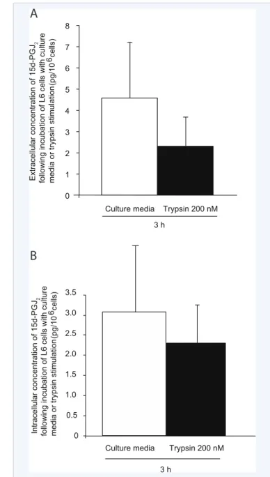
Figure 3 (A) – Extracellular and (B) – intracellular concentration of 15∆-PGJ2 following a 3 h incubation of L6 cells with or without trypsin,
a PAR-2 agonist.
[31], activation of this nuclear receptor can stimulate myoblast proliferation and it can be assumed that 15∆-PGJ2 may target this intracellular nuclear receptor to induce its proliferative effect. However, this field deserves a more complete investigation.
The second objective of the study was to verify if 15∆-PGJ2 is produced by L6 muscle cells following protease-activated receptor (PAR)-2 activation, as seen with fibroblasts in culture. 15∆-PGJ2 was detected in L6 cell culture media under basal conditions after 3 h in culture (Figure 3A). However, this extracellular concentration of 15∆-PGJ2 remained statistically unchanged when L6 cells were stimulated with trypsin 200 nM (Figure 3A). These results prompted us to verify if 15∆-PGJ2 is produced intracellularly by L6 cells following PAR-2 activation without being thereafter externalized. Culture media
surrounding L6 cells was thus removed at the end of incubation period and cells were lysed. A level of 15∆-PGJ2 comparable to the one observed in extracellular media was measured in L6 cell homogenates (Figure 3B) and trypsin-induced PAR-2 activation [15] did not influence the intracellular concentration (Figure 3B). Overall, the experimental protocol used clearly allowed to show that L6 cells can produce 15∆-PGJ2, but we were unable to
demonstrate that its production was sensitive to PAR-2 receptor activation. The complex and costly procedures involved in the extraction and measurement of this molecule makes it difficult to perform protocols with various time course designs in terms of duration of stimulation and sampling post-stimulation. Further work is needed before one can conclude solidly on the responsiveness of 15∆-PGJ2 production to PAR-2 activation.
Thus, key elements of the signaling cascade leading to skeletal muscle cell proliferation following PAR-2 activation remain to be discovered. We were able to demonstrate that 1) 15∆-PGJ2 is produced by skeletal muscle cell and 2) DP1 and DP2 receptors are expressed in skeletal muscle cells. We have thus identified intra- and extracellular receptors potentially used by this metabolite to induce its proliferative effect. Even if we were unable to demonstrate that PAR-2 activation leads to an increased 15∆-PGJ2 production, our results remain very physiologically relevant
since 15∆-PGJ2 can also be released by other cell types found in skeletal muscle, such as fibroblasts and mast cells, under basal and inflammatory conditions [13,32]. The discovery of receptors used by the metabolite 15∆-PGJ2 can lead to the development of new therapeutic approaches in the treatment of musculoskeletal injuries relying on endogenous molecules synthesized during the period of resolution of inflammation. Further studies focusing on the interaction of 15∆-PGJ2with nonreceptor protein following its entry into cells, possibly by an active transport system [11], are needed to complete our observations. Indeed, as demonstrated by Oliva et al. (2003), the direct activation of Ras/Erk MAP kinase pathway by 15∆-PGJ2 at cytosolic level may be a key element in the myogenic process [33].
CONCLUSION
The treatment of musculoskeletal injuries takes an enormous place in the field of sport medicine and there is obviously room to improve clinical conduct which is not always evidence-based. The fine understanding of the processes leading to tissue repair following acute injury is critical for ensuring an optimal treatment and healing. This is particularly true for the athlete population, where complete recovery of original tissue strength must be aimed to avoid re-rupture of structures submitted to high mechanical stress. Lipid mediators have been largely studied in disease models, but their implication in the treatment of acute musculoskeletal injury remains relatively unexplored. It seems promising to study the effect of 15∆-PGJ2 on muscle repair since in addition to its anti-inflammatory properties, mitogenic effects have been associated to this metabolite.
ACKNOWLEDGEMENTS
This work was supported by a grant to Claude H. Côté from the Natural Sciences and Engineering Research Council of Canada.
REFERENCES
1. Segawa M, Fukada S, Yamamoto Y, Yahagi H, Kanematsu M, Sato M,
et al. Suppression of macrophage functions impairs skeletal muscle regeneration with severe fibrosis. Exp Cell Res. 2008; 314: 3232-3244. 2. Arnold L, Henry A, Poron F, Baba-Amer Y, Van Rooijen N, Plonquet A,
et al. Inflammatory monocytes recruited after skeletal muscle injury switch into anti-inflammatory macrophages to support myogenesis. J Exp Med. 2007; 204:1057-1069.
3. Summan M, Warren GL, Mercer RR, Chapman R, Hulderman T, Van Rooijen N, et al. Macrophages and skeletal muscle regeneration: a clodronate-containing liposome depletion study. Am J Physiol Regul Integr Comp Physiol. 2006; 290: R1488-1495.
4. Chazaud B, Brigitte M, Yacoub-Youssef H, Arnold L, Gherardi R, Sonnet C, et al. Dual and beneficial roles of macrophages during skeletal muscle regeneration. Exerc Sport Sci Rev. 2009; 37: 18-22.
5. Tidball JG, Villalta SA. Regulatory interactions between muscle and the immune system during muscle regeneration. Am J Physiol Regul Integr Comp Physiol. 2010; 298: R1173-1187.
6. Villalta SA, Rinaldi C, Deng B, Liu G, Fedor B, Tidball JG. Interleukin-10 reduces the pathology of mdx muscular dystrophy by deactivating M1 macrophages and modulating macrophage phenotype. Human molecular genetics. 2011; 20: 790-805.
7. Serhan CN, Chiang N. Endogenous pro-resolving and anti-inflammatory lipid mediators: a new pharmacologic genus. Br J Pharmacol. 2008; 153 Suppl 1: S200-215.
8. Serhan CN, Chiang N, Van Dyke TE. Resolving inflammation: dual anti-inflammatory and pro-resolution lipid mediators. Nat Rev Immunol. 2008; 8: 349-361.
9. Seki H, Tani Y, Arita M. Omega-3 PUFA derived anti-inflammatory lipid mediator resolvin E. Prostaglandins Other Lipid Mediat. 2009; 89: 126-130.
10. Gilroy DW, Colville-Nash PR, Willis D, Chivers J, Paul-Clark MJ, Willoughby DA. Inducible cyclooxygenase may have anti-inflammatory properties. Nat Med. 1999; 5: 698-701.
11. Scher JU, Pillinger MH. 15d- PGJ2: the anti-inflammatory prostaglandin? Clin Immunol. 2005; 114: 100-109.
12. Pettipher R, Hansel TT, Armer R. Antagonism of the prostaglandin D2 receptors DP1 and CRTH2 as an approach to treat allergic diseases. Nat Rev Drug Discov. 2007; 6: 313-325.
13. Frungieri MB, Weidinger S, Meineke V, Köhn FM, Mayerhofer A. Proliferative action of mast-cell tryptase is mediated by PAR2, COX2, prostaglandins, and PPAR gamma: Possible relevance to human fibrotic disorders. Proc Natl Acad Sci U S A. 2002; 99: 15072-15077. 14. Kliewer SA, Lenhard JM, Willson TM, Patel I, Morris DC, Lehmann JM. A
prostaglandin J2 metabolite binds peroxisome proliferator-activated receptor gamma and promotes adipocyte differentiation. Cell. 1995; 83: 813-819.
15. Duchesne E, Tremblay MH, Côté CH. Mast cell tryptase stimulates myoblast proliferation; a mechanism relying on protease-activated receptor-2 and cyclooxygenase-2. BMC Musculoskelet Disord. 2011; 12: 235.
16. Town MH, Casals-Stenzel J, Schillinger E. Pharmacological and cardiovascular properties of a hydantoin derivative, BW 245 C, with high affinity and selectivity for PGD2 receptors. Prostaglandins. 1983; 25: 13-28.
17. Hirai H, Tanaka K, Takano S, Ichimasa M, Nakamura M, Nagata K. Cutting edge: agonistic effect of indomethacin on a prostaglandin D2 receptor, CRTH2. J Immunol. 2002; 168: 981-985.
D, et al. Novel selective small molecule agonists for peroxisome proliferator-activated receptor delta (PPARdelta)--synthesis and biological activity. Bioorg Med Chem Lett. 2003; 13:1517-1521. 19. Duchesne E, Bouchard P, Roussel MP, Côté CH. Mast cells can regulate
skeletal muscle cell proliferation by multiple mechanisms. Muscle Nerve. 2013; 48: 403-414.
20. Brose SA, Thuen BT, Golovko MY. LC/MS/MS method for analysis of Eâ‚ series prostaglandins and isoprostanes. J Lipid Res. 2011; 52: 850-859.
21. Yang P, Felix E, Madden T, Fischer SM, Newman RA. Quantitative high-performance liquid chromatography/electrospray ionization tandem mass spectrometric analysis of 2- and 3-series prostaglandins in cultured tumor cells. Anal Biochem. 2002; 308:168-177.
22. Tidball JG, Wehling-Henricks M. Macrophages promote muscle membrane repair and muscle fibre growth and regeneration during modified muscle loading in mice in vivo. J Physiol. 2007; 578 : 327-336.
23. Bondesen BA, Mills ST, Kegley KM, Pavlath GK. The COX-2 pathway is essential during early stages of skeletal muscle regeneration. Am J Physiol Cell Physiol. 2004; 287: C475-483.
24. Harris WS, Mozaffarian D, Rimm E, Kris-Etherton P, Rudel LL, Appel LJ, et al. Omega-6 fatty acids and risk for cardiovascular disease: a science advisory from the American Heart Association Nutrition Subcommittee of the Council on Nutrition, Physical Activity, and Metabolism; Council on Cardiovascular Nursing; and Council on Epidemiology and Prevention. Circulation. 2009; 119: 902-907. 25. Myles IA. Fast food fever: reviewing the impacts of the Western diet on
immunity. Nutr J. 2014; 13: 61.
26. Grygiel-Górniak B. Peroxisome proliferator-activated receptors and their ligands: nutritional and clinical implications--a review. Nutr J. 2014; 13: 17.
27. Pettipher R. The roles of the prostaglandin D(2) receptors DP(1) and CRTH2 in promoting allergic responses. Br J Pharmacol. 2008; 153 Suppl 1: S191-199.
28. Veliça P, Khanim FL, Bunce CM. Prostaglandin D2 inhibits C2C12 myogenesis. Mol Cell Endocrinol. 2010; 319: 71-78.
29. Leduc M, Breton B, Galés C, Le Gouill C, Bouvier M, Chemtob S, et al. Functional selectivity of natural and synthetic prostaglandin EP4 receptor ligands. J Pharmacol Exp Ther. 2009; 331: 297-307. 30. Petrova TV, Akama KT, Van Eldik LJ. Cyclopentenone prostaglandins
suppress activation of microglia: down-regulation of inducible nitric-oxide synthase by 15-deoxy-Delta12,14-prostaglandin J2. Proc Natl Acad Sci U S A. 1999; 96: 4668-4673.
31. Bonala S, Lokireddy S, Arigela H, Teng S, Wahli W, Sharma M, et al. Peroxisome proliferator-activated receptor beta/delta induces myogenesis by modulating myostatin activity. J Biol Chem. 2012; 287: 12935-12951.
32. Tanaka A, Nomura Y, Matsuda A, Ohmori K, Matsuda H. Mast cells function as an alternative modulator of adipogenesis through 15-deoxy-delta-12, 14-prostaglandin J2. Am J Physiol Cell Physiol. 2011; 301: C1360-1367.
33. Oliva JL, Pérez-Sala D, Castrillo A, Martínez N, Cañada FJ, Boscá L, et al. The cyclopentenone 15-deoxy-delta 12,14-prostaglandin J2 binds to and activates H-Ras. Proc Natl Acad Sci U S A. 2003; 100: 4772-4777.
Côté CH, Bouchard P, Duchesne E (2015) Identification of New Targets Used by 15-deoxy-delta-12,14-Prostaglandin J2 to Stimulate Skeletal Muscle Cell Prolifera-tion. Ann Sports Med Res 2(4): 1027.