HAL Id: hal-02412662
https://hal.archives-ouvertes.fr/hal-02412662
Submitted on 5 Jul 2020
HAL is a multi-disciplinary open access
archive for the deposit and dissemination of
sci-entific research documents, whether they are
pub-lished or not. The documents may come from
teaching and research institutions in France or
abroad, or from public or private research centers.
L’archive ouverte pluridisciplinaire HAL, est
destinée au dépôt et à la diffusion de documents
scientifiques de niveau recherche, publiés ou non,
émanant des établissements d’enseignement et de
recherche français ou étrangers, des laboratoires
publics ou privés.
Mutual influence of gold and silver nanoparticles on
Tris-(2,2’bipyridine)-Ru(II) core complexes:
Post-functionalization processes, optical and
electrochemical investigations
Frédéric Dumur, Audrey Guerlin, Anaïs Lehoux, P.R. Selvakannan, Fabien
Miomandre, Rachel Méallet-Renault, Mateusz Rebarz, Michel Sliwa, Eddy
Dumas, Loic Le Pleux, et al.
To cite this version:
Frédéric Dumur, Audrey Guerlin, Anaïs Lehoux, P.R. Selvakannan, Fabien Miomandre, et al..
Mu-tual influence of gold and silver nanoparticles on Tris-(2,2’bipyridine)-Ru(II) core complexes:
Post-functionalization processes, optical and electrochemical investigations. Applied Surface Science,
Else-vier, 2020, 499, pp.143847. �10.1016/j.apsusc.2019.143847�. �hal-02412662�
UNCORRECTED
PROOF
Contents lists available at ScienceDirect
Applied Surface Science
journal homepage: www.elsevier.com
Mutual influence of gold and silver nanoparticles on Tris-(2,2′bipyridine)-Ru(II) core
complexes: Post-functionalization processes, optical and electrochemical investigations
Frédéric Dumur
a, ⁎, Audrey Guerlin
f, Anaïs Lehoux
c, P.R. Selvakannan
g, Fabien Miomandre
d,
Rachel Méallet-Renault
h, Mateusz Rebarz
i, Michel Sliwa
i, Eddy Dumas
c, Loïc Le Pleux
e, Yann Pellegrin
e,
Fabrice Odobel
e, Cédric R. Mayer
b, ⁎aAix Marseille Univ, CNRS, ICR, UMR 7273, F-13397 Marseille, France
bDépartement de Chimie, UFR des Sciences, Université de Versailles Saint-Quentin-en-Yvelines, 78035 Versailles, France
cInstitut Lavoisier de Versailles, UMR-CNRS 8180, Université de Versailles Saint-Quentin-en-Yvelines, 78035 Versailles, France
dLaboratoire de Photophysique et Photochimie Supramoléculaires et Macromoléculaires, UMR CNRS 8531, ENS Paris-Saclay, 94235 Cachan, France
eChimie et Interdisciplinarité: Synthèse, Analyse, Modélisation (CEISAM), UMR-CNRS 6230, Université de Nantes, 44322 Nantes, France
fUniversité Paris Diderot, UFR de Chimie, 15 Rue Jean Antoine de Baïf, 75013 Paris, France
gResearch Fellow School of Science, RMIT University, Melbourne, 3001, VIC, Australia
hInstitut des Sciences Moléculaires d'Orsay (ISMO) - UMR 8214, Université Paris-Saclay, Université Paris-Sud, 91405 Orsay Cedex, France
iUniv. Lille, CNRS, UMR 8516 - LASIR - Laboratoire de Spectrochimie Infrarouge et Raman, F-59000 Lille, France
A R T I C L E I N F O
Keywords:
Polypyridyl ruthenium complexes Ag Au Nanoparticles Spectroscopies Electrochemistry A B S T R A C T
The synthesis, reactivity and properties of a series of four polypyridyl ruthenium complexes have been studied. These complexes were used to post-functionalize preformed 3nm silver and gold nanoparticles (NPs) in water and in dichloromethane (DCM). We studied the influence of the grafted complexes on the formation process and stability of the colloidal solutions and we investigated the optical and electrochemical properties of the fi-nal nanocomposites. Among the series of four ruthenium complexes, three novel heteroleptic complexes (1–3) bearing one pyridine, one amine or two carboxydithioic acid pendant groups were synthesized and reacted with preformed Au-NPs and Ag-NPs. Results were compared to those obtained with the model [Ru(bpy)3]2+complex
(4). The strength of the interaction between the anchoring group and the surface of NPs influenced the size, shape and stability of the final nanocomposites. Polar solvent such as water induced aggregation and lead to unstable nanocomposites. Stationary and time resolved luminescence of grafted nanocomposites (1–3) showed that the luminescence of complexes were completely quenched (lifetime and emission quantum yield) in water by electron transfer processes, moreover electrical measurements rationalize that Ag nanocomposites exhibit the stronger quenching due to a lower oxidation potential. It also showed a current enhancement associated with double layer charging of the metal nanoparticle cores.
1. Introduction
Over the past three decades, tris-(2,2′-bipyridine)ruthenium(II) has been one of the most studied coordination complex in photochemistry [1]. The reasons are numerous but its chemical and electrochemical ro-bustness [2], the facile design and the modularity of the ligands [3] and its long-lived emission motivated the synthesis of various deriva-tives [4]. The use of Ru(II) polypyridine complexes is broad and covers a wide range of properties and applications such as optoelectronics de
vices [5], nonlinear optics [6], bio-imaging [7], bio-analysis [8], catal-ysis [9], solar cells [10] and nanosciences [11]. Even if considerable efforts have been devoted to the design of new molecular derivatives for the above mentioned applications, molecular building blocks (MBBs) have also received a great deal of interest for material sciences. This interest is motivated by the unique properties brought by the Ru-MBB to the host matrixes and the multifunctional character of the result-ing materials. They have been incorporated in liquid crystals [12], hy-brid polymers (metallopolymers) [13], dendrimers (metallodendrimers)
⁎ Corresponding authors.
Email address: frederic.dumur@univ-amu.fr (F. Dumur)
https://doi.org/10.1016/j.apsusc.2019.143847
Received 10 July 2019; Received in revised form 12 August 2019; Accepted 30 August 2019 Available online xxx
0169-4332/ © 2019.
Full length article
UNCORRECTED
PROOF
[14], micelles [15], sol-gel materials [16], silica nanomaterials [17] and more recently grafted onto oxometallic [18] and metallic nanoparticles [19]. Parallel to Ru(II) coordination complexes, silver and gold nanopar-ticles (NPs) constitute another active field of research. Both types of NPs are of particular interest in nanosciences and they know an ever increasing research effort due to their intrinsic physicochemical proper-ties (in particular electronic and optical properproper-ties) [20] and their facile functionalization [21]. Domains of applications are ranging from catal-ysis [22], opto-electronics [23] to biomedical materials [24]. The graft-ing of redox active and/or fluorophore probes onto metallic nanoparti-cles has recently drawn an intense research interest [25]. Electrochem-ical studies realized by Murray's group clearly demonstrated the role of electron reservoir played by the nanoparticles and the possibility to modulate the charge states of the NPs by means of the applied poten-tial [26]. The ability to confine and control the number of redox/fluo-rophore centres on the NPs also leads to characteristic electrochemical behaviours showing a concerted redox process combined with a fluo-rophore emission quenching [27]. In all these reports, the redox active centres are not electronically connected to the NPs and these systems are comparable to functionalized dendrimers, the NPs simply acting as linkers [28]. Polypyridyl Ru(II) complexes are excellent redox and lumi-nescent probes. Consequently, the grafting of these molecular units onto the surface of nano-objects with an effective control of the communica-tion between both components of the nanocomposites is of great interest for fundamental research but also for potential applications. Studies of the interactions between polypyridyl ruthenium complexes and metal-lic nanoparticles appeared only recently in the literature. Most of these ruthenium tris-bipyridine complexes are either interacting with the NPs via electrostatic interactions [29], or they are grafted onto the NPs via long aliphatic chains functionalized by various anchoring end-groups such as thiols [30], dithiocarbamate [31], or aromatic triamine [32]. However, both strategies are not suitable for efficient electronic trans-fers due to the absence of conjugation between both partners.
In the past five years, our group has been involved in the synthesis of gold and silver NPs stabilized and functionalized by ruthenium com-plexes via rigid and fully conjugated bridges [33]. The physicochemi-cal properties of these nanocomposites and more specifiphysicochemi-cally their elec-trochemical and luminescence properties were first discussed [34]. To date, few works have thoroughly been devoted to the study of the al-teration of the luminescence of inorganic fluorophores when grafted onto metallic nanoparticles. The quenching of luminescence is often observed because different deactivation pathways, inter/intramolecular
interaction, energy and/or electron transfer are possible between the fluorophore and the NP. In both cases, these processes are sensitive to the nature of the bridge (length, conjugation, rigidity …) between the fluorophore and the NP and are also sensitive to the size and the shape of the NP. Different approaches are possible to functionalize Ag-NPs or Au-NPs but the most common method remains the place exchange of the stabilizing agent, initially required to generate the NPs, by the de-sired functionalizing agent [35]. The place exchange approach allows for a better control of the doping amount of functionalizing agent on the NPs and is applicable in water and organic solvents such as chloroform or toluene. In these media, the initial stabilizing agent (sodium citrate in water and long alkylamine or alkylcarboxylic acid chains in organic solvent) is easily substitutable by the functionalizing agent.
Herein, we investigated the post-functionalization of preformed Ag-NPs and Au-NPs by tris-(2,2′-bipyridine)Ru(II) derivatives and the reciprocal influence between the NPs and the ruthenium complexes on the stability and on the physicochemical properties of the final nanocomposites. Au-NPs and Ag-NPs were used to investigate the influ-ence of oxidation potential and of the plasmon band energy (lower and higher energy respectively) compare to the one of the MLCT band of
tris-(2,2′-bipyridine)Ru(II) derivatives. Four complexes have been used
in this work, namely [(bpy)2Ru(4,4′-(carboxydithioic
acid)-2,2′-bipyri-dine)](X)2 (1.X2), [(bpy)2
Ru((4-(4-pyridinyl)phenyl)imi-dazo[4,5-f][1,10]phenanthroline)](X)2 (2.X2),
[(bpy)2Ru((4-aminophenyl)imidazo[4,5-f][1,10]phenanthroline)](X)2
(3.X2) and [(bpy)3Ru](X)2(4.X2), X−=PF6−or Cl−, in which we varied the nature of the anchoring group as well as the distance between the complex and the NP (Scheme 1). Complex 4 was prepared as a model to control the role of the anchoring groups (pyridine, amine and carboxy-dithioic acid) on the post-functionalization process. The mutual influ-ence between metal-NPs and ruthenium complexes has been analyzed in two solvents, water and dichloromethane (DCM). The choice of both sol-vents was motivated by the fact that redox and luminescence properties of the resulting nanocomposites can be investigated in both organic and aqueous media simply by counteranion exchange. This influence has been investigated by TEM, UV/Vis, luminescence, time resolved spectro-scopies and electrochemistry.
2. Results and discussion
2.1. Synthesis of the proligands and complexes
Four complexes have been used in this study (Scheme 1). The first
one, 1, is structurally closely related to the well-known [Ru(bpy)3]2+
UNCORRECTED
PROOF
(4) except that one bipyridine was functionalized by two carboxy-dithioic acid groups. The choice of carboxycarboxy-dithioic acid as anchoring group was motivated by its strong affinity for soft metals [31,36,37], which should ensure strong bonding to metal surfaces [38]. The prepa-ration of 1 is relatively straightforward and consists of the oxidation of 4,4′-bis(chloromethyl)-2,2′-bipyridine directly on 5 [39] by elemental sulphur in the presence of sodium methanolate (Scheme 2) [40].
Complexes 2 and 3 were functionalized with phenylpyridine for 2 and aniline for 3, since these groups proved to be effective anchor-ing groups to stabilize and to bind to metal NPs [33,41,42]. Conven-tional aliphatic thiols have not been used in this work to link the ruthenium complexes to the metal NPs because this type of binding has already been abundantly reported by Kamat et al. [43] and we wanted to favor electronic interactions by maintaining a conjugated linkage between both partners. Complex 2 was synthesized by reaction
of [(bpy)2Ru(phendione)]Cl2(6.Cl2) with 4-(4-pyridinyl)benzaldehyde.
Complexes 1, 2, 3 and 4 were isolated as hexafluorophosphate or chlo-ride salts depending on the solvent further used to generate the final nanocomposites.
2.2. Synthesis of the nanocomposites
As already mentioned, the conventional method to functionalize Au-NP or Ag-NPs consists in the post-functionalization of preformed NPs. This method was also selected here. In previous works reported by our group, other methods have been described to functionalize metal NPs with metal complexes: the direct functionalization [33a], the post-functionalization by phase transfer [33c] or the generation of the metal complexes directly on NPs pre-functionalized with the appro
priate proligands [33b]. In the first two cases, the colloidal solutions contained a significant excess of free complexes which renders the lu-minescence investigations difficult. This problem is avoided with the third method but requires proligands able to stabilize the NPs before complexation and this strategy is not applicable to a large variety of complexes. These considerations prompted us to develop the post-func-tionalization of NPs by place exchange reaction (Scheme 3). Besides, this approach provides a better control of the size of the NPs and a better control of the amount of metal complexes grafted on the NPs. The place exchange process was performed in water and in DCM but can be extended to other solvents such as chloroform or toluene. The choice of both solvents is supported by the potential applications of the nanocomposites studied in this work. Indeed, Ru(II) complexes and metal NPs have potential applications in biology and optoelectronics. In the context of our work, one of the major difficulties was to obtain homogeneous colloidal solutions with uniform NPs of narrow size dis-tribution and morphology stabilized with easily exchangeable surfac-tants. In aqueous solution, sodium citrate was used as initial surfactant in diluted medium. Ag-NPs and Au-NPs were obtained with a size dis-tribution centred around 2.5nm. In DCM, 1-hexadecylamine (HDA) or a mixture of HDA and oleic acid (OA) were used to prepare Au-NPs and Ag-NPs, respectively. In the case of Ag-NPs, OA was essential to obtain stable and homogeneous colloidal solutions. The mean size dis-tribution of Au-NPs and Ag-NPs was determined to be 3±1nm. The post-functionalization was performed by mixing the preformed metal NPs with the appropriate ruthenium complex. The amount of ruthe-nium complexes added in each colloidal solution was chosen follow-ing this calculation: we first estimated the ratio between the surface of one NP and the projected surface of each complex onto a plane surface. Then, for all colloidal solutions, the amount of complexes introduced
Scheme 2. Synthesis of 1 and 2.
UNCORRECTED
PROOF
was about 20% of the theoretical amount required for the maximal cov-erage. Except for the electrochemistry measurements, we voluntarily added a low concentration of ruthenium complexes in order to limit the communication between them once grafted onto the surface of the NPs because it was already observed by Kamat et al. that electron transfer
between different Ru(bpy)32+could be observed [43].
2.3. NPs in water
In aqueous solution, the nature of the metal NPs had a strong in-fluence on the stability of the final nanocomposites. Noticeable ligand effects were also evidenced with a strong impact on the form, the size and the stability of the functionalized NPs. Immediately after addition of the ruthenium complexes, and depending on the anchoring group, the colour of the gold colloidal solutions changed, more or less rapidly, from red to purple and finally blue. The colour of the solutions is highly indicative of the stability of the colloidal solutions. Without anchor-ing group, as in the case of complex 4, aggregates lead to a blue so-lution and a precipitate was rapidly observed. The stability of the col-loidal solutions increased with the strength of the interaction between the anchoring group and the surface of the Au-NP in the following or-der: amine (3), pyridine (2) and carboxydithioic acid (1) (see Fig. S1). However, even with strong anchoring groups such as phenylpyridine or carboxydithioic acid, Au-NPs flocculated after one night. The instability of the gold colloidal solution can be explained by these mechanisms: in the case of complexes 1 and 2, these ruthenium complexes first replace citrate anions onto the surface of Au-NPs. Then, in a second step, the positive charges brought by the complexes are neutralized by citrates anions, remaining in solution, leading to the progressive aggregation of Au-NPs (Scheme 4a). This type of neutralization, due to the presence of citrates anions, was recently described with other cationic molecules [44]. The aggregation phenomenon was confirmed by TEM, as shown in Fig. 1 for complex 2. For complex 3, for which a weaker interaction is expected between the anchoring group and the surface of the Au-NP, and for complex 4 an even faster aggregation was observed compared to complexes 1 and 2. In the case of cationic complex 4, electrostatic interactions with citrate anions surrounding the Au-NPs are responsible for the aggregation of the Au-NPs (Scheme 4a).
Concerning the functionalization of Ag-NPs in water, a specific strat-egy was developed due to the difference of reactivity between Au-NPs and Ag-NPs. This difference originates from the surface charge of the NPs: Ag-NPs are positively charged while Au-NPs are negatively charged [45]. Due to this major difference, addition of 4, for example, did not immediately induce the aggregation of Ag-NPs and the colloidal solu-tion was stable for several days (Fig. S1b). The influence of the an-choring group on the size of the final NPs is more pronounced with Ag-NPs compared to Au-NPs (Scheme 4b). For example, after addition of complex 4, the solution remained stable without modification of the colour. For all the other complexes, the colour of the solutions turned to brown yellow for 3, to red for 2 and dark red for 1. The stabil-ity of these solutions was maintained for a few days before aggrega-tion. Interpretation of these different colors, depending on the nature of the anchoring group, has been given using DLS, TEM and UV/Vis spectroscopy. DLS clearly showed a modification of the size of the silver nanocomposites after addition of the ruthenium complexes. This mod-ification was even more significant when the anchoring group had a higher affinity for the metallic surface (Fig. 2). 1h after addition of 4, no evolution of the size of the nanocomposites was observed (Fig. 2). For the other complexes, two populations of nanocomposites were
de-tected; one with an average hydrodynamic diameter (dh) centered at
2–3nm for all three complexes and a second one with dhvarying with
the nature of the anchoring group. For complex 3, the dhof the
sec-ond population was centered around 20nm, and for complexes 2 and 1, the size distribution was broader and centered around 35 and 50nm, respectively. For complexes 1, 2 and 3, DLS analysis shows that the evolution of the size of the silver nanocomposites follows a two steps process (Figs. S2 and S3 and Scheme 4b). In the first step, two pop-ulations of nanocomposites were observed, after five minutes of
stir-ring, with average dhvalues centered around 7 and 20nm for 1 and
around 4 and 9nm for 2. During the second step, the systems evolved to-wards the two final populations observed with the largest nanocompos-ites clearly growing at the expense of the smallest ones (Ostwald ripen-ing process). The final composition of the colloidal suspensions was con-firmed by TEM analysis (one drop of the solution studied by DLS 1h after addition of the complexes was taken and dried on a copper grid). TEM pictures (Figs. 3 and S3) showed Ag-NPs with a size distribution in accordance with the results obtained by DLS. Small Ag-NPs, with a
Scheme 4. Mechanisms proposed to explain the stability and the evolution of Au-NPs and Ag-NPs in aqueous medium, using ruthenium complexes 1, 2 and 3 containing different
UNCORRECTED
PROOF
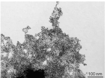
Fig. 1. TEM image of Au-NPs in water 1h after addition of 2.mean diameter of about 2nm, surround bigger Ag-NPs with a mean di-ameter varying with the nature of the anchoring group. The size of the bigger particle follows the strength of the interaction between the an-choring group and the surface of nanoparticles. The post-functionaliza-tion of Ag-NPs in aqueous medium with ruthenium complexes clearly induced a modification of the preformed Ag-NPs leading to two dis-tinct populations in size, following an Ostwald ripening process. The ex-tent of this phenomenon is obviously correlated with the nature of the anchoring group and therefore with the strength of the interaction be-tween the ruthenium complex and the silver surface. Kamat group ob-served by TEM surface modification of gold nanorods after
post-func-tionalization with Ru(bpy)3-C5-SH which lead to some aggregation. This
was explained as an induced surface corrosion from the thiol due to the
redox nature of the Ru(bpy)32+[46]. Similar hypothesis could be also
involved in our case because the aggregation process (size of nanopar-ticles) increases with the strength of the interaction between the an-choring groups. Finally, it has to be noticed that, as for Au-NPs, Ag-NPs precipitated after several days. The redox corrosion is stronger in our
case because no aliphatic bond was used between Ru(bpy)32+and NPs
and it could explained that the NPs are not stable in water after sev-eral days in comparison to DCM, electron transfer and redox corro-sion are facilitated in water. This precipitation can also been attrib-uted to the presence of citrate anions screening the repulsive positive charges brought by the ruthenium complexes located at the surface of
the Ag-NPs and responsible for the temporary stability of the colloidal solutions (Scheme 4b).
2.4. NPs in DCM
In DCM, the final colloidal solution showed a greater stability com-pared with the corresponding aqueous solutions and this can be ex-plained by the absence of aggregation due to corrosion process and also to electrostatic interactions between charged species remaining in solution and the charged NPs. All solutions were stirred for 48h be-fore analysis. TEM images evidenced significant differences of reactiv-ity depending on the nature of the metal NP and also depending on the nature of the ruthenium complex. TEM images of preformed Au-NPs, stabilized with HDA, showed spatially well-ordered Au-NPs due to in-terdigitation of the long alkyl chains of HDA surrounding nearby NPs [47]. In the same conditions, preformed Au-NPs reacted with 4 did not induced significant modification of the size of the Au-NPs or even of the organization of the Au-NPs on the grid. In contrast, reaction of the preformed Au-NPs with the other ruthenium complexes induced a noticeable alteration of the spatial organization of the Au-NPs ex-plained by the partial replacement of HDA by ruthenium complexes onto the surface of the Au-NPs. The stronger was the interaction be-tween the anchoring group of the ruthenium complexes and the sur-face of the Au-NPs, the more efficient was the substitution of HDA by ruthenium complexes (Fig. 4a). Indeed, this substitution is more effec-tive in the order 1>2>3. It has to be noticed that for all complexes, no noticeable modification of the size of the Au-NPs was observed af-ter post-functionalization. This implied that solvent polarity is playing a big role in the stability and the absence or existence of aggregation process. As it was mentioned previously the corrosion process due to
the redox nature of Ru(bpy)32+ through the anchoring group is
hap-pening more strongly in polar solvent. In the case of Ag-NPs, the ini-tial spaini-tial organization of the preformed Ag-NPs is no longer observed by TEM with 1, 2 and 3. While the post-functionalization process us-ing 1 and 3 has only a minor effect on the size and shape of the final nanocomposites, reaction of 2 on preformed Ag-NPs drastically modi-fied both the size and the shape of the latter (Fig. 4b). Two populations of Ag-NPs were observed, one similar to the preformed Ag-NPs and a second population of much bigger size, centered around 20nm, with facetted Ag-NPs (truncated nanotriangles, nanohexagons, nanorods …). The reason why only complex 2 induced such a drastic modification of the size and shape of the preformed Ag-NPs remains yet unexplained.
Fig. 2. DLS analysis of preformed silver nanocomposites in water (purple, diamonds) and 1h after addition of 1 (green, circles), 2 (red, crosses), 3 (blue, triangles) and 4 (black,
UNCORRECTED
PROOF
Fig. 3. TEM images of Ag-NPs in water 1h after addition of 1.However, in both solvents, water and DCM, it is obvious that the nature of the ruthenium complex, and more precisely the strength and the na-ture of the anchoring point of the complex, has much more influence on the size and shape of the final nanocomposites in the case of Ag-NPs than in the case of Au-NPs.
2.5. UV/Vis stationary and time resolved spectroscopy
Absorption spectra were recorded in two solvents, water and DCM, at room temperature using chloride or hexafluorophosphate salts of the ruthenium complexes. The most relevant spectroscopic results are sum-marized in Table 1.
Electronic absorption spectra of 1–4 in water and in DCM (Fig. 5a and d, respectively and see also Supporting Information) show intense bands in the UV region of the spectrum (250–310nm). These bands can be attributed to π→π* intraligand transitions from the bipyridine groups. In the 310–400nm region, spectra of 2 and 3 present a fea-ture band around 350nm attributed to π→π* intraligand transitions in-volving the imidazo(4,5-f)-(1,10)phenanthroline (PhenIm) moiety [48]. In the 400–600nm region, ruthenium complexes exhibit a less intense
and broader d(RuII)→π*(L) MLCT band, with L corresponding to
bipyri-dine and PhenIm [49]. As previously observed this MLCT band is slightly red-shifted compared to the reference compound 4 [50].
In aqueous solution, UV–visible spectra of Au° and Ag° nanocompos-ites are in agreement with results obtained by DLS and TEM analyses (Fig. 5b and c). The amount of complexes added corresponded to one fifth of the calculated theoretical maximum covering. The starting gold colloidal solutions exhibited a surface plasmon band (SPB) at 509nm in agreement with the size of the Au-NPs determined by TEM [41,51]. After addition of the different ruthenium complexes, the MLCT band was not discernible in the absorption spectra and only the π→π* intrali-gand transitions were observed in the 230–300nm region. Concerning the SPB, addition of 4 induced an important broadening of the band. This diffuse band seems to result from the contribution of two bands, centred around 540 and 655nm. This observation is in agreement with the rapid aggregation observed after addition of 4. Addition of 3 and 2 caused a broadening and a red shift of the SPB (556 and 548nm, respec-tively) while the broadening and the red shift of the SPB is less impor-tant after addition of 1 (527nm). These results are in agreement with the previous observations on the stability of the different colloidal so-lutions. After 1h, all solutions exhibited the same diffuse SPB illustrat-ing the irreversible aggregation of the nanoparticles. Concernillustrat-ing Ag-NPs (Fig. 5c), the starting Ag-NPs exhibited a SPB at 395nm. After addition of 4, the SPB was only slightly shifted. This result is in agreement with the previous observations, meaning that no interaction occurs between the cationic ruthenium complexes and the Ag-NPs. After addition of 3, the SPB was slightly shifted to 390nm but the most striking feature was the appearance of a second SPB centred at 511nm. The influence of the addition of 2 and 1 was even more pronounced with the presence of the second SPB observed at 573 and 583nm, respectively. The small differ-ences noticed in the spectra of 2 and 1 are in agreement with TEM and DLS observations, confirming in both cases the progressive growth of bigger Ag-NPs coexisting with smaller ones and the formation of aggre-gates in solution (Fig. S4).
Absorption spectra in DCM are gathered in Fig. 5d–f. With Au-NPs and with Ag-NPs the presence of the ruthenium complexes was only as-serted by the π→π* transition in the 235–300nm region. Concerning Au-NPs (Fig. 5e), the position of the SPB was only slightly modified af-ter addition of the ruthenium complexes, with a maximum red shift of 12nm for 2. These observations are in full agreement with the TEM analyses showing the absence of noticeable modification of the size of the Au-NPs after functionalization. In the case of Ag-NPs, the
UNCORRECTED
PROOF
Table 1Electronic absorption data and assignment.
Complexesa λmax[nm] ε[M−1cm−1]±15% Assignment Solvent Metal λmaxSPB [nm]
CH2Cl2 Ag 431 CH2Cl2 Au 509 H2O Ag 395 H2O Au 509 1 457 13,140 d→π* CH2Cl2 Ag-1 419 291 66,160 π→π* CH2Cl2 Au-1 517 251 23,260 π→π* H2O Ag-1 398/583b H2O Au-1 527c 2 458 17,320 d→π* CH2Cl2 Ag-2 423 334 27,515 π→π* CH2Cl2 Au-2 521 290 83,640 π→π* H2O Ag-2 393/573b 244 26,120 π→π* H2O Au-2 548c 3 463 14,670 d→π* CH2Cl2 Ag-3 420 342 26,530 π→π* CH2Cl2 Au-3 515 290 65,250 π→π* H2O Ag-3 390/511b 247 23,265 π→π* H2O Au-3 556c 4 453 12,300 d→π* CH2Cl2 Ag-4 416 289 73,800 π→π* CH2Cl2 Au-4 511 250 17,170 π→π* H2O Ag-4 402 H2O Au-4 540–655c a In DCM, at 298K.
b Absorption of both components of the SPB.
c Broad band.
Fig. 5. Absorption spectra in water at 298K of a) 1 (green), 2 (red), 3 (blue), 4 (black), b) Au-NPs (purple), Au°-1 (green), Au°-2 (red), Au°-3 (blue), Au°-4 (black) and c) (after 1h of
stirring) Ag-NPs (purple), Ag°-1 (green), Ag°-2 (red), Ag°-3 (blue), Ag°-4 (black). Absorption spectra in DCM at 298K of d) 1 (green), 2 (red), 3 (blue), 4 (black), e) Au-NPs (purple), Au°-1 (green), Au°-2 (red), Au°-3 (blue), Au°-4 (black) and f) Ag-NPs (purple), Ag°-1 (green), Ag°-2 (red), Ag°-3 (blue), Ag°-4 (black). (For interpretation of the references to colour in this figure legend, the reader is referred to the web version of this article.)
functionalization induced a small blue shift of the SPB together with a broadening of the peak (Fig. 5f). This broadening of the peak is more important after addition of 2, in agreement with the higher polydisper-sity in size and shape observed by TEM analyses.
Luminescence properties (stationary and time resolved data) of the ruthenium complexes are summarized in Table 2 (Fig. 6 and support-ing information). It should be stressed here that a full account of the
solvent effect on luminescent properties of Ru-bipyridine complexes is not an easy task because a minimum of three processes, solvent and temperature dependent, are involved: radiative and non-radiative de-cay from the lowest triplet MLCT emissive state (quenched by the oxy-gen) as well as the transition to a low lying d-d state [52,53]. The en-ergy levels of the MLCT states depend on the polarity of the solvent and influence the transition efficiency to the ground state (radiatively
UNCORRECTED
PROOF
Table 2Luminescence data of free ruthenium complexes in solution (without NPs).
λem,max[nm] Φr(x 10−2) τ [ns] kr[104s−1] knr[104s−1]
ACN DCM Water ACN DCM Water ACN DCM Water ACN DCM Water ACN DCM Water
1 648 603 642 1.6 9.2 3.9 860 602 552 1.8 15.2 7.0 114.4 150.8 174.0
2 625 594 624 2.3 12.5 9.4 771 735 656 2.9 17.0 14.3 126.7 119.0 138.1
3 617 597 617 0.7 11.7 7 352 914 570 1.9 12.8 12.2 282.1 96.6 163.1
4 617 600 623 11 10.7 8.7 905 575 588 12.1 18.6 14.7 98.3 155.3 155.2
Fig. 6. Stationary luminescence of a) 1 (green), 2 (red), 3 (blue), 4 (black), b) Au°-1 (green), Au°-2 (red), Au°-3 (blue), Au°-4 (black) and c) Ag°-1 (green), Ag°-2 (red), Ag°-3 (blue), Ag°-4
(black) in DCM with 460nm excitation. (For interpretation of the references to colour in this figure legend, the reader is referred to the web version of this article.)
and non radiatively) and to the thermally activated d-d states (tem-perature dependent). It was also shown that OH vibrations of water play the role of energy acceptors and thus enhance radiationless decay for Ru-bipyridine complexes [54]. Attention should be also paid to the properties of the anchoring group: imidazole groups are known to be electron donor groups and sensitive to pH. Dithioic group is an acid whereas aniline is a weak base and also an electron donor group. Al-together these features will influence the MCLT level and its emissive properties. Therefore luminescence of ruthenium complexes 1–4 was measured in water, acetonitrile and DCM at 298K in deoxygenated con-ditions to try to understand the influence of the polarity and protic-ity of the solvent. Complex 4 was used as the reference in the whole study to determine luminescence quantum yields of the other complexes
[52,53,55]. Assuming that the intersystem crossing quantum yield (ΦT)
for the formation of the3MLCT triplet state is equal to unity, and
know-ing the lifetimes (τ) and emission quantum yield (ϕr), the radiative (kr)
and non-radiative (knr) rate constants for the series of complexes can be
calculated using the following expressions:
In DCM a quantum yield about 10% is found for all the complexes. The lifetime of the MLCT excited state of 1 is very close to the value for the reference complex 4 (about 600ns) whereas it is slightly longer for complexes 2 and 3 (735 and 914ns, respectively). This is mainly due to a lower non radiative constant which can be explained on the basis of a higher energy gap between the lowest excited and the ground state. For complexes 1–3 a general result is a lower lumines-cence quantum yield combined with a lower radiative constant and a bathochromic shift of the emission maximum of the triplet MCLT ex-cited state from DCM, apolar solvent (ε=9.1) to acetonitrile (aprotic, polar solvent, ε=37.5). In the case of the reference complex 4, the lu-minescence quantum yield and radiative constant are almost constant
whereas non radiative constant decrease from 155 to 98 104s−1
con-comitantly with an increase of its lifetime to 905ns. This can be ex
plained by a higher energy gap between MLCT and d-d states (bathochromic shift of the emission maximum) and then a lower non radiative constant because of less effective thermal activation of d-d state quenching pathway. In acetonitrile complexes 1 and 2 present slightly lower lifetime and higher non radiative constant (about 800ns
and 110.104s−1) in comparison to complex 4. The main difference is
a lower radiative constant which leads to a lower luminescence quan-tum yield. Longer groups which increase vibrational relaxation path-way and lower energy gap with the ground state is also responsible of these results. Moreover the electron donor effect, which is more impor-tant in polar solvent, of different anchoring groups (dithioic groups and imidazole group) changes and reduces the radiative constant as previ-ously observed for methyl- or dimethyl-substitution on bipyridyl ligand [56]. Complex 3 in acetonitrile has the lowest quantum yield which is characterized by a short lifetime (350ns), and a non-radiative constant
with a maximum value of 282.105s−1, more than two times the value
for the other complexes whereas the radiative constant is quite similar to complexes 1 and 2 (similar electron donor effect). The former effect comes from the presence of the aniline moiety and the already envi-sioned photo-induced electron transfer mechanism which is more effi-cient in polar solvent. Finally the luminescence results in water should be rationalized combining polar solvent influence in addition with spe-cific interactions with the solvent because the anchoring groups are pH sensitive. Water molecule reduces the electron donor effect of the imi-dazole group (hydrogen bond with the nitrogen of the imiimi-dazole group) and leads for complexes 2 and 3 to similar radiative constant in com-parison to reference complex 4. For complex 3 the lone pair of the ni-trogen can be involved in hydrogen bond and diminish the existence of the photoinduced electron transfer. Therefore similar non radiative con-stant, luminescence quantum yield and lifetime are found for complexes 2, 3 and 4. For complex 1 a lower radiative constant rate is observed and leads to a lower luminescence quantum yield compare to other com-plexes in water. The electron donor effect of dithioic acid is quite similar in water and explains this result.
Stationary and time resolved luminescence data after post-function-alization of the metal NPs with a surface coverage of 20% (see above §
UNCORRECTED
PROOF
Synthesis of the nanocomposites) are summarized in Table 3. Such low coverage percentage was used to avoid intermolecular quenching from different complexes attached to the same NP. Luminescence spec-tra are similar for the free and bound to NPs complexes, while, as ex-pected from the literature, luminescence of the different complexes are quenched by the NPs (Fig. 6). The reason of such quenching is com-plex and it was shown that energy transfer and electron transfer can quench the luminescence and that contribution of each process is de-pendant of the size of NPs; for small size NPs (below 8nm) the electron transfer quenching is dominant [43,46,57]. It was also mentioned that the quenching for chromophores bound to NPs can be due to a dras-tic decrease of the radiative constant rate [58]. Analysis of Fig. 7 and Table 3 leads to several comments: 1) bound complexes (1, 2 and 3) show a higher luminescence quenching compare to complex 4 which in-teracts with NPs only through electrostatic interactions, 2) the lumines-cence quenching is higher in polar solvent (almost 99% in water) which is characteristic of a quenching by electron transfer, 3) the quenching is more important for complex 3 than complex 2 and this result can be explained by a longer distance between the NPs and the ruthenium cen-tres, 4) pyridine and aniline anchoring groups show a higher quenching which is due to an additional specific photo-induced electron transfer which increases in the vicinity of the NPs, 5) the quenching is more im-portant for Ag-NPs due to a lower oxidation potential of Ag-NPs com-pared to Au-NPs and this reinforces a mechanism by electron transfer. The luminescence lifetime in DCM (the quenching was too strong to be able to measure luminescence lifetime in water) is multiexponen-tial in agreement with the existence of several quenching processes and was calculated by the mean lifetime procedure [59]. The mean life-time is about 10ns (about 500ns for free complexes), in agreement with the strong decrease of luminescent quantum yield. As the lumi-nescence is less quenched for Au-NPs compared to Ag-NPs, the mean lifetime is longer for Au-NPs. An interesting result is that while the lu-minescence quantum yield of the post-functionalized NPs is the low-est for complexes 2 and 3, the mean lifetime is slightly longer com-pared to complexes 1 and 4, a slower back electron transfer (longer distance between the NPs and the ruthenium centres) can be involved for the long component. It should be emphasized here that no long component was found for free complexes (few hundreds of nanosec
onds) and therefore it confirms that the complexes are either attached (complexes 1, 2 and 3) or in the vicinity of the NPs (complex 4). Our experiments lead to the main conclusion that electron transfer should be the main parameter in the strong luminescent quenching of complexes attached directly to the NPs. This agrees with previous reports about NPs with sizes below 8nm. An interesting result is that a longer distance between the NPs and the ruthenium complexes leads to a longer mean lifetime.
2.6. Electrochemistry
It must be emphasized that the as-prepared post-functionalized Au-NPs and Ag-NPs do not exhibit any electrochemical response when dispersed in DCM solution, due to their too low concentration in elec-troactive complex (ca. 1μM). In order to record cyclic voltammetry on these nanocomposites, a modified synthetic procedure had to be
imple-mented with the formation of NPs directly in a 2.10−4M solution of the
complex [34]. Fig. 8 displays the electrochemical behaviour, on gold electrode, of 1 either free or grafted onto Au-NPs or Ag-NPs. The
ox-idation peak at +1V can be attributed to the RuII/RuIIIredox couple.
From the shape of the CV it can be noticed that the oxidized form of the free complex tends to strongly adsorb on the electrode surface: the diffusion tail visible on the forward peak totally vanishes on the back-ward peak that displays a symmetrical shape. The same redox couple grafted on Au-NPs or Ag-NPs displays a very different behaviour: while oxidation takes place at nearly the same potential as for the free
com-plex, the corresponding reduction (RuIII→RuII) is no more visible. The
same behaviour is observed for 2, and whatever the metallic core of the NP. It is likely that a passivation phenomenon occurs at the elec-trode surface upon oxidation of the complex. This was confirmed by ro-tating disk electrode (RDE) experiments that are more likely to main-tain the nanocomposites in solution to the detrimental of adsorption Table 3
Luminescence data of NPs in DCM and water.a
Compounds τNPs in DCM[ns] with Au- τNPs in DCM[ns] with Ag- Luminescence Quenching %of Au-NPs in DCM Luminescence Quenching %of Ag-NPs in DCM Luminescence Quenching %of Au-NPs in water Luminescence Quenching %of Ag-NPs in water
1 9.97 6.98 68.8 86.8 99.3 91.7
2 10.51 8.07 75.4 94.3 99.8 98.1
3 12.19 10.36 78.1 97.6 99.8 97.8
4 6.57 5.73 59 80.8 87 85.9
a The lifetime is a mean lifetime calculation and luminescence quenching percentage is calculated by a comparison with the luminescence of free complexes in the corresponding
solvent.
Fig. 7. Luminescence lifetime of a) a) 1 (green), 2 (red), 3 (blue), 4 (black), and b) Au°-1 (green), Au°-2 (red), Au°-3 (blue), Au°-4 (black) in DCM with 460nm excitation. (For
UNCORRECTED
PROOF
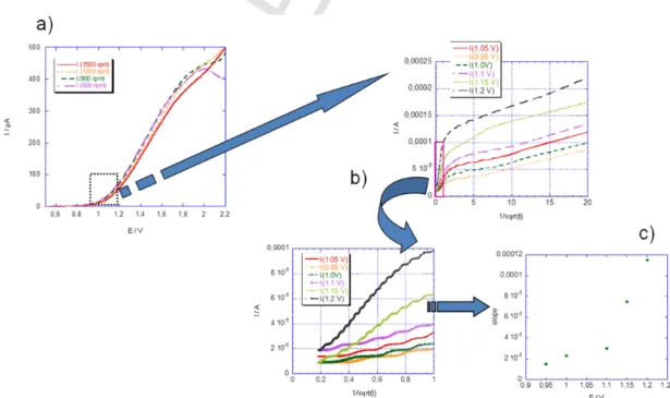
Fig. 8. CVs in DCM of free complex 1 (solid line), complex 1 grafted on Au-NPs (dottedline) and complex 1 grafted on Ag-NP (dashed line).
processes (Fig. 9a): the limiting current does not follow the expected linear dependence with the square root of the rotation rate. The reason for this passivation phenomenon is not fully elucidated but it might be related to the high concentration of NPs required to maintain the con-centration in ruthenium complexes to sufficiently high values to be de-tected, and to an aggregation process induced by the charge increase on the complex (Scheme 4). When the nanoparticles are adsorbed on the electrode surface, the electroactive species become less accessible, lead-ing to successive scans showlead-ing no more signal until the electrode sur-face is cleaned by polishing.
The forward peak current (Ip) variation with the scan rate shows a
square root dependence characteristic of diffusion limited process both for free complex or nanoparticle functionalized ones but current values do not scale with the diffusion coefficient estimated from Stokes-Ein-stein equation. As already observed an exalted current due to charge storage by the nanoparticles is recorded [34]. Indeed, based on Ran-dles-Sevcik equation [60], one obtains for the ratio of the apparent dif-fusion coefficients:
where α is the slope of the Ipvs. v1/2straight line, C is the bulk
concen-tration in electroactive species, and ‘NP’ and ‘free’ indexes correspond to the complex on nanoparticle and in solution, respectively. From ex-perimental data recorded with the same concentration in free and bound ruthenium complexes, one obtains a ratio of nearly 400, which is by far larger than what can be predicted from the relative sizes of NP and free complex. The reason for this extra-current is double layer charging of the nanoparticle [61], but modification of the electroactive area involv-ing physisorption of nanoparticles is likely to be also involved. To inves-tigate further this phenomenon, we performed chronoamperometry at various final potentials corresponding to the foot of the current-poten-tial wave. In that potencurrent-poten-tial range, mainly metallic nanoparticle cores are concerned by the electrochemical reaction. The results (Fig. 9b) show a Cottrellian behaviour, as expected for a diffusion limited current associ-ated with species in solution, with a slope that depends on the electrode potential (Fig. 9c). Assuming that no modification of the electroactive area of the electrode occurs in that potential range (i.e. adsorption oc-curs only when the complex is fully oxidized), one can relate this slope to both the (starting) faradaic oxidation of the ruthenium centres and the double layer charging of the nanoparticle [62], according to the fol-lowing equation derived from Murray's analysis [63]:
(1)
where A is the electrode area, ANPis the nanoparticle area, NAis the
Avogadro number, θRuis the ruthenium per nanoparticle ratio, F is the
Faraday constant (f=F/RT), Eiand E are the initial and final
poten-tials, E° is the standard potential for RuII/RuIIIand C
NPdlis the double layer capacitance per unit area for the nanoparticle. In Eq. (1), the first
Fig. 9. a) CV of Au-2 in DCM on Pt RDE (2mm diameter) at scan rate of 10mVs−1. Potentials are vs. aqueous SCE. b) Cottrell plots extracted from chronoamperometry at various
poten-tials (see inset) of Au-NP 2 in DCM on a 1mm diameter gold electrode. c) Plot of the slopes of the Cottrell plots as functions of potential extracted from figure b). (For interpretation of the references to colour in this figure legend, the reader is referred to the web version of this article.)
UNCORRECTED
PROOF
term of the sum is the faradaic oxidation of ruthenium which can be ne-glected (at least for the lowest values of the investigated potentials in the chronoamperometric measurements) taking into account the stan-dard potential drawn from Fig. 9a. In that model, the curve of Fig. 9c should display the variation of the double layer charging of the nanopar-ticle with the applied potential (second term in Eq. (1)). From the
val-ues of the various parameters involved in Eq. (1) (DNP=10−6cm2s−1,
ANP=2 10−13cm2) and the measured slope, one can draw an estimation
of the value of the double layer capacitance CNPdlequal to 10μFcm−2, in
agreement with reported values for gold functionalized clusters [63]. Finally Table 4 displays the redox potentials deduced from CV for the various free and bound complexes. The oxidation potentials are all in the same range for Au-NPs and Ag-NPs and close to the value of the free complex excepted for 3 (see Table 4). In this latter case, the dif-ference can arise from the aniline group, which can be oxidized in the free complex at lower potential than the ruthenium centre but not in the nanocomposite where it is involved in the NP surface functionaliza-tion. Further investigations are in progress on the controlled deposition of these functionalized nanoparticles on metallic surfaces to highlight some contributions from the linker or the nanoparticle size on the elec-trochemical features of the attached redox moiety.
3. Conclusion
A series of three ruthenium (II) complexes with different pendant groups, namely pyridine, aniline and carboxydithioic acid, have been reacted with preformed Au-NPs and Ag-NPs in water and in dichloromethane. Their reactivity and properties have been compared
to the ones of the model [Ru(bpy)3]2+complex which does not own
any anchoring group. We showed that water was not an appropriate solvent to obtain stable colloidal solutions. We also showed that the three polypyridyl ruthenium complexes possessing anchoring groups can drastically modify the size as well as the morphology of preformed Ag-NPs. Comparatively, alterations of preformed Au-NPs are less impor-tant using the same complexes, due to higher oxidation potentials. The strength of the interaction between the anchoring group and the sur-face of NPs influenced the size, shape and stability of the final nanocom-posites. Polar solvent like water induced aggregation and lead to un-stable nanocomposites. Stationary and time resolved luminescence of grafted nanocomposites (1, 2 and 3) showed that the luminescence of complexes were completely quenched (lifetime and emission quan-tum yield) in water by electron transfer processes. Current enhance-ment due to charge storage by the metal NPs and passivation phenom-ena of the electrode surface have also been characterized. The cur-rent enhancement can be associated with double layer charging of the nanoparticle cores. An interesting result is that while the NPs lumi-nescence quantum yield is the lowest for pyridine and aniline anchor Table 4
Oxidation potentials deduced from CV experiments. Potentials refer to Ag+/Ag reference.
Compound Ep,ox/V (scan rate/mVs−1)a
1 1.05 (100) 2 0.97 (20) 3 0.76 (20) 4 0.96 [64] Au°-1 1.10 (100) Ag°-1 0.99 (100) Au°-2 0.98 (20) Ag°-2 0.94 (20) Ag°-3 0.93 (20) Au°-4 0.90 (20) Ag°-4 0.92 (20)
a All potentials are measured in acetonitrile+TBAPF60.1M dried over molecular
sieves.
ing groups, the mean lifetime is slightly longer. A slower back electron transfer (longer distance between the NPs and the ruthenium centres) can be involved for the long component. Moreover, it should be em-phasized here that no long component from free complexes were found (few hundreds of nanoseconds) and therefore it confirms that the com-plexes are attached (comcom-plexes 1, 2 and 3) or in the vicinity of the NPs (complex 4). Interpretation of the optical properties' measurements may be also done from a broader perspective. In particular, the widths and the spectral positions of the observed plasmon resonances in ab-sorbance may be connected with the shape of the nanoparticles, the size of their aggregates and the permittivity of the solvents and ligands. Similarly, fluorescence quenching and enhancement may have electro-dynamics nature rather than being solely connected with charge trans-fer processes. To get a better knowledge of the nature of the quenching processes and to be able to quantify them, ultrafast spectroscopy stud-ies are needed (femtosecond and picosecond time resolved experiments) and are under progress.
4. Experimental section
4.1. Chemicals
All reagents and solvents were purchased from Aldrich, Alfa Ae-sar or SDS and used as received without further purification. All aque-ous solutions were prepared using ultrapure water purified with a
Millipore-Q+system. 1,10-phenanthroline-5,6-dione (phendione) [65],
[(bpy)2Ru(phendione)]Cl2 (7) [66], [(bpy)2RuCl2] (8) [67],
[Ru(bpy)3](X)2 (X=Cl− (4.Cl2) or PF6− (4.(PF6)2)) [53],
[(bpy)2Ru(4,4′-bis-(chloromethyl)-2,2′-bipyridine)]Cl2(5.Cl2) [39], and
[(bpy)2Ru((4-aminophenyl)imidazo[4,5-f][1,10]phenanthroline)](PF6)2
(3.(PF6)2) [68] were synthesized following procedures previously
re-ported in the literature, without modification and with similar yields.
4.2. Instrumentation and methods
ESI-MS measurements were carried out with an API 3000 (ESI/MS/ MS) PE-SCIEX triple quadrupole mass spectrometer and a HP 5989B sin-gle quadrupole mass spectrometer equipped with an electrospray source from Analytica of Branford. Both instruments were operated in the pos-itive ion mode. For the API 3000 (ESI/MS/MS) PE SCIEX triple quadru-pole mass spectrometer, the experiments were performed either by
di-rect infusion with a syringe pump with a flow rate of 10μl·min−1 or
by flow injection acquisition with a flow rate of 200μl·min−1.
Stan-dard experimental conditions were the followings: sample
concentra-tion: 10−4to 10−5M, nebulising gas N
2: 7units flow rate on a range of
10, ion spray voltage: −5.00kV, temperature: 200–400°C, declustering potential: −20V, focusing potential: −200V, entrance potential: 10V. UV/Vis absorption spectra and luminescence spectra were recorded on a Varian CARY 5000 and Fluoromax 3 spectrophotometer. Lumines-cence decay curves, in the nanosecond domain, were obtained with a time-correlated single- photon-counting method using a titanium-sap-phire laser pumped by an argon ion laser (Tsunami, by Spectra-Physics, 82MHz, 1ps pulse width, repetition rate lowered to 4MHz thanks to a pulse-peaker, a doubling crystals was used to reach 495nm excitation) [69]. Luminescence decays in the microsecond range were recorded us-ing a laser flash photolysis apparatus [70]. Excitation pulses (460nm, fwhm 4ns, 1mJ, 0.5Hz) were provided by a 10-Hz Nd:YAG laser (Con-tinuum Surelite II) coupled to an OPO (Con(Con-tinuum Panther EX OPO) and SH05 shutter (Thorlabs). The emitted light was collected at 90°, dispersed by a monochromator (Horiba Jobin-Yvon, iHR320) and an-alyzed with a photomultiplier (R1477-06, Hamamatsu) coupled to a digital oscilloscope (LeCroy 454, 500MHz). Synchronization of excita-tion pulses and acquisiexcita-tion time was secured with PCI-6602 8 Channel counter/timer (National Instruments). The experiment was controlled
UNCORRECTED
PROOF
by the home made software written in LabView environment. The recorded traces were averaged for several pulses and repeated for dif-ferent wavelengths. The deconvolutions of the individual decays with experimentally measured instrument response function (IRF) lead to 4ns time resolution. Single wavelengths as well as global analyses of
the transient absorption data were performed using Igor Pro 6.20.1H
NMR spectra were recorded at room temperature in 5mm o.d. tubes on a Bruker Avance 300 spectrometer equipped with a QNP probe head. Elemental analyses were performed by the “Service Central d'-Analyse du CNRS”, Vernaison (France). Electrochemical measurements were performed in a three electrode cell equipped with a 1mm di-ameter platinum disk as the working electrode, platinum wire as the
counter electrode and Ag+(0.01M)/Ag as the reference electrode (SCE
for RDE experiments). The reference potential of Ag+/Ag was checked
vs. ferrocene as recommended by IUPAC (E°(Fc)=+86mV) [71]. The supporting electrolyte was tetrabutylammonium hexafluorophosphate (Fluka, puriss) and the solutions were deaerated by argon bubbling prior to each experiment. Cyclic voltammograms were recorded with a 600 CH Instruments potentiostat connected to a PC. The size of the NPs was obtained using transmission electron microscope (TEM) images treat-ment and dynamic light scattering (DLS). TEM images were obtained with a JEOL JEM 100 CXII transmission electron microscope at an ac-celerating voltage of 100kV, a Zeiss EM902 at 70kV and for high res-olution measurements with a JEOL JEM 2010 at 200kV. DLS measure-ments were performed with a MALVERN Zetasizer nanoZS.
4.3. [(bpy)2Ru(4,4′-(carboxydithioic acid)-2,2′-bipyridine)](PF6)2(1.
(PF6)2)
Elemental sulphur (69mg, 2.15mmol) and of sodium methanolate (116mg, 2.15mmol) were placed in anhydrous methanol (50mL) under argon atmosphere and refluxed until elemental sulphur was completely
dissolved (ca. 2h). 5.(PF6)2(512mg, 0.54mmol) was then added and
the reaction mixture was refluxed overnight. The solvent was then evap-orated and the red solid obtained was dissolved in water. Acidification with diluted HCl led to the precipitation of the complex, which was fil-tered and washed several times with water. The product was dissolved in a minimum amount of acetonitrile. Addition of a saturated aqueous
solution of NH4PF6led to the precipitation of 1.(PF6)2(465mg, 86%) as
a dark orange solid, which was collected by filtration, washed with H2O
and dried under vacuum.1H NMR (300MHz, CD3CN): δ=8.49 (m, 6H),
8.05 (m, 6H), 7.72 (m, 4H), 7.39ppm (m, 6H); elemental analysis calcd (%) for C36H35F12N7O2P2RuS4(1.(PF6)2.(CH3OH)2.(CH3CN)): C 38.7, H 3.2, N 8.7, S 11.5; found: C 38.5, H 2.8, N 8.4, S 11.3.
4.4. [(bpy)2
Ru((4-(4-pyridinyl)phenyl)imidazo[4,5-f][1,10]phenanthroline)](PF6)2(2.(PF6)2)
7 (0.115g, 0.16mmol), 4-(4-pyridinyl)benzaldehyde (0.040g,
0.2mmol) and NH4OAc (0.3g, 4mmol) were stirred at reflux overnight
in glacial acetic acid (30mL). After cooling, the dark red solution was concentrated under reduced pressure to ca. 5mL and the crude prod-uct was precipitated using a concentrated aqueous solution of am-monium hexafluorophosphate. The product was purified by dissolu-tion in acetonitrile and precipitadissolu-tion by slow addidissolu-tion of diethyl ether.
2.(PF6)2was filtered, washed with ethanol (2×10mL), diethyl ether
(2×10mL) and dried under vacuum (0.078g, 78%). 1H NMR
(DMSO‑d6): δ=13.5 (s, 1H), 8.76 (d, 2H), 8.54 (m, 6H), 8.27 (d, 2H),
8.15 (m, 2H), 8.12 (m, 2H), 8.05 (m, 4H), 7.93 (m, 2H), 7.85 (m, 2H), 7.78 (m, 2H), 7.61 (m, 2H), 7.46 (dd, 2H), 7.22ppm (dd, 2H); ESI-MS:
m/z (positive mode): 393.7 ([M]2+/2) elemental analysis calcd (%) for
C44H31N9RuP2F12(2.(PF6)2): C 49.03, H 3.09, N 11.71, Ru 9.39, P 5.75, F 21.17; found: C 49.26, H 2.97, N 11.76, Ru 9.23, P 5.83, F 21.32.
4.5. Synthesis of 1.Cl2, 2.Cl2and 3.Cl2
Hexafluorophosphate salts of 1, 2 and 3 were converted to the corre-sponding chloride salts by dissolution of the hexafluorophosphate salts in acetone and addition of an excess of tetrabutylammonium chloride [72].
Preparation of the starting hydrosols of Au-NPs and Ag-NPs:
Au-NPs in water: Aqueous solutions of HAuCl4.3H2O (1mL, 0.025M) and sodium citrate (1mL, 0.025M) were poured into 98mL of water. Then, a
cooled aqueous solution of NaBH4(1.3mL, 0.1M) was added dropwise.
During the addition, the colour of the mixture turned to red-wine. The mean diameter of Au-NPs determined from TEM images was 2.5nm.
Ag-NPs in water: Aqueous solutions of AgNO3(2.36mL, 0.01M) and sodium citrate (1mL, 0.025M) were poured into 250mL of water. Then,
a cooled aqueous solution of NaBH4(4.2mL, 0.1M) was added
drop-wise, followed by another addition of an aqueous solution of NaBH4
(0.3mL, 0.1M) ten minutes later. During the addition, the colour of the mixture turned yellow. The mean diameter of Ag-NPs, calculated from TEM images, was 2.5nm.
4.6. Post-functionalization of Au-NPs and Ag-NPs hydrosols using the chloride salts of ruthenium complexes 1, 2, 3 and 4
As already mentioned in the article, the amount of ruthenium com-plexes added to post-functionalize the metal NPs was about 20% of the theoretical amount needed for the maximal covering. This low concen-tration of ruthenium complexes was chosen to limit the communication between them once attached to the surface of the NPs. The surface of
the Au-NPs and Ag-NPs was 19.63nm2, considering the mean diameter
of 2.5nm determined using TEM images. The footprint area of the
ruthe-nium complexes was determined to be ~1nm2. As a result, a very simple
calculation led to a maximum number of ruthenium complexes per par-ticle of 19. The post-functionalization was realized using either 10mL of the previous hydrosol of Au-NPs or 26.5mL of the previous hydrosol of Ag-NPs that were further diluted with water to obtain a final volume of 60mL. Both solutions contained the same amount of metal NPs (5.26
10−9mol using a tight-packed spherical model [73]). Then aqueous
so-lutions of 1.Cl2(70μL, 2.65 10−4M), 2.Cl2(70μL, 2.32 10−4M), 3.Cl2
(70μL, 2.51 10−4M) or 4.Cl
2(70μL, 2.9 10−4M) were added to each
hy-drosol and the resulting solutions were stirred for 48h at 25°C before analysis.
Preparation of the starting DCM solutions Au- and Ag-NPs:
Au-NPs in DCM: HAuCl4.3H2O (0.010g, 2.5 10−5mol) and
1-hexadecy-lamine (0.200g, 8.3 10−4mol) were dissolved in DCM (10mL). The
fi-nal volume of the solution was adjusted to 60mL using DCM. A cooled
aqueous solution of NaBH4(400μL, 1M) was then added and the
result-ing mixture was stirred for 1h. The water phase was removed usresult-ing a micropipette. The mean diameter of Au-NPs determined from TEM im-ages was ~3nm.
Ag-NPs in DCM: AgNO3(0.004g, 2.5 10−5mol) and 1-hexadecylamine
(0.075g, 3.1 10−4mol) were dissolved in DCM (5mL). Oleic acid (10μL)
was then added and the volume of the solution was adjusted to 60mL
using DCM. A cooled aqueous solution of NaBH4(100μL, 1M) was then
added and the resulting mixture was stirred for 1h. The water phase was removed using a micropipette. The mean diameter of Ag-NPs deter-mined from TEM images was deterdeter-mined to be ~3nm.
UNCORRECTED
PROOF
4.7. Post-functionalization of Au-NPs and Ag-NPs in DCM using the hexafluorophosphate salts of ruthenium complexes 1, 2, 3 and 4
The same approach was used in DCM and in water with the aim of adding an amount of ruthenium complexes of about 20% of the theoretical amount needed for the maximum covering. DCM solutions of 1.(PF6)2(70μL, 2.32 10−4M), 2.(PF6)2(70μL, 1.9 10−4M), 3.(PF6)2
(70μL, 2.0 10−4M) and 4.(PF
6)2(70μL, 2.2 10−4M) were added to 6mL
of the previous solution of Au-NPs or Ag-NPs in DCM. The different mix-tures were stirred during 48h at 25°C before analysis.
Acknowledgements
We thank Christine Longin and Sophie Chat for TEM analyses, and Julien Dubois for help in the nanosecond lifetime experiments. This work was supported by the PRES UniverSud Paris and the ANR “Jeunes Chercheuses et Jeunes Chercheurs” in the frame of AURUS program. Appendix A. Supplementary data
Supplementary data to this article can be found online at https:// doi.org/10.1016/j.apsusc.2019.143847.
References
[1] a) U. Schatzschneider, Eur. J. Inorg. Chem. 10 (2010) 1451; b) M.J. Rose, P.K. Mascharak, Coord. Chem. Rev. 252 (2008) 2093. [2] J.F. Endicott, Y.-J. Chen, Coord. Chem. Rev. 251 (2007) 328.
[3] a) L. Spiccia, G.B. Deacon, C.M. Kepert, Coor. Chem. Rev. 248 (2004) 1329; b) Q. Sun, S. Mosquera-Vazquez, Y. Suffren, J. Hankache, N. Amstutz, L.M. Lawson Daku, E. Vauthey, A. Hauser, Coord. Chem. Rev. 282 (2015) 87.
[4] a) S. Bonnet, J.-P. Collin, Chem. Soc. Rev. 8 (2008) 1490;
b) E. Baranoff, J.P. Collin, L. Flamigni, J.-P. Sauvage, Chem. Soc. Rev. 33 (2004) 147–155;
c) P.P. Laine, S. Campagna, F. Loiseau, Coordination Chemistry Reviews 252 (2008) 2552–2571.
[5] W.K. Chan, Coord. Chem. Rev. 251 (2007) 2104. [6] O. Maury, H. Le Bozec, Acc. Chem. Soc. Res. 38 (2005) 691.
[7] L. Moriggi, A. Aebischer, C. Cannizzo, A. Sour, A. Borel, J.-C. G, L. Helm Bünzli, Dalton Trans. (12) (2009) 2088.
[8] S. Zanarini, E. Rampazzo, L. Della Ciana, M. Marcaccio, E. Marzocchi, M. Montalti, F. Paolucci, L. Prodi, J. Am. Chem. Soc. 131 (2009) 2260.
[9] J.K. Hurst, J.L. Cape, A.E. Clark, S. Das, C.Y. Qin, Inorg. Chem. 47 (2008) 1753. [10] M.K. Nazeeruddin, S.M. Zakeeruddin, J.J. Lagref, P. Liska, P. Comte, C. Barolo, G.
Viscardi, K. Schenk, M. Grätzel, Coord. Chem. Rev. 248 (2004) 1317. [11] M.J. Li, Z.F. Chen, V.W.W. Yan, Y.B. Zu, ACS Nano 2 (2008) 905.
[12] R. Kawano, M.K. Nazeerudin, A. Sato, M. Grätzel, M. Watanabe, Electrochem. Commun. 9 (2007) 1134.
[13] a) C.G. Cameron, P.G. Pickup, J. Am. Chem. Soc. 121 (1999), 11773; b) M. Abe, H. Futagawa, T. Ono, T. Yamada, N. Kimizuka, Y. Hisaeda, Inorg. Chem. 54 (2015), 11061.
[14] a) T. Le Bouder, O. Maury, A. Bondon, K. Costuas, E. Amouyal, I. Ledoux, J. Zyss, H. Le Bozec, J. Am. Chem. Soc. 125 (2003), 12284;
b) F. Puntoriero, F. Nastasi, M. Cavazzini, S. Quici, S. Campagna, Coord. Chem. Rev. 251 (2007) 536;
c) J.-L. Wang, X. Li, C.D. Shreiner, X. Lu, C.N. Moorefield, S.R. Tummalapalli, D.A. Medvetz, M.J. Panzner, F.R. Fronczek, C. Wesdemiotis, G.R. Newkome, New J. Chem. 36 (2012) 484.
[15] a) A. Guerrero-Martinez, Y. Vida, D. Dominguez-Gutierrez, R.Q. Albuquerque, L. De Cola, Inorg. Chem. 47 (2008) 9131;
b) R. Sangiliapillai, R. Arumugam, R. Eswaran, R. Seenivasan, Luminescence 31 (2016) 30–37.
[16] a) X.D. Wu, Y. Cong, Y.H. Liu, J. Ying, B. Li, J. Sol-Gel Sci. Technol. 49 (2009) 355;
b) P. Innocenzi, H. Kozuka, T. Yoko, J. Phys. Chem. B 101 (1997) 2285. [17] a) D.L. Ma, A.J. Kell, S. Tan, Z.J. Jakubek, B. Simard, J. Phys. Chem. C 113 (2009),
15974;
b) M. Frasconi, Z. Liu, J. Lei, Y. Wu, E. Strekalova, D. Malin, M.W. Ambrogio, X. Chen, Y.Y. Botros, V.L. Cryns, J.-P. Sauvage, J.F. Stoddart, J. Am. Chem. Soc. 135 (2013) 11603–11613.
[18] a) S. Verma, P. Kar, A. Das, D.K. Palit, N.H. Ghosh, Chem. Eur. J. 16 (2010) 611; b) N.-N. Zhang, F. Bigdeli, Q. Miao, M.-L. Hu, A. Morsali Journal of Organometallic Chemistry 878 (19) (2018) 11–18.
[19] See for example: a) J. Zhao, J. A. Dieringer, X. Y. Zhang, G. C. Schatz, R. P. Van Duyne J. Phys. Chem. B. 2008, 112, 19302.b G. Lemercier, M. Four, S. Chevreux, Coord. Chem. Rev. 368 (2018) 1.
[20] M.C. Daniel, D. Astruc, Chem. Rev. 104 (2004) 293.
[21] M. Brust, M. Walker, D. Bethell, D.J. Schiffrin, R. Whyman, Chem. Commun. (1994) 801.
[22] J.L. Gong, C.B. Mullins, Acc. Chem. Res. 42 (2009) 1063.
[23] a) J.H. Lee, M.A. Mahmoud, V. Sitterle, J. Sitterle, J.C. Meredith, J. Am. Chem. Soc. 131 (2009) 5048;
b) A.M. Schwartzberg, J.Z. Zhang, J. Phys. Chem. B 112 (2008), 10323. [24] a) A. Franca, B. Pelaz, M. Moros, C. Sanchez-Espinel, A. Hernandez, C.
Fernan-dez-Lopez, V. Grazu, J.M. de la Fuente, I. Pastoriza-Santos, L.M. Liz-Marzan, A. Gonzalez-Fernandez, Small 131 (2009) 5048;
b) P.K. Jain, X. Huang, I.H. El-Sayed, M.A. El-Sayed, Acc. Chem. Res. 41 (2008) 1578.
[25] a) A.N. Shipway, M. Lahav, R. Blonder, I. Willner, Chem. Mater. 11 (1999) 13; b) M. Lahav, V. Heleg-Shabtai, J. Wasserman, E. Katz, I. Willner, H. Dürr, Y.-Z. Hu, S.H. Bossmann, J. Am. Chem. Soc. 122 (2000), 11480;
c) T.B. Norsten, B.L. Frankamp, V.M. Rotello, Nano Lett. 2 (2002) 1345; d) X.-H.N. Xu, S. Huang, W. Brownlow, K. Salaita, R.B. Jeffers, J. Phys. Chem. B 108 (2004) 15543;
e) P.P.H. Cheng, D. Silvester, G. Wang, G. Kalyuzhny, A. Douglas, R.W. Murray, J. Phys. Chem. B 110 (2006) 4637.
[26] a) J. F. Hicks, D. T. Miles, R. W. Murray, J. Am. Chem. Soc. 2002, 124, 13322;b) R.L. Wolfe, R. Balasubramanian, J.B. Tracy, R.W. Murray, Langmuir 23 (2007) 2247.
[27] a) B. I. Ipe, K. Yoosaf, K. G. Thomas, J. Am. Chem. Soc. 2006, 128, 1907;b) T. Huang, R.W. Murray, Langmuir 18 (2002) 7077.
[28] a) W.S. Baker, B.I. Lemon, R.M. Crooks, J. Phys. Chem. B 105 (2001) 8885; b) J.D.E.T. Wilton-Ely, Dalton Trans. 25 (2008).
[29] W.R. Glomm, S.J. Moses, M.K. Brennaman, J.M. Papanikolas, S. Franzen, J. Phys. Chem. B 109 (2005) 804.
[30] a) M. Ito, T. Tsukatani, H. Fujihara, J. Mater. Chem. 15 (2005) 960; b) N. Vilvamani, M. Chhatwal, I. Bhowmick, R. Devi Gupta, S.K. Awasthi, RSC Adv. 6 (2016), 55507.
[31] C. Querner, P. Reiss, J. Bleuse, A. Pron, J. Am. Chem. Soc. 126 (2004), 11574. [32] C. Pérez Leon, L. Kador, B. Peng, M. Thelakkat, J. Phys. Chem. B 109 (2005) 5783. [33] a) C.R. Mayer, E. Dumas, F. Sécheresse, Chem. Commun. (2005) 345;
b) C.R. Mayer, E. Dumas, A. Michel, F. Sécheresse, Chem. Commun. 40 (2006) 4183;
c) C.R. Mayer, E. Dumas, F. Sécheresse, J. Coll. Interf. Sci. 328 (2008) 452. [34] C.R. Mayer, E. Dumas, F. Miomandre, R. Méallet-Renault, F. Warmont, J.
Vi-gneron, R. Pansu, A. Etcheberry, F. Sécheresse, New J. Chem. 30 (2006) 1628. [35] N. Nerambourg, M.H.V. Werts, M. Charlot, M. Blanchard-Desce, Langmuir
23 (2007) 5563.
[36] C. Querner, A. Benedetto, R. Demadrille, P. Rannou, P. Reiss, Chem. Mater. 18 (2006) 4817.
[37] T.-H. Park, M.J. Therien, Org. Lett. 9 (2007) 2779.
[38] M.S. Vickers, J. Cookson, P.D. Beer, P.T. Bishop, B. Thiebaut, J. Mater. Chem. 16 (2006) 209.
[39] J.E. Collins, J.J.S. Lamba, J.C. Love, J.E. McAlvin, C. Ng, B.P. Peters, X. Wu, C.L. Fraser, Inorg. Chem. 38 (1999) 2020.
[40] S.R. Ramadas, P.S. Srinivasan, J. Ramachandran, V.V.S.K. Sastry, Synthesis (1983) 605.
[41] S. Rucareanu, V.J. Gandubert, R.B. Lennox, Chem. Mater. 18 (2006) 4674. [42] J.D.S. Newman, G.J. Blanchard, Langmuir 22 (2006) 5882.
[43] P. Pramod, P.K. Sudeep, K.G. Thomas, P.V. Kamat, J. Phys. Chem. B 110 (2006), 20737.
[44] I. Ojea-Jiménez, V. Puntes, J. Am. Chem. Soc. 131 (2009), 13320. [45] a) L.L. Rouhana, J.A. Jaber, J.B. Schlenoff, Langmuir 23 (2007), 12799;
b) S. Tan, M. Erol, S. Sukhishvili, H. Du, Langmuir 24 (2008) 4765. [46] M. Jebb, P.K. Sudeep, P. Pramod, K.G. Thomas, P.V. Kamat, J. Phys. Chem. B
111 (2007) 6839.
[47] a) A. Kumar, S. Mandal, P.R. Selvakannan, R. Pasricha, A.B. Mandale, M. Sastry, Langmuir 19 (2003) 6277;
b) M. Yamamoto, Y. Kashiwagi, M. Nakamoto, Langmuir 22 (2006) 8581. [48] a) H. Chao, R.-H. Li, C.-W. Jiang, H. Li, L.-N. Ji, X.-Y. Ji, Dalton Trans.
2001 (1920);
b) M. Mariappan, B.G. Maiya, Eur. J. Inorg. Chem. (2005) 2164.
[49] See for example: L.-F. Tan, F. Wang, H. Chao, Hel. Chim. Acta 2007, 90, 205 and references therein.
[50] K. Nakamaru, Bull. Chem. Soc. Jpn. 55 (1982) 2697.
[51] a) K.-C. Lee, S.-J. Lin, C.-H. Lin, C.-S. Tsai, Y.-J. Lu, Surf. Coat. Techn. 202 (2008) 5339;
b) W. Wang, X. Chen, S. Efrima, J. Phys. Chem. B 103 (1999) 7238; c) E. Oh, K. Susumu, R. Goswami, H. Mattoussi, Langmuir ASAP (2010). [52] J.V. Caspar, T.J. Meyer, J. Am. Chem. Soc. 105 (1983) 5583.
[53] B. Durham, J.V. Caspar, J.K. Nagle, T.J. Meyer, J. Am. Chem. Soc. 104 (1982) 4803.
[54] a) T.J. Meyer, Pure Appl. Chem. 58 (1986) 1193;
b) A. Masschelein, L. Jacquet, A. Kirsch-De Mesmaeker, J. Nasielski, Inorg. Chem. 29 (1990) 855.
[55] H. Ishida, S. Tobita, Y. Hasegawa, R. Katoh, K. Nozaki, Coord. Chem. Rev. 254 (2010) 2449.