HAL Id: hal-02360573
https://hal.archives-ouvertes.fr/hal-02360573
Submitted on 16 Nov 2020
HAL is a multi-disciplinary open access
archive for the deposit and dissemination of
sci-entific research documents, whether they are
pub-lished or not. The documents may come from
teaching and research institutions in France or
abroad, or from public or private research centers.
L’archive ouverte pluridisciplinaire HAL, est
destinée au dépôt et à la diffusion de documents
scientifiques de niveau recherche, publiés ou non,
émanant des établissements d’enseignement et de
recherche français ou étrangers, des laboratoires
publics ou privés.
Heptylmannose-functionalized cellulose for the binding
and specific detection of pathogenic E. coli
Madeleine Cauwel, Adeline Sivignon, Clarisse Bridot, Medy Nongbe, David
Deniaud, Benoît Roubinet, Ludovic Landemarre, Francois-Xavier Felpin, Julie
Bouckaert, Nicolas Barnich, et al.
To cite this version:
Madeleine Cauwel, Adeline Sivignon, Clarisse Bridot, Medy Nongbe, David Deniaud, et al..
Heptylmannose-functionalized cellulose for the binding and specific detection of pathogenic E.
coli.
Chemical Communications, Royal Society of Chemistry, 2019, 55 (68), pp.10158-10161.
�10.1039/c9cc05545b�. �hal-02360573�
Heptylmannose‐functionalized cellulose for the binding and
specific detection of pathogenic E.coli.
Madeleine Cauwel,
aAdeline Sivignon,
bClarisse Bridot,
cMedy C. Nongbe,
aDavid Deniaud,
aBenoit
Roubinet,
dLudovic Landemarre,
dFrançois‐Xavier Felpin,
aJulie Bouckaert,
cNicolas Barnich
b,
Sébastien G. Gouin.*
aWe developed a chemical method to covalently functionalize cellulose nanofibers and cellulose paper with mannoside ligands displaying a strong affinity for the FimH adhesin from pathogenic E. coli strains. Mannose‐grafted cellulose proved efficient to selectively bind FimH lectin and discriminate pathogenic E. coli strains from non‐pathogenic ones. These modified papers are valuable tools for diagnosing infections promoted by E.coli, such as cystitis or inflammatory bowel diseases, and the concept may be applicable to other life‐ threatening pathogens.
The worldwide spread of antibiotic resistances raises serious health problems, and has driven the identification of new virulence factors and development of alternative antibacterial therapeutics. Mannose‐binding FimH adhesin, expressed by Escherichia coli strains has been extensively studied as a target for disrupting bacterial attachment to the host cells.1 Impressive results were obtained in the context
of urinary tract infections (UTI), a prevalent infection type generally mediated by the attachment of uropathogenic E.
coli strains (UPEC) to the highly mannosylated uroplakin
transmembrane protein of urothelial lining cells. FimH antagonists orally administered to in vivo UTI mouse models, were shown to decrease the E.coli load in the bladder by several orders of magnitude,2–4 making them competitive
with conventional antibiotic treatment.5,6 This concept was
more recently extended to Crohn’s disease (CD), an inflammatory disorder of the intestine where an altered gut microbiota, particularly the presence of adherent‐invasive E.
coli strains (AIEC), is suspected to play a key role in the
pathogenesis.7 Synthetic derivatives of heptylmannoside
(HMan), a nanomolar FimH antagonist,8 were shown to
lower the AIEC bacterial level, signs of colitis and gut inflammation when administered per os (10 mg/kg) in CD mouse models.9,10
Sensitive and rapid diagnostic systems are essential to evaluate the presence of E. coli expressing FimH adhesin in gut microenvironments in order to properly stratify patients before treatment. While the high prevalence of UPEC in the normally sterile urinary tract environment facilitates diagnosis, the AIEC niche is more complex, located at the ileal mucosa in 21 to 63% of CD patients,11,12 within an
ecological community of hundreds of symbiotic
microorganisms. Furthermore, no specific biomarkers are currently effective at distinguishing AIEC from other commensal E. coli of the gut microbiota. Previously, it has been shown that AIEC pathobiont possesses specific allelic variants in the fimH gene, conferring them a high ability to
adhere in vitro and to colonize the gut of CEABAC10 mice.13
Establishing an approach to discriminate the strong mannose‐binders from other bacteria would therefore be of tremendous importance for efficient diagnosis.
In this work, we developed a heterogeneous support to specifically trap and accumulate pathogenic E.coli from biological fluids. Heptyl‐α‐D‐ mannoside (HMan) was grafted by click chemistry techniques onto cellulose nanofibers (CN) and cellulose paper (CP). HMan was previously identified as a potent binder of the isolated FimH lectin domain.8 It should
be noted that the lectin domain represents the high‐affinity state of FimH under mechanical force and that full‐length FimH display a lower affinity for mannosides.14,15 Covalently
functionalized CN or CP were characterized by Fourier transform spectroscopy (FTIR), elemental analysis, X‐ray photo‐electron spectrometry (XPS), and scanning electron microscopy (SEM). HMan‐CN was first compared in vitro against CN grafted with lower FimH affinity ligands i.e. Man‐ CN lacking the hydrophobic heptyl chain, and HGlc‐CN an analog with a glucose sugar that is not recognized by FimH lectin (Scheme 1). In addition, we switched the anomeric oxygen atom to a sulfur and synthesized HSMan‐CN to prevent potential sugar hydrolysis from the surface by mannosidases. The modified CN were first assessed for their faculty to bind FimH and to prevent AIEC adhesion to intestinal cells. HMan‐CN was then orally administered to the CEABAC10 mouse model of CD to assess its capacity to decrease AIEC in the feces of AIEC‐infected mice and to act as a potential CD therapeutic. HM was next coated on CP and the capacity of the HMan‐CP to selectively catch AIEC in solution was analyzed. Scheme 1. Chemical synthesis of the sugar‐coated cellulose fibers.
Cellulose fibers are inexpensive and biocompatible materials composed of β (1‐4) linked D‐glucose units. Synthetic methods relying on the functionalization of cellulose fibers16,17 have been reported for various applications in
environmental sciences,18 sensing,19 catalysis20 and
medicine.21 Having previously shown that HMan derivatives
terminated with triazol moieties could still efficiently bind FimH,22,23 the HMan ligands were grafted to CN or CP by the
robust copper‐catalyzed azide alkyne cycloaddition (CuAAc). Azidation of the cellulose materials was done using a recently reported tosylation‐azidation sequence using pyridine/tosyl chloride and sodium azide.24 The reaction
conditions were optimized to achieve degrees of substitution (DS) with azide of around 30 and 40% for CN and CP, respectively. DS were determined by elemental analysis and the reactions could be followed by FT‐IR, showing appearance of characteristic bands during the first and second step as(SO2) = 1363 cm‐1, s (SO2) = 1177cm‐1 and
as (N3) = 2108 cm‐1.Extensive washing under ultrasound was
required to remove the excess sodium azide (as (N3) = 2034
cm‐1) trapped in the CN fibers. Mannosides were armed with
the corresponding alkynyl chain to form 1,25 2,22 3 and 4
(synthesis in Figure S1).
After N3‐CN lyophilization, the support was engaged in
CuAAC with an excess of 1‐4, using copper sulfate and sodium ascorbate as catalyst. A successful disappearance of the as (N3) band was observed by FT‐IR after CuAAc,
indicating complete conversion (Figure S4a). This was further demonstrated by XPS analysis, providing information on the surface composition. Deconvolution of the high‐resolution spectrum of the N1s region showed disappearance of the peak at 404.2 ev, which was assigned to the electropositive atom (Figure S4b). For HSMan‐CN, the sulfur atom could be tracked by XPS with the appearance of the spin‐orbit components S2p1/2 and S2p3/2 at 169.9 and 168.7 eV,
respectively (Figure S9), and by elemental analysis where the sulfur ratio of 2.57% matches a theoretical DS value between 20 and 30%. As shown by the SEM images (Figure S4c), the structure of the cellulosic material was conserved after the critical CuAAc step.
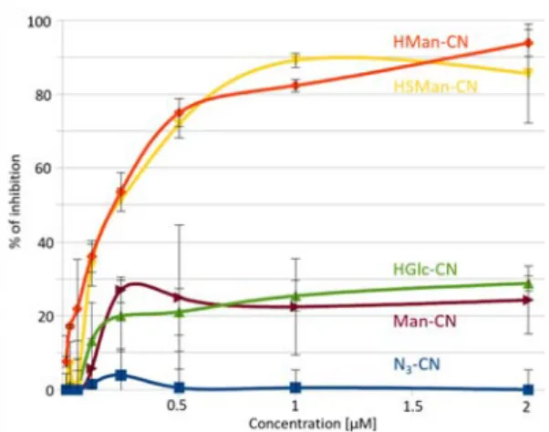
The functionalized CN were first assessed using a system adapted from a “lectin glycoprofiling technologies (LectPROFILE kit)”. In brief, FimH lectin domain (amino acids 1‐158) was coated onto a microplate surface and different concentrations of the functionalized CN (expressed in µmol of the coated sugars) were challenged with labelled BSA coated with mannopyrannosyl residues. In the concentration range tested (0‐2µM), the uncoated N3‐CN was unable to
disrupt the Man‐BSA – FimH interaction (Figure 2). The functionalized CN with low‐affinity ligands (Man‐CN and HGlc‐CN) inhibited FimH binding by less than 30% at 2 µM. In contrast, functionalized CN with the high‐affinity ligands HMan and HSMan nearly abrogated the interaction, with a virtually identical dose‐response that was fully coherent with their similar affinity for FimH lectin. HMan‐CN and HSMan‐ CN are able to efficiently bind to FimH lectin, opening the possibility to capture AIECs in solution.
Figure 2. HMan‐CN and HSMan‐CN efficiently disrupt the labelled ManBSA
interaction with FimH coated on a microplate surface.
The CN derivatives were then assessed for their ability to disrupt the adhesion of AIEC LF82, isolated from an ileal biopsy of a CD patient, to T84 intestinal epithelial cells. Briefly, N3‐CN, HMan‐CN, HSMan‐CN, HGlc‐CN and HMan
were incubated with AIEC LF82 and added to T84 cells for 3 hours at an infection multiplicity of 10 bacteria/cell. After washings, the level of AIEC remaining associated with the cells (not trapped by the CN derivatives) was quantified after cell lysis, by culturing on Luria Bertani agar plates. Levels of LF82 bacteria adhering to the cells in the presence of the CN derivatives were compared to the adhesion levels in absence of any treatment (non‐treated condition, NT). Normalized results expressed in percentages of residual adhesion according to the concentrations of mannose (or N3 for N3‐
CN) are shown in Figure 3. No anti‐adhesive effect was observed using N3‐CN, even at the highest concentration of
100µM, which demonstrates that cellulose does not interfere in the bacteria‐cell interaction process. In contrast, HMan‐CN or HSMan‐CN nearly abrogated the adhesion at 100µM, with just 3.4% and 2.8% of bacteria still adhered to the cells respectively. The dose‐dependent inhibition profile was very similar to that observed with free HMan (2.1%) meaning that no loss of inhibitory capacity is observed when the HMan or HSMan ligands are grafted on the CN. Glucosides are not recognized by FimH. HGlc‐CN was ineffective in preventing AIEC binding (Figure S6), validating the key role of FimH in the cell binding process.
Figure 3. Effects of the compounds on the adhesion ability of AIEC bacteria to T84 intestinal cells. Results are expressed as percentages of residual adhesion (n=6 experiments, mean±SEM; *p<0.05, **p<0.01, ***p<0.001). LF82 infection in the absence of treatment (non‐treated, NT) was normalized to 100%. Next, HMan‐CN was studied in vivo to evaluate its capacity to decrease AIEC bacteria in the gut microbiota. The non‐ toxic biocompatible CN is a particularly well‐suited scaffold for developing a potential CD treatment. The high molecular weight of HMan‐CN should confine the anti‐adhesive treatment to the gut, thus preventing its systemic dissemination and lowering the risk of potential side effects. The AIEC‐infected CEABAC10 murine model is a preclinical model suitable to mimic the abnormal colonization of the gut by AIEC bacteria observed in CD patients. CEABAC10 mice express the highly mannosylated CEACAM6 human protein that is overexpressed at the ileal mucosa of CD patients. Mice were first infected intragastrically with 5x10⁹ LF82
bacteria. After 2 hours, HMan‐CN was orally administered to the mice (n=9 per group) at the dose of 30 mg/kg. A second administration was realized 24 hours later. The level of AIEC in the feces was quantified at days 1 and 2 post‐infection for the treated and non‐treated (NT) mice and is presented in Figure S7. The bacterial clearance was more pronounced in the HMan‐CN group compared to the NT group, with a 724 vs 86‐fold decrease, respectively, in the bacterial load between D1 and D2 (Figure S3). These results suggest that HMan‐CN can bind AIEC in the gut microbiota and could be further evaluated as a treatment for AIEC‐induced colitis. With the capacity of HMan‐CN to selectively bind AIEC established, we focused our attention on the evaluation of potential paper sensors for AIEC detection. We investigated whether HMan‐CP could function as a paper sensor to detect
E. coli expressing high‐affinity FimH adhesin. Binding of AIEC
to HMan‐CP was first confirmed by SEM where single or bacterial aggregates were efficiently trapped by the HMan coated fibers (Figure 4). Figure 4. SEM images showing AIEC bacteria at the surface of the cellulose fibers of HMan‐CP. The AIEC reference strain LF82, the non‐piliated LF82‐ΔfimH mutant and the E. coli K12 C600 non‐pathogenic strain were then included in the following assays. Calibrated papers (28, 27 mm²), HMan‐CP and N3‐CP were incubated with bacterial
suspensions (108 bacteria/mL) with gentle shaking for one
hour. After extensive washings and cellulose fibers homogenization, bacteria were quantified by plating on LB agar plates. The percentages of recovered bacteria in the CP are presented in Figure 5. Results clearly showed that HMan‐ CP is much more effective than N3‐CP to trap the LF82
adherent strain. The bacterial binding to CP is fully promoted by FimH as HMan‐CP does not bind the non‐piliated LF82∆fimH mutant. Furthermore, HMan‐CP is able to selectively discriminate the E. coli strain displaying a high‐ binding affinity variant of FimH (LF82) from a FimH harboring by a non‐pathogenic E. coli K12. Figure 5. Percentage of bacterial strains AIEC LF82, AIEC LF82∆fimH and K12 C600 trapped into N3‐CP or HMan‐CP; n=4 to 5, *p<0.05. NT 100 10 100 10 1 0,1 100 10 1 0,1 100 10 1 0,1 0 20 40 60 80 100 120 140 160
HMan-CN HSMan-CN HMan
N3-CN ** * *** *** * (µM) R esi d u al a dh esi o n ( % )
Conclusions
In this work, we developed the first paper sensors that selectively bind AIEC pathotype‐expressing FimH usable as non‐invasive companion test for AIEC carriage. This method could be easily converted for the detection of other pathogens. Since AIEC and UPEC share amino acid substitutions in FimH, a similar detection strategy could be used in the context of UTI. More broadly, this approach could be extended by coating the CP with various sugar ligands able to interact with lectin‐type adhesin expressed by other pathogens (e.g. galactose for PA2L from Pseudomonas
aeruginosa, fucose for FleA from Aspergillus fumigatus, sialic acid for the hemagglutinin from influenza virus).