doi: 10.1136/ard.2008.099564
2010 69: 644-647 originally published online May 10, 2009
Ann Rheum Dis
P G Conaghan, M A D'Agostino, M Le Bars, et al.
from a large, 3-year, prospective EULAR study
replacement for knee osteoarthritis: results
Clinical and ultrasonographic predictors of joint
http://ard.bmj.com/content/69/4/644.full.html
Updated information and services can be found at:
These include:
References
http://ard.bmj.com/content/69/4/644.full.html#ref-list-1
This article cites 15 articles, 9 of which can be accessed free at:
service
Email alerting
box at the top right corner of the online article.
Receive free email alerts when new articles cite this article. Sign up in the
Notes
http://ard.bmj.com/cgi/reprintform
To order reprints of this article go to:
http://ard.bmj.com/subscriptions
go to:
Annals of the Rheumatic Diseases
Ann Rheum Dis 2010;69:644–647. doi:10.1136/ard.2008.099564 644
ABSTRACT
Objectives To determine clinical and ultrasonographic predictors of joint replacement surgery across Europe in primary osteoarthritis (OA) of the knee.
Methods This was a 3-year prospective study of a painful OA knee cohort (from a EULAR-sponsored, multicentre study). All subjects had clinical evaluation, radiographs and ultrasonography (US) at study entry. The rate of knee replacement surgery over the 3-year follow-up period was determined using Kaplan–Meier survival data analyses. Predictive factors for joint replacement were identifi ed by univariate log-rank test then multivariate analysis using a Cox proportional-hazards regression model. Potential baseline predictors included demographic, clinical, radiographic and US features.
Results Of the 600 original patients, 531 (88.5%), mean age 67±10 years, mean disease duration 6.1±6.9 years, had follow-up data and were analysed. During follow-up (median 3 years; range 0–4 years), knee replacement was done or required for 94 patients (estimated event rate of 17.7%). In the multivariate analysis, predictors of joint replacement were as follows: Kellgren and Lawrence radiographic grade (grade ≥III vs <III, hazards ratio (HR) = 4.08 (95% CI 2.34 to 7.12), p<0.0001); ultrasonographic knee effusion (≥4 mm vs <4 mm) (HR = 2.63 (95% CI 1.70 to 4.06), p<0.0001); knee pain intensity on a 0–100 mm visual analogue scale (≥60 vs <60) (HR = 1.81 (95% CI 1.15 to 2.83), p=0.01) and disease duration (≥5 years vs <5 years) (HR=1.63 (95% CI 1.08 to 2.47), p=0.02). Clinically detected effusion and US synovitis were not associated with joint replacement in the univariate analysis. Conclusion Longitudinal evaluation of this OA cohort demonstrated signifi cant progression to joint replacement. In addition to severity of radiographic damage and pain, US-detected effusion was a predictor of subsequent joint replacement.
INTRODUCTION
Osteoarthritis (OA) of the knee is a major problem for ageing Western populations.1 A major part of
the economic burden is related to joint replacement surgery.2 It would be advantageous to have
predic-tors of subsequent joint replacement in order to pri-oritise research in these patients, examine reversible risk factors and provide cohorts for evaluating puta-tive disease-modifying treatments.3 The limited
prospective studies on joint replacement for OA suggest that radiographic severity, pain and global disease assessments, and willingness to consider surgery are the strongest predictors of subsequent
Clinical and ultrasonographic predictors of joint
replacement for knee osteoarthritis: results from
a large, 3-year, prospective EULAR study
P G Conaghan,
1M A D’Agostino,
2M Le Bars,
3G Baron,
4,5N Schmidely,
3R Wakefi eld,
1P Ravaud,
5W Grassi,
6E Martin-Mola,
7A So,
8M Backhaus,
9M Malaise,
10P Emery,
1M Dougados
111Section of Musculoskeletal
Disease, University of Leeds and NIHR Leeds Musculoskeletal Biomedical Research Unit Leeds, UK
2Rheumatology Department,
Ambroise Pare Hospital, UVSQ University, Boulogne-Billancourt, France
3Bristol Myers-Squibb, France 4Epidemiology, Biostatistics and
Clinical Research Department, Bichat Hospital, France
5INSERM E0357 and University
Paris 7, Paris, France
6Rheumatology Department,
Jesi Hospital, Jesi, Italy
7Rheumatology Department, La
Paz Hospital, Madrid, Spain
8Rheumatology Department,
Vaudois Hospital, Lausanne, Switzerland
9Rheumatology Department,
Charité University Hospital, Berlin, Germany
10Rheumatology Department,
Saint Tiltman Hospital, Liege, Belgium
11Paris-Descartes University,
Medicine Faculty, UPRES-EA 4058 & APHP, Cochin Hospital, Rheumatology B Department, Paris, France
Correspondence to Professor Maxime Dougados, Paris-Descartes University, Medicine Faculty, UPRES-EA 4058 & APHP, Rheumatology B Department, Cochin Hospital, 27, rue du Faubourg St Jacques, 75014 Paris, France; maxime. dougados@cch.ap-hop-paris.fr Accepted 3 March 2009 Published Online First 10 May 2009
joint replacement surgery.4 5 Such research
high-lights the complexity of joint replacement as an outcome measure in clinical trials, as patient per-ceptions of the need for surgery and potential side effects affect willingness to undergo a procedure; socioeconomic features are important and these factors are refl ected in regional and national varia-tions in use of joint replacement.3 5
Previous work has demonstrated the prevalence of synovitis in OA,6 and studies of predictors of
joint replacement have been unable to adequately evaluate the infl ammatory component of OA as they did not employ modern imaging techniques such as ultrasonography (US), which are the most sensitive way of evaluating the presence of syno-vitis. The importance of such synovitis in disease progression or its relationship to subsequent need for joint replacement is not clear.
We have previously reported the results of a large, cross-sectional, multinational EULAR study that employed clinical examination and US in a cohort with painful OA of the knee.7 This study
demonstrated that synovial infl ammation (either synovial hypertrophy or effusion) was very com-mon and much more frequently detected with US than by clinical examination (47% vs 30%). However, it was not possible to derive useful clin-ical decision rules by which to identify patients with synovial infl ammation,8 suggesting that only
an imaging technique such as US will be useful in identifying these patients.
The aim of this study was to prospectively fol-low-up this EULAR cohort of patients with pain-ful OA of the knee and determine the rate of joint replacement at 3 years and the factors predicting surgery; in particular, whether US fi ndings of syno-vial infl ammation would be predictive of subse-quent surgery. Joint replacement was judged by the performance of partial or total joint arthroplasty and also by patients being listed for surgery, or being considered as deserving of such surgery but being medically unfi t.
PATIENTS AND METHODS Patients
The cohort followed up in this study was from a multicentre European study conducted under the umbrella of the EULAR Standing Committee for International Clinical Studies Including Therapeutics Trials (ESCISIT), enrolling subjects from seven European countries (Belgium, France, Germany,
04_ar099564.indd 644
Ann Rheum Dis 2010;69:644–647. doi:10.1136/ard.2008.099564 645
Italy, Spain, Switzerland and the UK) who were recruited by 50 rheumatologists.7 Appropriate ethics committee permission
was obtained in each country and written informed consent was obtained from every patient before study participation. The main inclusion criteria were as follows: age ≥18, primary knee OA according to the American College of Rheumatology defi nition,9
with radiographic Kellgren and Lawrence (K&L) grade 1–4,10 11
symptoms of at least 6 months’ duration, functional capacity 1–3 according to the Steinbrocker functional score12 and pain
inten-sity at study entry ≥30 mm on a 100 mm visual analogue scale that asked about pain in the previous 48 h related to physical activities. Exclusion criteria included any known cause for sec-ondary OA, infl ammatory arthritis, recurrent pseudogout and surgery on the study joint within the previous 12 months.
Methods
At baseline, all patients were examined by a rheumatologist, then a radiologist or second rheumatologist performed US of the study knee according to prespecifi ed US parameters (details in D’Agostino et al7). Data were recorded by the investigators on
two separate case report forms that were forwarded to a central data entry and quality-control service. Subsequently, the patients were invited for annual review, where information was recorded on whether joint replacement (total or unicompartmental) had been performed, or whether the patient had been listed for such surgery, or whether such surgery would have been performed if the patient had been medically fi t.
Statistical analysis
Descriptive statistics were calculated for follow-up characteris-tics of the overall cohort, as well as the percentage of patients having knee replacement performed/required. The survival analysis was performed on all subjects with at least one con-tact. In order to determine the rate of joint replacement over the 3-year follow-up for the overall population, a survival curve was established using the Kaplan–Meier estimator. For subjects who were not prescribed knee replacement, the following censoring rules were applied: subjects who did not require knee replace-ment after years 1, 2 and 3 were censored at the date of contact of year 3; subjects who did not require knee replacement at year 1 and for whom no data were available at year 2 (no contact or missing data concerning knee replacement) were censored at date of contact of year 1; and subjects who did not require knee replacement at year 2 and for whom no data were available at year 3 (no contact or missing data concerning knee replacement) were censored at date of contact of year 2.
The relationship between prescription of knee replacement and baseline characteristics was evaluated and no missing data were replaced, so only complete observations were used in the analyses. Predictive factors for joint replacement at 3 years were identifi ed by univariate analysis using a log-rank test and HR. All pertinent patients’ baseline characteristics identifi ed in the uni-variate analysis (p<0.20) were integrated in a multiuni-variate analy-sis (Cox proportional-hazards regression model) in which the dependent variable was the time to performance/requirement for knee replacement, and the independent variables were the signifi cant patient characteristics.
RESULTS
Of 643 subjects enrolled in the study, 600 patients were analy-sed in the cross-sectional part of the study (16 were missing a case report from either clinical examination or ultrasound and 27 had major protocol deviations). Out of these, 565 patients
entered the extension phase and had at least one follow-up visit: of these, 33 could not be contacted for the 3-year follow up and one patient was excluded because the studied knee at follow-up was different from baseline. Therefore at 3 years, 531 patients were analysed, including 94 patients (18%) with a knee replace-ment done (64 (12%)) or required (30 (6%)). Six of the knee operations were unicompartmental replacements.
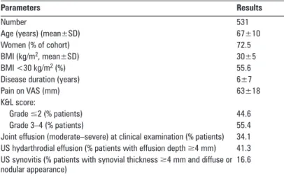
The baseline characteristics of those completing 3 years of follow-up are presented in table 1, with the annual requirement for surgery according to defi ned surgical end points presented in table 2. The 3-year survival rate was 82.3% (standard error 0.017). The survival curve is presented in fi gure 1.
The baseline characteristics included and the results for the univariate analysis are presented in table 3. Of note, the WOMAC physical function subscale, age, clinical joint effusion, body mass index, gender, night awakening due to knee pain and
Figure 1 Survival curve for knee replacement.
Table 1 Baseline characteristics of the subjects completing the 3-year
follow-up
Parameters Results
Number 531
Age (years) (mean±SD) 67±10
Women (% of cohort) 72.5
BMI (kg/m2, mean±SD) 30±5
BMI <30 kg/m2 (%) 55.6
Disease duration (years) 6±7
Pain on VAS (mm) 63±18
K&L score:
Grade ≤2 (% patients) 44.6
Grade 3–4 (% patients) 55.4
Joint effusion (moderate–severe) at clinical examination (% patients) 34.1 US hydarthrodial effusion (% patients with effusion depth ≥4 mm) 41.3 US synovitis (% patients with synovial thickness ≥4 mm and diffuse or nodular appearance)
16.6 BMI, body mass index; K&L, Kellgren and Lawrence; US, ultrasonography; VAS, visual analogue scale.
Table 2 Numbers (percentage) of patients reaching knee replacement
over 3 years
Time
Knee replacement Knee replacement
performed Indication for surgery Total
Year 0–year 1 44 (8) 20 (4) 64 (12)
Year 0–year 2 53 (10) 26 (5) 79 (15)
Year 0–year 3 64 (12)* 30 (6) 94 (18)
n=31 analysed at year 3.
*Six had a unicompartmental knee replacement.
04_ar099564.indd 645
Ann Rheum Dis 2010;69:644–647. doi:10.1136/ard.2008.099564 646
DISCUSSION
This longitudinal study of painful knee OA has confi rmed the importance of the extent of radiographic damage and patient symptoms in predicting subsequent need for joint replacement. In addition, it has demonstrated for the fi rst time that an US fea-ture of synovial infl ammation, the US effusion, is also an inde-pendent predictor of joint replacement.
This study does not of course imply causation—that is, that synovial infl ammation causes symptomatic/structural deteriora-tion leading to joint replacement. The presence of joint infl am-mation may just refl ect structural OA severity: it is plausible that synovial infl ammation is associated with the degree of chon-dropathy, and increased levels of synovitis have been associ-ated with more advanced cartilage loss.6 Synovial effusion and
hypertrophy on MRI have also been associated with knee pain in OA.13 14
It is not surprising that US-detected effusion, but not clini-cally detected effusion, was a predictor of the outcome; we have previously demonstrated the increased sensitivity of US for this infl ammatory feature.7 However, it may appear
confus-ing that effusion but not synovial hypertrophy was associated with subsequent joint replacement need. Our previous work7
also demonstrated that larger effusions generally meant greater degree of synovial hypertrophy. However, because of the mul-tiple ultrasonographer requirement for this multicentre study, we employed a conservative defi nition of synovial hypertrophy that may have made it diffi cult to demonstrate an association with synovial hypertrophy. A possible limitation of this work is that the ultrasound assessment included was only performed at a single time point; although anecdotal clinical reports sug-gest that infl ammation varies over time in OA, little has been reported on the longitudinal evaluation of synovitis using sensi-tive imaging modalities.
Using joint replacement as an outcome measure for OA is of course a complex issue that relates to national and local health priorities, individual surgeon choices, as well as patient will-ingness and comorbidities.3 15 One limitation of this study is
therefore the pragmatic outcome we have chosen. Additionally, we did not measure psychosocial factors such as anxiety and depression to assess their role in prediction. Certainly there is no international consensus on when joint replacement should be considered, and an ongoing OMERACT–OARSI initiative is developing a multidomain tool that will allow agreement on the level of OA severity that will act as a surrogate measure for the need for joint replacement.16
In summary, this large cohort of patients with painful knee OA has confi rmed the importance of presenting features of pain and radiographic damage in predicting subsequent need for joint replacement. But it has also demonstrated that the presence of a large US-detected effusion may also be included in clinical mod-els to predict surgical need.
Acknowledgements We gratefully acknowledge the contribution of all investigators. Italy: Dr Patrizia Blasetti, Dr Rossella De Angelis, Dr Antonella Farina, Dr Emilio Filippucci, Dr Gianni Lamanna. Spain: Dr Gema Bonilla, Dr Eugenio De Miguel. Germany: Dr Eugen Feist, Dr Sonja Kary, Dr Uwe Lang, Dr Anett Reißhauer, Dr Udo Schneider. Switzerland: Professeur Jean-Charles Gerster, Dr Laurent Malterre, Dr Nicholas Theumann, Dr Daniel Van Linthoudt, Dr Pascal Zufferey. Belgium: Dr Béatrice Andre, Dr Marie-Joëlle Kaiser, Dr Olivier Kaye, Dr Stefaan Marcelis. Competing interests None.
Funding This study was supported in part by an unrestricted grant from Bristol Myers-Squibb.
Patient consent Obtained.
Ethics approval All involved multinational centres had local approval. Provenance and peer review Not commissioned; externally peer reviewed.
US synovitis were not associated with joint replacement in the univariate analysis.
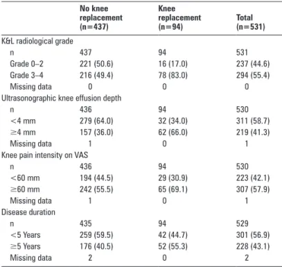
The following features were included in the multivariate analysis: K&L radiological grade; US knee effusion; physi-cian’s global assessment of clinical knee OA severity; knee pain intensity; patient’s global assessment of clinical knee OA; dis-ease duration; US knee synovitis; WOMAC stiffness and pain subscales. The fi nal predictors of joint replacement were K&L radiographic grade (HR=4.08 (95% CI 2.34 to 7.12), p<0.0001); ultrasonographic knee effusion (HR=2.63 (95% CI 1.70 to 4.06), p<0.0001); knee pain intensity on a 0–100 mm visual analogue scale (HR=1.81 (95% CI 1.15 to 2.83), p=0.01) and disease dura-tion (HR=1.63 (95% CI 1.08 to 2.47), p=0.02). Table 4 presents the baseline characteristics of these fi nal predictors.
Table 3 Characteristics included and results of the univariate analysis HR (95% CI) p Value
Sex 1.10 (0.71 to 1.72) 0.6582
Age 1.24 (0.81 to 1.89) 0.3144
BMI 1.15 (0.76 to 1.73) 0.5121
Disease duration 1.70 (1.13 to 2.55) 0.0109
K&L radiological grade 4.67 (2.73 to 8.01) <0.0001
Knee pain intensity on VAS 1.82 (1.17 to 2.82) 0.0074
Joint effusion at clinical examination 1.15 (0.75 to 1.76) 0.5153
Sudden aggravation of knee pain 0.74 (0.49 to 1.13) 0.1617
Duration of morning stiffness 1.41 (0.86 to 2.32) 0.1686
Night awakening due to knee pain 1.07 (0.69 to 1.65) 0.7730
WOMAC pain subscale 1.45 (0.94 to 2.22) 0.0894
WOMAC stiffness subscale 1.36 (0.88 to 2.08) 0.1614
WOMAC function subscale 1.27 (0.81 to 2.00) 0.3053
Patient’s global assessment of clinical knee OA severity on VAS
1.46 (0.95 to 2.25) 0.0832 Physician’s global assessment of clinical
knee OA severity on VAS
1.88 (1.24 to 2.84) 0.0031 Ultrasonographic knee effusion depth 3.06 (2.00 to 4.69) <0.0001
Ultrasonographic knee synovitis 1.54 (0.95 to 2.50) 0.0826
BMI, body mass index; HR, hazard ratio; K&L, Kellgren and Lawrence; OA, osteoarthritis; VAS, visual analogue scale; WOMAC, Western Ontario and McMaster Universities Osteoarthritis Index.
Table 4 Baseline characteristics of knee osteoarthritis predicting
subsequent knee replacement No knee replacement (n=437) Knee replacement (n=94) Total (n=531)
K&L radiological grade
n 437 94 531
Grade 0–2 221 (50.6) 16 (17.0) 237 (44.6)
Grade 3–4 216 (49.4) 78 (83.0) 294 (55.4)
Missing data 0 0 0
Ultrasonographic knee effusion depth
n 436 94 530
<4 mm 279 (64.0) 32 (34.0) 311 (58.7)
≥4 mm 157 (36.0) 62 (66.0) 219 (41.3)
Missing data 1 0 1
Knee pain intensity on VAS
n 436 94 530 <60 mm 194 (44.5) 29 (30.9) 223 (42.1) ≥60 mm 242 (55.5) 65 (69.1) 307 (57.9) Missing data 1 0 1 Disease duration n 435 94 529 <5 Years 259 (59.5) 42 (44.7) 301 (56.9) ≥5 Years 176 (40.5) 52 (55.3) 228 (43.1) Missing data 2 0 2
Results are shown as number or number (%). K&L, Kellgren and Lawrence; VAS, visual analogue scale.
04_ar099564.indd 646
Ann Rheum Dis 2010;69:644–647. doi:10.1136/ard.2008.099564 647
9. Altman R, Asch E, Bloch D, et al. Development of criteria for the classifi cation and reporting of osteoarthritis. Classifi cation of osteoarthritis of the knee. Diagnostic and Therapeutic Criteria Committee of the American Rheumatism Association. Arthritis
Rheum 1986;29:1039–49.
10. Kellgren JH, Lawrence JS. Radiological assessment of osteo-arthrosis. Ann Rheum
Dis 1957;16:494–502.
11. Kellgren JH, Jeffrey MR, Ball J. Atlas of standard radiographs. In: The epidemiology of
chronic rheumatism. Vol 2. Oxford: Blackwell Scientifi c, 1963.
12. Steinbrocker O, Traeger CH, Batterman RC. Therapeutic criteria in rheumatoid arthritis. J Am Med Assoc 1949;140:659–62.
13. Hill CL, Gale DG, Chaisson CE, et al. Knee effusions, popliteal cysts, and synovial thickening: association with knee pain in osteoarthritis. J Rheumatol 2001;28:1330–7.
14. Hill CL, Hunter DJ, Niu J, et al. Synovitis detected on magnetic resonance imaging and its relation to pain and cartilage loss in knee osteoarthritis. Ann Rheum Dis 2007;66:1599–603.
15. Maillefert JF, Dougados M. Is time to joint replacement a valid outcome measure in clinical trials of drugs for osteoarthritis? Rheum Dis Clin North Am 2003;29:831–45.
16. Gossec L, Hawker G, Davis AM, et al. OMERACT/OARSI initiative to defi ne states of severity and indication for joint replacement in hip and knee osteoarthritis.
J Rheumatol 2007;34:1432–5. REFERENCES
1. Sharma L, Kapoor D, Issa S. Epidemiology of osteoarthritis: an update. Curr Opin
Rheumatol 2006;18:147–56.
2. Parsons IM, IV, Sonnabend DH. What is the role of joint replacement surgery? Best
Pract Res Clin Rheumatol 2004;18:557–72.
3. Maillefert JF, Hawker GA, Gossec L, et al. Concomitant therapy: an outcome variable for musculoskeletal disorders? Part 2: total joint replacement in osteoarthritis trials. J Rheumatol 2005;32:2449–51.
4. Gossec L, Tubach F, Baron G, et al. Predictive factors of total hip replacement due to primary osteoarthritis: a prospective 2 year study of 505 patients. Ann Rheum Dis 2005;64:1028–32.
5. Hawker GA, Guan J, Croxford R, et al. A prospective population-based study of the predictors of undergoing total joint arthroplasty. Arthritis Rheum 2006;54:3212–20. 6. Ayral X, Pickering EH, Woodworth TG, et al. Synovitis: a potential predictive factor of
structural progression of medial tibiofemoral knee osteoarthritis – results of a 1 year longitudinal arthroscopic study in 422 patients. Osteoarthr Cartil 2005;13:361–7. 7. D’Agostino MA, Conaghan P, Le Bars M, et al. EULAR report on the use of
ultrasonography in painful knee osteoarthritis. Part 1: prevalence of infl ammation in osteoarthritis. Ann Rheum Dis 2005;64:1703–9.
8. Conaghan P, D’Agostino MA, Ravaud P, et al. EULAR report on the use of ultrasonography in painful knee osteoarthritis. Part 2: exploring decision rules for clinical utility. Ann Rheum Dis 2005;64:1710–14.
04_ar099564.indd 647