C
ERVICAL
T
UMORAL
C
ALCIUM
P
YROPHOSPHATE
D
IHYDRATE
D
EPOSITION
D
ISEASE
28 Y
EARS AFTER
S
UBOCCIPITAL
C
RANIOTOMY
: C
ASE
R
EPORT
OBJECTIVE:
To describe a rare case of tumoral cervical chondrocalcinosis that appeared
28 years after the patient had undergone suboccipital craniotomy.
CLINICAL PRESENTATION:
A 42-year-old woman suffered from cervicalgia
associ-ated with a firm mass at the occipitocervical region. Plain x-ray and computed
tomo-graphic and magnetic resonance images revealed a calcified lesion in a scar from a
previous suboccipital craniotomy.
INTERVENTION:
The patient underwent tumorectomy and histopathology, which
revealed an exuberant tumoral chondrocalcinosis. Laboratory test results revealed no
secondary cause for the chondrocalcinosis.
CONCLUSION:
Identification of chondrocalcinosis beyond the cervical region is very
rare. Localization of chondrocalcinosis in a scar from a previous suboccipital
cran-iotomy has not been previously reported. Surgery appears to be the treatment of choice
for this form of chondrocalcinosis.
KEY WORDS: Calcium pyrophosphate dihydrate disease, Cervical spine, Craniotomy, Tumoral calcinosis Neurosurgery 60:E1151, 2007 DOI: 10.1227/01.NEU.0000255477.06247.B8 www.neurosurgery-online.com
N
EUROSURGERY VOLUME 60 | NUMBER 6 | JUNE 2007 | E1151CASE REPORTS
Didier Scavarda, M.D.
Department of Pediatric Neurosurgery, Children’s Hospital, La Timone, Marseille, France Claude F. Litre, M.D. Department of Neurosurgery, Hospital Maison-Blanche, Cedex, France Stéphane Froelich, M.D. Department of Neurosurgery, Centre Hospitalier Louis Pasteur, Cedex, France
Robin Srour, M.D.
Department of Neurosurgery, Centre Hospitalier Louis Pasteur, Cedex, France
Pascal Rousseaux, M.D.
Department of Neurosurgery, Hospital Maison Blanche, Cedex, France
Reprint requests:
Didier Scavarda, M.D.
Department of Pediatric Neurosurgery, Hospital des Enfants, La Timone, 264 Rue Saint Pierre, 13005, Marseille, France. Email: didier.scavarda@ap-hm.fr
Received, November 26, 2006. Accepted, February 2, 2007.
A
rthropathies that result from the deposit of pyrophosphate crystals in soft tissues have been the subject of numerous pub-lications since the early 1960s. The term “chon-drocalcinosis” was introduced in 1963 by Zitnan and Sitaj (35); however, their contempo-raries, McCarty et al. (20), preferred to call this pathology a “pseudo-gout syndrome.” In 1977, after he reviewed the literature on the subject, McCarty (19) proposed the designation “cal-cium pyrophosphate dihydrate crystal dis-ease.” This arthritic disorder is generally found in the elderly, and in the great majority of cases it affects the joints of the pelvis and the limb extremities (24). Neurological deficit or bone lysis occurs when the spine is affected (21, 22). There are two interesting points concerning this case. First, in this patient we have the only reported case of cervical chondrocalcinosis that includes neither neurological symptoms nor osseous disruption and is associated with intense cervicalgia that was partially relieved by surgical treatment. Second, it is interesting for neurosurgeons to note that the patient’s lesion appeared on a scar from a suboccipital cran-iotomy that was performed 28 years earlier.CASE REPORT
In 2004, a 42 year-old woman with cervical-gia was admitted to the hospital. Her pain had been radiating to the occipital and scapulary areas for 4 months. In this time, the pain pro-gressively worsened and proved refractory to standard analgesics.
In 1976, when the patient was 14 years old, she underwent surgery to relieve symptoms of intracranial hypertension syndrome. This involved exposure of the posterior cranial fossa, which revealed an arachnoid cyst.
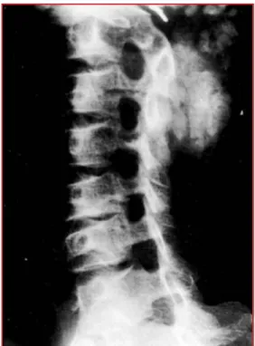
A neurological assessment in 2004 yielded normal findings, although it revealed a sensi-tive and firm swelling located at the occipito-cervical junction. Standard x-rays of her spine (Fig. 1) showed a flaky, apparently calcified lesion that extended from the inion to the superior border of the spinous process of C4 at the midline. The lesion appeared to develop within the soft tissue rather than the bone structures. Computed tomographic imaging of the cervical spine (Fig. 2) confirmed that the structure was calcified in nature. The lesion had a heterogeneous aspect and extended from
the suboccipital region to the spinous process of C3 without canal invasions.
The lesion was detected on magnetic resonance images as an isointensity area in T1-weighted sequences (Fig. 3).
In April 2004, the tumor was removed via a posterior approach. The resection of the calcium pyrophosphate dihy-drate disease was complete. Surgery revealed a crumbly, whitish tissue that was easily distinguishable but hard to sep-arate from the surrounding muscles. The growth had not pen-etrated into the region in front of the laminae. Total excision
was accomplished. The anatomopathological assessment revealed large, purplish, crystalline deposits that were identi-fied as calcium pyrophosphate dihydrate crystals in high-magnification microphotographs (square-tipped crystals that were weakly refringent under polarized light). A strong, dis-solving macrophage reaction surrounding the deposits was apparent, as was giant multinucleate cell proliferation. We found no sign of malignancy and diagnosed this as cervical chondrocalcinosis.
Standard x-ray images of the wrists, hips, and pelvis showed no other stigma of chondrocalcinosis. Laboratory test results revealed no secondary causes for chondrocalcinosis.
The follow-up duration after the second surgery was 18 months. At our last clinical examination, the patient reported she was free of pain and demonstrated good cervical mobility. Her scar was clean.
Standard x-ray control images did not reveal any calcification.
DISCUSSION
Chondrocalcinosis belongs to the arthritis family because of the inherent role of microcrystalline deposits in the disease (6, 16, 17, 23, 29): calcium pyrophosphate dihydrate crystals deposit within the joints. It is a pathology mostly confined to the elderly population except for certain unusual cases such as hypomagnesemia or Coffin-Lowry syndrome (24). Its prevalence increases with age, reaching close to 45% in sub-jects over 85 years old (24), generally with no symptomatic manifestation. The most common sites for the chondrocalci-nosis include the hip, elbow, shoulder, and foot. In the
major-E1151| VOLUME 60 | NUMBER 6 | JUNE 2007 www.neurosurgery-online.com
S
CAVARDA ET AL.
FIGURE 1. This sagittal standard x-ray of the cervi-cal spine shows a pseudotumoral cervi-calcified lesion behind the spinous process of C1–C2 and C3.
FIGURE 2.Axial computed tomography reveals a cal-cified lesion within the soft tissue with no indication of bone erosion.
FIGURE 3. A sagittal T1-weighted magnetic reso-nance image indicates the presence of a lesion within the soft tissue that has no contact with bone (with respect to the epidural space at C1 level).
ity of cases, its onset is diffused and involves more than two joints, usually symmetrically (24).
This pathology of the elderly, which is particularly predom-inant in females, should be approached with a rigorous initial etiological workup to search for the presence of morbid condi-tions that are sometimes associated with this disorder, such as psoriasis, hyperparathyroidism, hemochromatosis, hypophos-phatasemia, hypomagnesemia, hyperuricemia, Wilson’s dis-ease, traumatisms, and past history of surgery (4, 6, 8, 24). However, primitive and idiopathic forms of chondrocalcinosis are the most frequently encountered; family-related forms are uncommon.
Chondrocalcinosis is a risk factor for neck pain. It frequently involves the cervical spine, and as such, may be associated with development of neck pain. This diagnosis should be con-sidered when a patient presents with neck pain (9).
For this patient, surgery was the logical indication; she had a mass within a previous scar, and her clinical examination revealed abnormal findings. The clinical and radiological data showed strong correlation. Surgery was important, both to con-firm the anatomopathological assessment and to relieve her clinical symptoms.
Localization of calcium pyrophosphate dihydrate crystals in joints is most common, but extra-articular deposits have also been reported to occur, within the ligamentum flavum (1, 3, 7, 18, 19, 20), the dura mater (12, 14), and the tendon calcaneus (10). The presence of such intra-articular deposits does not nec-essarily cause symptoms that are either clinically or radi-ographically recognizable (24), and clinical symptoms can occur that do not show in x-rays. These conditions often render the diagnosis somewhat tricky.
Spinal localizations of these crystals have rarely been reported in the literature. Those that have been reported describe cervical (2, 11–13, 15) and dorsal medullary com-pression (26), lumbar sciatica (22, 27), and pseudospondy-lolisthesis (30). They are extra-articular and lie within the lig-amentum flavum, posterior common ligament (23, 28), atlanto-occipital ligament (5), vertebral body (21), and annu-lus fibrosus of intervertebral discs (21). Such deposits are always nodular and of low volume, but because they are located within the spinal canal, they can induce cord com-pression and neurological deficits. All vertebral levels are involved, from the foramen magnum (5, 27) to the lum-bosacral hinge (22). Cervical localizations occur with the highest incidence. In 1993, Baba et al. (1) reviewed 91 cases of cervical myelopathies, to which he added 8 personal cases. According to him, 8 times out of 10, this myelopathy has a slow and gradual evolution, but it can suddenly decompen-sate after minor trauma. Baba et al. confirmed the predomi-nance of this condition in females, as it affected 84 women and only 15 men. A tumoral form similar to that in our patient was described only once, by Kokubun et al. (16): they reported the case of a 68-year-old woman who suffered from cervicalgia associated with restrained motion of the cervical spine in all directions. Radiographic evaluation and inter-vention revealed a calcified, multilobular, pseudotumoral
lesion located in the interlaminar portions of C1 and C2. During surgery, the lesion proved to be very adherent to the dura mater. More chondrocalcinosis nodular deposits were found in the ligamentum flavum at the same level, which indicates that the ligamentum flavum may have been the point of origin.
CONCLUSION
Our observations are interesting because they report an unusual localization of cervical chondrocalcinosis: it touched neither the dura mater nor the ligamentum flavum. That this tumor was strictly extracanalar accounts for the unusual clin-ical manifestations that we encountered, including the exis-tence of considerable pain in the patient with no sign of spinal cord compression, and the patient’s positive response to surgi-cal treatment.
The link between a preexisting scar and this kind of tumor has already been described, but it is not completely understood.
REFERENCES
1. Baba H, Maezawa Y, Kawahara N, Tomita K, Furusawa N, Imura S: Calcium crystal deposition in the ligamentum flavum of the cervical spine. Spine 18:2174–2181, 1993.
2. Berghausen EJ, Balogh K, Landis WJ, Lee DD, Wright AM: Cervical myelopa-thy attributable to pseudogout. Case report with radiographic, histologic, and crystallographic observations. Clin Orthop Relat Res 214:217–221, 1987. 3. Brown TR, Quinn SF, D’Agostino AN: Deposition of calcium pyrophosphate dihydrate crystals in the ligamentum flavum: Evaluation with MR imaging and CT. Radiology 178:871–873, 1991.
4. Bywaters EG, Hamilton EB, Williams R: The spine in idiopathic haemochro-matosis. Ann Rheum Dis 30:453–465, 1971.
5. Chen CF, Chang MC, Wang ST, Liu CL, Chen W: Calcium pyrophosphate dihydrate crystal deposition disease in cervical radiculomyelopathy. J Chin
Med Assoc66:256–259, 2003.
6. Ciricillo SF, Weinstein PR: Foramen magnum syndrome from pseudogout of the atlanto-occipital ligament. Case report. J Neurosurg 71:141–143, 1989. 7. Doherty M, Dieppe P: Clinical aspects of calcium pyrophosphate dihydrate
crystal deposition. Rheum Dis Clin North Am 14:395–414, 1988.
8. Ellman MH, Vazquez T, Ferguson L, Mandel N: Calcium pyrophosphate dep-osition in ligamentum flavum. Arthritis Rheum 21:611–613, 1997. 9. Finckh A, Van Linthoudt D, Duvoisin B, Bovay P, Gerster JC: The cervical
spine in calcium pyrophosphate dihydrate deposition disease. A prevalent case-control study. J Rheumatol 31:545–549, 2004.
10. Flores J, Munoz J, Gallego S, Bujan A, Ferreiro I, Gonzalez A: Spinal cord com-pression due to tumoral idiopatic calcinosis. Spinal Cord 41:413–416, 2003. 11. Gerster JC, Baud CA, Lagier R, Boussina I, Fallet GH: Tendon calcifications in
chondrocalcinosis: A clinical, radiologic, histologic, and crystallographic study. Arthritis Rheum 20:717–722, 1977.
12. Gomez H, Chou SM: Myeloradiculopathy secondary to pseudogout in the cervical ligamentum flavum: Case report. Neurosurgery 25:298–302, 1989. 13. Grahame R, Sutor DJ, Mitchener MB: Crystal deposition in
hyperparathy-roidism. Ann Rheum Dis 30:597–604, 1971.
14. Iwasaki Y, Akino M, Abe H, Tsuru M, Tashiro K, Miyasaka K, Kaneda K, Isu T, Ito T: Calcification of the ligamentum flavum of the cervical spine. Report of four cases. J Neurosurg 59:531–534, 1983.
15. Kawano N, Matsuno T, Miyazawa S, Uchiyama H, Ohtaka H, Mii K, Tachibana S: New knowledge on the calcium pyrophosphate dihydrate (CPPD) crystal deposition disease in the cervical ligamentum flavum [in Japanese]. No Shinkei Geka 15:181–190, 1987.
N
EUROSURGERY VOLUME 60 | NUMBER 6 | JUNE 2007 | E115116. Kokubun S, Ozawa H, Sakurai M, Tanaka Y: Tumoral calcinosis in the upper cervical spine: A case report. Spine 6:249–252, 1996.
17. Kubota M, Baba I, Sumida T: Myelopathy due to ossification of the ligamen-tum flavum of the cervical spine. A report of two cases. Spine 6:553–559, 1981.
18. Matsukado K, Amano T, Itou O, Yuhi F, Nagata S: Tumoral calcinosis in the upper cervical spine causing progressive radiculomyelopathy—Case report.
Neurol Med Chir (Tokyo)41:411–4144, 2001.
19. McCarty DJ: Calcium pyrophosphate dihydrate crystal deposition disease: Nomenclature and diagnostic criteria. Ann Intern Med 87:240–242, 1977. 20. McCarty D, Kohn N, Faires J: The significance of calcium pyrophosphate
crystals in the synovial fluid of arthritic patients. The pseudogout syndrome. I. Clinical aspects. Ann Intern Med 56:711–737, 1962.
21. Miyasaka K, Kaneda K, Sato S, Iwasaki Y, Abe S, Takei H, Tsuru M, Tashiro K, Abe H, Fujioka Y: Myelopathy due to ossification or calcification of the lig-amentum flavum: Radiologic and histologic evaluations. AJNR Am J
Neuroradiol4:629–632, 1983.
22. Muthukumar N, Karuppaswamy U: Tumoral calcium pyrophosphate dihy-drate deposition disease of the ligamentum flavum. Neurosurgery 53: 103–109, 2003.
23. Nagashima C, Takahama M, Shibata T, Nakamura H, Okada K, Morita H, Kubo H: Calcium pyrophosphate dihydrate deposits in the cervical liga-menta flava causing myeloradiculopathy. J Neurosurg 60:69–80, 1984. 24. Nakajima K, Miyaoka M, Sumie H: Cervical radiculomyelopathy due to
cal-cification of the ligamenta flava. Surg Neurol 21:479–488, 1984.
25. Pola E, Pola R, Gaetani E, De Santis E: Tumoral calcinosis. Lancet 359:1818, 2002.
26. Resnick D, Pineda C: Vertebral involvement in calcium pyrophosphate dihy-drate crystal deposition disease. Radiographic-pathological correlation.
Radiology153:55–60, 1984.
27. Riemenschneider PA, Ecker A: Sciatica caused by tumoral calcinosis: A case report. J Neurosurg 9:304–307, 1952.
28. Ryan LM: Calcium pyrophosphate dihydrate crystal deposition and other crystal deposition diseases. Curr Opin Rheumatol 5:517–521, 1993. 29. Salcman M, Khan A, Symonds DA: Calcium pyrophosphate arthropathy of
the spine: Case report and review of the literature. Neurosurgery 34:915–918, 1994.
COMMENTS
S
cavarda et al. report a rare case of calcium pyrophosphate deposi-tion disease (pseudogout) that caused cervicalgia in a patient who had suboccipital craniotomy 28 years earlier. The lesion is rare because it arose within a scar of a previous surgery rather than within a more typical spinal location, such as the ligamentum flavum. Spinal surgeons should note that this lesion was readily visible on a lateral cervical x-ray, and its removal can potentially relieve neck pain in affected patients.Praveen V. Mummaneni
San Francisco, California
S
cavarda et al. describe a most unusual case of a woman who devel-oped a symptomatic mass in the posterior cervical region, which was ultimately found to be consistent with calcium pyrophosphate. The patient responded well to excision of the mass. This case is also unique in that this patient had a posterior fossa craniectomy 28 years before the development of this mass. Whether the previous surgery was related to the calcium pyrophosphate deposition is unclear.Surgeons should be aware that soft-tissue deposition of calcium pyrophosphate may occur, and those patients who are symptomatic may well respond to surgical resection, as did this patient.
Vincent C. Traynelis
Iowa City, Iowa
S
cavarda et al. present a rare case of calcium pyrophosphate deposi-tion. Its association with previous surgery is most certainly suspect from an etiological perspective. This patient fits into the archival cate-gory of rare cases. It is important to keep this imagery in mind when dealing with similar patients in the future.Edward C. Benzel
Cleveland, Ohio
E1151| VOLUME 60 | NUMBER 6 | JUNE 2007 www.neurosurgery-online.com