Role of insulin-like growth factor binding proteins in
limitation of IGF-I degradation into the N-methyl-
D
-aspartate receptor antagonist GPE: evidence from
gonadotrophin-releasing hormone secretion in vitro at two
developmental stages
Jean-Pierre Bourguignon*, Arlette Gérard
Developmental Neuroendocrinology Unit, Department of Pediatrics, University of Liège, C.H.U. Sart Tilman, B-4000 Liège, Belgium
* Corresponding author. Division of Pediatric and Adolescent Medicine, C.H.U. Sart Tilman, B35, B-4000 Liege, Belgium. Fax: + 32-4-366-72-46; e-mail: jean-pierre.bourguignon@chu.ulg.ac.be
KEYWORDS: onadotrophin-releasing hormone; N-Methyl-D-aspartate receptor; Insulin-like growth
factor-1; Insulin-like growth factor-binding protein 2; Insulin-like growth factor-binding protein 3; GPE or (1–3)IGF-I
ABSTRACT
We showed previously that insulin-like growth factor-I (IGF-I) could inhibit the secretion of gonadotrophin-releasing hormone (GnRH) evoked in vitro by N-methyl-D-aspartate (NMDA) or veratridine depolarization. Such an IGF-I effect appeared to be mediated by its physiological breakdown product, the N-terminal tripeptide GPE. That effect was developmentally regulated since IGF-I could inhibit GnRH secretion from hypothalamic explants of 50-day-old adult rats but not from immature 15-day-old explants. We hypothesized that the IGF-binding proteins (BPs) could limit the peptide availability to endopeptidases and account for the absent IGF-I effects at 15 days. In this paper, we show that the inhibition of GnRH secretion by 10−10 M of IGF-I at 50 days is prevented in a dose-dependent manner by 0.3 to 3 nM of IGF-BP2 as well as IGF-BP3. The inhibition caused by 10−10 M of GPE is not affected under similar conditions. Using explants obtained at 15 days, a significant inhibition of GnRH secretion can be obtained by 10−10 M of IGF-I in the presence of an anti IGF-BP2 antiserum used at 1:3000 and 1:1000 concentrations. These data indicate that in the immature rat brain, the IGF-BPs could act as modulators of IGF-I degradation into its subproduct GPE, a possible endogenous antagonist at NMDA receptors.
1. Introduction
In the central nervous system, insulin-like growth factor-I (IGF-I) is synthesized and cleaved by brain peptidases into two major peptides, the des(1–3)IGF-I and the N-terminal tripeptide, (1– 3)IGF-I, also referred to as GPE based on its amino acid sequence glycine, proline and glutamate [25]. Des(1–3)IGF-I has reduced affinity to IGF-binding proteins (IGF-BP) and shows therefore increased biopotency as a neurotrophic factor through the classical IGF receptors [27]. GPE was proposed as a possible agonist at N-methyl-D-aspartate (NMDA) receptors [17,24]. In contrast, using the in vitro secretion of gonadotropin-releasing hormone (GnRH) as a paradigm of NMDA-receptor-mediated effect, we reported that IGF-I resulted in NMDA receptor antagonism mediated through GPE, since that effect was prevented by peptidase inhibitors and not observed using des(1–3)IGF-I [7].
In our previous developmental studies, we found that the NMDA receptors were equally active in the mechanism of GnRH secretion at 15 and 50 days of age, as evidenced from the similar sensitivity to increasing concentrations of the agonist NMDA and the antagonist MK-801 [4,6]. GPE was also equally effective in inhibiting GnRH secretion at both ages [7]. There was, however, a striking age dependency of the IGF-I effect which was obvious at 50 days but absent at 15 days [7]. Such a difference could be explained by developmental changes in IGF-I endopeptidase activity. Alternatively or additionally, the BPs could prevent GPE generation through a reduction of IGF-I availability to enzymatic degradation. Testing the latter hypothesis was the aim of the present study.
2. Materials and methods
Individual explants (12 to 15 in each experiment) of the retrochiasmatic hypothalamus of 15-day- and 50-day-old male Wistar rats were studied using a static incubation system described in detail previously [2,3,6]. In 0.5 ml fractions of the incubation medium collected every 7.5 min for 4–6 h, the radioimmunoassay of GnRH was performed as described earlier in detail [2,3]. The secretion of GnRH was studied during a 7.5-min exposure to veratridine (Sigma, St. Louis, MO), a depolarizing agent through Na+ channel opening. Veratridine was used repeatedly at intervals of 37.5 min (50 days) or 52.5 min (15 days), in order to prevent inhibition of GnRH secretion resulting from the inhibitory autofeedback [5]. The secretory response of GnRH was calculated as the difference between the concentrations measured immediately before and during exposure to veratridine. For each explant, the data were transformed into percentages of the initial secretory response considered as 100%. The effects of recombinant human IGF-I (Pharmacia, Stockholm, Sweden) or GPE (Peninsula, Merseyside, UK) were studied through incubation for 15 min including 7.5 min immediately prior to and 7.5 min during veratridine stimulation. Using explants of 50-day- old rats, the effects of increasing concentrations of IGF-BP2 (courtesy of Dr. P. Ramage, Novartis Pharma,
Basle, Switzerland) and IGF-BP3 (courtesy of Dr. A. Sommer, Celtrix Pharmaceuticals, San José, CA) were studied through incubation for 15 min together with IGF-I or GPE. Using explants of 15- and 50-day-old rats, the effects of increasing concentrations of a mouse antiserum against IGF-BP2 (courtesy of Dr. P. Ramage, Novartis Pharma, Basle, Switzerland) were studied through incubation for 15 min together with IGF-I. For control purposes, equally increasing concentrations of mouse immunoglobulins G (Sigma, St. Louis, MO) were used. Statistical analysis was performed using ANOVA with correction for repeated measurement and Dunnett’s multiple comparison test. The level of significance was at p < 0.05.
3. Results
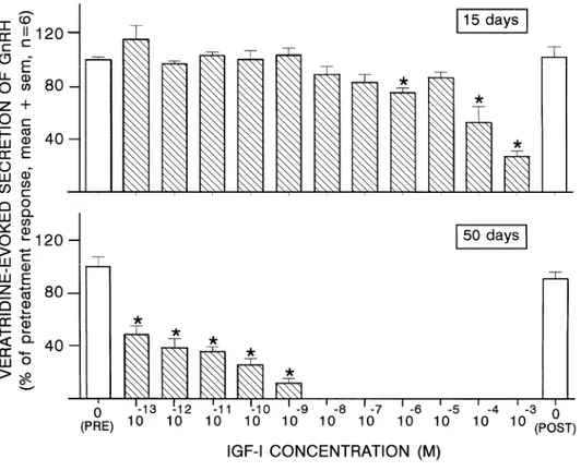
Using explants from 15-day-old rats, IGF-I did not affect the veratridine-evoked secretion of GnRH unless concentrations ≥ 10−6 M were used (Fig. 1). In contrast, a concentration as low as 10−13 M of IGF-I was already effective in reducing GnRH secretion by more than 50%, at 50 days (Fig. 1).
Fig. 1. Effects of increasing IGF-I concentrations on the veratridine-evoked secretion of GnRH from hypothalamic explants of 15- and 50-day-old male rats. *p < 0.05 vs. pretreatment control response.
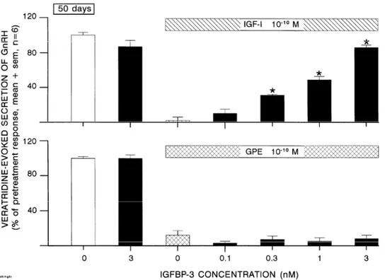
Using explants from 50-day-old rats, the inhibition of GnRH secretion by 10−10 M of IGF-I was prevented in a dose-related manner by IGF-BP2 (Fig. 2) as well as IGF-BP3 (Fig. 3). In the absence of IGF-I, the highest concentration of BP used (3 nM) did not affect GnRH secretion. The inhibition of GnRH secretion by 10−10 M of GPE was not affected by IGF-BP2 (Fig. 2) or IGF-BP3 (Fig. 3).
Fig. 2. Effects of increasing IGF-BP2 concentrations on the inhibition of veratridine-evoked GnRH secretion by IGF-I (upper panel) or GPE (lower panel) using hypothalamic explants from 50-day-old rats. *p < 0.05 vs. IGF-I or GPE in the absence of BP.
Fig. 3. Effects of increasing IGF-BP3 concentrations on the inhibition of veratridine-evoked GnRH secretion by IGF-I (upper panel) or GPE (lower panel) using hypothalamic explants from 50-day-old rats. *p < 0.05 vs. IGF-I or GPE in the absence of BP.
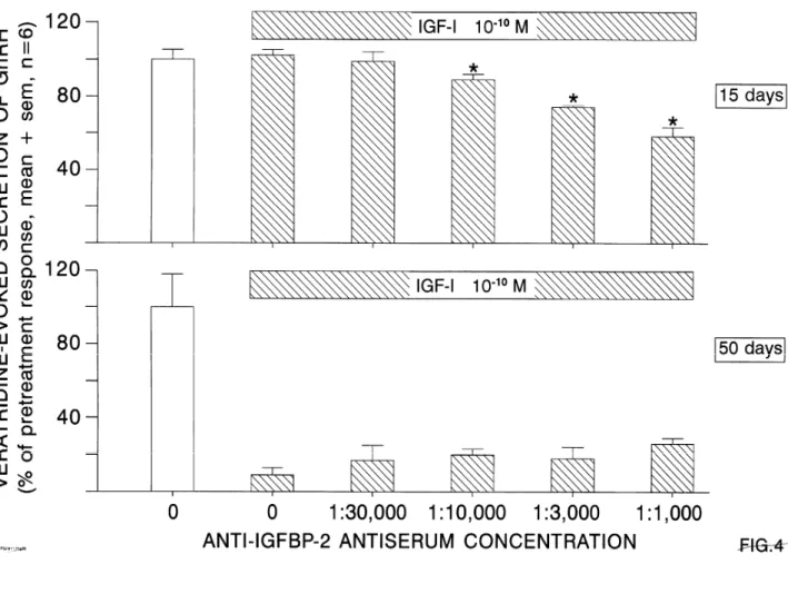
Using explants from 15-day-old rats, 10−10 M of IGF-I did not influence GnRH secretion, except when IGF-BP2 was passively immunoneutralized by the antiserum at the three highest concentrations used (Fig. 4). At 50 days, the inhibition of GnRH secretion by IGF-I was unchanged during passive immunoneutralization of IGF-BP2. At both ages, no effects were seen using increasing concentrations of mouse immunoglobulins (data not shown).
Fig. 4. Effects of increasing concentrations of mouse anti-IGF-BP2 antiserum (Ab) on the veratridine-evoked GnRH secretion which is studied in the presence of IGF-I at 15 days (upper panel) and 50 days (lower panel). *p < 0.05 vs. IGF-I in the absence of Ab.
4. Discussion
In this study, we used the GnRH neurosecretory system as a paradigm of NMDA-–receptor modulated neuronal function. During the past 10 years, we accumulated evidence that NMDA receptors were involved in pulsatile GnRH secretion as well as GnRH release evoked by veratridine from the explanted rat hypothalamus in vitro [2–6]. Recently, using the antisense strategy, we concluded that facilitation of GnRH secretion was mediated by receptors involving the NR2A subunit which were expressed in the mediobasal hypothalamus and encoded in the preoptic area, possibly in GnRH neuron cell bodies [9]. Using that paradigm, we aimed at further characterizing the interaction of IGF-I at NMDA receptors.
The role of IGF-I in the CNS is particularly complex. IGF-I has trophic brain effects mediated through its classical receptors. These effects were mimicked by the endogenous truncated derivative des(1–3)I [23] which was more potent than I due to reduced binding to the IGF-BPs [1,13,27]. IGF-I also has neuroprotective effects which were suggested by increased IGF-I expression at the site of hypoxic–ischemic or traumatic brain injury [14,15,19,28] and the positive effects of IGF-I on neuronal survival in vivo and in vitro [14,16,21]. Such in vitro neuroprotective effects were observed using the GT-1 cell line of immortalized GnRH neurons [26]. Some neuroprotective effects resulted from the IGF-I molecule and did not require IGF-I degradation into its physiological subproducts des(1–3)IGF-I and GPE since the IGF-I analog Gln3, Ala4, Tyr15, Leu16 IGF-I was neuroprotective in vitro [16]. The observation, however, that the neuroprotective effects of IGF-I were not mimicked by des(1–3)IGF-I [14] suggested the possible involvement of a mechanism different from the classical IGF-I receptors.
An alternative mechanism for IGF-I effects in the CNS was through GPE, its post-translational side-product of peptidase degradation. This was initially suggested by Sara et al. [24], who proposed that GPE worked as a competitive agonist at NMDA receptors involved in dopamine release, while acetylcholine release could be stimulated by GPE through another type of receptor. Because the NMDA receptors were involved in neurotoxicity and IGF-I in neuroprotection, it appeared critical to elucidate further the relationship between both systems. The data on interactions of IGF-I or GPE with glutamate or NMDA receptors were equivocal, however. IGF-I was found to have inhibitory effects on glutamate-evoked release of GABA [12], as well as sensitizing effects on the neurotoxic glutamate effects [10]. GPE was found to act as a NMDA receptor agonist in cortical neurons [25] and retinal glial cells [17], whereas we found GPE as well as other peptide subproducts to antagonize the NMDA-receptor-mediated secretion of GnRH [7,8]. The reason for those discrepancies was unclear, particularly since the precise mechanism of GPE interaction at the receptor level was not elucidated. Anyhow, this indicated that the conclusions drawn using a particular paradigm of NMDA-receptor-mediated effect could not be extrapolated to other models. While we found that GPE was similarly effective in inhibiting GnRH secretion in immature and adult rats, IGF-I showed no effect in immature rats but was extremely potent in adults [7]. We show in this paper that at 15 days, IGF-I concentrations 107 to 109 times higher than at 50 days are required to obtain a similar effect. While some developmental difference in IGF-I peptidase activity could contribute to the observations, we provide here evidence of a possible role of the IGF-BPs. This complex system could act as a dual modulator enhancing as well as decreasing IGF-I effects [31]. It has been proposed that the IGF-BPs protect IGF-I from peptidase degradation [11]. It was shown recently that in urine, which contains relatively low IGF-I peptidase activity, the 50-kDa BP-associated IGF-I fraction contained only 6% of des(1–3)IGF-I whereas 64% of the truncated peptide were obtained from the free IGF-I fraction [30]. Thus, the BPs could be viewed as a modulator of the production of a bioactive IGF-I degradation subproduct. We used the IGF-BP2 and IGF-BP3 because they have been shown to be prominently expressed in the CNS [20,22]. In addition, IGF-BP2 and, to a lesser extent, IGF-BP3 are increasingly expressed during the week following an hypoxic–ischemic or traumatic brain injury [14,15,18,19,28,29]. It could be that the initial phase of IGF-I neuroprotective effects involved an effect of the GPE subproduct at NMDA receptors, the
delayed increase in IGF-BP2 expression acting as a possible inhibitory modulator of that effect. A role for endogenous IGF-BP2 is suggested by our passive immunoneutralization experiments. The prominent effect of IGF-BP2 in the immature rat are consistent with the observation that the 29 and 32 kDa BP complex which should include BP2, showed a steadily decreasing expression between postnatal period and adulthood [21].
Acknowledgements
We are grateful to Dr. V.D. Ramirez (Urbana, IL) for the generous supply of the anti-GnRH antiserum. We are indebted to Dr. P. Ramage (Novartis Pharma, Basle, Switzerland) for the gift of IGF-BP2 and anti-IGF-BP2 and Dr. A. Sommer (Celtrix, San Jose, CA) for the gift of IGF-BP3. This study was supported by the Belgian F.R.S.M. (grant 3.4529.97) and by the Faculty of Medicine at the University of Liege. We thank Mr. E. Vandersmissen for assistance in the statistical analysis and Mrs. J. Laurent for excellent secretarial work.
References
1. F.J. Ballard, G.L. Francis, M. Ross, C.J. Bagley, B. May, J.C. Wallace, Natural and synthetic forms of insulin-like growth factor-1 (IGF-I) and the potent derivative, destripeptide IGF-I: biological activities and receptor binding, Biochem. Biophys. Res. Commun. 149 (1987) 398–404.
2. J.P. Bourguignon, A. Gérard, J. Mathieu, J. Simons, P. Franchimont, Pulsatile release of gonadotropin-releasing hormone from hypothalamic explants is restrained by blockade of N-methyl-D,L-aspartate receptors, Endocrinology 125 (1989) 1090–1096.
3. J.P. Bourguignon, A. Gérard, P. Franchimont, Direct activation of GnRH secretion through different receptors to neuroexcitatory amino acids, Neuroendocrinology 49 (1989) 402–408.
4. J.P. Bourguignon, A. Gérard, J. Mathieu, A. Mathieu, P. Franchimont, Maturation of the hypothalamic control of pulsatile gonadotropin-releasing hormone secretion at onset of puberty: I. Increased activation of N-methyl-D-aspartate receptors, Endocrinology 127 (1990) 873–881.
5. J.P. Bourguignon, A. Gérard, P. Franchimont, Maturation of the hypothalamic control of pulsatile gonadotropin-releasing hormone secretion at onset of puberty: II. Reduced potency of an inhibitory autofeedback, Endocrinology 127 (1990) 2884–2890.
6. J.P. Bourguignon, A. Gérard, M.L. Alvarez Gonzalez, P. Franchimont, Neuroendocrine mechanism of onset of puberty. Sequential reduction in activity of inhibitory and facilitatory N-methyl-D-aspartate receptors, J. Clin. Invest. 90 (1992) 1736–1744.
7. J.P. Bourguignon, A. Gérard, M.L. Alvarez Gonzalez, P. Franchimont, Acute suppression of gonadotropin-releasing hormone secretion by Insulin-like growth factor-I and subproducts: an age-dependent endocrine effect, Neuroendocrinology 58 (1993) 525–530.
8. J.P. Bourguignon, M.L. Alvarez Gonzalez, A. Gérard, P. Franchimont, Gonadotrophin-releasing hormone inhibitory autofeedback by subproducts antagonist at N-methyl-D-aspartate receptors: a model of autocrine regulation of peptide secretion, Endocrinology 134 (1994) 1589–1592.
9. J.P. Bourguignon, A. Gérard, G. Purnelle, V. Czajkowski, C. Yamanaka, M. Lemaître, J.M. Rigo, G. Moonen, P. Franchimont, Duality of glutamatergic and gabaergic control of pulsatile GnRH secretion by rat hypothalamic explants: I. Effects of antisense oligodeoxynucleotides and role of explant size, J. Neuroendocrinol. 9 (1997) 183–191.
10. P. Calissano, M.T. Ciotti, L. Battistini, C. Zona, A. Angelini, D. Merlo, D. Mercanti, Recombinant human insulin-like growth factor I exerts a trophic action and confers glutamate sensitivity on glutamate-resistant cerebellar granule cells, Proc. Natl. Acad. Sci. U.S.A. 90 (1993) 8752–8756.
11. P.G. Campbell, J.F. Novak, T.B. Yanosick, J.H. McMaster, Involvement of the plasmin system in dissociation of the insulin-like growth factor-binding protein complex, Endocrinology 130 (1992) 1401– 1412.
12. M.A. Castro-Alamancos, I. Torres-Aleman, Long-term depression of glutamate-induced gamma-aminobutyric acid release in cerebellum by insulin-like growth factor I, Proc. Natl. Acad. Sci. U.S.A. 90 (1993) 7386–7390.
13. M.M. Giacobini, L. Olson, B.J. Hoffer, V.R. Sara, Truncated IGF-1 exerts trophic effects on fetal brain tissue grafts, Experimental Neurology 108 (1990) 33–37.
14. P. Gluckman, N. Klempt, J. Guan, C. Mallard, E. Sirimanne, M. Dragunow, M. Klempt, K. Singh, C. Williams, K. Nikolics, A role for IGF-1 in the rescue of CNS neurons following hypoxic–ischemic injury, Biochem. Biophys. Res. Commun. 182 (1992) 593–599.
15. J. Guan, C. Williams, S.J.M. Skinner, E.C. Mallard, P.D. Gluckman, The effects of insulin-like growth factor-I, IGF-2, and des-IGF-1 on neuronal loss after hypoxic–ischemic brain injury in adult rats: evidence for a role for IGF binding proteins, Endocrinology 137 (1996) 893–898.
16. S.J. Harper, A.J. Macaulay, R.G. Hill, T. Priestley, The effects of insulin-like growth factor analogues on survival of cultured cerebral cortex and cerebellar granule neurones, Brain Res. 709 (1996) 303–310. 17. T. Ikeda, R.J. Waldbillig, D.G. Puro, Truncation of IGF-I yields two mitogens for retinal Müller glial cells,
Brain Res. 686 (1995) 87–92.
18. N.D. Klempt, M. Klempt, A.J. Gunn, K. Singh, P.D. Gluckman, Expression of insulin-like growth factor-binding protein 2 (IGF-BP2) following transient hypoxia–ischemia in the infant rat brain, Mol. Brain Res. 15 (1992) 55–61.
19. W.H. Lee, C. Bondy, Insulin-like growth factors and cerebral ischemia, Ann. N. Y. Acad. Sci. 679 (1993) 418–422.
20. I. Ocrant, C.T. Fay, J.T. Parmelee, Characterization of insulin-like growth factor binding proteins produced in the rat central nervous system, Endocrinology 127 (1990) 1260–1267.
21. S. Pons, M.T. Rejas, I. Torres-Aleman, Ontogeny of insulin-like growth factor I, its receptor, and its binding proteins in the rat hypothalamus, Dev. Brain Res. 62 (1991) 169–175.
22. R.G. Rosenfeld, B.A. Bengtsson, Effects of growth hormone and insulin-like growth factors on the central nervous system, Acta Paediatr. Suppl. 406 (1994) 89–91.
23. V.R. Sara, C. Carlsson-Skwirut, C. Andersson, E. Hall, B. Sjögren, A. Holmgren, H. Jörnvall, Characterization of somatomedins from human fetal brain: identification of a variant form of insulin-like growth factor I, Proc. Natl. Acad. Sci. U.S.A. 83 (1986) 4904–4907.
24. V.R. Sara, C. Carlsson-Skwirut, T. Bergman, H. Jornvall, P.J. Roberts, M. Crawford, L. Nilsson Hakansson, I. Civalero, A. Nordberg, Identification of gly-pro-glu (GPE), the aminoterminal tripeptide of insulin-like growth factor I which is truncated in brain, as a novel neuroactive peptide, Biochem. Biophys. Res. Commun. 165 (1989) 766–771.
25. V.R. Sara, C. Carlsson-Skwirut, K. Drakenberg, M.B. Giacobini, L. Hakansson, M. Mirmiran, A. Nordberg, L. Olson, M. Reinecke, P.A. Stahlbom, A.C. Sandberg Nordqvist, The biological role of truncated insulin-like growth factor-1 and the tripeptide GPE in the central nervous system, Ann. N. Y. Acad. Sci. 692 (1993) 183–191.
26. M.A. Sortino, P.L. Canonico, Neuroprotective effect of insulin-like growth factor I in immortalized hypothalamic cells, Endocrinology 137 (1996) 1418–1422.
27. L. Szabo, D.G. Mottershead, F.J. Ballard, J.C. Wallace, The bovine insulin-like growth factor binding protein purified from conditioned medium requires the N-terminal tripeptide in IGF-1 for binding, Biochem. Biophys. Res. Commun. 151 (1988) 214–270.
28. H.J. Walter, M. Berry, D.J. Hill, A. Logan, Spatial and temporal changes in the insulin-like growth factor axis indicate autocrine/paracrine actions of IGF-I within wounds of the rat brain, Endocrinology 138 (1997) 3024–3034.
29. H.J. Walter, M. Berry, D.J. Hill, S. Cwyfan-Hughes, J.M.P. Holly, A. Logan, Distinct sites of insulin-like growth factor-II expression and localization in lesioned rat brain: possible roles of IGF binding proteins in the mediation of IGF-II activity, Endocrinology 140 (1999) 520–532.
30. H. Yamamoto, L.J. Murphy, N-terminal truncated insulin-like growth factor-I in human urine, J. Clin. Endocrinol. Metab. 80 (1995) 1179–1183.
31. J. Zapf, Physiological role of the insulin-like growth factor binding proteins, Eur. J. Endocrinol. 132 (1995) 645–654.