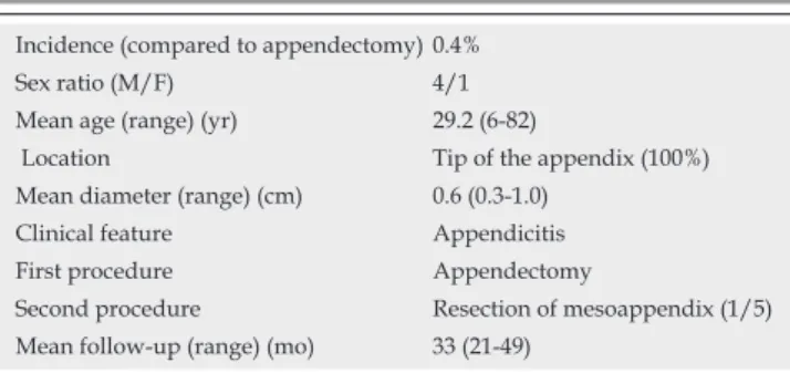
Carcinoid tumor of the appendix: A consecutive series from 1237 appendectomies
Texte intégral
Figure
Documents relatifs
Five of these boulders (with six carvings of stepped shrines), which were photographed by Tashi Ldawa in 2007, were broken in recent years to be used as construction material –
C) Individual weighted residuals (IWRES) with respect to time. Population analysis of experimental tumor growth kinetics. A) Visual predictive checks assess goodness-of-fit for
Interpolated recall vs precision averages plot Average precision statistics and box plot Average precision comparison to median plot Document cutoff levels vs precision at DCL
Interpolated recall vs precision averages plot Average precision statistics and box plot Average precision comparison to median plot Document cutoff levels vs precision at DCL
Range: The difference between the maximum and the minimum of a set of observations C.Var: Coefficient of variation, the spread of a set of data as a proportion of its mean Geom.:
Interpolated recall vs precision averages plot Average precision statistics and box plot Average precision comparison to median plot Document cutoff levels vs precision at DCL
AH-ROBUST-CLEF2008 Track Overview Results and Graphs AH-ROBUST-MONO-EN-TEST-CLEF2008 Statistics for Precision at the 0% interpolated recall level.. Topic Min 1st Q 2nd Q 3rd Q Max
Interpolated recall vs precision averages plot Average precision statistics and box plot Average precision comparison to median plot Document cutoff levels vs precision at DCL
