Bile acid-based drug delivery systems for enhanced
doxorubicin encapsulation: Comparing hydrophobic
and ionic interactions in drug loading and release
Alexander J. Cunningham1, Mattieu Robinson3, Xavier Banquy2, Jeanne Leblond-Chain2, and X.X. Zhu1,*
1 Département de Chimie, Université de Montréal, C.P. 6128, Succ. Centre-ville, Montréal, QC,
H3C 3J7, Canada
2 Faculté de Pharmacie, Université de Montréal, C.P. 6128, Succ. Centre-ville, Montréal, QC,
H3C 3J7, Canada
3 Département de Gérontologie, Université de Sherbrooke, Sherbrooke, QC, J1H 4C4, Canada
ABSTRACT
Doxorubicin (Dox) is a drug of choice in the design of drug delivery systems directed towards breast cancers, but is often limited by loading and control over its release from polymer micelles. Bile acid-based block copolymers present certain advantages over traditional polymer-based systems for drug delivery purposes, since they can enable a higher drug loading via the formation of a reservoir through their aggregation process. In this study, hydrophobic and electrostatic interactions are compared for their influence on Dox loading inside cholic acid-based block copolymers. Poly(allyl glycidyl ether) (PAGE) and poly(ethylene glycol) (PEG) were grafted from the cholic acid (CA) core yielding a star-shaped block copolymer with 4 arms
(CA-(PAGE-b-PEG)4) and then loaded with Dox via a nanoprecipitation technique. A high Dox loading of 14
wt% was achieved via electrostatic as opposed to hydrophobic interactions with or without oleic acid as a cosurfactant. The electrostatic interactions confer a pH responsiveness to the system. 50% of the loaded Dox was released at pH 5 in comparison to 12% at pH 7.4. The nanoparticles with Dox loaded via hydrophobic interactions, did not show such a pH responsiveness. The systems with Dox loaded via electrostatic interactions showed the lowest IC50 and highest
cellular internalization indicating the pre-eminence of this interaction in Dox loading. The blank formulations are biocompatible and did not show cytotoxicity up to 0.17 mg/mL. The new functionalized star block copolymers based on cholic acid show great potential as drug delivery carriers.
Keywords: Bile acids, doxorubicin, pH-responsive, drug delivery systems
INTRODUCTION
Breast cancer is the second most prevalent and deadly form of cancer in women. It is currently treated with a combination of surgery and chemotherapy.1 Over the course of chemotherapy, not
only will the patient suffer greatly from adverse effects, but the cancer often develops resistance mechanisms limiting the therapeutic outcome of the treatment.2 A common strategy to diminish
drug delivery system. Doxorubicin (Dox) is currently used for breast cancer treatment, but is characterized with severe toxicity due to its off-target effects. Over the past decades, a great deal of research effort has been directed at encapsulating chemotherapeutics in drug delivery systems to increase their safety and transport efficiency to the cancer tissues. Toward this end, polymer-based micelles have attracted much attention with several formulations reaching clinical trials.3, 4
Polymer-based micelles of a core-shell structure are typically aggregates of polymer chains with a hydrophobic core and hydrophilic corona. They are often the preferred platform for antitumor drug formulations due to their unique core-shell structure, tunable size and shape, ease of synthesis and freedom in design and structure.5, 6 Much research work has focused on the
loading of therapeutically relevant molecules with hydrophobic character inside the core of these micelles.7-10 For these systems, the drug loading level is greatly influenced by the hydrophobic
interaction of the core-forming block and the drug.11 Unfortunately, these formulations are often
plagued with a low drug loading and instability in vivo.12-14 Strategies to circumvent this
limitation include adjustment of the hydrophobic-hydrophilic balance,15-17 improvement of the
polymer-drug interactions,18-21 cross-linking,22-25 or conjugation of the drug to the polymer via
covalent bonding.15, 26-29 However, these strategies are often met with challenges, particularly
slow, insufficient release. For example, release of a drug conjugated to the polymer necessitates the cleavage of a covalent bond, which may slow down the release. Non-covalent interactions such as ionic interactions and hydrogen bonding may also favour drug loading.30-38 Dox bears a
positive charge at physiological pH and is composed of aromatic rings favoring non-covalent interactions via aromatic stacking. Many reports demonstrate the superiority of ionic interactions offering higher drug loading and better stability, and stimuli-sensitive release in response to the environmental pH.30, 32-34 Despite recent advancements in polymer-based systems, achieving high
drug loading and improving tissue biodistribution remain limiting factors.6, 15, 39 In an effort to
address the first issue, we intend to study the interaction and forces that govern Dox loading in the context of bile acid-based polymer micelles.
We have previously shown the advantage of using bile acids in the design of drug delivery systems.40-42 Micelles formed by bile acid-based polymers are less densely packed than linear
polymers of similar structure,43 creating an internal reservoir that may hold a larger amount of
bile acid-based nanoparticles.41 These formulations reached high loading contents (35 wt%) and
high encapsulation efficiencies (>89 %) as well as improved bioavailability and pharmacokinetics of the drug. Dox presents a different challenge with different interaction forces. Therefore, we modified the design of the copolymer based on cholic acid. Hydrophobic poly(allyl glycidyl ether) (PAGE) and hydrophilic poly(ethylene glycol) (PEG) blocks were attached sequentially via anionic polymerization onto a cholic acid (CA) core to yield a star-shaped copolymer of 4 arms, CA-(PAGE-b-PEG)4 (Scheme 1). The star-shaped polymers based
on bile acids have demonstrated their advantages in biocompatibility and drug loading.44 The
allyl groups of PAGE can be functionalized post-polymerization. Incorporating COOH moieties in the polymer backbone can facilitate Dox loading via electrostatic interactions and improve the stability of the complex. The different interactions between Dox and the polymer aggregates (hydrophobic, intrinsic electrostatic, and extrinsic electrostatic) are studied to compare their impact on drug loading, stability, drug release, and in vitro cytotoxicity of the new formulations.
MATERIALS AND METHOD Materials
All chemical reagents were purchased from Sigma-Aldrich and used without further purification unless stated otherwise. For the anionic polymerization, dimethyl sulfoxide (DMSO) and allyl glycidyl ether (AGE) was dried overnight with calcium hydride and distilled immediately prior to use. Ethylene oxide gas was passed through a column of calcium hydride and condensed with dry ice and acetone for quantification before adding to the reaction vessel. Tetrahydrofuran (THF) was dried using sodium under reflux, and methanol was dried using magnesium sulphate. Doxorubicin hydrochloride was purchased from Tecoland.
Synthesis of CA-(PAGE-b-PEG)4 via anionic polymerization
The synthesis and functionalization of the polymers are shown in Scheme 1. For the anionic polymerizations, all glassware was flame-dried under vacuum and purged three times with argon before use. The ethanolamine derivative of cholic acid with four hydroxyl groups was synthesized according to a previous method.45, 46 For anionic polymerization, 5β-cholanoamide
in THF was added (0.44 mol/L, 1.66 mmol, 1 eq., 3.74 mL) dropwise using a canula under high pressure. AGE was distilled and added (7.6 g, 66.4 mmol, 40 eq.) dropwise using a canula under high pressure. The anionic polymerization was initiated by immersing in an oil bath at 40 °C and allowed to proceed for 24 h to allow the consumption of all the AGE monomers. Then, dry ethylene oxide (17 mL, 332.2 mmol, 200 eq.) chilled in dry ice/acetone was introduced into the flask and polymerized for another 24 h. The reaction was stopped by quenching the reaction with concentrated hydrochloric acid. The DMSO solution was extracted with hexane (3 x 10 mL) to remove the naphthalenide. Distilled water was added to the DMSO solution and the mixture was dialyzed against distilled water (48 h) through a membrane with 3,500 Da molecular weight cut-off (MWCO) to remove all unreacted monomers.
Scheme 1. Synthesis of cholic acid-based star polymers via anionic polymerization and the
Functionalization of CA-(PAGE-b-PEG)4 to CA-(PAGE-COOH-b-PEG)4
The purified and dried CA-(PAGE-b-PEG)4 polymers (2 g, 0.14 mmol) were dissolved in dry
methanol (20 mL). Azobisisobutyronitrile (0.5 g, 3.2 mmol), and 3-mercaptopropionic acid (9.0 g, 84.4 mmol) were dissolved in the solution which was refluxed at 70 °C overnight. The reaction was stopped by cooling and methanol was removed. Finally, distilled water was added to dissolve product and remaining reactants and the solution was dialyzed against distilled water for 48 h and dried to yield CA-(PAGE-COOH-b-PEG)4.
Characterization methods
The molar mass of the polymers was determined by size exclusion chromatography (SEC) running on THF as eluent on a Breeze system from Waters equipped with a 717 plus autosampler, a 1525 Binary HPLC pump, and a 2410 refractive index detector and three consecutive styragel columns with a flow rate of 1.0 mL/min and at 30°C. Polystyrene standards were used for calibration. All samples were filtered on a PTFE 0.2 μm filters prior to injection.
1H-NMR spectra were recorded on a Bruker AV400 spectrometer operating at 400 MHz and
samples were dissolved in d6-DMSO. Dynamic light scattering (DLS) measurements were
performed on a Malvern Zetasizer NanoZS instrument equipped with a He-Ne laser with a wavelength of 633 nm, and at a scattering angle of 173.5°. Intensity-averaged hydrodynamic diameters of the dispersions were obtained using the non-negative least-squares algorithm (NNLS). Disposable cuvettes were used, and the samples were filtered using 0.45 μm Nylon filters prior to measurements. Sample concentration used was 1 mg/mL for both blank and loaded micelles and samples were run at room temperature. Dox concentration was determined by recording the fluorescence spectra on a Cary eclipse fluorescence spectrophotometer with excitation and emission wavelengths of 485 and 590 nm, respectively, and a bandwidth of 5 nm. The transmission electron microscope (TEM) images were obtained on a FEI Tecnai 12 at 80kV and equipped with an AMT XR80C CCD camera system. TEM samples were prepared by drop-casting blank micelles on a carbon-coated copper grid (300 mesh, Carbon Type-B, Ted Pella Inc.).
Doxorubicin loading and release
The encapsulation of Dox into the micelles is illustrated in Fig. 1. To compare hydrophobic and electrostatic interactions, different loading strategies were selected. Hydrophobic interaction was used for Dox loading into the CA-(PAGE-b-PEG)4 system and TEA was added to remove
the charge on Dox, followed by the removal of the organic salt after the formation of the encapsulates. Electrostatic interactions were used for the other two systems with CA-(PAGE-b-PEG)4 + OA and CA-(PAGE-COOH-b-PEG)4) for which the charged form of Dox was used
directly without reacting with TEA. Briefly, for the CA-(PAGE-COOH-b-PEG)4 sample, the
copolymers (10 mg) were dissolved in methanol (1 mL) along with Dox (2 mg) with gentle stirring. The solution was added dropwise into 10 mL PBS buffer (pH 7.4, 10 mM) over a period of 10 min using a syringe pump. The resulting PBS solution was stirred gently for 10 min before purification via dialysis (MWCO 6,000-8,000 Da) against 1 L PBS (pH 7.4, 10 mM) for 24 h, changing the outer media twice. Samples of the outer media were taken before replenishment to quantify the amount of Dox released. The same procedure was repeated for the CA-(PAGE-b-PEG)4 + OA sample, with a slight modification by using 10 mg of copolymer and 0.126 mg of
OA. For the CA-(PAGE-b-PEG)4 system, the same procedure was followed by using 10 mg of
CA-(PAGE-b-PEG)4 alone and Dox which was reacted overnight with 1.5 eq TEA prior to the
nanoprecipitation. Dox loading was measured by quantifying the amount of Dox released during the purification and subtracting this value from the amount of Dox in the feed. The quantification was achieved by taking 1 mL of the outer water, mixing with 1 mL DMSO, measuring the fluorescence intensity on a Cary fluorescence spectrometer (excitation 485 nm, emission 590 nm), and comparing with a calibration curve for Dox in DMSO/PBS (1:1 Vol.).
DLC (wt %)=weight of loaded drug
weight of copolymer x 100 % EE (%)=weight of loaded drug
weight of drug∈feed x 100 %
The Dox release from the different formulations was determined with a dialysis method at different pH. Briefly, 2 mL of the drug-loaded formulations were placed in a dialysis bag (MWCO 6,000-8,000 Da) and dialyzed against 225 mL of the appropriate buffer at 37 °C; PBS
pH 7.4 or acetate buffer pH 5 10 mM for the experiments conducted at pH 7.4 and 5, respectively. 1 mL of the outer media was taken at regular intervals and replenished with fresh buffer. The amount of Dox released was quantified by fluorescence spectroscopy with the appropriate calibration curve. All experiments were conducted in triplicates and the results are presented as average values.
Figure 1. Formulations based on different drug-polymer interactions. For
CA-(PAGE-b-PEG)4, Dox is loaded via hydrophobic interactions between the drug and the core of the micelle
composed of cholic acid and hydrophobic PAGE. For CA-(PAGE-COOH-b-PEG)4, Dox is
loaded via intrinsic electrostatic interactions between the drug and the pendant COOH groups. For CA-(PAGE-b-PEG)4 + OA, the electrostatic interactions are perceived as extrinsic because
they appear on OA only. The hydrophobicity of the OA aliphatic chains may drive the loading of Dox more toward the hydrophobic core.
In vitro cytotoxicity assay
HeLa cells were grown in Dulbecco’s Modified Eagle’s Medium (DMEM) medium supplemented with 10% Fetal Bovine Serum (FBS). MTT assay was used to determine the cytotoxicity of the blank formulations and the IC50 for free Dox and all three Dox-loaded
formulations. MTT assay was conducted according to manufacturer’s protocol. HeLa cells were plated on a 96-well plate at a seeding density of 5,000 cells per well in 200 μL DMEM and allowed to adhere overnight (37°C, 5% CO2, 12 h). 40 μL of the formulation to be tested was
added to each well. The plates were incubated for 48 h (37°C, 5% CO2). Medium was removed
and replaced with 100 μL of fresh medium. 10 μL of MTT solution (5 mg/mL) was added to each well and incubated (37°C, 5% CO2) for 4 h. DMEM medium was carefully removed from
each well and replaced with DMSO. MTT crystals were dissolved and the absorbance was read at 590 nm using a plate reader. Cytotoxicity is reported as compared to absorbance measured for control (untreated cells). To calculate the IC50, a linear regression was intrapolated by drawing a
straight line through the inflection in the viability curve. The IC50 value was obtained by setting y
= 50% from the linear equation obtained and solving for x.
Fluorescence activated cell sorting (FACS) assay
HeLa cells were grown in DMEM medium supplemented with 10% FBS. Cells were seeded in a 24-well plate at a seeding density of 5 x 10⁴ cells/well and allowed to adhere overnight (37°C,
5% CO2). The cells were washed with PBS and replenished with fresh medium. Free Dox and
Dox-loaded formulations were added to the cells to yield a final concentration of 1 and 10 μM Dox in 550 μL DMEM. After 1 h incubation, the cells were washed with PBS, trypsinized (100 μL of 0.25 % Trypsin/EDTA), and suspended in FACS buffer (400 μL, 95% PBS, 5% FBS, 1 mM EDTA). The cells were observed on a FACScalibur flow cytometer (BD Biosciences, San Jose, CA, USA). Dox concentration internalized in the cells was calculated with respect to control samples (untreated cells). The experiment was done in triplicates.
RESULTS AND DISCUSSION Drug formulation
To study the interactions driving the Dox loading, three formulations were prepared (Figure 1). The first is composed of CA-(PAGE-b-PEG)4 alone, in which only hydrophobic interactions are
present to ensure Dox loading inside the core of the micelles. The hydrophobicity is conferred from the hydrophobic face of the cholic acid and the hydrophobic PAGE block, while the hydrophilic PEG block promotes micelle formation and stability in the aqueous environment. Numerous studies point toward the use of PEG chain length of about 2,000 g/mol as the optimal chain length for successful drug delivery.47 Our previous results showed that the ratio of
PAGE/PEG controls the hydrophobicity/hydrophilicity of the system and affects the micelle aggregation.48 When the PAGE block is too long compared to the PEG block (a PAGE/PEG ratio
of 0.7 or greater), the hydrophobicity of the individual polymer chains causes the micelles to aggregate into species with large diameters. A block length of 19 for PAGE and of 30 for PEG (ratio of 0.63) should be appropriate for the polymer to maintain micellar stability and maximize Dox loading. The second formulation is composed of CA-(PAGE-b-PEG)4 supplemented with
oleic acid (OA). OA is used to mimic the carboxylic acid content on the CA-(PAGE-COOH-b-PEG)4 formulation. The carboxylic acid groups of OA can complex electrostatically to Dox and
the aliphatic chain of OA imparts an enhanced hydrophobicity thereby increasing Dox loading while reducing its premature release at physiological pH.49 Here the electrostatic interactions are
extrinsic to the system; the COOH group of OA and the increased hydrophobicity imparted by OA drive the loading of Dox inside the nanoparticles. The third formulation is composed of functionalized PAGE blocks bearing carboxylic acid groups (CA-(PAGE-COOH-b-PEG)4).
Here, electrostatic interactions between the NH2 groups of Dox and the COOH groups attached
on PAGE drives the loading of Dox into the middle block in addition to the hydrophobic core. In this situation, the electrostatic interactions may be tuned by varying the PAGE block length.
Polymer synthesis and characterization
The star-shaped block copolymers were synthesized as shown in Scheme 1. An ethanolamine derivative of CA with four hydroxyl groups was used as initiator for the anionic polymerization of allyl glycidyl ether (AGE), followed by the addition of ethylene glycol (EG) to afford a
star-shaped CA-(PAGE-b-PEG)4 with 4-arms composed of hydrophobic PAGE and hydrophilic PEG
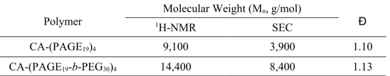
blocks. The copolymer obtained was analyzed by SEC and 1H-NMR (Figures S1 and S2). The
SEC traces show monomodal, narrow distributions and an increase in Mn when going from
CA-(PAGE)4 to CA-(PAGE-b-PEG)4. The NMR signals at 0.58, 0.81, and 0.92 ppm were assigned to
the 18-CH3, 19-CH3, and 21-CH3 groups, respectively, on the cholic acid backbone. These peaks
were used as reference for the analysis of the peaks at 5.79-5.92 and 3.35-3.60 ppm, which correspond respectively to the CH2 protons on the PAGE block and on both PAGE and PEG. The
block copolymer was determined to be CA-(AGE19-b-EG30)4 with 19 monomer units of AGE and
30 monomer units of EG (Table 1). The presence of four arms was confirmed by reacting the polymers with trifluoroacetic anhydride and by the appearance of the methylene protons at 4.5 ppm, corresponding to protons of the PEG terminal CH2 unit coupled with the trifluoroacetic
group. Then, the allyl side chains from the PAGE block were functionalized with mercaptopropionic acid with close to 100% functionalization as confirmed by 1H-NMR (Figure
S2).
Table 1. Characteristics of the polymer samples obtained from the anionic polymerization as
studied by SEC and 1H-NMR.
Polymer
Molecular Weight (Mn, g/mol)
Đ
1H-NMR SEC
CA-(PAGE19)4 9,100 3,900 1.10
CA-(PAGE19-b-PEG30)4 14,400 8,400 1.13
The critical micellar concentration (CMC) of the bile acid derivative in water was measured with fluorescence spectroscopy using pyrene as a probe.50 Pyrene fluorescence is measured with
increasing concentration of the block copolymers and the CMC is calculated from a plot of the excitation intensity ratio I383/I373 as a function of polymer concentration. The CMC for
CA-(PAGE-b-PEG)4 is determined to be 20 μg/mL (Figure S3), comparable to typical polymer-based
micellar systems found in the literature.43, 51-53
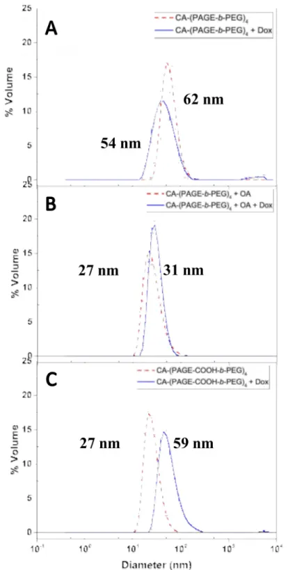
Particle size and distribution were analyzed with dynamic light scattering (DLS) and transmission electron microscopy (TEM), before and after Dox loading. The DLS results show
that CA-(PAGE-b-PEG)4 forms larger aggregates (d = 62 ± 18 nm) than (CA-(PAGE-b-PEG)4
with OA, and CA-(PAGE-COOH-b-PEG)4) (Figure 2). CA-(PAGE-b-PEG)4 mixed with OA, a
surfactant, forms mixed micelles smaller in diameter (27 ± 10 nm). The negatively charged COOH moieties may induce an electrostatic repulsion between individual polymer chains leading to smaller micelles. These results are corroborated with the TEM images showing spherical aggregates with a dense core (Figure 3). Bile acid-based micelles have a hydrophobic β-face that drives the micellization process. This micellization is affected by the presence of polymer chains on the surface of the bile acids. In the case of CA-(PEG)4, the literature shows
that the length of the PEG blocks has effect on the size of the cavity formed during micellization.43 The size of the cavity is governed by steric repulsion between individual PEG
chains; the longer the PEG chains, the stronger the repulsion which results in a smaller cavity and a smaller micelle. The current system should be expected to behave similarly, i.e., the electrostatic repulsion between the individual polymer chains governs the cavity size. The micelles obtained with the CA-(PAGE-COOH-b-PEG)4 are smaller than those obtained with
CA-(PAGE-b-PEG)4 because the electrostatic repulsion between the individual polymer chains is
stronger than the steric repulsion which leads to a smaller micelle. However, the hydrophobic β-face of cholic acid causes micellarization of the star-shaped block copolymers and leads to the appearance of a dense core as shown in the TEM images (Fig. 3).
Table 2 shows the zeta-potentials measured for the nanoparticles. The CA-(PAGE-b-PEG)4
micelles have a zeta-potential close to neutral with a value of -3.9 ± 0.4 mV, whereas the presence of the negatively charged COOH moieties lead to larger negative values, -9.2 ± 0.7 mV for CA-(PAGE-b-PEG)4 with OA and -17.2 ± 0.1 mV for the CA-(PAGE-COOH-b-PEG)4
Figure 2. The volume-size distribution of (A) CA-(PAGE-b-PEG)4 with and without Dox, (B) CA-(PAGE-b-PEG)4 + OA with and without Dox, and (C) CA-(PAGE-COOH-b-PEG)4 with and
without Dox as determined by DLS. Blank micelles and formulations are obtained at 1 mg/mL and filtered with 0.45 µm PES filters prior to DLS measurements.
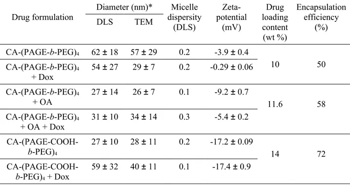
Table 2. Micellar size, zeta-potential and Dox-loading of the formulations based on star-shaped block copolymers. Drug formulation Diameter (nm)* Micelle dispersity (DLS) Zeta-potential (mV) Drug loading content (wt %) Encapsulation efficiency (%) DLS TEM CA-(PAGE-b-PEG)4 62 ± 18 57 ± 29 0.2 -3.9 ± 0.4 10 50 CA-(PAGE-b-PEG)4 + Dox 54 ± 27 29 ± 7 0.2 -0.29 ± 0.06 CA-(PAGE-b-PEG)4 + OA 27 ± 14 26 ± 7 0.1 -9.2 ± 0.7 11.6 58 CA-(PAGE-b-PEG)4 + OA + Dox 31 ± 10 34 ± 14 0.3 -5.4 ± 0.2 CA-(PAGE-COOH-b-PEG)4 27 ± 10 28 ± 11 0.2 -17.2 ± 0.09 14 72 CA-(PAGE-COOH-b-PEG)4 + Dox 59 ± 32 40 ± 11 0.1 -17.4 ± 0.9
*Diameters of the blank and Dox-loaded micelles are listed. The DLS samples were studied at a concentration of 1 mg/mL in 10 mM PBS at 7.4 pH with 154 mM NaCl. The same samples were dried and used for the TEM experiments.
Doxorubicin loading and release
We hypothesize that electrostatic interactions enhance the loading of Dox in comparison to Dox via hydrophobic interaction. To study these interaction forces, three different formulations were loaded with equal amount of Dox but with different loading strategies. The encapsulation efficiencies and drug loading contents are listed in Table 1. The CA-(PAGE-b-PEG)4 aggregates
showed drug loading content of 10 wt% and an encapsulation efficiency of 50%. Adding OA to the formulation enabled a higher drug loading (11.6 wt%) and encapsulation efficiency (58%), most likely due to an increased hydrophobicity of the Dox complex.49 Functionalizing the bile
acid-based polymers with pendant COOH moieties resulted in an improvement of the drug loading to 14 wt% and loading efficiency to 72%.
In the case of CA-(PAGE-b-PEG)4 and CA-(PAGE-b-PEG)4 with OA, there is no significant
systems. The decrease in micelle size observed for CA-(PAGE-b-PEG)4 falls within the error of
the instrument and this small decrease observed with the DLS is not significant. A similar observation was made previously for bile acid-based systems;54 there are no appreciable changes
in micelle sizes when the Dox loading is carried out using hydrophobic interactions. TEM images in Figure 3 show that upon Dox loading, both CA-(PAGE-b-PEG)4 and
CA-(PAGE-b-PEG)4 with OA retain their spherical shape. In contrast, the Dox-loaded
CA-(PAGE-COOH-b-PEG)4 micelles showed the formation of aggregates larger in size (Figure 2) and the spherical
micelles changed to vesicular-shaped polymersomes (Figure 3).
Zeta-potential measurements showed a slight increase in the surface charge of the micelles upon Dox loading when compared to the blank micelles (Table 1) due to the presence of positively charged Dox loaded in the reservoir of the polymersome and adsorbed on the surface of the micelles. Dox has amphiphilic properties and has been shown to be distributed at the core-shell interface with its anthracycline ring inserted in the hydrophobic core and the amine exposed at the surface.55 Moreover, Dox has a pKa of 9.53, therefore at pH 7.4 of PBS, the amine is
positively charged. For the case of CA-(PAGE-b-PEG)4 system there was a significant change
upon Dox loading where the zeta-potential increased from -3.9 to -0.29. Here, we believe that Dox is found in the core and at the interface of the PAGE and PEG blocks, which causes the zeta-potential of the micellar surface to become less negative. In the case of
CA-(PAGE-COOH-b-PEG)4, 14 wt% of Dox loaded in 10 mg polymer is equivalent to 76 negative charges of the
polymer vs 3.7 positive charges of Dox. Therefore, even after loading, the surface charge of the micelles remains highly negatively charged due to the presence of the high amount of negative charges and this is what is observed in the zeta-potential experiment. In the case of
CA-(PAGE-b-PEG)4 + OA system, the negative charges of OA seem to be less exposed due to micellar
packing, showing in general a less negative zeta-potential.
In vitro Dox release profiles at 37 °C were obtained at two different pH values, mimicking the endosomal pH at 5 encountered by the nanoparticles upon cellular entry and the physiological pH at 7.4 sensed by the nanoparticles during transport. The objective of this experiment is to compare the release kinetics of the different formulations to assess the impact of the interaction forces on the release profile. Moreover, the presence of the COOH moieties in the CA-(PAGE-COOH-b-PEG)4 formulation bestows pH-responsiveness to the system, an advantage compared
to hydrophobic entrapment alone. The results shown in Figure 4 compare the Dox release profiles at both pH values for all three formulations. As a control, free Dox was loaded in the dialysis bag and its release was monitored over time. The declivity observed for free Dox at pH 7.4 can be explained by the possible degradation of the drug as reported previously.56, 57 At pH 5
the free Dox is more stable.58 The degradation of Dox was also observed for the
CA-(PAGE-b-PEG)4 with OA formulation at pH 7.4, but not for the other two formulations for which the
quantity of Dox released was small so that the degradation was not measured at an appreciable amount.
Figure 3. Transmission electron micrograph images of the micelles formed: (top) blank
formulations and (bottom) Dox-loaded formulations (1 mg/mL samples). The enlarged inserts show more clearly the shape of the aggregates and the bars in the inserts are 50 nm in length.
All formulations show a biphasic release with an initial burst release within the first 8 h, followed by a slower, continuous release. The CA-(PAGE-b-PEG)4 formulation should be
pH-irresponsive but shows an increase in the release from 7.2 to 23% when the pH changes from 7.4 to 5. The higher solubility of Dox at pH 5 facilitates its release.59 For the CA-(PAGE-b-PEG)
4
3). In this case, Dox interacts electrostatically with the negative COOH moieties on the OA and the long aliphatic chain of OA provides a higher hydrophobicity for Dox to remain in the core.49
At pH 5, the COOH groups are partially protonated weakening their electrostatic interaction with Dox, while the core remains hydrophobic and keeps the drug inside. Therefore, lowering the pH does not significantly impact the release of Dox from the core of the micelle.60 OA and the
CA-(PAGE-b-PEG)4 forms mixed micelles that have a higher hydrophobic core. We believe that the
system with OA imparts enough hydrophobicity to Dox through its long aliphatic chain that after protonation the hydrophobic interaction between Dox and OA maintains Dox in the hydrophobic core. Moreover, not all of the carboxylic acid groups of OA are exposed to the surface (as can be seen in the zeta-potential results) such that at low pH not all are deprotonated and this maintains the Dox in the micelles. Finally, the CA-(PAGE-COOH-b-PEG)4 formulation shows the highest
difference between the two pHs, with a change in release from 16 to 51% when the pH changed from 7.4 to 5. For the CA-(PAGE-COOH-b-PEG)4 system, the carboxylic acid groups are more
exposed to the surface and are more easily protonated and this breaks its interaction with Dox causing a higher release at pH 5. This demonstrates the pH responsiveness of the formulation. The COOH groups on the polymer protonate at pH 5, thereby disrupting the electrostatic interaction between the polymer and the drug helping with its release. Moreover, this formulation shows a more stable drug loading at physiological pH than the other two formulations.
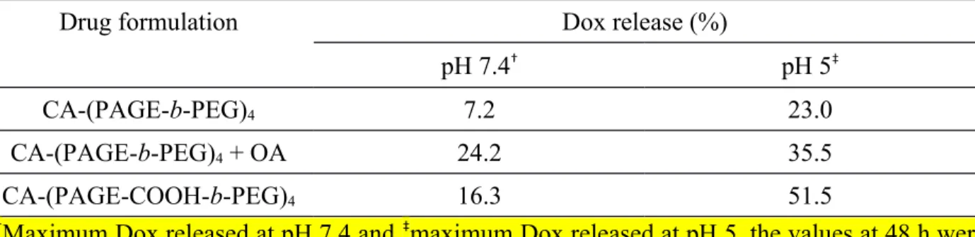
Table 3. The maximum amount of Dox released in response to pH changes.
Drug formulation Dox release (%)
pH 7.4† pH 5‡
CA-(PAGE-b-PEG)4 7.2 23.0
CA-(PAGE-b-PEG)4 + OA 24.2 35.5
CA-(PAGE-COOH-b-PEG)4 16.3 51.5
†Maximum Dox released at pH 7.4 and ‡maximum Dox released at pH 5, the values at 48 h were
Figure 4. Cumulative release of Doxorubicin from formulations at 37 °C (A) in PBS buffer at
pH 7.4 and (B) in acetate buffer at pH 5. Results show pH-responsiveness for the CA-(PAGE-COOH-b-PEG)4 formulation but not for the other two formulations.
In vitro cytotoxicity
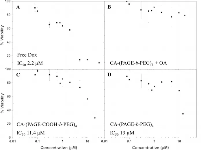
The toxicity of the blank formulations was tested in HeLa cells and the results are presented in Figure S6. All the formulations are non-toxic up to 0.17 mg/mL with over 80% cell viability;
consistent with the previous reports on bile acid micelles.44 To determine the efficiency of the
new bile acid-based drug delivery system, the IC50 of the different formulations were obtained on
HeLa cells and compared with that of free Dox. The IC50 of free Dox is 2.2 μM, in agreement
with values reported in the literature.22, 61 The CA-(PAGE-b-PEG)
4 formulation has an IC50 of 13
μM which is ca. 6 times higher than that of free Dox. For the CA-(PAGE-b-PEG)4 with OA
formulation, no IC50 was observed at the concentrations tested. Finally, for the
CA-(PAGE-COOH-b-PEG)4 formulation, the IC50 obtained is 11.4 μM, comparable to that of the
CA-(PAGE-b-PEG)4 formulation.
Figure 5. In vitro cytotoxicity obtained on HeLa cells after 48 h incubation of (A) free Dox
compared with the three blank formulations: (B) CA-(PAGE-b-PEG)4 with OA, (C)
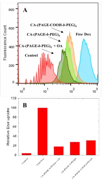
To clarify these results, cellular entry for the formulation was studied and compared with that of free Dox using flow cytometry (Figure 6). Free Dox is readily internalized inside the cell, but Dox in aggregates of both CA-(PAGE-b-PEG)4 and CA-(PAGE-COOH-b-PEG)4 is less readily
internalized. Although previous reports indicate the advantage of spherical over vesicular-shaped nanoparticles for cellular entry, the negative charge of the nanoparticles had a greater impact.62, 63
Presumably, the negative charges of CA-(PAGE-COOH-b-PEG)4 may interact with extracellular
proteins to facilitate membrane interaction and internalization, and consequently slightly lower the IC50.63 The higher IC50 observed may be a result of cellular entry. For the purpose of cellular
uptake Dox-loaded nanoparticles, the pH responsiveness of the CA-(PAGE-COOH-b-PEG)4
formulation does not bring an advantage over the neutral, non-responsive system. Dox release was not sufficiently rapid to promote endosomal escape and cellular uptake was the determining factor for the lower IC50. Surprisingly, the formulation with OA showed even less cellular entry
than the other two formulations. The presence of OA affected the cellular entry hence increasing further the IC50, probably due to its impact on the structure of the micelles or the surface
properties. The TEM images shows that the micelles prepared from OA are much smaller than those of the other two formulations, but are also dense and irregular in shape. Reports demonstrated the advantage of smaller nanoparticles for cellular entry, but also the effect of particle shape on cellular entry.62, 63 It is believed that the irregular shape of the Dox-loaded
nanoparticles caused by the presence of the OA reduced the cellular internalization process observed via FACS. The drug delivery systems used here demonstrated the advantage of achieving a higher stability avoiding unspecific uptake of Dox, but further studies are necessary to improve the specific nanoparticle internalization in cancer cells and optimize the pH-responsiveness to enable quick and adequate endosomal escape.
Figure 6. Flow cytometry uptake profiles for formulations of free Dox and with
CA-(PAGE-b-PEG)4, CA-(PAGE-b-PEG)4 with OA, and CA-(PAGE-COOH-b-PEG)4. Experiment was
performed in triplicates with HeLa cells with 2 h incubation; non-treated HeLa cells were used as control.
CONCLUSION
Cholic acid-based micelles formed stable micellar aggregates with high Dox loading. Electrostatic interactions used in the formulation with CA-(PAGE-COOH-b-PEG)4 led to higher
of Dox via electrostatic interactions confers pH-responsiveness to the drug delivery system, increasing Dox release from 12 to 50% when the pH was lowered from 7.4 to 5. We have previously used OA to increase loading of hydrophobic itraconazole in a CA-(PEG)4 system.44 In
this study, OA did not provide an advantage for Dox loading in comparison to the CA-(PAGE-COOH-b-PEG)4 system, since some of the OA molecules may be buried close to the core of the
micelle and become less accessible for electrostatic binding with Dox. Cholic acid-based drug delivery systems present advantages of higher Dox loading and pH-responsiveness in release. The proposed system can be used to alleviate side effects caused by premature release of Dox during systemic administration. Dox became more stable and was released at 50% at pH 5 but only 16% at pH 7.4. Therefore, the system can be used to release Dox at the tumor site since tumor micro-environments are known to be more acidic than the healthy tissues. The blank nanoparticles (without Dox) used in this study are non-toxic with over 80% cell viability at concentrations up to 0.17 mg/mL. After loading with Dox, the nanoparticles formulated with both CA-(PAGE-b-PEG)4 and CA-(PAGE-COOH-b-PEG)4 formulations showed higher IC50
values than free Dox, probably due to the lower cellular internalization of the Dox-loaded micellar aggregates. The Dox-loaded CA-(PAGE-b-PEG)4 formulation with OA did not show an
IC50 in the measured concentration range and had lower cellular internalization than the other
two formulations. Clearly, cellular entry is an issue that needs to be addressed to achieve an overall successful drug delivery platform. For this purpose, the attachment of targeting ligand on the surface of the nanoparticle is currently being tested. Further studies are underway to determine the efficiency of the new bile acid-based drug delivery system on drug resistant cancer cell lines and cytotoxic drugs with poor cellular entry.
ACKNOWLEDGMENTS
Financial support from NSERC of Canada is gratefully acknowledged. AC and XXZ are members of CSACS funded by FRQNT and GRSTB funded by FRSQ. AC thanks the Camille Sandorfy, Charron Lam, and GRSTB Ph.D. scholarships. The authors thank Mrs. Mouna Rabeb-Derbali and Dr. Wilms E. Baille for their technical support.
ASSOCIATED CONTENT
Supporting information including the following figures is available. SEC traces and 1H-NMR
spectra of block copolymers, critical micellar concentration, Dox calibration curves and in vitro cytotoxicity of formulations.
REFERENCES
1. Global Burden of Disease Cancer, C. Global, regional, and national cancer incidence, mortality, years of life lost, years lived with disability, and disability-adjusted life-years for 32 cancer groups, 1990 to 2015: A systematic analysis for the global burden of disease study. JAMA
Oncology 2017, 3, (4), 524-548.
2. Gonzalez-Angulo, A. M.; Morales-Vasquez, F.; Hortobagyi, G. N. Overview of resistance to systemic therapy in patients with breast cancer. Adv Exp Med Biol 2007, 608, 1-22.
3. Cabral, H.; Kataoka, K. Progress of drug-loaded polymeric micelles into clinical studies. J
Control Release 2014, 190, 465-476.
4. Gong, J.; Chen, M.; Zheng, Y.; Wang, S.; Wang, Y. Polymeric micelles drug delivery system in oncology. J Control Release 2012, 159, (3), 312-323.
5. Chan, J. M.; Valencia, P. M.; Zhang, L.; Langer, R.; Farokhzad, O. C., Polymeric Nanoparticles for Drug Delivery. In Cancer Nanotechnology: Methods and Protocols, Grobmyer, S. R.; Moudgil, B. M., Eds. Humana Press: Totowa, NJ, 2010; pp 163-175.
6. Kataoka, K.; Harada, A.; Nagasaki, Y. Block copolymer micelles for drug delivery: design, characterization and biological significance. Adv Drug Deliver Rev 2001, 47, (1), 113-131. 7. Kim, D. W.; Kim, S. Y.; Kim, H. K.; Kim, S. W.; Shin, S. W.; Kim, J. S.; Park, K.; Lee, M. Y.; Heo, D. S. Multicenter phase II trial of Genexol-PM, a novel Cremophor-free, polymeric micelle formulation of paclitaxel, with cisplatin in patients with advanced non-small-cell lung cancer. Ann of Oncol 2007, 18, (12), 2009-2014.
8. Lee, K. S.; Chung, H. C.; Im, S. A.; Park, Y. H.; Kim, C. S.; Kim, S.-B.; Rha, S. Y.; Lee, M. Y.; Ro, J. Multicenter phase II trial of Genexol-PM, a Cremophor-free, polymeric micelle formulation of paclitaxel, in patients with metastatic breast cancer. Breast Cancer Res Tr 2008,
108, (2), 241-250.
9. Hamaguchi, T.; Matsumura, Y.; Suzuki, M.; Shimizu, K.; Goda, R.; Nakamura, I.; Nakatomi, I.; Yokoyama, M.; Kataoka, K.; Kakizoe, T. NK105, a paclitaxel-incorporating micellar
nanoparticle formulation, can extend in vivo antitumour activity and reduce the neurotoxicity of paclitaxel. Br J Cancer 2005, 92, (7), 1240-1246.
10. Alexander, S.; Cosgrove, T.; Prescott, S. W.; Castle, T. C. Flurbiprofen Encapsulation Using Pluronic Triblock Copolymers. Langmuir 2011, 27, (13), 8054-8060.
11. Varshosaz, J.; Hasanzadeh, F.; Eslamdoost, M. Optimization of self-assembling properties of fatty acids grafted to methoxy poly(ethylene glycol) as nanocarriers for etoposide. Acta
Pharm 2012, 62, (1), 31-44.
12. Wang, X.; Wu, G.; Lu, C.; Zhao, W.; Wang, Y.; Fan, Y.; Gao, H.; Ma, J. A novel delivery system of doxorubicin with high load and pH-responsive release from the nanoparticles of poly (α,β-aspartic acid) derivative. Eur J Pharm Sci 2012, 47, (1), 256-264.
13. Shen, Y.; Jin, E.; Zhang, B.; Murphy, C. J.; Sui, M.; Zhao, J.; Wang, J.; Tang, J.; Fan, M.; Van Kirk, E.; Murdoch, W. J. Prodrugs Forming High Drug Loading Multifunctional
Nanocapsules for Intracellular Cancer Drug Delivery. JACS 2010, 132, (12), 4259-4265.
14. Zhang, Y.; Ren, T.; Gou, J.; Zhang, L.; Tao, X.; Tian, B.; Tian, P.; Yu, D.; Song, J.; Liu, X.; Chao, Y.; Xiao, W.; Tang, X. Strategies for improving the payload of small molecular drugs in polymeric micelles. J Control Release 2017, 261, 352-366.
15. Ke, X.; Ng, V. W. L.; Ono, R. J.; Chan, J. M. W.; Krishnamurthy, S.; Wang, Y.; Hedrick, J. L.; Yang, Y. Y. Role of non-covalent and covalent interactions in cargo loading capacity and stability of polymeric micelles. J Control Release 2014, 193, 9-26.
16. Gadelle, F.; Koros, W. J.; Schechter, R. S. Solubilization of Aromatic Solutes in Block Copolymers. Macromolecules 1995, 28, (14), 4883-4892.
17. Soliman, G. M.; Sharma, R.; Choi, A. O.; Varshney, S. K.; Winnik, F. M.; Kakkar, A. K.; Maysinger, D. Tailoring the efficacy of nimodipine drug delivery using nanocarriers based on A2B miktoarm star polymers. Biomaterials 2010, 31, (32), 8382-92.
18. Letchford, K.; Liggins, R.; Burt, H. Solubilization of hydrophobic drugs by methoxy poly(ethylene glycol)-block-polycaprolactone diblock copolymer micelles: theoretical and experimental data and correlations. J Pharm Sci 2008, 97, (3), 1179-90.
19. Sharma, A.; Soliman, G. M.; Al-Hajaj, N.; Sharma, R.; Maysinger, D.; Kakkar, A. Design and Evaluation of Multifunctional Nanocarriers for Selective Delivery of Coenzyme Q10 to Mitochondria. Biomacromolecules 2012, 13, (1), 239-252.
20. Yan, J.; Ye, Z.; Chen, M.; Liu, Z.; Xiao, Y.; Zhang, Y.; Zhou, Y.; Tan, W.; Lang, M. Fine Tuning Micellar Core-Forming Block of Poly(ethylene glycol)-block-poly(ε-caprolactone) Amphiphilic Copolymers Based on Chemical Modification for the Solubilization and Delivery of Doxorubicin. Biomacromolecules 2011, 12, (7), 2562-2572.
21. LeDevedec, F.; Houdaihed, L.; Allen, C. Anionic polymerization of an amphiphilic
copolymer for preparation of block copolymer micelles stabilized by π-π stacking interactions. J
Vis Exp 2016, 116, e54422.
22. Chen, W.; Meng, F.; Cheng, R.; Deng, C.; Feijen, J.; Zhong, Z. Facile construction of dual-bioresponsive biodegradable micelles with superior extracellular stability and activated
intracellular drug release. J Control Release 2015, 210, 125-133.
23. Shuai, X.; Merdan, T.; Schaper, A. K.; Xi, F.; Kissel, T. Core-Cross-Linked Polymeric Micelles as Paclitaxel Carriers. Bioconjugate Chem 2004, 15, (3), 441-448.
24. Wu, Y.; Chen, W.; Meng, F.; Wang, Z.; Cheng, R.; Deng, C.; Liu, H.; Zhong, Z. Core-crosslinked pH-sensitive degradable micelles: A promising approach to resolve the extracellular stability versus intracellular drug release dilemma. J Control Release 2012, 164, (3), 338-345. 25. Lin, W.; Kim, D. pH-Sensitive Micelles with Cross-Linked Cores Formed from
Polyaspartamide Derivatives for Drug Delivery. Langmuir 2011, 27, (19), 12090-12097. 26. Zhong, Y.; Goltsche, K.; Cheng, L.; Xie, F.; Meng, F.; Deng, C.; Zhong, Z.; Haag, R. Hyaluronic acid-shelled acid-activatable paclitaxel prodrug micelles effectively target and treat CD44-overexpressing human breast tumor xenografts in vivo. Biomaterials 2016, 84, 250-61. 27. Yin, S.; Huai, J.; Chen, X.; Yang, Y.; Zhang, X.; Gan, Y.; Wang, G.; Gu, X.; Li, J.
Intracellular delivery and antitumor effects of a redox-responsive polymeric paclitaxel conjugate based on hyaluronic acid. Acta Biomater 2015, 26, 274-85.
28. Xu, R.; Fisher, M.; Juliano, R. L. Targeted albumin-based nanoparticles for delivery of amphipathic drugs. Bioconjug Chem 2011, 22, (5), 870-8.
29. Prabaharan, M.; Grailer, J. J.; Pilla, S.; Steeber, D. A.; Gong, S. Amphiphilic multi-arm-block copolymer conjugated with doxorubicin via pH-sensitive hydrazone bond for tumor-targeted drug delivery. Biomaterials 2009, 30, (29), 5757-5766.
30. Lv, S.; Song, W.; Tang, Z.; Li, M.; Yu, H.; Hong, H.; Chen, X. Charge-Conversional PEG-Polypeptide Polyionic Complex Nanoparticles from Simple Blending of a Pair of Oppositely Charged Block Copolymers as an Intelligent Vehicle for Efficient Antitumor Drug Delivery.
Mol Pharm 2014, 11, (5), 1562-1574.
31. Wang, C. H.; Wang, W. T.; Hsiue, G. H. Development of polyion complex micelles for encapsulating and delivering amphotericin B. Biomaterials 2009, 30, (19), 3352-8.
32. Eckman, A. M.; Tsakalozou, E.; Kang, N. Y.; Ponta, A.; Bae, Y. Drug release patterns and cytotoxicity of PEG-poly(aspartate) block copolymer micelles in cancer cells. Pharm Res 2012,
29, (7), 1755-67.
33. Xu, H.; Yang, D.; Cai, C.; Gou, J.; Zhang, Y.; Wang, L.; Zhong, H.; Tang, X.
Dual-responsive mPEG-PLGA-PGlu hybrid-core nanoparticles with a high drug loading to reverse the multidrug resistance of breast cancer: an in vitro and in vivo evaluation. Acta Biomater 2015, 16, 156-68.
34. Xu, H.; Cai, C.; Gou, J.; Sui, B.; Jin, J.; Xu, H.; Zhang, Y.; Wang, L.; Zhai, Y.; Tang, X. Self-Assembled Monomethoxy (Polyethylene Glycol)-b-P(D,L-Lactic-co-Glycolic Acid)-b-P(L-Glutamic Acid) Hybrid-Core Nanoparticles for Intracellular pH-Triggered Release of
Doxorubicin. J Biomed Nanotechnol 2015, 11, (8), 1354-69.
35. Yang, C.; Tan, J. P. K.; Cheng, W.; Attia, A. B. E.; Ting, C. T. Y.; Nelson, A.; Hedrick, J. L.; Yang, Y.-Y. Supramolecular nanostructures designed for high cargo loading capacity and kinetic stability. Nano Today 2010, 5, (6), 515-523.
36. Yang, C.; Ebrahim Attia, A. B.; Tan, J. P. K.; Ke, X.; Gao, S.; Hedrick, J. L.; Yang, Y.-Y. The role of non-covalent interactions in anticancer drug loading and kinetic stability of
polymeric micelles. Biomaterials 2012, 33, (10), 2971-2979.
37. Su, W.; Luo, X.-h.; Wang, H.-f.; Li, L.; Feng, J.; Zhang, X.-Z.; Zhuo, R.-x. Hyperbranched Polycarbonate-Based Multimolecular Micelle with Enhanced Stability and Loading Efficiency.
Macromol Rapid Comm 2011, 32, (4), 390-396.
38. Kim, S. H.; Tan, J. P. K.; Nederberg, F.; Fukushima, K.; Colson, J.; Yang, C.; Nelson, A.; Yang, Y.-Y.; Hedrick, J. L. Hydrogen bonding-enhanced micelle assemblies for drug delivery.
Biomaterials 2010, 31, (31), 8063-8071.
39. Tsoi, K. M.; MacParland, S. A.; Ma, X.-Z.; Spetzler, V. N.; Echeverri, J.; Ouyang, B.; Fadel, S. M.; Sykes, E. A.; Goldaracena, N.; Kaths, J. M.; Conneely, J. B.; Alman, B. A.; Selzner, M.; Ostrowski, M. A.; Adeyi, O. A.; Zilman, A.; McGilvray, I. D.; Chan, W. C. W. Mechanism of hard-nanomaterial clearance by the liver. Nat Mater 2016, 15, (11), 1212-1221.
40. Le Dévédec, F.; Fuentealba, D.; Strandman, S.; Bohne, C.; Zhu, X. X. Aggregation Behavior of Pegylated Bile Acid Derivatives. Langmuir 2012, 28, (37), 13431-13440.
41. Le Devedec, F.; Strandman, S.; Baille, W. E.; Zhu, X. X. Functional Star Block Copolymers with a Cholane Core: Thermo-Responsiveness and Aggregation Behavior. Polymer 2013, 54, 3898-3903.
42. Shao, Y.; Jia, Y.-G.; Shi, C.; Luo, J.; Zhu, X. X. Block and Random Copolymers Bearing Cholic Acid and Oligo(ethylene glycol) Pendant Groups: Aggregation, Thermosensitivity, and Drug Loading. Biomacromolecules 2014, 15, (5), 1837-1844.
43. Despa, F.; Luo, J. T.; Li, J.; Duan, Y.; Lam, K. S. Cholic acid micelles-controlling the size of the aqueous cavity by PEGylation. Phys Chem Chem Phys 2010, 12, (7), 1589-1594.
44. Le Dévédec, F.; Strandman, S.; Hildgen, P.; Leclair, G.; Zhu, X. X. Pegylated bile acids for use in drug delivery systems enhanced solubility and bioavailability of itraconazole. . Mol
Pharm 2013, 10, 3057-3066.
45. Juo, J.; Giguère, G.; Zhu, X. X. Asymmetric Poly(ethylene glycol) Star Polymers with a Cholic Acid Core and Their Aggregation Properties. Biomacromolecules 2009, 10, 900-906. 46. Gouin, S.; Zhu, X. X. Synthesis of 3 alpha- and 3 beta-dimers from selected bile acids.
Steroids 1996, 61, (11), 664-9.
47. Knop, K.; Hoogenboom, R.; Fischer, D.; Schubert, U. S. Poly(ethylene glycol) in drug delivery: pros and cons as well as potential alternatives. Angew Chem Int Ed Engl 2010, 49, (36), 6288-308.
48. Li, C.; Lavigueur, C.; Zhu, X. X. Aggregation and thermoresponsive properties of new star block copolymers with a cholic acid core. Langmuir 2011, 27, (17), 11174-9.
49. Zhang, X.; Sun, X.; Li, J.; Zhang, X.; Gong, T.; Zhang, Z. Lipid nanoemulsions loaded with doxorubicin-oleic acid ionic complex: characterization, in vitro and in vivo studies. Die
Pharmazie 2011, 66, (7), 496-505.
50. Topel, Ö.; Çakır, B. A.; Budama, L.; Hoda, N. Determination of critical micelle
concentration of polybutadiene-block-poly(ethyleneoxide) diblock copolymer by fluorescence spectroscopy and dynamic light scattering. J Mol Liq 2013, 177, 40-43.
51. Allen, C.; Maysinger, D.; Eisenberg, A. Nano-engineering block copolymer aggregates for drug delivery. Colloid Surface B 1999, 16, (1), 3-27.
52. Shao, Y.; Shi, C.; Xu, G.; Guo, D.; Luo, J. Photo and redox dual responsive reversibly cross-linked nanocarrier for efficient tumor-targeted drug delivery. ACS Appl Mater Interfaces 2014, 6, (13), 10381-92.
53. Li, Y.; Xiao, K.; Luo, J.; Xiao, W.; Lee, J. S.; Gonik, A. M.; Kato, J.; Dong, T. A.; Lam, K. S. Well-defined, reversible disulfide cross-linked micelles for on-demand paclitaxel delivery.
Biomaterials 2011, 32, (27), 6633-45.
54. Xiao, K.; Luo, J.; Li, Y.; Lee, J. S.; Fung, G.; Lam, K. S. PEG-oligocholic acid
telodendrimer micelles for the targeted delivery of doxorubicin to B-cell lymphoma. J Control
Release 2011, 155, (2), 272-81.
55. Wang, J.; Xing, X.; Fang, X.; Zhou, C.; Huang, F.; Wu, Z.; Lou, J.; Liang, W. Cationic amphiphilic drugs self-assemble to the core-shell interface of PEGylated phospholipid micelles and stabilize micellar structure. Philos Trans A Math Phys Eng Sci 2013, 371, (2000), 20120309. 56. Janssen, M. J. H.; Crommelin, D. J. A.; Storm, G.; Hulshoff, A. Doxorubicin decomposition on storage. Effect of pH, type of buffer and liposome encapsulation. Int J Pharm 1985, 23, (1), 1-11.
57. Wu, D. C.; Ofner, C. M. Adsorption and Degradation of Doxorubicin from Aqueous Solution in Polypropylene Containers. AAPS PharmSciTech 2013, 14, (1), 74-77.
58. Zutshi, A. Physicochemical characterization and stability of doxorubicin in aqueous solutions. University of Florida, Fl, 1994.
59. Fritze, A.; Hens, F.; Kimpfler, A.; Schubert, R.; Peschka-Süss, R. Remote loading of
doxorubicin into liposomes driven by a transmembrane phosphate gradient. BBA-Biomembranes
2006, 1758, (10), 1633-1640.
60. Thorat, N. D.; Bohara, R. A.; Noor, M. R.; Dhamecha, D.; Soulimane, T.; Tofail, S. A. M. Effective Cancer Theranostics with Polymer Encapsulated Superparamagnetic Nanoparticles: Combined Effects of Magnetic Hyperthermia and Controlled Drug Release. ACS Biomater Sci
61. Plourde, K.; Derbali, R. M.; Desrosiers, A.; Dubath, C.; Vallée-Bélisle, A.; Leblond, J. Aptamer-based liposomes improve specific drug loading and release. J Control Release 2017,
251, 82-91.
62. Salatin, S.; Maleki Dizaj, S.; Yari Khosroushahi, A. Effect of the surface modification, size, and shape on cellular uptake of nanoparticles. Cell Biol Int 2015, 39, (8), 881-890.
63. Zhao, J.; Lu, H.; Wong, S.; Lu, M.; Xiao, P.; Stenzel, M. H. Influence of nanoparticle shapes on cellular uptake of paclitaxel loaded nanoparticles in 2D and 3D cancer models. Polym Chem