HAL Id: hal-03153604
https://hal-cnrs.archives-ouvertes.fr/hal-03153604
Submitted on 3 Mar 2021
HAL is a multi-disciplinary open access
archive for the deposit and dissemination of
sci-entific research documents, whether they are
pub-lished or not. The documents may come from
teaching and research institutions in France or
abroad, or from public or private research centers.
L’archive ouverte pluridisciplinaire HAL, est
destinée au dépôt et à la diffusion de documents
scientifiques de niveau recherche, publiés ou non,
émanant des établissements d’enseignement et de
recherche français ou étrangers, des laboratoires
publics ou privés.
Biochemical analysis of α-synuclein extracted from
control and Parkinson’s disease colonic biopsies
Anne-Gaëlle Corbillé, Cécile Preterre, Malvyne Rolli-Derkinderen, Emmanuel
Coron, Michel Neunlist, Thibaud Lebouvier, Pascal Derkinderen
To cite this version:
Anne-Gaëlle Corbillé, Cécile Preterre, Malvyne Rolli-Derkinderen, Emmanuel Coron, Michel Neunlist,
et al.. Biochemical analysis of α-synuclein extracted from control and Parkinson’s disease colonic
biopsies. Neuroscience Letters, Elsevier, 2017, 641, pp.81-86. �10.1016/j.neulet.2017.01.050�.
�hal-03153604�
Research article
Biochemical analysis of a-synuclein extracted from control and
Parkinson’s disease colonic biopsies
Anne-Gaëlle Corbillé
a,b,c,1, Cécile Preterre
a,b,c,1, Malvyne Rolli-Derkinderen
a,b,d,
Emmanuel Coron
a,b,d, Michel Neunlist
a,b,d, Thibaud Lebouvier
e, Pascal
Derkinderen
a,b,c,∗a Inserm, U913, Nantes F-44035, France b Nantes University, Nantes F-44035, France
c CHU Nantes, Department of Neurology, Nantes F-44093, France
d CHU Nantes, Institut des Maladies de l’Appareil Digestif, Nantes F-44093, France e Centre Mémoire de Ressources et de Recherche (CMRR), Lille F-59037, France
h i g h l i g h t s
• Alpha-synuclein deposits are found in the gut in PD.
• Alpha-synuclein expression levels and phosphorylation status were analyzed by biochemical means in 17 PD patients.
• No differences in the expression levels, phosphorylation and aggregation status of alpha-synuclein was observed between controls and PD.
• Our study suggests that the biochemical methods tested are not adequate for the prediction of PD using gastrointestinal biopsies.
a r t i c l e i n f o a b s t r a c t
Article history:
Received 20 December 2016
Received in revised form 18 January 2017 Accepted 20 January 2017
Available online 23 January 2017
Keywords: Alpha-synuclein Phosphorylation Western blot Two-dimensional immunoblot Gastrointestinal biopsy Enteric nervous system
Lewy bodies and neurites, the pathological hallmarks found in the brain of Parkinson’s disease (PD) patients, are primarily composed of aggregated and hyperphosphorylated alpha -synuclein. The obser- vation that alpha-synuclein inclusions are also found in the gut of the vast majority of parkinsonian patients has led to an increasing number of studies aimed at developing diagnostic procedures based on the detection of pathological alpha-synuclein in gastrointestinal biopsies. The previous studies, which have all used immunohistochemistry for the detection of alpha -synuclein, have provided conflicting results. In the current survey, we used a different approach by analyzing the immunoreactivity pattern of alpha-synuclein separated by one- and two-dimensional electrophoresis, in colonic biopsies from PD subjects and healthy individuals. We did not observe any differences between controls and PD in the expression levels, phosphorylation or aggregation status of alpha-synuclein. Overall, our study suggests that the two biochemical methods tested here are not adequate for the prediction of PD using gastroin- testinal biopsies. Further studies, using other biochemical approaches, are warranted to test whether there exists specific forms of pathological alpha-synuclein that distinguish PD from control subjects.
1. Introduction
In recent years, the neuronal 140 amino acid protein alpha- synuclein has attracted much attention because of its involvement
∗ Corresponding author at: Inserm U913, 1 place Gaston Veil, 44035 Nantes, France.
E-mail addresses: derkinderenp@yahoo.fr, pascal.derkinderen@chu-nantes.fr
(P. Derkinderen).
1 Equal contributors.
in neurodegenerative diseases and more specifically in Parkinson’s disease (PD). Five independent mutations in human alpha- synuclein cause familial PD, and wild type alpha-synuclein is one of the main components of Lewy bodies, the characteristic intra- neuronal inclusions found in the brain of parkinsonian patients [1]. When deposited in Lewy bodies, alpha-synuclein is aggregated and hyperphosphoryaled at serine 129 [2,3].
Alpha-synuclein inclusions that resemble those observed in the brain are also found in the gut of the vast majority of PD patients
[4,5]. These findings prompted a substantial amount of research to determine if the immunohistochemical detection of aggregated
−
±
±
±
Table 1
Demographic data for patients with Parkinson’s disease (PD).
Patient number Gender Age (y) Disease duration (y)
PD1 F 56 7 PD2 F 64 10 PD3 M 67 13 PD4 F 55 5 PD5 M 71 12 PD6 M 67 9 PD7 M 70 14 PD8 F 70 11 PD9 F 53 6 PD10 F 52 8 PD11 M 58 10 PD12 F 72 3 PD13 F 48 7 PD14 F 69 12 PD15 M 63 8 PD16 F 61 7 PD17 M 62 6 Table 2
Demographic data for control subjects. CRC: colorectal cancer.
Control number Gender Age (y) Reason for colonoscopy
C1 F 61 CRC screening C2 F 67 Rectorrhagia C3 F 63 CRC screening C4 M 45 CRC screening C5 F 19 Abdominal pain C6 M 76 CRC screening C7 F 63 CRC screening C8 M 62 CRC screening C9 F 69 CRC screening C10 F 63 CRC screening C11 M 72 CRC screening C12 F 69 CRC screening C13 M 74 CRC screening
and/or phosphorylated alpha-synuclein in routine gastrointesti- nal biopsies could be used for in vivo pathological diagnosis of PD [6–11]. These studies gave conflicting results regarding the sensitivity and specificity of colonic biopsies for the detection of pathological alpha-synuclein inclusions. This was confirmed by a more recent multi-center study, which showed limited diagnostic power of quantifying alpha-synuclein deposition in gastrointesti- nal biopsies by immunohistochemistry [12]. No studies have yet used biochemical methods such as immunoblotting to detect pathological alpha-synuclein in gastrointestinal biopsies, despite the generally higher sensitivity of these techniques when compared to immunohistochemistry. We therefore undertook the current study to determine if analysis of alpha-synuclein expression and its phosphorylation by one- and two-dimensional immunoblotting would allow the differentiation of colonic samples from PD subjects relative to healthy controls.
2. Patients and methods
2.1. Human colonic biopsies and brain samples
Seventeen patients with PD who were diagnosed according to the United Kingdom PD Society Brain Bank criteria (Table 1, mean age 62.3 7.7) and 13 healthy subjects (Table 2, mean age 61 15) participated in the study. Control subjects underwent a colonoscopy for colorectal cancer screening or for investiga- tion of gastrointestinal symptoms (Table 2). All controls subjects underwent a detailed neurological examination to rule out PD symptoms and cognitive deficiency. Biopsies were taken in the sig- moid/descending colon during the course of a rectosigmoidoscopy for PD. The study protocol was approved by the local Committee
on Ethics and Human Research (Comité de Protection des Person-
nes Ouest IV and VI), conformed to the Code of Ethics of the World
Medical Association (Declaration of Helsinki) and was registered on ClinicalTrials.gov (identifier NCT01353183 and NCT01748409). Written informed consent was obtained from each patient and from each normal volunteer. Biopsies were snap frozen in liquid nitrogen at the time of collection and stored at 80 ◦C until required. Samples of frozen temporal cortex from two postmortem human brains, one with dementia with Lewy bodies (DLB) and one devoid of neurode- generation, were obtained from the Neuropathology Department of Angers (Dr. Franck Letournel).
2.2. One-dimensional immunoblotting
The biopsies from 10 PD patients (Table 1, PD1 to PD10) and 9 control subjects (Table 2, C1–C9) were lysed in NETF buffer (100 mM NaCl, 5 mM EDTA, 50 mM Tris-Cl, pH 7.4, and 50 mM NaF) containing 1% (v/v) Igepal CA-630 (Sigma-Aldrich, Saint-Quentin Fallavier, France), phosphatase inhibitor cocktail II (Roche, Neuilly sur Seine, France) and protease inhibitor cocktail (Roche), homogenized using the “Precellys 24” tissue homog- enizer (Bertin technologies, Saint Quentin-en-Yvelines, France), sonicated and centrifugated at 16,000g for 20 min at 4 ◦C. Super- natants were analyzed by Western Blot as described previously [13]
using rabbit polyclonal anti-alpha-synuclein (C20)-R (Santa Cruz Biotechnology, Heidelberg, Germany, 1:1000) and mouse mon- oclonal anti-alpha-synuclein Syn-1 (BD Bioscience, Le pont de Claix, France, 1:1000) antibodies to detect total alpha-synuclein, a mouse monoclonal Psyn#64 (Wako, Sodioba, France, 1:500) anti- body for the detection of phosphorylated alpha-synuclein and mouse monoclonal antibody anti-Protein Gene Product 9.5 (PGP 9.5; Ultraclone, Isle of Wight, UK, 1:1000) as a general neuronal marker. Enhanced membrane retention of alpha-synuclein was achieved by treating the sample lysate with the cross linker dithio- bis[succinimidylpropionate] (DSP) (Sigma-Aldrich, Saint-Quentin Fallavier, France) at 1 mM [13,14]. The relevant immunoreac- tive bands were quantified with laser-scanning densitometry and analyzed using NIH Image J software. The abundance of total alpha-synuclein was normalized to the amount of PGP 9.5 in each sample, and is expressed as percentage average control. Amounts of phosphorylated alpha-synuclein were normalized to total alpha- synuclein in each sample. All data shown are mean standard error of the mean (SEM). For statistical comparison between groups, a Mann–Whitney test was performed. Differences were deemed sta- tistically significant if p < 0.05.
2.3. Two-dimensional immunoblotting
The remaining biopsies (7 PD, PD11–PD17 and 4 controls, C10–C13, Tables 1 and 2, respectively) were lysed in UTC buffer (7 M urea, 2 M thiourea, 4% CHAPS, all from Sigma) containing 0.8% ZOOM® Carrier 3–10 or 4–7 ampholyte buffer (NuPAGE, Life
technologies, Cergy-Pontoise, France), 60 mM dithiothreitol (DTT) (Sigma) and protease inhibitor cocktail (Roche), then homogenized using the “Precellys 24” tissue homogenizer (Bertin technolo- gies, Saint Quentin-en-Yvelines, France). Samples (48 µl) were then diluted in a rehydration sample buffer with a trace of bro- mophenol blue and Isoelectric focusing was performed using the ZOOM® IPGRunnerTM System on 3–10 or 4–7 immobilized pH
Gradient gels (Life technologies). Gels were equilibrated twice for 15 min with Lithium dodecyl sulfate Sample Buffer (NuPage). The first equilibration step contained 50 mM DTT and the sec- ond 125 mM iodoacetamide (Sigma). Lysates were separated on 4– 12% sodium dodecyl sulfate polyacrylamide gel electrophoresis and proteins electrotransfered to polyvinylidene difluoride (PVDF) membranes (NuPAGE, Life technologies). Post-transfer membranes
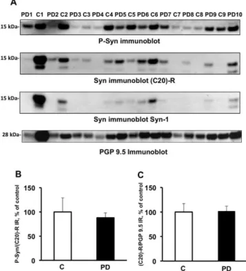
Fig. 1. There are no differences in the expression levels or phosphorylation state
of alpha-synuclein in colonic biopsies between control subjects and patients with Parkinson’s disease. (A) Colonic biopsies from 10 PD patients (Table 1, PD1–PD10) and 9 control subjects (Table 2, C1–C9) were lysed in NETF/Igepal CA-630 buffer. Biopsy lysates (20 µg of protein per sample) were subjected to immunoblot anal- ysis using antibodies against total alpha-synuclein (Syn immunoblot, C20-R and Syn-1), phosphorylated alpha-synuclein (P-Syn immunoblot) and PGP 9.5 (PGP 9.5 immunoblot). The optical densities of (C) total alpha-synuclein (C20-R) and (B) phos- phorylated alpha-synuclein immunoreactive bands were measured, normalized to tively, and expressed as percentage control. Data correspond to mean ± SEM of 9 the optical densities of PGP 9.5 and alpha-synuclein immunoreactive bands, respec-samples for control subjects (C) and 10 respec-samples for Parkinson’s disease (PD) patients. No significant changes were observed between the two groups, (p = 0.9 and 0.75, respectively).
were fixed with 0.4% (v/v) paraformaldehyde (PFA, Sigma) for 30 min and alpha-synuclein immublotting was performed as pre- viously described [13].
3. Results
In a first set of experiments, we analyzed by Western blotting the expression levels and phosphorylation status of alpha-synuclein in colonic biopsies from control and PD subjects. A band migrating to 15 kDa on Western blot was observed using (C20)-R and Syn- 1 antibodies that recognize different epitopes on alpha-synuclein, thus confirming its identity (Fig. 1A). In some samples, a faster migrating 12–13 kDa band was also observed (Fig. 1A). Such a band has already been described in platelets [15] and cerebrospinal fluid [16] and might correspond to a caspase-truncated form of the protein [17]. Despite equal protein loading, the 15-kDa alpha- synuclein band density was extremely variable (Fig. 1A), with no significant difference between PD patients and controls being found when alpha-synuclein amounts were normalized to PGP 9.5 in each sample (Fig. 1C). Phosphorylated alpha-synuclein was iden- tified in all PD and control samples (Fig. 1A), but in both groups the amount detected was also highly variable and following nor- malization to total alpha-synuclein no significant differences were found between PD and control cases (Fig. 1B). No difference in the
immunoreactivity of the 12–13 kDa band was observed between PD and controls (data not shown).
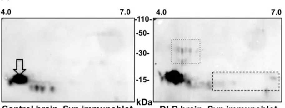
The small amount of pathological aggregated and hyper- phosphorylated alpha-synuclein present in biopsy samples may have remained largely unsolubilized because of the relatively low-stringency buffer that was used in our one-dimenional immunoblotting experiments. This prompted us to perform a sec- ond set of experiments in which samples were lysed with the high stringency UTC buffer then subsequently analyzed by two- dimensional immunoblotting. As previously reported [3], we first showed that this technique was capable of detecting a series of alpha-synuclein species that are unique to DLB brain when compared to control brain, including high molecular weight alpha- synuclein species as well as acidic full-length species (Fig. 2A). However, when this technique was applied to colonic biopsies, the patterns from control subjects and PD patients were qualitatively indistinguishable from each other, with only total unmodified alpha-synuclein being detected in both conditions (Fig. 2B and sup- plementary Fig. 1).
4. Discussion
Based on the topographic distribution of Lewy bodies and neurites established after autopsy from PD patients, Braak and coworkers hypothesized that the gastrointestinal tract and the olfactory bulb might be trigger sites of alpha-synuclein pathol- ogy [18,19]. The pathology would then subsequently spread to the brainstem via the vagus nerve and connected brain regions
[20]. This hypothesis has been reinforced by the discovery that alpha-synuclein is capable of spreading transcellularly in a “prion- like” manner, thereby providing a cellular basis to Braak’s staging scheme that describes PD progression [21]. These ‘gut to brain’ and ‘nose to brain’ scenarios have ignited heated debates within the movement disorders community and have prompted a large number of studies in both humans and animals. Two recent studies showed that injection of fibrillar alpha-synuclein into the intestinal wall [22] or into the olfactory bulb [23] in rodents leads to prop- agation of alpha-synuclein pathology in the brain. In human, the accessibility of both the olfactory and enteric nervous systems to biopsies led to interest in utilizing these tissues as more accessible biomarker material for PD and a substantial amount of research has now been conducted to develop original histopathological mark- ers of PD in these tissues [24,25]. Being the only component of the olfactory system accessible to biopsy, the olfactory epithelium has been screened for Lewy pathology. Several independent autopsy studies showed that alpha-synuclein deposits were observed in the olfactory mucosa in subjects with PD [26–29]. However, a pilot
in vivo study reported no evidence of disease-specific pathology
in the olfactory epithelium in seven hypo/anosmic PD patients
[30]. Conflicting results have been gained following analysis of gas- trointestinal biopsies, all of which have used immunohistochemical methods to detect alpha-synuclein, and there is not yet any fully validated biochemical method for detecting aggregated and/or phosphorylated alpha-synuclein in peripheral tissues [12,31].
Despite the growing interest in peripheral tissue for the detec- tion of PD pathology [24], very few studies have used biochemical approaches including immunoblotting to examine pathological alpha-synuclein in peripheral autonomic networks. One likely explanation is that alpha-synuclein detection by Western blot has proven difficult because the protein is easily washed off from both PVDF or nitrocellulose membranes, making it difficult to detect especially in tissues expressing low levels of the protein [32]. This prompted several groups to develop methods for improv- ing alpha-synuclein retention on blot membranes by increasing its hydrophobicity. This included fixation of membranes with PFA
Fig. 2. There are no differences in the two-dimensional immunoblotting pattern of alpha-synuclein extracted from colonic biopsies from control subjects and patients
with Parkinson’s disease. (A) Immunoblots of brain proteins from a control subject (Control brain) and a DLB patient lysed in UTC buffer and resolved by two-dimensional electrophoresis using pH 4–7 isoelectric focusing gradients were probed with an antibody against Syn-1 (Syn immunoblot). White arrows indicate full-length alpha-synuclein. The boxes in the DLB brain immunoblot indicate differences between control and DLB brain: acidic modifications of full-length monomer (dashed) and high molecular weight species (dotted). (B) Colonic biopsies from 2 PD patients (Table 1, PD 12 and PD14 colonic biopsy) and 2 control subjects (Table 2, C10 and C12 colonic biopsy) were lysed in UTC buffer and resolved by two-dimensional electrophoresis using pH 3–10 isoelectric focusing gradients and probed with the C20-R antibody against total alpha-synuclein. The figure shown is representative of the 2-D immunoblot experiments performed in 7 PD and 4 control subjects.
or treatment of samples with the cross-linker DSP [14,32]. We have recently shown that these two methods are indeed efficient at improving the sensitivity of western blotting to detect alpha- synuclein immunoreactivity in gut samples or primary ENS cultures
[13].
We therefore took advantage of these two methods to ana- lyze the expression levels and the phosphorylation status of alpha-synuclein in colonic biopsies from control and PD sub- jects. The cross-linker DSP was used in experiments involving one-dimensional immunoblotting while PFA-membrane fixation was used in conjunction with two-dimensional immunoblotting. Using one-dimensional immunoblotting, we showed that alpha- synuclein was detected in samples from all PD and control samples, however in both groups the amounts detected were highly variable and no differences between groups were found following normal- ization of alpha-synuclein amounts to that of the neuronal marker
PGP 9.5. Given that alpha-synuclein is found in nerve terminals and cell bodies of submucosal neurons captured by the biopsy forceps [33– 35], it might be postulated that this variability between biop- sies relates to variation in the tissues sampled including omission of submucosal neuronal processes [12]. Immunoreactivity for phos- phorylated alpha-synuclein was also observed in PD and control, a finding that is likely to explained by the fact that a small amount (around 4 %) of alpha-synuclein is physiologically phosphorylated at serine 129 in the brain [36] and in the gut [34]. After quantifica- tion of phosphorylated alpha-synuclein amounts as a proportion of total alpha-synuclein, we did not observe any difference between PD and healthy subjects.
Two-dimensional immunoblotting is a sensitive technique for identifying protein posttranslational modifications and aggregates, and we therefore used this technique to assess a second set of colonic samples. As previously reported, this approach enabled
the detection of acidic and high molecular weight alpha-synuclein species in DLB brain, that likely represent hyperphosphorylated and aggregated forms of the protein, respectively [3]. However, these species were not observed in colonic samples from PD subjects, which contained only full-length unmodified alpha-synuclein.
Overall, our study suggests that the two biochemical methods investigated here are not sufficient to detect pathological alpha- synuclein species in gastrointestinal biopsies, and therefore have limited utility for predicting PD. This may be due to the relative rar- ity of neuronal structures and/or alpha-synuclein inclusions in the gastrointestinal samples analyzed, or as a result of the relatively low sensitivity of the methods tested. Such limitations could be overcome in future studies by using ultrasensitive techniques such as protein misfolding cyclic amplification (PMCA) and real-time quaking-induced conversion (RT-QuIC) assays, which have been recently shown to efficiently amplify aggregated alpha-synuclein
[37,38].
Conflict of interest
The authors declare no actual or potential conflict of interest.
Author’s contribution
EC performed the colonoscopy and biopsies in PD patients. MRD performed the biopsies for control individuals. AGC, CP and TL per- formed and analyzed the biochemical experiments. PD and MN designed the study. PD supervised the study, wrote the first draft and the final version of the manuscript.
Acknowledgements and sources of support
This work was supported by a grant from centre d’entraide et
de coordination des associations de parkinsoniens (CECAP) and by
a Master’s grant of DHU2020 to PD. AGC is a recipient of a poste d’accueil Inserm. We thank Dr. Wendy Noble, Institute of Psychiatry,
King’s College, London, UK, for language editing.
Appendix A. Supplementary data
Supplementary data associated with this article can be found, in the online version, at http://dx.doi.org/10.1016/j.neulet.2017.01. 050.
References
[1]M.G. Spillantini, R.A. Crowther, R. Jakes, M. Hasegawa, M. Goedert,
Alpha-synuclein in filamentous inclusions of Lewy bodies from Parkinson’s disease and dementia with lewy bodies, Proc. Natl. Acad. Sci. U.S.A. 95 (1998) 6469–6473.
[2]H. Fujiwara, M. Hasegawa, N. Dohmae, A. Kawashima, E. Masliah, M.S. Goldberg, et al., Alpha-synuclein is phosphorylated in synucleinopathy lesions, Nat. Cell Biol. 4 (2002) 160–164.
[3]J.P. Anderson, D.E. Walker, J.M. Goldstein, R. de Laat, K. Banducci, R.J. Caccavello, et al., Phosphorylation of Ser-129 is the dominant pathological modification of alpha-synuclein in familial and sporadic Lewy body disease, J. Biol. Chem. 281 (2006) 29739–29752.
[4]T.G. Beach, C.H. Adler, L.I. Sue, L. Vedders, L. Lue, C.L. White Iii, et al., Multi-organ distribution of phosphorylated alpha-synuclein histopathology in subjects with Lewy body disorders, Acta Neuropathol. 119 (2010) 689–702.
[5]T.G. Beach, A.-G. Corbillé, F. Letournel, J.H. Kordower, T. Kremer, D.G. Munoz, et al., Multicenter assessment of immunohistochemical methods for
pathological alpha-Synuclein in sigmoid colon of autopsied parkinson’s disease and control subjects, J. Parkinson’s Dis. 6 (2016) 761–770.
[6] T. Lebouvier, T. Chaumette, P. Damier, E. Coron, Y. Touchefeu, S. Vrignaud, et al., Pathological lesions in colonic biopsies during Parkinson’s disease, Gut 57 (2008) 1741–1743, http://dx.doi.org/10.1136/gut.2008.162503.
[7]T. Lebouvier, M. Neunlist, S. Bruley des Varannes, E. Coron, A. Drouard, J.-M. Nguyen, et al., Colonic biopsies to assess the neuropathology of Parkinson’s disease and its relationship with symptoms, PLoS One 5 (2010) e12728.
[8]H. Pouclet, T. Lebouvier, E. Coron, S.B. des Varannes, T. Rouaud, M. Roy, et al., A comparison between rectal and colonic biopsies to detect Lewy pathology in Parkinson’s disease, Neurobiol. Dis. 45 (2012) 305–309.
[9]N.P. Visanji, C. Marras, D.S. Kern, A. Al Dakheel, A. Gao, L.W.C. Liu, et al., Colonic mucosal a-synuclein lacks specificity as a biomarker for Parkinson disease, Neurology 84 (2015) 609–616.
[10] K.M. Shannon, A. Keshavarzian, E. Mutlu, H.B. Dodiya, D. Daian, J.A. Jaglin, et al., Alpha-synuclein in colonic submucosa in early untreated Parkinson’s disease, Mov. Disord. 27 (2012) 709–715.
[11] D. Hilton, M. Stephens, L. Kirk, P. Edwards, R. Potter, J. Zajicek, et al., Accumulation of a-synuclein in the bowel of patients in the pre-clinical phase of Parkinson’s disease, Acta Neuropathol. 127 (2013) 235–241.
[12] A.-G. Corbillé, F. Letournel, J.H. Kordower, J. Lee, E. Shanes, M. Neunlist, et al., Evaluation of alpha-synuclein immunohistochemical methods for the detection of Lewy-type synucleinopathy in gastrointestinal biopsies, Acta Neuropathol. Commun. 4 (2016) 35.
[13] C. Preterre, A.-G. Corbillé, G. Balloy, F. Letournel, M. Neunlist, P. Derkinderen, Optimizing western blots for the detection of endogenous a-synuclein in the enteric nervous system, J, Parkinson’s Dis. 5 (2015) 765–772.
[14] A.J. Newman, D. Selkoe, U. Dettmer, A new method for quantitative immunoblotting of endogenous a-synuclein, PLoS One 8 (2013) e81314.
[15] Q.-X. Li, B.C.V. Campbell, C.A. McLean, D. Thyagarajan, W.P. Gai, R.M. Kapsa, et al., Platelet alpha- and gamma-synucleins in Parkinson’s disease and normal control subjects, J. Alzheimers Dis. 4 (2002) 309–315.
[16] R. Borghi, R. Marchese, A. Negro, L. Marinelli, G. Forloni, D. Zaccheo, et al., Full length alpha-synuclein is present in cerebrospinal fluid from Parkinson’s disease and normal subjects, Neurosci. Lett. 287 (2000) 65–67.
[17] W. Wang, L.T.T. Nguyen, C. Burlak, F. Chegini, F. Guo, T. Chataway, et al., Caspase-1 causes truncation and aggregation of the Parkinson’s disease-associated protein a-synuclein, Proc. Natl. Acad. Sci. U. S. A. 113 (2016) 9587–9592.
[18] H. Braak, K. Del Tredici, U. Rüb, R.A.I. de Vos, E.N.H. Jansen Steur, E. Braak, Staging of brain pathology related to sporadic Parkinson’s disease, Neurobiol. Aging 24 (2003) 197–211.
[19] H. Braak, R.A.I. de Vos, J. Bohl, K. Del Tredici, Gastric alpha-synuclein immunoreactive inclusions in Meissner’s and Auerbach‘s plexuses in cases staged for Parkinson’s disease-related brain pathology, Neurosci. Lett. 396 (2006) 67–72.
[20] H. Braak, K. Del Tredici, Potential pathways of abnormal tau and a-synuclein dissemination in sporadic alzheimers and parkinsons diseases, Cold Spring Harb. Perspect. Biol. 8 (2016) a023630, http://dx.doi.org/10.1101/cshperspect. a023630.
[21] P. Brundin, J. Ma, J.H. Kordower, How strong is the evidence that Parkinson’s disease is a prion disorder? Curr. Opin. Neurol. 29 (2016) 459–466.
[22] S. Holmqvist, O. Chutna, L. Bousset, P. Aldrin-Kirk, W. Li, T. Björklund, et al., Direct evidence of Parkinson pathology spread from the gastrointestinal tract to the brain in rats, Acta Neuropathol. 128 (2014) 805–820.
[23] N.L. Rey, J.A. Steiner, N. Maroof, K.C. Luk, Z. Madaj, J.Q. Trojanowski, et al., Widespread transneuronal propagation of a-synucleinopathy triggered in olfactory bulb mimics prodromal Parkinson’s disease, J. Exp. Med. 213 (2016) 1759–17784.
[24] T. Lebouvier, M. Tasselli, S. Paillusson, H. Pouclet, M. Neunlist, P. Derkinderen, Biopsable neural tissues: toward new biomarkers for Parkinson’s disease? Front. Psychiatry 1 (2010) 128.
[25] J.M. Lee, P. Derkinderen, J.H. Kordower, R. Freeman, D.G. Munoz, T. Kremer, et al., The search for a peripheral biopsy indicator of a-Synuclein pathology for parkinson disease, J. Neuropathol. Exp. Neurol. (2017) nlw103, http://dx. doi.org/10.1093/jnen/nlw103.
[26] J.E. Duda, U. Shah, S.E. Arnold, V.M. Lee, J.Q. Trojanowski, The expression of alpha- beta-, and gamma-synucleins in olfactory mucosa from patients with and without neurodegenerative diseases, Exp. Neurol. 160 (1999) 515–522.
[27] S.E. Arnold, E.B. Lee, P.J. Moberg, L. Stutzbach, H. Kazi, L.-Y. Han, et al., Olfactory epithelium amyloid-beta and paired helical filament-tau pathology in Alzheimer disease, Ann. Neurol. 67 (2010) 462–469.
[28] Y. Saito, A. Shioya, T. Sano, H. Sumikura, M. Murata, S. Murayama, Lewy body pathology involves the olfactory cells in Parkinson’s disease and related disorders, Mov. Disord. 31 (2016) 135–138.
[29] S. Funabe, M. Takao, Y. Saito, H. Hatsuta, M. Sugiyama, S. Ito, et al., Neuropathologic analysis of Lewy-related a-synucleinopathy in olfactory mucosa, Neuropathology 33 (2013) 47–58.
[30] M. Witt, K. Bormann, V. Gudziol, K. Pehlke, K. Barth, A. Minovi, et al., Biopsies of olfactory epithelium in patients with Parkinson’s disease, Mov. Disord. 24 (2009) 906–914.
[31] A.-G. Corbillé, T. Clairembault, E. Coron, L. Leclair-Visonneau, C. Preterre, M. Neunlist, et al., What a gastrointestinal biopsy can tell us about Parkinson’s disease, Neurogastroenterol. Motil. 28 (2016) 966–974.
[32] B.R. Lee, T. Kamitani, Improved immunodetection of endogenous a-synuclein, PLoS One 6 (2011) e23939.
[33] M. Böttner, D. Zorenkov, I. Hellwig, M. Barrenschee, J. Harde, T. Fricke, et al., Expression pattern and localization of alpha-synuclein in the human enteric nervous system, Neurobiol. Dis. 48 (2012) 474–480.
[34] M. Barrenschee, D. Zorenkov, M. Böttner, C. Lange, F. Cossais, A.B. Scharf, et al., Distinct pattern of enteric phospho-alpha-synuclein aggregates and gene expression profiles in patients with Parkinson’s disease, Acta Neuropathol. Commun. 5 (2017) 1.
[35] H. Pouclet, T. Lebouvier, E. Coron, S.B. des Varannes, M. Neunlist, P. Derkinderen, A comparison between colonic submucosa and mucosa to detect Lewy pathology in Parkinson’s disease, Neurogastroenterol. Motil. 24 (2012) e202–5.
[36] Y. Hirai, S.C. Fujita, T. Iwatsubo, M. Hasegawa, Phosphorylated
alpha-synuclein in normal mouse brain, FEBS Lett. 572 (2004) 227–232.
[37] G. Fairfoul, L.I. McGuire, S. Pal, J.W. Ironside, J. Neumann, S. Christie, et al., Alpha-synuclein RT-QuIC in the CSF of patients with alpha-synucleinopathies, Ann. Clin. Transl. Neurol. 3 (2016) 812–818.
[38] M. Shahnawaz, T. Tokuda, M. Waragai, N. Mendez, R. Ishii, C. Trenkwalder, et al., Development of a biochemical diagnosis of parkinson disease by detection of a-synuclein misfolded aggregates in cerebrospinal fluid, JAMA Neurol. (2016), http://dx.doi.org/10.1001/jamaneurol.2016.4547.