AVIS
Ce document a été numérisé par la Division de la gestion des documents et des archives de l’Université de Montréal.
L’auteur a autorisé l’Université de Montréal à reproduire et diffuser, en totalité ou en partie, par quelque moyen que ce soit et sur quelque support que ce soit, et exclusivement à des fins non lucratives d’enseignement et de recherche, des copies de ce mémoire ou de cette thèse.
L’auteur et les coauteurs le cas échéant conservent la propriété du droit d’auteur et des droits moraux qui protègent ce document. Ni la thèse ou le mémoire, ni des extraits substantiels de ce document, ne doivent être imprimés ou autrement reproduits sans l’autorisation de l’auteur.
Afin de se conformer à la Loi canadienne sur la protection des renseignements personnels, quelques formulaires secondaires, coordonnées ou signatures intégrées au texte ont pu être enlevés de ce document. Bien que cela ait pu affecter la pagination, il n’y a aucun contenu manquant.
NOTICE
This document was digitized by the Records Management & Archives Division of Université de Montréal.
The author of this thesis or dissertation has granted a nonexclusive license allowing Université de Montréal to reproduce and publish the document, in part or in whole, and in any format, solely for noncommercial educational and research purposes.
The author and co-authors if applicable retain copyright ownership and moral rights in this document. Neither the whole thesis or dissertation, nor substantial extracts from it, may be printed or otherwise reproduced without the author’s permission.
In compliance with the Canadian Privacy Act some supporting forms, contact information or signatures may have been removed from the document. While this may affect the document page count, it does not represent any loss of content from the document.
EFFECTS OF INTERLEUKIN-27 ON
HUMAN CD8 T CELLS
par
Teodora Yaneva
Départen'lent de microbiologie et immunologie
Faculté de médecine
Mémoire présenté à la Faculté des études supérieures
en vue de l'obtention du grade de
llId':::il~~nide t9,~
.
b' 1 "
.
~~""
en n'liCrO
10ogie et Imn'lUnO ogie
.
'1f 8 NOV. 2008
Juin, 2008
Faculté des études supérieures
Ce mémoire intitulé:
EFECTS OF INTERLEUKIN-27 ON HUMAN CD8 T CELLS
présenté par:
Teodora Yaneva
a été évalué par un jury composé des personnes suivantes:
Dr. Jean-Francois Gauchat
Président-rapporteur
Dr. Nathalie Arbour
Directrice de recherche
Dr. Nathalie Labrecque
Membre du jury
L'orchestration des médiateurs cellulaires et solubles du système immunitaire inné est essentielle pour le maintien de la santé. Les lymphocytes T contribuent de part leurs fonctions cytolytiques et la sécrétion de nombreux médiateurs solubles et à cet équilibre.
L'interleukine-27 (IL-27) est composée de deux sous-unités, EBI3 et p28, qui sont liées à une façon non-covalente. Des effets pro- et anti-inflammatoires sur des cellules T murines ont été décrits pour cette cytokine. Toutefois, peu d'informations sont disponibles pour les cellules T humaines.
L'impact potentiel de l'IL-27 sur les fonctions des cellules T humanes a été étudié en utilisant des cellules mononucléaires du sang périphérique (PB MC) des donneurs sains. Une petite quantité des cellules CD4 et CD8 exprimaient le récepteur de l'IL-27 (IL-27R), composé de deux chaines : TCCR et gp130, ex vivo, surtout les CD8, et cette proportion augmentait site à une activation in vitro. Des PBMC et des cellules T CD8 purifiées, naïves et mémoires, ont été activées brièvement in vitro et leur prolifération et production des médiateurs ont été étudiées par cytométrie en flux. L'ajout d'IL-27 à une stimulation polyclonale a augmenté de façon significative et dose-dépendante la prolifération et la production d'interféron-y et de granzyme B par les cellules T. De plus, une analyse par RT-PCR a démontré que les cellules CD8 humanes ne peuvent pas produire d'IL-27 même après activation.
Ces résultats démontrent l'impact pro-inflammatoire de l'IL-27 sur des cellules T (CD8) humanes et leur plus grande susceptibilité aux effets de cette cytokine après activation via l'augmentation de l'expression de l'IL-27R.
Mots clés: cytokine, interféron, cytométrie en flux, granzyme, récepteur de cytokine
Orchestration of the innate and adaptive immune systems through numerous cell types and their secreted mediators is crucial to maintain health. Cytokines, soluble protein mediators, have crucial role in shaping immune responses. T lymphocytes through the secretion of soluble mediators and cytolytic functions are important players of the adaptive immune responses.
Interleukin-27 (IL-27) consists of two non-covalently linked subunits: EBI3 and p2S. This recently described cytokine has been shown to exert both pro- and anti-inflammatory effects, especially on mouse T cells. However, information on human T cells is lacking.
The potential impact of IL-27 on human T cell functions was assessed using, peripheral blood mononuclear cells (PBMC) from healthy donors. Surface expression of both chains of IL-27 receptor (IL-27R) (TCCR and gp130) was assessed on ex vivo and in vitro activated PBMC. A small proportion of CD4 and CDS T cells expressed detectable IL-27R ex vivo, with the latter subset having a greater proportion and increased levels upon activation. PBMC or purified naïve and memory CDS T cells were shortly stimulated in vitro and then analyzed using flow cytometry~based assays assessing proliferation and mediators. Addition of IL-27 to anti-CD3 stimulated cells led to a significant dose-dependent increase of proliferation, interferon-y and granzyme B production by T cells. Moreover, in contrast to mouse ceIls, RT -PCR analysis showed that human CDS T cells can not be a source of IL-27.
These results underscore the pro-inflammatory impact of IL-27 on human CDS T cells and their increased susceptibility upon activation.
TABLE OF CONTENTS TABLE OF CONTENTS ... 1 LIST OF FIGURES ... 4 LIST OF TABLES ... 5 LIST OF ABBREVIATIONS ... 6 DEDICATION ... 10 ACKNOWLEDGEMENTS ... 11 LITERATURE REVIEW ... 12 1.0 T lymphocytes ... 13
1.1 DistinctT lymphocyte subsets ... 13
1.2 Transition from naïve to effector and memory T ceIls ... 15
1.3 Roles of cytokines in T ceIl polarization and acquisition of effector functions ... 20
2.0 ANTIGEN PRESENTING CELLS (APC) ... 24
2.1 Monocytes and Macrophages (Mcp) ... 24
2.2 Dendritic cells ... 26
2.3 B cells ... 27
3.0 INTERLEUKIN-27 (IL-27) ... 28
3.1 Characteristics of IL-27 protein ... 30
3.2 IL-27 expression in human tissues ... 33
3.3 Receptor of IL-27 ... 34
3.4 Downstream signalling of IL-27R ... 36
3.5 Effects of IL-27 on T lymphocytes ... 37
3.7 Impact of IL-27 on B lymphocytes ... 44
3.8 Impact of IL-27 on other ceU types ... 45
3.9 Roles of IL-27 in mouse models of human diseases ... 46
3.10 Roles of IL-27 in human disorders ... 50
3.11 Interleukin-35: EBI3 + IL-12p35 ... 51
HYPOTHESES AND OBJECTIVES ... 53
Hypotheses ... 53
Objectives ... 53
MATERIALS AND METHODS ... 54
1.0 Isolation of human blood cells ... 55
1.1 Peripheral mononuclear ceH isolation ... 55
1.2 Total CD8 T ceH isolation ... 56
1.3 Naïve CD45RA+ CD8 T ceH isolation (negative selection) ... 56
1.4 Memory CD45RO+ CD8 T ceU isolation ... 57
2.0 Functional assays on human T cells ... 58
2.] CFSE labeUing ... 58
2.2 Cell stimulation ... 58
2.2.] PBMC ... 58
2.2.2 Naïve CD8 T ceUs (plate-bound) ... 59
2.2.3 Memory CD8 T ceUs (plate-bound) ... 60
2.3 Flow cytometry ... 60
2.4 Ex-vivo expression of IL-27R (gp130 and TCCR) ... 61
2.5 ELISA for IFN-y ... 62
3.1 RNA extraction ... 63
3.2 Complementary DNA (cDNA) ... 64
3.3 Real time-Polymerase Chain Reaction ... 64
4.0 Statistica! analysis ... 66
RESULTS ... 67
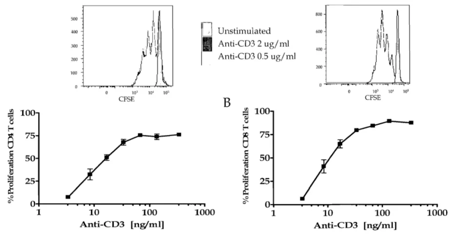
1.0 Optimization of anti-CD3 activation of human PBMC. ... 68
1.1 Proliferation of human T cells in response to a-CD3 Ab ... 68
2.0 Expression of IL-27 signalling receptor (IL-27R) by human PBMC ... 70
2.1 Ex vivo expression of IL-27R by human T lymphocytes and monocytes ... 71
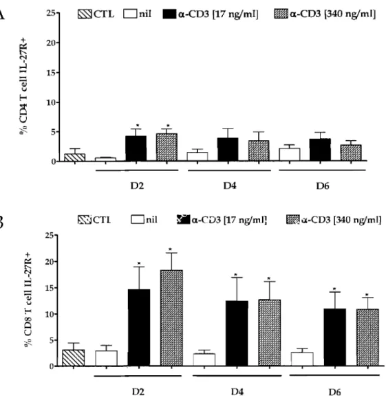
2.2 IL-27R expression upon activation ... 73
3.0 Effects of IL-27 on T cell functions ... 75
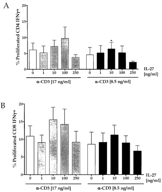
3.1 PBMC ... 75
3.2 Naïve CD8 T cells ... 84
3.3 Memory CD8 T cells ... 88
4.0 CD8 T cells as a source of IL-27 ... 91
DISCUSSION ... 93
1.0 Expression and modulation of IL-27R ... 94
1.1. Ex-vivo IL-27R expression ... 94
1.2 IL-27R modulation upon activation ... 98
2.0 Effects of IL-27 on T cell functions ... 99
3.0 CD8 AS A SOURCE OF IL-27 ... 107
4.0 FUTURE DIRECTIONS ... 108
BlBLIOGRAPHY ... 110
LIST OF FIGURES
Figure 1: Effects of IL-27 signaIling ... 42
Figure 2: a-CD3 Ab dose response by PB MC as assessed by proliferation ... 69
Figure 3: Expression of IL-27 receptor chains on ex-vivo monocytes (CD14), CD4 and CD8 T cells ... 72
Figure 4: Expression of IL-27R on T cells upon activation ... 74
Figure 5: Illustration of IL-27 effects on the proliferation of, and IFN-y and granzyme B production by human CD8 T ceIls ... 76
Figure 6: IL-27 boosts the proliferation of T ceIls ... 78
Figure 7: IL-27 augments the IFN-y producing T cells ... 80
Figure 8: IL-27 increases granzyme B production by CD8 T cells ... 81
Figure 9: IL-27 augments IFN-y production by a-CD3 stimulated PBMC ... 83
Figure 10: IL-27 enhances naïve CD8 T cell proliferation upon a-CD3 stimulation in the presence or absence of co-stimulation ... 85
Figure 11: Effects of IL-27 on effector functions of naïve CD8 T ceIls ... 86
Figure 12: IL-27 enhances memory CD8 T cell proliferation upon a-CD3 stimulation ... 89
Figure 13: Effects of IL-27 on effector functions of memory CD8 T cells ... 90
Figure 14: IL-27 is not produced by CD8 T ceIls upon stimulation ... 92
LIST OF TABLES
Table 1. Naïve and memory specifie markers for human T cells ... 19
Table 2. Different effector T celllineages ... 20
Table 3: Stimuli, inducing mRNA expression of both subunits of IL-27 ... 32
Table 4. List of f1uorochroms used for flow cytometry analysis ... 61
A647 A700 Ab Ag APC APC-Cy? APCy BCR CD CFSE CNTF CT-1 CTL d.s. DC EBI3 ELISA Eomes PITC GATA-3 G-CSF GM-CSF gp130 IFN-a IFN-y IL-27 IL-27R LIF LPS M-CSF mRNA Mcp NF-kB
NK
OSM PAMPs PB PBMC PE PE-Cy7LIST OF ABBREVIATIONS
Alexa Fluor®647 (fluorochrome) Alexa Fluor®700 (fluorochrome) Antibody
Antigen
Antigen presenting cell
AIlophycocyanin-Cy™7 (fluorochrome) Allophycocyanin
B ceIl receptor
Cluster of differentiation
5-(and 6-) carboxyfluorescein diacetate succinimidyl ester Ciliary neurotrophic factor
Cardiotrophin 1
Cytotoxic T lymphocytes (CDS+)
Double stranded deoxyribonucleic acid Dendritic cells
Epstein-Barr virus-induced gene 3 Enzyme linked immunosorbent assay Eomesodermin (transcription factor) Fluorescein (fluorochrome)
"GATA" DNA sequence recognizing transcription factor 3 Granulocyte colony stimulating factor
Granulocyte-macrophage colony stimulating factor Glycoprotein 130
Interferon alpha Interferon gamma Interleukin 27
Receptor of interleukin-2? Lymphocyte inhibitory factor Lipopolysaccharide
Macrop hage-colony -stimula ting factor Messenger ribonucleic acid
Macrophages Nuclear factor kB Natural killer ceUs Oncostatin M
Pathogen associated molecular patterns Pacific Blue™ (fluorochrome)
Peripheral blood mononuclear ceUs Phycoerythrin (fluorochrome)
PRRs ROI ROR-y RT-PCR s.s. DNA sgp130 SOCS STAT T-bet Tc TCR TGF-p Th TLR TNF Treg A647 A700 Ab Ag APC APC-Cy7 APCy BCR CD CFSE CTL d.s. DC EBI3 ELISA Eomes FITC FnIII GATA-3 G-CSF GM-CSF gp130 IFN-a IFN-y IL-27 IL-27R
Pattern recognition receptors Reactive oxygen intermediates Retinoid orphan receptor y
Real-time polymerase chain reaction Single stranded deoxyribonuc1eic add Soluble gp130
Suppressor of cytokine signaling
Signal transducer and activator of transcription T -box expressed in T ceUs
Cytotoxic T cells (CD8+) T ceU receptor
Transforming growth factor- beta T helper cells (CD4+)
Toll-like receptor Tumor necrosis factor Regulatory T cells
Alexa Fluor®647 (fluorochrome) Alexa Fluor®700 (fluorochrome) Antibody
Antigen
Antigen presenting cell
Allophycocyanin-Cy™7 (fluorochrome) Allophycocyanin
B cell receptor
Cluster of differentiation
5-(and 6-) carboxyfluorescein diacetate succinimidyl ester Cytotoxic T lymphocytes (CD8+)
Double stranded deoxyribonuc1eic add Dendri tic cells
Epstein-Barr virus-induced gene 3 Enzyme linked immunosorbent assay Eomesodermin (transcription factor) Fluorescein (fluorochrome)
Fibronectin type III domain
IIGATA" DNA sequence recognizing transcription factor 3 Granulocyte colony stimulating factor
Granulocyte-macrophage colony stimulating factor Glycoprotein 130
Interferon alpha Interferon gamma Interleukin 27
LIF Lymphocyte inhibitory factor LPS Li popolysaccharide
M-CSF Macrop hage-co Ion y -stimula ting factor mRNA Messenger ribonucleic acid
Mcp Macrophages
NF-kB Nuclear factor kB NK Natural killer cells
PAMPs Pathogen associated molecular patterns PB Pacific Blue™ (fluorochrome)
PB MC Peripheral blood mononuclear cells PE Phycoerythrin (fluorochrome)
PE-Cy7 Phycoerythrin-Cy TM7 (fl uorochrome) PRRs Pattern recognition receptors
ROI reactive oxygen intermediates ROR-y Retinoid orphan receptor y
RT-PCR Real-time polymerase chain reaction s.s. DNA Single stranded deoxyribonucleic acid sgp130 Soluble gp130
SOCS Suppressor of cytokine signaling
STAT Signal transducer and activator of transcription T-bet T -box expressed in T cells
Tc Cytotoxic T cells (CD8+) TCR T cell receptor
TGF-~ Transforming growth factor- beta Th T helper cells (CD4+)
TLR Toll-like receptor TNF Tumor necrosis factor Treg Regulatory T cells
DEDICATION
"Y ou are never given a wish without being given the power to make it true." Richard Bach
"The future belongs to those who believe in the beauty of their dreams. I l
Eleanor Roosevelt
To my mother, Tsvete, the magic flower with spirit of steel, who taught me to dream ...
To my father, Yanko, who taught me to fight for my dreams ...
To my brother, Yavor, who leads me in the invisible world of beliefs ...
ACKNOWLEDGEMENTS
l would like to express my gratitude to my supervisor, Dr. Nathalie Arbour, for giving me the opportunity to work with her, for teaching me and being so patient and full of love, for believing in me and for keeping the gui ding light in sight till the very end.
l would like also to thank Neuro-Immuno team at CHUM Notre-Dame Hospital:
Our lab technician, Diane Beauseigle, for being my second pair of eyes and for providing me with help and guidance ...
My colleagues Camille Pittet, Dr. Raphael Schneider and Lama EI-Khoury for their love, assistance and moral support ....
Dr. Alexandre Prat and his team - Monique Bernard, Igal Ifergan, Hania Kébir, Romain Cayrol, Aurore Dodelet-Devillers, Dr. Mike Sabbagh and Simone Terouz for their support, for making the lab my home and for turning the time of work into such an incredible experience ...
The immune system comprises two complementary arms: the innate and the adaptive systems. The innate immune system acts as the first line of defence and consists of cells that recognize pathogen associated molecular patterns (PAMPs) through pattern recognition receptors (PRRs), for example Toll-like receptors (TLRs). Although it is rapidly activated in response to insult or pathogen, the innate system lacks the ability to improve the host' s resistance upon re-encounter of the same Ag (Ag). This immune system compartment is composed of mediators including the lysosyme, the complement, acute phase proteins, and cells such as phagocytes (dendritic cells (DC), macrophages (Mcp) and neutrophils) and natural killer cells (NK). When this first line of defence does not successfully get rid of the pathogen threat in the first few days, the innate immune system activates the adaptive immune system, which recognizes specific Ag of the infectious agent through the B cell receptor (BCR) and the T cell receptor (TCR) leading respectively to antibodies (Ab) secretion by B lymphocytes and cellular immune responses by T lymphocytes. The adaptive immune system through the activation and expansion of specifie B cells and T cells is responsible for the immunological memory towards the pathogen-derived Ag.
1.0 T LYMPHOCYTES
1.1 Distinct T lymphocyte subsets
T lymphocytes are cells of the adaptive immune system ex pressing the cluster of differentiation (CD). marker CD3, which consists of the invariable signal transducing su bu nits (CD3y, CD30, CD3r and CD3~ [CD247]) (Lin and Weiss, 2001; Werlen and Palmer, 2002; Risueno et a1., 2008). The CD3 chains have negatively charged acidic residues in their transmembrane domains which ensure interaction with the positively charged ligand-binding subunits (TCRa and TCR~)
of the T cell receptor (TCR). The TCR of most T cells consists of an a- and a ~
chains, whereas a min or population expresses an alternative form made of a y-and a O-chains. These ligy-and-binding chains are immunoglobulin like y-and have a constant and a variable region. The a~TCR recognizes peptides presented by
major histocompatibility complex (MHC). a~TCR T lymphocytes fall into two classes based on distinct cell-surface co-receptors: CD4 and CD8, thus are called CD4 T lymphocytes or CD8 T lymphocytes. CD4 is a single chain molecule binding the classical MHC class II mole cule at a different site than the TCR. Thus, CD4 T lymphocytes, also named T helper cells (Th), recognize peptidic Ag presented by MHC class II molecule. The CD8 mole cules in most T lymphocytes is a heterodimer consisting of an a- and a ~-chains and interacts with MHC class 1
molecules in a way similar to the interaction CD4 ~ MHC class II. Thus, CD8 T lymphocytes, also named cytotoxic T cells (Tc), recognize peptidic Ag presented by classical MHC class 1 molecules or non-classical ones such as HLA-G (Gomes et aL, 2007) and HLA-E (Godfrey et aL, 2008). However, T cells can also recognize glycolipids, complexed to CD1d molecule (Godfrey et aL, 2008). CD1 mole cules are cell surface glycoproteins expressed mainly by B lymphocytes, macrophages and dendritic cells. They consist of two chains: ~2 microglobulin (~2m), similarly to MHC class 1 molecules, and a heavy chain containing three extracellular domains (a1-a3). The a1-a2 super domain for ms the antigen-binding groove consisting of two ahelices (al and a2) whereas the membrane proximal a3 domain binds ~2m. Humans have five CD1 isotypes (CD1a-e). Many natural killer T cells are specifie for CD1d presenting several types of glycolipids (Zajonc and Kronenberg, 2007).
Both classes of MHC mole cules are heterodimers anchored in the cell membrane and bear a groove where the presented peptide binds. The MHC class 1
molecule consists of a larger a-chain spanning the membrane, and a smaller, non-covalently linked ~2-microglobulin, which does not have a transmembrane domain. The peptide that fits in the groove is usually 8-10 amino acids (aa) long and gets anchored at both ends by its free amino- and carboxy- termini. The MHC class II molecule comprises two non-covalently associated chains (a- and ~-) both spanning the cytoplasmic membrane. The peptide-binding groove is more open than the cleft of the MHC class 1 molecule resulting in presentation of longer peptides (13-17 aa) and lack of anchoring at the ends of the peptide. Both classes of
MHC molecules are differentially expressed throughout the body: almost aIl nucleated ceUs express MHC class l molecules, whereas MHC class II molecules are restricted to professional Ag presenting cells (APC) (e.g. B lymphocytes, DC and Mer). MHC class land class II expression could be enhanced upon activation and inflammatory conditions.
1.2 Transition from naïve to effector and memory T cells
Based on experimental observations mainly obtained using mouse models of infection, the in vivo T cell response has been dissected into four main phases: i)initial activation ii) expansion phase iii) contraction phase, and iv) memory phase.
The initial activation consists in the activation of rare naïve T ceUs specifie for the particular Ag into effector T cells. Three signaIs are required for initiation of a program leading to strong expansion, development of effector functiol1S, and survival of an efficient memory cell population. The first signal is the MHC-peptide complex recognized by the TCR, the second signal isprovided by co-stimulatory molecules (e.g. CD80 or CD86) concurrently provided with the MHC-peptide complex on the surface of the APC. Cytokines present near-by the T ceIl-APC interaction provide the third signal (Mescher et al., 2006; Hart y and Badovinac, 2008). Stimulation with these three signaIs synergize and cause the naïve T cells to enter an irreversible differentiation program through activation of specifie transcription factors (T-bet, GATA-3, Foxp3 or RORyt) Ieading to the acquisition of tissue homing receptors and distinct effector functions. Cytokines provided in the vicinity have a tremendous impact on the differentiation of T cells. Skewing towards different T ceIl responses will be described in more details below.
The expansion phase consists in the proliferation and differentiation of specifie T cells, which will become effector or memory T cells that will then travel throughout the host' s body to find their specific cognate peptide-MHC complex. Chroma tin remodeling of effector cytokine genes can be inherited through mitosis
and can contribute to the maintenance of specifie states of gene activity between cell generations (Agarwal et al., 1998; Sallusto et al., 2004) through a process, called "cycling" in the absence of cognate Ag (demonstrated for memory CD8 T ceUs).
The contraction phase describes the transition from a large effector population to a smaller population of memory T cells in order to maintain long-lasting immunologieal memory. This is necessary given the limited space and resources available for aIl T cells expanded throughout the host' s life. Moreover, protection front the highly active effector cells, which can turn into detrimental autoimmune weapon once the pathogen has been cleared (Hart y and Badovinac, 2008) is provided by this contraction phase. The mechanisms involved in this phase are not fully elucidated but could include apoptosis of Ag-specifie T ceUs and a balance between pro- and anti-apoptotic BCL-2 family members. In addition, after each division, chromos omal telomeres shorten determining the cellular lifespan and limiting the number of divisions that cells can undergo (Hart y and Badovinac, 2008).
The memory phase describes the long-term maintenance of memory T ceUs within the host. Cytokines play crucial roI es in this process especially the COlnmon y chain cytokines: lL-2, lL-7 and lL-15. lL-7 provides the second signal required for survival of both memory and naïve T ceUs, which makes it a limiting factor (Schluns et aL, 2000; Vivien et aL, 2001; Guimond et aL, 2005; Boyman et al., 2007; Prlic et al., 2007). The memory T ceUs are highly heterogeneous in terms of surface markers, cytokines they are able to produce as weIl as their lytic enzyme content. This heterogeneity could be ascribed to several factors including the signaIs provided during the priming (type of pathogen, presence of cytokine milieu, and APC) determining the bouquet of cytokines produced by the effectors and thus by the memory T cells (Seder et al., 2008), and survival signaIs received later on (including survival cytokines).
Some of the markers used for phenotyping T cells according to their state of activation are shown in Table 1 including the function of these mark ers with (+)
and (-) representing the relative expression in a given subpopulation. Thus, (+) means that the marker is usually expressed by the entire subpopulation, (++1-)
means that most cells in the subpopulation express the marker, whereas (+1--)
means that most cens in the subpopulation do not express the marker, but that a sman subset do. One of these mark ers is CD45 which has two isoforms: CD45RA, expressed on naïve T celIs, and CD45RO typical for memory T celIs. A small subset of CD8 T cells called effector memory CD45RA+ (TEMRA) express CD45RA, are devoid of CCR7, and are characterized by the largest content of perforin among the memory subsets. The transition between both isoforms occurs upon T-cell activation with a shift from RA to RO as this change is reversible. CD62L is a lymph node homing receptor, which in combination with the CC-chemokine receptor 7 (CCR7) is used for distinguishing the central memory (TCM, expressing CD62LhiCCR7hi) from the effector memory (T EM, expressing CD62UowCCR7Iow) subset. TCM represent the long-term memory cens which maintain the memory pool due to their good proliferative capacity. Human TCM express CD45RO, CCR7, and are found mainly in secondary lymphoid organs (lymph nodes, spleen) and tissues. They have high sensitivity to Ag stimulation, are less dependent on co-stimulation than naïve T celIs, and up-regulate CD40L to a greater extent to provide more effective co-stimulatory feedback to DC and B cells. Upon activation they produce mainly IL-2, but after proliferation they differentiate into effector cells and produce large amounts of cytokines. The other subset (TEM) have less efficient proliferative capacity upon re-encounter of the Ag but can rapidly and efficiently respond to it via cytokine production (Sallusto et al., 2004). This compartment consists of effector memory T cells that have lost the constitutive expression of CCR7, are heterogeneous for CD62L, show different chemokine receptor and adhesion molecule expression that lead them to inflamed tissues. These cells reside mainly in the spleen, blood, and non-lymphoid tissues (lung, liver, and gut) but are not found in the lymph nodes and have rapid effector functions because are charged with great amounts of perforin. Both CD4 and CD8 TEM produce cytokines very shortly after reactivation (Sallusto et a1., 2004;
Badovinac and Hart y, 2006). Other markers used are the co-stimulatory molecules CD27 and CD28. In addition, memory T cells are characterized by their effector functions including their cytokine profile and lytic enzyme content. Thus, to identify the memory phenotype a combination of multiple markers is used. However, regardless of their phenotype, good memory T cells: (i) persist and outnumber the naïve T cell repertoire; (ii) are able to rapidly respond to re-infection by vigorous proliferation and various effector mechanisms (cytolysis and cytokine production) in an Ag dose-dependent manner; (iii) provide rapid protection after pathogen re-encounter (Badovinac and Hart y, 2006).
Table 1. Naïve and Inemorl/ svecifie markers for hwnan T cells
CD4 CDS Functions
•
Marker T TCM TEM T TCM TEM TEMRA
naïve naïve
Chemokine receptor binding CCL19 and CD197
(+) (+) (-) (+) (+) (-) (-) CCL21; Homing to lymph nodes;
Down-(CCR7) regulated upon naïve T cell activation;
Re-expressed after stimulation.
CD45RA (+) 1 C\ (+ / --) (+ ) (-) (-) (+) high-molecular Transmembrane
l ' ,
weight phosphate regulating the low-molecular TCR-CD3 complex weight, increases signalling; T-cell CD45RO (-) (+) (+ )
H
(+ ) (+) (+ /-) activation leads toefficiency of TCR- a
reversible shift from RA CD3 signalling
toRO.
Cellular adhesion molecule from selectin CD62L (+) (++ /-) (+ /-) (+ ) (++ /-) (+ / --) (+ / --) family, homing to lymph nodes via interaction
with 6-sulpho-Lewis X,
Binds CD70; Present on activated T cells and CD27 (+) (++ /-) (+ /-) (+) (++ /-) (+ /-)
DC; Promotes effector functions and memory (+ /-) cell formation; CD70 engagement or prolonged stimulation leads to 10ss of CD27 expression; no re-exrressed uron activation.
Binds CD80 and CD86 expressed by APC; CD28 (+) (+) (+) (+) (++/-) (+ /-) (+ /-) Potent transducer of co-stimula tory signaIs enhancing IL-2 production, proliferation, survival, and effector functions.
1.3 Roles of cytokines in T œIl polarization and acquisition of effector functions The timing and the intensity of several signaIs (signal 1: TCR, signal 2: co-stimulation, signal 3: cytokines) regulate the development of effector as weIl as memory T cells (Hart y and Badovinac, 2008). Cytokines produced by innate immune ceIls significantly contribute to shape subsequent T ceIl activation by regulating the expression of many genes in T lymphocytes promoting optimal proliferation, survival, commitment to a lineage (a summary of ThljTc1, Th2jTc2, and Th17 jTc17 lineages is shown in Table 2), effector functions, adhesion and trafficking (Meseher et al., 2006). The skewing of T cells towards type 1, type 2 or type 17 has been considerably studied for CD4 T cells but mueh less is known for CD8 T cell differentiation. Moreover, numerous publications suggest that factors dictating the activation, expansion, and development of memory differ between these two T cell subsets (Seder and Ahmed, 2003). Thus, the following description of lineage commitment has been mainly deseribed for CD4 T cells, but when appropriate information regarding CD8 T cells is mentioned. In addition to the ThljTc1, Th2jTc2, or Th17 jTc17 lineages, other T cell differentiation patterns can be observed. For example, presence of TGF-p during TCR activation can induce the expression of forkhead box transcription factor Foxp3 by CD4 T eeIls leading to the development of a T cell subset bearing regulatory functions (Shevach et al., 2008), but these regulatory T cell subsets will not be discussed herein.
Table 2. Different effector T celllineaçres
LI'
Cytokine profile Role in immunityProinflammatory action against intracellular ThljTc1 IFN-y, TNF pathogens (e.g. viruses)
Stimulate humoral immunity by activating B IL-4, IL-5, IL-ID, cells, contribute to fight extracell ular Th2jTc2 IL-13 pathogens
•
• Implicated 111 autoimmunity, inflammation, •
IL-17 IL-17F, IL- cancer, protect from extracellular pathogenic Th17 jTc17 21, IL-22, bacteria
Presence of IL-12 in the vicinity of CD4 T ceUs being activated and the signalling and induction of transcription factor 'T-box expressed in T ceUs' (T-bet) and STAT-4 (Signal Transducer and Activator of Transcription-4) lead to the development of Thl celIs (Seder and Ahmed, 2003). Similarly, it has been shown that naïve CD8 T cells exposed to their cognate Ag in the presence of co-stimulation but in absence of IL-12 exp and but these celIs do not acquire cytolytic activity (Mescher et al., 2006), supporting the crucial role of IL-12 also in efficient CD8 T ceIl effector functions. In Table 2 are represented the basic T subsets according to their secreted cytokines. Type 1 T celIs (Thl/Tel) produce pro-inflammatory cytokines such as IFN-y and TNF as weIl as lytic enzymes and are implicated in clearance of viral infections and extracellular pathogens. An uncontrolled expansion of Thl ceUs has been associated with the development of autoimmune diseases (Gonzalez-Rey and Delgado, 2006; Guilherme et al., 2007; Skurkovich and Skurkovich, 2007).
In contrast, presence of IL-4 and signalling and induction of the transcription factor GATA-3 (the name cornes from the "GATA" DNA sequence recognized) and activation of ST AT -6 (Signal Transducer and Activator of Transcription-6) favour the development of Th2 cells. Type 2 T lymphocytes (Th2/Tc2) produce cytokines such as IL-4, - S, -10 and IL-13, suppressing pro-inflammatory Thl cytokines, and promoting humoral immunity and responses to intracellular pathogens. An uncontrolled expansion of Th2 ceUs has been linked with the development of allergy (Robinson, 2000; Bullens et al., 2004; Haczku, 2006).
Differentiation of naïve CD4 T cells into Th17 occurs in different conditions in mouse and human (McGeachy and Cu a, 2008). Naïve mouse CD4 T cells activated in the presence of TGF-p and IL-6 up-regulate the transcription factor ROR-y (Retinoid-related orphan receptor-gamma) and adopt the Th17 lineage (Bettelli et al., 2006; Mangan et al., 2006; Veldhoen et al., 2006). Cytokines implicated in the differentiation of human CD4 T celIs into Th17 cells is not fully elucidated. Contradictory results have been published regarding the role of TGF-p and IL-6.
According to one group (Yang et al., 2008) Th17lineage results from stimulation in the presence of TGF-~ and IL-21, whereas others (Acosta-Rodriguez et al., 2007a; Wilson et al., 2007; McGeachy and Cua, 2008) observed that the combined action of IL-23 and IL-1 is necessary for the differentiation of human Th17 which can be further enhanced by IL-23 and/ or IL-6. Th17/Tc17 secrete IL-17, IL-17F, IL-21 and IL-22 and have been implicated in man y human diseases like multiple sclerosis, systemic lupus erythematosus, asthma, rheumatoid arthritis (Afzali et al., 2007; Aujla et al., 2007; Hirota et al., 2007; Kebir et al., 2007; Roark et al., 2007; McGeachy and Cua, 2008; Wong et a1., 2008). IL-22 and IL-17 have been related to bacterial protection in lungs and gut, and regulate immune responses of cells in non-lymphoid tissues. IL-21 has been also implicated in humoral immunity in the lymph node germinal center reactions (Dong, 2008). It has been shown that Th17 cells, especiaIly human Th17 ceIls ex pressing specific chemokine receptors (CCR6 and CXCR3) can also pro duce IFN-y suggesting that the segregation of cytokine profile is not complete (Acosta-Rodriguez et a1., 2007b; McGeachy and Cua, 2008).
There is extensive cross-talk between each T ceIl subsets through the secretion of cytokines that can negatively regulate the differentiation of other subsets. For example, IL-12, IFN-y or IL-4 can prevent the differentiation induced by IL-23 or
TGF-~+ IL-6 of human or mouse Th17 respectively (Murphy et a1., 2003;
Harrington et al., 2005; Annunziato et al., 2007; Wilson et al., 2007; McGeachy and Cua, 2008).
Cytokines are also crucial in the survival and maintenance of naïve and memory T ceIls, but CD4 and CD8 T cells have different requirements, probably because each subset has intrinsic proliferative capacity (more prominent in the CD8 T ceIl compartment) (Se der and Ahmed, 2003). The homeostatic proliferation of both naïve CD4 and CD8 T ceIl populations is strongly reduced when transferred into IL-7-deficient mice (Schluns et al., 2000; Tan et al., 2001; Goldrath et al., 2002) demonstrating that this cytokine is crucial for the maintenance of these ceIls in mice. IL-15 deficiency selectively affects the homeostasis of naïve mouse CD8 T cells leading to a diminished expansion of these ceIls but does not affect
1
Il
II '1 l' Il ! ! .1 li Ilnaïve CD4 T ceUs, supporting a selective role for IL-15 in the survival and proliferation of murine naïve CD8 T ceUs (Zhang et al., 1998; Tan et al., 2001; Judge et al., 2002; Tan et al., 2002; Berard et al., 2003). IL-7 and IL-15 have non-redundant roI es in murine naïve CD8 T ceU proliferation as treatment of IL-15 deficient mice with IL-7Ra blocking mAbs completely abolishes naïve CD8 T ceU division (Goldrath et al., 2002). Similarly, using in vitro studies, human naïve CD4 and CD8 T ceU subset survival and expansion are induced by IL-7, whereas IL-15 favors mainly naïve CD8 T ceU (Geginat et al., 2001; Alves et al., 2003; Geginat et al., 2003).
IL-7 has been shown to be important for the survival but not for the homeostasis proliferation of murine memory CD4 T ceUs (Lantz et al., 2000; Tan et al., 2002; Kondrack et al., 2003; Li et al., 2003; Seddon et al., 2003)~ Although IL-15 is not crucial in the maintenance of this ceU subset, it can also promo te their survival (MueUer et al., 2003). On the other hand, homeostasic proliferation of murine memory CD8 T ceUs is induced by both IL-7 and IL-15, but IL-15 seems to play a more crucial role (Schluns et al., 2000; Goldrath et al., 2002; Judge et al., 2002; Kieper et al., 2002; Tan et al., 2002). Homeostatic proliferation of human memory CD4 and CD8 T ceUs is driven by 7 and 15 respectively although IL-7 can also affect human CD8 T ceUs (Alves et al., 200IL-7). Furthermore, IL-15 enhances cytotoxic T lymphocyte responses by inducing IFN-y, perforin and granzyme B, and up-regulating co-stimulatory receptors (Alves et a1., 2007).
T lymphocytes can also acquire the capacity to kill target ceUs via two distinct ways: lytic enzymes (perforin-granzyme)-mediated and Fas-mediated (including FADD-recruitment, caspase-8, TNFR1 and TRAILR) mechanisms. The lytic enzyme-mediated pathway is mainly used by cytotoxic T lymphocytes (CD8 Tc) and NK cells (Russell and Ley, 2002). NK cells contain storage of lytic enzymes in specific granules, but naïve CD8 T cells lack such enzymes. However, upon complete activation (TCR, co-stimulation and cytokine activation such as IL-2) CD8 T ceUs express granule components, including perforin and granzymes. In human five types of granzymes are known. It has been demonstrated that upon its
release, perforin rapidly polymerizes in the presence of Ca2+ in a ring-like
structure that is inserted into the target cell' s membrane. First, perforin-induced damage of the target cell' s membrane was thought to facilitate entry of granzymes. It was further clarified that perforin is needed not only for creating an entry pore for granzyme into the target cell' s membrane, but also to release lytic enzymes from the endocytosed membrane in the created endosome (Barry and Bleackley, 2002). Granzyme A and granzyme B are released by cytotoxic T lymphocytes bound to proteoglycans which protect it from inactivation (Russell and Ley, 2002). Granzyme H can enter target cell in a perforin-independent way, whereas granzyme K is dependent on perforin (Barry and Bleackley, 2002).
2.0 ANTIGEN PRESENTING CELLS (APC)
Although most nucleated cells have the capacity to present Ag from the translated polypeptides the y produce in the context of MHC class 1, they do not have the capacity to activate naïve CD8 T cells. However, professional Ag presenting ceUs (APC) have the capacity to take-up external Ag, process them, and then efficiently present them via the MHC class II molecules to naïve CD4 T cens and via MHC class l to naïve CD8 T cens leading to the activation of these adaptive immune cens. In addition to provide TCR stimulation, professional APC supply the necessary extra signaIs for a full activation of T cells: co-stimula tory molecules and cytokines.
2.1 Monocytes and Macrophages (M<p)
Monocytes are a subset of peripheral white blood cells that can be distinguished by their bean-shaped nucleus. They originate from a bone marrow myeloid progenitor common with neutrophils. They circulate for several days in the peripheral blood and then enter in different tissues to replenish local macrophage (Mcp) populations or De. Circulating monocytes and Mcp, which the y give rise to, are heterogeneous populations and different monocyte subsets appear to represent different developmental stages with distinct physiological roles
(recruitment to inflammatory sites or entry to normal tissues). Monocytes typically express the differentiation marker CD14, which is part of the receptor for lipopolysaccharide (LPS). Additional markers are used to distinguish different monocyte subsets: including CD16 (Fc receptor), chemokine receptors such as CD192 (CCR2 - receptor for CCL2, a chemokine involved in monocyte infiltration in inflamed sites), CXC3CR1 (receptor for the chemokine CX3CL1), CCR5 (receptor for CCL3, CCL4, and CCL5) (Dorner et al., 2002». The classical monocytes (Strauss-Ayali et al., 2007) also called inflammatory monocytes by other authors (Gordon and Taylor, 2005) represent the vast majority of circulating monocytes and bear the following phenotype: CD14hiCD16-MHC class n+CCR2+CX3CR1Io. The second subset of monocytes, less abundant in the
circulation is called the non-classical or resident monocytes and has the phenotypic profile CD14+CD16+MHCclassIlhiCCRZCX3CR1hiCCR5+, Both monocyte subsets can differentiate into DC or M<p in the presence of adequate stimuli.
Recruitment of monocytes to peripheral sites can be enhanced by pro-inflammatory (which activates them through the classical pathway) or anti-inflammatory cytokines like IL-4 and lL-13 (this leads to their activation through the alternative pathway, leading to inflammation relief and persistence of infections with intracellular pathogens (Ruckerl et al., 2006», metabolic or immune stimuli leading to their differentiation into M<p and De. Tissue M<p are responsible for clearance of senescent ceUs, remodeling and repair of tissues after inflammation (Gordon, 1986; Gordon and Taylor, 2005). lt is still unknown whether tissue M<p are derived from random monocytes or from particular lineage-committed precursors (Gordon and Taylor, 2005). They are maintained both by entry of new monocytes and local proliferation. Heterogeneity within M<p results from the tissue specialization that these ceUs undergo upon each particular microenvironment (Gordon and Taylor, 2005). Tissue M<p include osteoclasts in bones, alveolar M<p in the lungs, microglia, perivascular and meningeal M<p in the
central nervous system (CNS), and Kuppfer ceUs in the liver (Gordon and Taylor, 2005).
2.2 Dendritic ceUs
Dendritic cells (DC) are a heterogeneous population of specialized migra tory APC and are dispersed throughout the body. They belong to the myeloid phagocyte system together with Mcp, brain microglia and osteoclasts (Jung, 2004) or have a lymphoid origin like Langerhans' cells and plasmacytoid DC These cells are very mobile allowing them to carry Ag from peripheral tissues to lymphoid organs and thus becoming highly immunogenic.
Immature DC very efficiently uptake Ag sample from their environment by phagocytosis, macro-pinocytosis, or receptor mediated endocytosis, without discrimination between foreign and self-Ag (Jung, 2004). These cells contain numerous intracellular MHC molecules in the non-Iysosomal compartments ready to be loaded with these processed Ag. Immature DC express a variety of sensors of inflammation and P AMPs, and upon stimulation via these sensors stop their Ag up-take activities and enter a maturation process leading to mature DC High expression levels of MHC molecules and co-stimula tory molecules of the B7 superfamily characterize mature De, that will then migrate to draining lymph nodes in or der to very efficiently activate naïve T ceUs. In fact, mature DC are the only cells capable of fully activating naïve T ceUs by efficiently presenting MHC-peptide complexes and co-stimlatory molecules. The initial stimulation of the latter is restricted to the tissue-draining lymph nodes. DC can be both Ag carriers and bystander stimulators and thus can prime directly, prime or cause cross-tolerance. Apart from being immunogenic in the context of complete maturation signalling in the presence of danger / foreign signalling, DC are also responsible for tolerance towards self-Ag. Whether DC will cause tolerization or immunostimulation depends on many factors including the maturation/ activation status of the DC such as the expression level of co-stimula tory molecules and tolerogenic cytokine production (such as TGF-~ and IL-10) (Jung, 2004), the
turnover of Ag and the amounts of ingested Ag. When low levels of Ag is uptaken, cross-tolerance could take place (Melief, 2003) depending on other conditions present. Fully activated mature DC upon dangerjforeign signaIs are the most efficient APC.
2.3 B cells
B ceUs originate from the bone marrow and their na me initiaUy came from the fact that they were found to mature in birds in a specific organ, called "Bursa of Fabricius". In human or mouse, after reaching a certain stage of maturation (a successful expression of p heavy chain with a concomitant expression of K or À
light chain) in the bone marrow, the y migrate to the spleen where they reach their full mature state. Each B ceU has a unique receptor protein (B ceU receptor (BCR)) on its surface, which is an immunoglobulin by nature and binding with good affinity to Ag leads to B ceU activation. However, to become fuUy activated B ceUs need co-stimulation through CD40 provided by CD40L (CD154) on the surface of activated CD4 T ceUs (Rodriguez-Pinto, 2005). B ceUs can also serve as APC and though being less efficient than DC, can still be very effective APC upon appropriate stimulation (Banchereau and Steinman, 1998; Gagro et al., 2006). Ag ligation on the surface of naïve B cells induces signalling through the BCR, leading to the internalization of the complex through the endocytic pathway towards the MHC class II rich compartments where peptide-MHC class II complex is formed, leading to up-regulated MHC class II expression and peptide presentation. The BCR signal also leads to growth, proliferation, survival, and expression of co-stimulatory molecules (e.g. CD86). CD40 signalling enhances the Ag processing (Faassen et al., 1995), increases the MHC class II expression and induces CD86 (Ranheim and Kipps, 1993; Kennedy et al., 1994; Wu et al., 1995; Mackey et al., 1998; Evans et al., 2000; Roy and Chaudhuri, 2008). Upon such activation, B cells can become efficient APC and subsequently activate T lymphocytes. FuUy activated B cells div ide and become plasma ceUs, producing different classes of antibodies through different splicing of mRNAs. It has been shown that cytokines
influence the class switching. In mouse, under Th1 conditions (IFN-y) isotype switching is from IgM to IgG2a and IgG3, in Th2 conditions (IL-4) the switch is towards IgG1 and IgE, while in the presence of TGF-~ the switch is towards IgG2b and IgA.
3.0 INTERLEUKIN-27 (IL-27)
Cytokines are soluble proteins released by cells acting as intercellular mediators in the generation of an immune response, and playing roles in cell and body homeostasis. The latter is the ability of the immune system to maintain normal cell counts following depletion or cell expansion. Cytokines include numerous pro teins coined lymphokines (produced by lymphocytes), monokines (produced by monocytes/Mcp), interleukins (first seen to be expressed by white blood cells (leukocytes, hence "leukin") as a means of communication ("inter-") and interferons.
Several members of class 1 helical cytokines share little pnmary sequence identity but have a common tertiary structure. It is characterised by a bundle of four tightly packed a-helices arranged in an "up-up-down-down" fashion. In most of the class-I cytokines the helix bundle is stabilized by up to three disulphide bridges or in sorne cases - by hydrophobic interactions (Huising et al., 2006). This group includes: interleukins-2 to 7, -9, -11 to -13, -15, -27, granulocyte-macrophage colony-stimulating factor (GM-CSF), granulocyte-colony stimulating factor (G-CSF), erythropoietin (EPO), ciliary neurotrophic factor (CNTF), leukaemia inhibitory factor (LIF), oncostatin M (OSM), 1 (CT-1), cardiotropin-like cytokine (CLC), thrombopoietin (TPO), leptin and others.
Class-I helical cytokines signal through surface receptors that share the same modular structure. The extracellular domain of the receptor includes at least one cytokine-binding domain of around 200 aa arranged in Fibronectin type -III domains (FnIII). Very often there is a second FnIII or immunoglobulin-like domain associated to the first one. The cytokine FnIII domain that is close to the membrane usually contains a characteristic WSXWS motif. Sorne of these receptors have long
signal trnsducing intracellular tails whereas others have short ones with reduced capacity for signalling. Thus, the class-I helical cytokine receptor complex contains at least one chain with long intracellular domain responsible for signalling (Huising et al., 2006).
The multimerisation of the cytokine receptor upon ligand binding brings together the long intracellular tails of two receptor chains. Each intracellular domain is constitutively associated with a tyrosine kinase from the Janus Kinase (JAK) Family and upon dimerisation JAKs get activated via transphosphorylation. The mammalian JAK family consists of four members: JAK1, 2, 3 and TYK2 (Shuai and Liu, 2003). Phosphorylated JAKs phosphorylate sorne intracellular membrane-distal tyrosine residues of the receptor chain. These phosphorylated tyrosines serve as docking sites for members of the signal transducer and activator of transcription (ST AT) family. It includes seven members in mammals: STAT1, 2, 3, 4, Sa Sb and 6. Upon docking to a phosphorylated tyrosine, ST ATs get phosphorylated through their Src homology 2 (SH2) domains. This leads to their dissociation from the receptor chain, with consecutive formation of homo- or heterodimers and translocation into the nucleus to induce transcription (Huising et al., 2006). The types of genes targeted by STATs depend on the composition of the ST AT dimer. STATs can be divided into two groups. The first one comprises STAT-2, -4 and -6 which are implicated in the T cell development and in IFN-y signalling. The second group consists of STAT-l, -3 and -5 which are activated by different ligands playing role in IFN signalling and embryogenesis (Calo et al., 2003). In mice, STAT-l mediates IFN-y signaling, STAT-2 pla ys anti-apoptotic role in IFN-y responses, STAT-3 mediates IL-10 effects (Matsukawa, 2007), STAT-4 plays role in Thl differentiation (Matsukawa, 2007), whereas ST AT -5 is activated by yc cytokines, EPO, IL-3, IL-5 and different growth factors (Ross et al., 2007).
3.1 Charaderistics of IL-27 protein
Interleukin-27 (IL-27) belongs to the IL-6jIL-12 family, together with IL-12 and IL-23. Cytokines of this family are composed of two distinct subunits not linked by a disulphide bond. This lack of covalent link theoretically permits production of the two subunits by different cells with a subsequent extracellular association (Batten and Ghilardi, 2007) or association with different partners. IL-27 subunits are named EBI3 and p28.
The EBI3 subunit of IL-27 has been described for the first time in 1996 as a lymphocyte - specifie G-protein coupled receptor family member related to the IL-12p40 subunit (Devergne et al., 1996) and ciliary neurotrophic factor receptor (CNTFR) (Becker et al., 2005). It was identified from Epstein-Barr virus (EBV) transformed B lymphocytes from which the name was coined Epstein-Barr Induced molecule 3. This secreted 34-kDa glycoprotein lacks membrane anchoring motif thus resembles a soluble cytokine receptor. Together with several other proteins involved in hematopoietic cell growth and differentiation, it is encoded on chromosome 19. EBI3 contains two pairs of conserved cysteine residues (positions 35, 46, 79, and 89), implicated in intramolecular disulfide linkage but lacks the cysteine that could mediate heterodimerization with other cytokine subunits. The mou se and human predicted aa sequences share 62% identity as estimated using BLAST from the NCBI webpage.
mRNA encoding for EBI3 is expressed at low levels in human lymphoid tissues, in higher levels in placenta, but is undetected on resting PBMC but significantly expressed in the latter upon activation (Devergne et aL, 1996; Pflanz et aL, 2002). Murine APC (DC, M<p, and B lymphocytes) express basal levels of EBI3 mRNA, but such expression is strongly boosted by signalling through the TLRs, via MyD88 and NF-kB p50jp65 (Wirtz et aL, 2005). The promoter of EBI3 con tains binding sites for the transcription factors NF-KB and PU-l, which have been shown to control its transcription (Wirtz et al., 2005). EBI3 association with IL-27p28 was demonstrated for the first time in 2002 (Pflanz et aL, 2002) and its
association with IL-12p35 subunit to form IL-35 has been reported (Devergne et al., 1997; Collison et al., 2007; Niedbala et al., 2007).
The p28 subunit was identified computationally through search for IL-6 helical cytokine homologues (Pflanz et al., 2002) and was named according to its apparent molecular mass, determined by SDS-P AGE. By sequence comparison p28 is very close to IL-11, novel neurotrophin-1 (NNT-1), cardiotrophin-like cytokine (CLC), and ciliary neurotrophic factor (CNTF). It is also structurally related to 6, IL-12p35 subunit (Molle et al., 2007), and IL-23p19 subunit. The human p28 (hp28) gene is located in chromosome 16 and encodes a 243 aa polypeptide with a calculated molecular mass of 24.5 kDa (the mouse gene encodes 234 aa and a 23.6 kDa calculated molecular mass). Whereas there are several O-glycosilation sites and no N-glycosilation sites on hp28, there are several N-glycosilation sites on the mouse p28 (mp28), which share 73% identity with its human counterpart (Pflanz et al., 2002). Four SNPs have been determined so far in the IL-27p28 gene: one in the promoter region (g. -964 A>G), one in exon 2 (g.2905 T>G), one between exons 3 and 4 (g.4603 G> A), and one in exon 4 (g.4730 T>C). The prevalence of the haplotype ATT to GGT in the regions of the g. -964 A>G, g.2905 T>G, and g.4730 T>C has been associated with asthma. However, this polymorphism in IL-27p28 gene is not related to changes in serum IgE lev el s, neither to counts of peripheral blood eosinophils (Chae et al., 2007) and remains to be confirmed by other groups.
IL-27 is rapidly produced by APC (DC or monocytes, or activated microglia) after stimulation, as decribed in Table 3 (Sonobe et al., 2005).
Table 3: Stimuli inducin:z mRNA exvression ofboth subunits
of
IL-27! Stimuli (Monocyte derived -DC) EBI3mRNA 1 p28mRNA
1 E.coli ++ 1 ++ Flu strain PR-8 +/-
-Pam3Cys (TLR2) -
-PIC (TLR3) + + LPS (TLR4) + + R-848 (TLR7-8) +
-CpG (TLR9)
-
-TNF, IL-1p, IL-6, PGE2
-1
- !
CD40L +
-1
14-18 hours of stimulation (Schnurr et aL, 2005)
APC including mouse microglia, do not express p28 under basal condition but upon stimulation with IFN-y, CD40L, LPS, or viruses quickly express the mRNA, such expression also rapidly declines (Pflanz et al., 2002). For example LPS-stimulated human monocyte-derived DC express the maximal p28 mRNA levels after 3-6 h of stimulation and such expression rapidly disappears after 24h (Pflanz et al., 2002). The expression of mouse p28 mainly controlled at the mRNA levels (Liu et al., 2007) via multiple distinct pathways. Both LPS (the ligand of TLR4) and IFN-y can induce p28 expression with LPS having a stronger effect, but both can further synergize (Sonobe et al., 2005; Liu et al., 2007). Whereas LPS-induced p28 expression is totally dependent on the TLR4-MyD88 (myeloid differentiation factor 88) pathway and partially dependent on the NF-kB c-Rel (nuclear factor kB c-Rel), the action of IFN-y on p28 expression is partially dependent on MyD88 and c-Rel-independent (Liu et al., 2007). The p28 promo ter contains at least two LPS response elements (Liu et al., 2007) and one interferon-stimulated response
element (ISRE), on which the transcription factor IRF-3 (interferon regulatory factor 3) binds. IFN-y needs the transcription factor IRF-l (the major IFN-y response element (IRE) is localized between -57 and -48) for p28 expression, LPS ls less dependent on it. IRF-3 was suggested to be a 'master switch' for p28 synthesis both in mou se and human myeloid cells (Molle et a1., 2007).
IL-27p28 is not secreted by human cells in the absence of EBI3 (Pflanz et a1., 2002). While mp28 can be secreted independently, hp28 requires the presence of EBI3 (Pflanz et al., 2002). Co-expression at both mRNA and protein levels of both subunits has been shown in DC (Pflanz et al., 2002; Coulomb-L'hermine et al., 2007), mouse NK and NKT ceUs, mou se CD8 T ceUs and CD4+CD25+ regulatory T ceUs (Treg) (Villarino et a1., 2005).
3.2 IL-27 expression in human tissues
In situ histochemical staining showed that in normal lymph nodes, co-expression of EBI3 and p28 was observed only in ceUs bearing a Mlf or an endothelial ceIl like morphology (Larousserie et al., 2004). However, ln
granulomatous diseases of Thl type (tuberculosis, sarcoidosis, and Crohn's disease) cells positive for p28 and EBI3 were more abundant and had morphology consistent with epithelioid, multinucleate giant cells, endotheliaI, plasma ceUs, and Mlf (Larousserie et al., 2004). Stainings were not convincing in aIl cases and no co-staining was performed to confirm any ceU type. The same group looked at the expression of IL-27 subunits in human fetai-maternai interface by looking at trophoblasts. They found that both subunits were detected in cells having the morphology of myeloid, endothelial, plasma ceUs and by the ceUs with fetal origin, that have direct contact with the maternaI immune system -syncitiotrophoblasts and extravillous trophoblasts (Coulomb-L'hermine et al., 2007). Syncitiotrophoblasts expressed highest levels of both subunits of IL-27 during the first trimes ter of pregnancy and lower although detectable levels during the following stages (Coulomb-L'hermine et al., 2007). And while EBI3 and p28 mRNA are detected in the placenta through all gestational stages, in the
serum of pregnant women only EBI3 can be detected at the protein level (ELISA), but no significant levels of p28 protein could be detected (Coulomb-L' hermine et al., 2007), but appropriate tools may be lacking. The role of IL-27 during human pregnancy remains to be elucidated. Both IL-27 subunits have been detected at the mRNA in human and mouse retina (Amadi-Obi et al., 2007) suggesting a role for this cytokine in immunopriviledged organs or situations (su ch as pregnancy).
3.3 Receptor of IL-27
The typical type 1 cytokine receptor is a multimeric complex, usually consisting of several cytokine receptor chains: a ligand-binding a-chain, with or without intracellular signalling capacity, and a signal transducing p-chain, which is shared by multiple receptor complexes (Daniel G. Remick, 1997; Huising et al., 2006). The complete signalling receptor for IL-27 (IL-27R) consists of two membrane chains: TCCR (also named WSX-1) and gp130, as neither subunit is sufficient to transduce a signal (Pflanz et al., 2004). "WSX-1" cornes from the WSXWS protein motif found in the carboxyl terminus of many type 1 cytokine receptor and TCCR stands for T ceIl cytokine receptor. The complete signalling receptor (as it will be called here-in the heterodimer of TCCR and gp130) is expressed on NK cells, monocytes, De, T and B lymphocytes, mast cells and endothelial cells (Szabo et al., 2003; Pflanz et al., 2004; Li et al., 2005).
The glycoprotein 130 receptor (gp130 or CD130) belongs to the structural family of IL-6jIL-12 signal transducing type 1 transmembrane pro teins together with WSX-1, granulocyte colony stimulating factor receptor (G-CSFR), IL-12RP1,
IL-12R~2, Lymphocyte inhibitory factor receptor (LIFR), Oncostatin M receptor ~
(OSMR~) and gp130-like monocyte receptor (Pflanz et al., 2004). It is a 918 aa
polypeptide with a single transmembrane domain (Hibi et al., 1990) and in human is encoded in chromosome 5 (Rodriguez et aL, 1995). The extracellular domain (597 aa) consists of six fibronectin type III modules, followed by a membrane spam1ing region of 22 aa, and a cytoplasmic region of 277 aa. There are four conserved cysteins (aa 134, 144, 172, and 182) and a WSXWS motif (aa 310-314).
The intracellular domain of gp130 contains the typieal protein kinase sequence Gly-X-Gly-X-X-Gly-X-Val without the usual catalytie domain motifs. gp130 is expressed by most cells in the body (Scheller et aL, 2005). Receptor association of JAKs (particularly JAKI and JAK2) to gp130 is mediated by the membrane-proximal boxlfbox2 regions. Boxl is a proline-rich motif of eight aa residues essential for JAK association. Box2 is a cluster of hydrophobie aa residues followed by positively charged aa and is necessary for JAK association only with sorne receptors. In sorne cases the region between these two boxes can also be important for the signalling (Heinrich et a1., 1998). A naturally occurring soluble murine form (sgpI30) has been shown to bind to IL-6 and thus inhibit its signalling. However, this soluble chain does not inhibit IL-27 signalling (Scheller et al., 2005).
TCCK a c1ass 1 cytokine receptor, has been described for the first time as a result of gpl30-sequence homology cDNA search (Sprecher et al., 1998). It is encoded in chromosome 19 (human) (like IL-12R~1 (Pflanz et aL, 2002» and has been identified in human infant brain after a search for the WSXWS motif. It encodes a receptor of 636 aa, consisting of a hydrophobie signal peptide, a conserved 200aa WSXWS-cytokine binding domain, three fibronectin type III domains, a 26 aa transmembrane domain and a 96 aa cytoplasmie domain. Apart from the conserved cysteine residues in the cytokine binding domain, there are other conserved resides (proline131, tryptophan151 and tyrosine202), as weIl as a box 1 motif (Apolar-X-X-X-Aliphatic-Pro-X-Pro)ss4-561 whieh are important for association with JAKs, and conserved tyrosine residues in the cytoplasmic domain, 2 for the human receptor and 3 for the murine one, with 1 tyrosine residue (Y609 (Takeda et al., 2003», conserved between both species. The identity between the human and the murine WSX-l is 63% (Sprecher et aL, 1998). The only tyrosine-based phosphorylation motif in the cytoplasmic domain of this chain is GYEKHF which closely resembles the ST AT motif found in the cytoplasmic region of IFN-yR (GYDKPH) (Takeda et aL, 2003; Pflanz et al., 2004). However, this molecule is not capable of transducing a signal on its own (Pflanz et al., 2004).
TCCR mRNA is expressed at low levels in aIl tissues (including bone marrow and brain) but at much higher levels in the lymphoid tissues (thymus, spleen, lymph nodes and peripheral blood leukocytes). In mouse, higher levels of TCCR mRNA have been described in CD4 than in CD8 T lymphocytes especially following activation such as 7 day mixed leukocyte reaction (MLR) (Sprecher et a1., 1998).
3.4 Downstream signalling of IL-27R
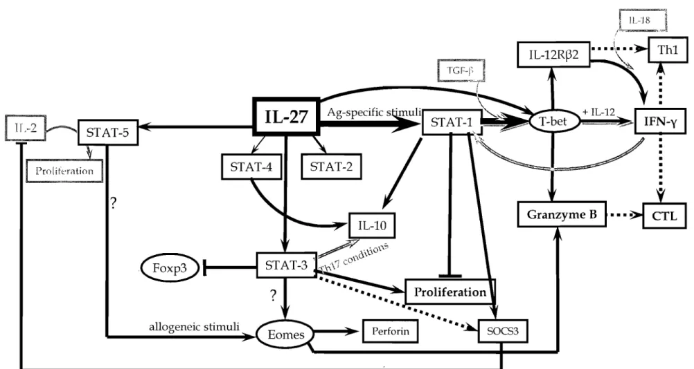
The downstream signalling of IL-27R has been mainly studied in the mouse system. Like an cytokines that bind to type I family cytokine receptors, IL-27 signaIs via the JAKjSTAT pathway. It causes phophorylation of JAK1 (through WSX-1 (Takeda et al., 2003)), JAK2, and TYK2 (tyrosine kinase 2) (Becker et a1., 2005), which further activate STAT-1, -2, -3, -5, and to a lesser extent STAT-4 in mouse naïve CD4 (Hibbert et al., 2003; Lucas et al., 2003; Takeda et a1., 2003; Kamiya et al., 2004) and CD8 T cells (Morishima et al., 2005). It has been suggested that STAT-2 and STAT-5 activation depends on STAT-1 whereas STAT-3 is independent of that transcription factor (Kamiya et al., 2004). Downstream STAT-1 in mouse T cells, the cascade continues with the induction of T-bet, IL-12R~2,
granzyme Band perforin in naïve CD8 T cells (Morishima et al., 2005). Moreover, IL-27 synergizes with IL-12 to boost IFN-y production in a T-bet dependent manner while IL-27 alone does not induce much IFN-y production (Lucas et al., 2003) in naïve (but not in memory) CD4 T cells (Pflanz et al., 2002; Takeda et al., 2003). It has been shown that T-bet and TCCR are critical for the IFN-y production by CD8 T cells during infections such as influenza A virus and Toxoplasma gondii (Mayer et a1., 2008). CD8 T cells require both T-bet and IFN-yR for optimal IFN-y production in neutral conditions whereas the presence of IL-12 renders both molecules dispensable (Mayer et al., 2008), suggesting an important role for IL-27 in CD8 T cell priming. Using chimeric models in which bone marrow derived cells from either IFN-yK/ -, T-bef /- or TCCK / - Yeti IFN-y reporter mice were transplanted into sub lethally irradiated wild type recipients, Mayer and colleagues dissected the contribution of each factor to IFN-y expression (at both
mRNA and protein levels) by CD8 T ceUs foUowing different infections (e.g. influenza virus, Sendai virus, 1. gondii) (Mayer et aL, 2008). They observed that T-bet induced by TCCR is necessary, whereas IFN-yR-mediated signaIs are not essential for priming, expansion or dissemination of Ag-specifie CTLs in these infection models (Mayer et aL, 2008). In addition, other STAT-1 activating receptors (e.g. IFN-yR type l IFNRs) can not compensate for direct IL-27R signaIs (Mayer et aL, 2008).
3.5 Effects of IL-27 on T lymphocytes
Like aU other cytokines, IL-27 has effects on multiple cen types including NK NKT, and T ceUs. Most studies describing the impact of IL-27 on immune cens have been performed using mouse ceUs and very few studies on human cens are available. More than haH of murine NK and NKT cells from naïve mice express TCCR but upon activation such as an in vivo T. gondii infection, these ceUs lose TCCR surface expression (Villarino et aL, 2005). However, in the same infected animais TCCR surface expression on CD4 and CD8 T lymphocytes is enhanced compared to naïve mice and both effector and memory T cells express comparable levels of TCCR (Villarino et al., 2005). Similarly, TCR signalling (anti-CD3 cross-linking antibodies) enhances the proportion of TCCR ex pressing T cells regardless of polarizing conditions (Villarino et al., 2005). TCR-ligation leads to IL-2 production and proliferation of ceUs (at least one round is necessary for optimal expression of TCCR), which over a prolonged period of time leads to down-regulation of TCCR (but not of gp130) on T cells. These observations suggest that immune cells can be distinctly affected by IL-27 depending on their activation status.
Vpon binding to its receptor, IL-27 activates JAK1, JAK2, TYK2, STAT-1, -2 slightly but constantly (Kamiya et al., 2004), -3 and STAT-5 in naïve murine CD4 T cells (Hibbert et aL, 2003; Lucas et aL, 2003; Kamiya et aL, 2004) . Further more, ST AT -2 and ST AT -5 (but not ST AT -3) activation depends on ST AT -1 as in ST AT-1 deficient mice the activation of these transcription factors is greatly reduced