The effects of electronic cigarettes on human gingival
cells and Candida albicans
Thèse
Humidah Alanazi
Doctorat en microbiologie-immunologie
Philosophiæ doctor (Ph. D.)
The effects of electronic cigarettes on human gingival
cells and Candida albicans
Thèse
Humidah Alanazi
Sous la direction de :
Résumé
Plusieurs alternatives ont été mises au point pour réduire les effets de la cigarette sur la santé buccale et générale. La plus récente de ces initiatives est la cigarette électronique. Plusieurs études montrent que la cigarette électronique contient moins de produits toxiques comparativement à la cigarette standard. Ces études concluent que la cigarette électronique est moins nocive pour la santé. Cependant, d’autres études émettent des doutes sur l’innocuité de la cigarette électronique étant donné la présence de multiples produits chimiques. Ces derniers peuvent interagir négativement avec plusieurs parties du corps, dont la cavité buccale.
Les objectifs de cette étude sont (i) d’évaluer les effets d’expositions répétées (1, 2 ou 3 fois) au condensé de cigarette électronique sur la morphologie, la croissance, la migration et l’apoptose des fibroblastes gingivaux humains (ii) d’évaluer les effets de la vapeur de la cigarette électronique sur la croissance, la production de chitine et l’expression de certains gènes codant pour des protéines de la famille des "secreted aspartyl proteinases (SAP par C. albicans avec des temps d’exposition de 15 min, deux fois par jour, pendant 2 et 3 jours. (iii) d’évaluer l’interaction des cellules épithéliales gingivales avec C. albicans préalablement exposé à la cigarette électronique. Nous avons utilisé différentes techniques de biologie cellulaire, de biologie moléculaire et de microbiologie.
Nos travaux montrent que les fibroblastes exposés au condensé de cigarette électronique ont une morphologie anormale (cellules plus grosses, vacuolées,.) et un taux de prolifération plus faible comparativement aux cellules non exposées. Ces observations sont consolidées par un taux plus élevé de cellules apoptotiques comparativement aux cellules non exposées. L’analyse de la migration cellulaire montre que le condensé de cigarette électronique réduit de façon significative la capacité de migration des fibroblastes. Il est à noter que les effets sont plus importants avec la cigarette, suivi de la cigarette électronique contenant la nicotine, puis celle sans nicotine. Les effets de la cigarette électronique sont moins importants
non exposées. Nos études montrent que l’exposition de C. albicans à la cigarette électronique entraîne une augmentation de sa croissance. Cette observation est supportée par un taux plus élevé de chitine produite par C. albicans exposé à la cigarette électronique. L’analyse de la transformation montre des formes hyphes plus longues après l’exposition à cigarette électronique. Nous avons aussi observé que la cigarette électronique augmente l’expression des gènes SAP2, SAP3 et SAP9 par C. albicans comparativement au contrôle (non exposé). L’exposition de C. albicans à la cigarette électronique favorise l'adhésion de la levure aux cellules épithéliales, augmente le taux de tyransformation de la levure. L’exposition de C. albicans à la cigarette électronique, puis son contact avec les cellules épithéliales cause une libération importante de la lactate deshydrogénase (LDH), et la différenciation des cellules épithéliales, mais réduit le taux de croissance de ces cellules gingivales.
Les résultats globaux indiquent que les cigarettes électroniques peuvent interagir avec le microbiome buccal de l’utilisateur. Parce que les cigarettes électroniques réduisent la croissance des cellules gingivales et augmentent l'apoptose cellulaire, cela peut diminuer l'immunité innée dans la cavité buccale, ce qui pourrait augmenter le risque d'infections buccales, telles que la candidose.
Abstract
Electronic cigarettes (e-cigarettes) were designed to replace regular cigarette smoking and to contribute to smoking cessation. E-cigarettes require the use of vaping liquid that contains propylene glycol (PG) and vegetable glycerin (VG) as well as nicotine in various concentrations and flavours. Several studies comparing e-cigarettes to conventional e-cigarettes show that e-e-cigarettes contain lower levels of toxic compounds and for this reason are deemed safer. However, a growing body of evidence shows that e-cigarettes contain many chemicals including formaldehyde, acetaldehyde, acrolein, and toluene, which may have adverse effects on different body parts, including the oral cavity.
The first objective of this study was to investigate the impact of repeated exposures (1, 2, or 3 times) to e-cigarette condensates with or without nicotine on normal human gingival fibroblast morphology, proliferation, migration, and apoptosis. The second objective was to evaluate the effect of e-cigarettes vapors on the growth changes of C. albicans from blastospore to hyphal form and the expression of secreted aspartic proteinases (SAPs) SAP2, SAP3, and SAP9 genes by C. albicans, with exposure times of 15 min twice a day for 2 and 3 days. The third objective was to shed light on the interaction between e-cigarette-exposed C. albicans and gingival epithelial cells. Various cell biology, molecular biology, and microbiology protocols were deployed.
Results show that exposure of gingival fibroblasts to nicotine-rich e-cigarette condensate altered both cell morphology and proliferation rate. Exposure to the e-cigarette condensate also increased the levels of apoptotic fibroblasts. Fibroblast migration was delayed after culture scratches were exposed to e-cigarette condensate. Although e-cigarettes are considered to be less harmful than are conventional cigarettes, e-cigarettes significantly harmed the fibroblasts compared to non-exposed cells. E-cigarette exposure also increased C. albicans growth and hyphal length. The exposed C. albicans produced high levels of chitin and expressed
epithelial cells, e-cigarette-exposed C. albicans adhered better compared to the controls. Indirect communication between e-cigarette-exposed C. albicans and gingival epithelial cells led to epithelial cell differentiation, reduced cell growth, and increased lactate dehydrogenase (LDH) activity.
Overall results indicate that e-cigarettes may interact with the user’s oral microbiome. Because e-cigarettes reduce gingival cell growth and increase cell apoptosis, this may decrease the innate immunity in the oral cavity, which could increase the risk of oral infections, such as candidiasis.
Table of Contents
Résumé ... ii
Abstract ... iv
Liste des figures ... xi
Liste des tableaux ... xiii
Liste des abréviations ... xiv
Remerciements ... xvii
Avant-propos ... xix
INTRODUCTION ... 1
Electronic cigarettes and their prevalence ... 1
Concerns about e-cigarettes ... 2
Hypotheses ... 3
CHAPTER 1: LITERATURE REVIEW ... 7
1.1. Tobacco smoke across civilisation ... 7
1.2. Smoking prevalence among youth and adult populations ... 9
1.3. Impact of smoking on health ... 12
1.3.1. Tobacco and cardiovascular diseases ... 14
1.3.2. Cigarette smoke and pulmonary diseases ... 15
1.3.3. Cigarette smoking and cancer ... 17
1.4. Cigarette smoke and oral health ... 18
1.4.1. Dental caries promotion ... 19
1.4.2. Periodontal disease promotion ... 19
1.4.3. Oropharyngeal candidiasis promotion ... 20
1.5. Smoking cessation and health improvement ... 21
1.6. Tobacco cessation strategies ... 22
1.6.1. Pharmacotherapy for smoking cessation ... 23
1.6.1.1. Bupropion ... 23
1.6.1.2. Varenicline ... 24
1.6.2. Nicotine Replacement Therapy ... 25
1.7.1.1. E-cigarette cartridge ... 27 1.7.1.2. E-cigarette atomizer ... 27 1.7.1.3. E-cigarette battery ... 29 1.7.2. E-cigarette generations ... 29 1.7.2.1. First generation ... 30 1.7.2.2. Second generation ... 30 1.7.2.3. Third generation ... 30 1.7.2.4. Fourth generation ... 32
1.7.2.5. The most recent e-cigarette device: JUUL ... 32
1.7.3. Refill solutions for e-cigarettes (e-liquids) ... 32
1.7.3.1. Nicotine levels in e-cigarettes ... 34
1.7.3.2. E-liquid pH ... 35
1.7.3.3. E-liquid flavours ... 36
1.7.4. Prevalence of e-cigarette Use ... 37
1.7.4.1. Prevalence in the U.S. ... 37
1.7.4.2. Prevalence in Europe ... 37
1.7.4.3. Prevalence in Australia ... 38
1.7.4.4. Prevalence in the United Kingdom ... 38
1.7.4.5. Prevalence in Canada ... 38
1.7.5. E-cigarettes and smoking cessation ... 39
1.7.6. Dual use of EC and SC ... 40
1.7.7. Benefits of e-cigarette use ... 42
1.7.7.1. Experimental data and animal model ... 42
1.7.7.2. Beneficial effects of e-cigarettes on general health ... 43
1.7.7.3. Beneficial effects of e-cigarettes on oral health ... 44
1.7.8. Concerns regarding e-cigarettes ... 45
1.7.8.1. E-cigarettes appear to target youth ... 46
1.7.8.2. Concerns regarding the e-cigarette device constituents ... 47
1.7.8.2.1. Possible harmful effects of e-cigarette batteries ... 47
1.7.8.2.2. Concerns regarding the vaping solutions/e-liquids ... 48
1.7.8.2.2.1. Vaping propylene glycol ... 48
1.7.8.2.2.2. Vaping vegetable glycerin (VG). ... 49
1.7.8.2.2.3. Vaping PG-VG e-liquids. ... 49
1.7.8.2.3. Concerns regarding e-liquid flavours ... 50
1.7.8.2.4. Concerns regarding nicotine dosing in e-cigarettes ... 52
1.7.9. Concerns regarding the effects of e-cigarettes on human health ... 53
1.7.9.3. Possible harmful effects of e-cigarettes on oral health ... 55
1.7.9.3.1. E-cigarettes and periodontal diseases ... 55
1.7.9.3.2. Effects on peri-implant parameters ... 56
1.7.9.3.3. Effects on e-cigarette users’ teeth ... 57
1.7.9.3.4. E-cigarette use and dry mouth/xerostomia ... 58
1.7.9.3.5. Effects on e-cigarette user saliva ... 58
1.7.9.4. Effects of e-cigarettes on oral microorganisms ... 59
1.7.9.4.1. Interaction with Staphylococcus aureus ... 59
1.7.9.4.2. Interaction with Streptococcus mutans ... 59
1.8. Context ... 61
1.9. Hypotheses ... 61
1.10. Objectives ... 61
CHAPTER 2: Comparative Study of the Effects of Cigarette Smoke and Electronic Cigarettes on Human Gingival Fibroblast Proliferation, Migration and Apoptosis. ... 63
2.1 Résumé ... 64
2.2 Abstract ... 64
2.3 Introduction ... 65
2.4 Material and Methods ... 66
2.4.1 Gingival fibroblast isolation and culture ... 66
2.4.2 Preparation of cigarette smoke condensate solution ... 67
2.4.3 Preparation of the e-vapor condensate solutions ... 67
2.4.4 Effect of cigarette smoke and e-vapor condensates on human gingival fibroblast adhesion and morphology ... 69
2.4.5 Effect of cigarette smoke and e-vapor condensates on cell growth ... 69
2.4.6 Effect of cigarette smoke and e-vapor condensates on fibroblast proliferation ... 70
2.4.7 Apoptotic cell analysis by DNA fragmentation assay ... 71
2.4.8 Effect of cigarette smoke and e-vapor condensates on gingival fibroblast migration 72 2.4.9 Statistical analyses ... 72
2.5 Results ... 73
2.5.1 Cigarette smoke and e-vapor condensates modulated fibroblast morphology but not early adhesion ... 73
2.5.3 Cigarette smoke and e-vapor condensates promoted gingival fibroblast apoptosis . 77 2.5.4 Cigarette smoke and e-vapor condensates delayed gingival fibroblast migration and
wound closure ... 78
2.6 Discussion ... 81
2.7 Conclusion ... 83
2.8 References ... 85
CHAPTER 3: E-cigarettes vapour increase Candida albicans growth and modulate its interaction with gingival epithelial cells ... 89
3.1. Résumé ... 90
3.2. Abstract: ... 90
3.3. Introduction ... 91
3.4. Material and Methods ... 92
3.4.1. Candida strain ... 92
3.4.2. E-cigarettes ... 93
3.4.3. Effect of e-vapor on C. albicans growth ... 93
3.4.4. Effect of e-vapor on C. albicans cell wall chitin content ... 94
3.4.5. Effect of e-vapor on C. albicans transition from blastospore to hyphal form ... 96
3.4.6. Effect of e-vapor on the expression of SAP2, SAP3, and SAP9 genes by C. albicans .. 96
3.4.7. Adhesion of e-vapor-exposed C. albicans to gingival epithelial cells ... 97
3.4.8. Growth of epithelial cells following contact with e-vapor-exposed C. albicans ... 98
3.4.9. Statistical analysis ... 99
3.5. Results and Discussion ... 100
3.5.1. E-cigarette vapor promoted C. albicans growth ... 100
3.5.2. Chitin content was high in e-cigarette vapor-exposed C. albicans ... 102
3.5.3. E-vapor-exposed C. albicans displayed an increase in hyphal length ... 104
3.5.4. E-vapor-exposed C. albicans expressed high virulent gene levels ... 105
3.5.5. E-vapor-exposed C. albicans adhered better to gingival epithelial cells ... 107
3.5.6. Cross- talk interactions between e-vapor-exposed C. albicans and epithelial cells promoted yeast growth and morphological changes ... 110 3.5.7. E-vapor-exposed C. albicans promoted morphological changes in epithelial cells and
3.6. Conclusion ... 116
3.7. References ... 117
CHAPTER 4: GENERAL DISCUSSION ... 122
CONCLUSION ... 128
Liste des figures
Figure 1.1: Experimental protocol for objective 1………..… 5
Figure 1.2: Experimental protocol for objective 2………..…… 6
Figure 1.3: Waterpipe smoking device………...……. 8
Figure 1.4: 2014 Europe smoking prevalence.……….. 10
Figure 1.5: 2015 Canadian adults smoking prevalence ……….. 11
Figure 1.6: Canadian smokers’ proportion 2017………..…... 13
Figure 1.7: 2007-2017 Global tobacco smoking prevalence by countries’ income level………..… 13
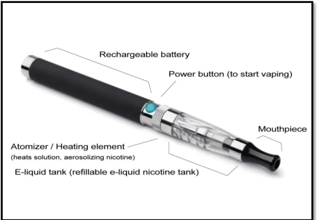
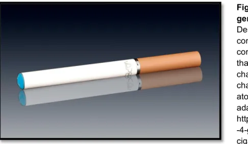
Figure 1.8: Electronic cigarette device………..…... 27
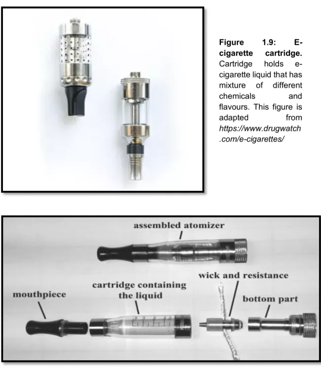
Figure 1.9: E-cigarette cartridge………...…. 28
Figure 1.10: E-cigarette atomizer……….... 28
Figure 1.11: E-cigarette battery………...…. 29
Figure 1.12: First generation of Electronic Cigarette……….... 30
Figure 1.13: Second generation of Electronic Cigarette………..… 31
Figure 1.14: Third generation of Electronic Cigarette………... 31
Figure 1.15: Fourth generation of Electronic Cigarette………. 33
Figure 1.16: JUUL electronic cigarette……….... 34
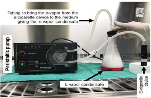
Figure 2.1: Schema showing the system used to generate the e-vapor condensates………..… 68
Figure 2.2: Early exposure to CSC and e-cigarette vapor condensate had no effect on fibroblast adhesion………. 74
Figure 2.3: CSC and e-cigarette vapor condensate modulated human gingival fibroblast morphology………..…….. 75
Figure 2.4: CSC and e-cigarette vapor condensate decreased human gingival fibroblast growth………..….. 76
Figure 2.5: CSC and e-cigarette vapor condensate downregulated human gingival fibroblast proliferation……….... 77
Figure 2.6: CSC and e-vapor condensate increased human gingival fibroblast apoptosis………. 79
Figure 2.7: CSC and e-vapor condensate modulated human gingival fibroblast migration……….. 80
Figure 3.1: Exposure protocol of Candida albicans to e-cigarette vapor or combustible cigarette smoke……….. 94
Figure 3.2: E-cigarette vapor promoted C. albicans………... 101
Figure 3.3: E-cigarette vapor increased the level of chitin produced by C. albicans………. 103
Figure 3.4: E-cigarette vapor increased the hyphal length of C. albicans cultured under cell morphology transition conditions………. 105
Figure 3.5: E-cigarette vapor increased the expression of secreted aspartyl proteinases SAPs 2, 3, and 9 genes……….... 108
Figure 3.6: C. albicans pre-exposed to e-cigarette vapor adhered better to gingival epithelial cells cultures……….. 109
Figure 3.7: Growth and transition of C. albicans pre-exposed to e-cigarette vapor then co-cultured with gingival epithelial cells……… 111
Figure 3.8: C. albicans pre-exposed to e-cigarette vapor promoted gingival
epithelial cell differentiation……….… 113 Figure 3.9: E-vapor pre-exposed C. albicans decreased gingival epithelial
cell viability………...… 114 Figure 3.10: Epithelial cells co-cultured with e-vapor pre-exposed
C. albicans displayed high levels of LDH activity………....
Liste des tableaux
Table 1.1: The different possibilities to help patients to stop smoking…… 23 Table 1.2: The pH of the e-liquids………. 36 Table 3.1: Primer sequences used for the qRT-PCR………. 97
Liste des abréviations
a4b2 alpha-4 beta-2A549 cells human alveolar cell cultures
ACT1 actin 1
AIDS acquired immunodeficiency syndrome APVs advanced personal vaporizers
BOP bleeding on probing bpm beats per minute BrdU bromodeoxyuridine C. albicans Candida albicans
Ca9-22 human gingival epithelial carcinoma cell line CCS conventional cigarette smoke
CDC Centers for Disease Control cDNA C° complementary DNA celsius cig-a-likes cigarette-like CO carbon monoxide Ctrl control
COPD chronic obstructive pulmonary disease CS
SC cigarette smoke smoke cigarette
CSC cigarette smoke condensate
CT cycle threshold
CVDs cardiovascular diseases
CXCL8 C-X-C motif chemokine ligand 8 DMEM Dulbecco's Modified Eagle's medium DNA Deoxyribonucleic acid
DNase Deoxyribonuclease e-cigarette/e-
cig
electronic cigarette
e-liquid electronic cigarette liquid e-vaping electronic cigarette vaping
e-VC electronic cigarette vapor condensate EAP1 Enhanced Adherence to Polystyrene EC electronic cigarette
ECM extracellular matrix
EDTA ethylenediaminetetraacetic acid ELISA enzyme-linked immunosorbent Assay
EU European Union
ex-smokers former smoker FBS fetal bovine serum
FDA Food and Drug Administration
hBD human β-Defensin
HSRRB Health Science Research Resources Bank HWP1 Hyphal Wall Protein 1
IL-1β Interleukin 1 beta IL-6 Interleukin 6
LDH lactate dehydrogenase Li-ion lithium ion
Li-poly lithium polymer LiMn lithium manganese
LL-37 cathelicidin antimicrobial peptides MEC mobile exam center
mRNA messenger RNA
MTT thiazolyl blue tetrazolium bromide nAChR nicotinic cetylcholine receptors NF/NR
e-vapour
nicotine free/ nicotine rich electronic cigarette vapor NHANES National Health and Nutrition Examination Survey NiCad nickel-cadmium
NiMh nickel metal-hydride
NIOSH National Institute for Occupational Safety and Health NRT nicotine replacement therapy
ns non-significant
OSCC oral squamous cell carcinoma P. gingivalis Porphyromonas gingivalis PBS phosphate-buffered saline PCR polymerase chain reaction PD periodontal disease PD probing depth PG propylene glycol pH power of Hydrogen PI plaque index ppm ppn
parts per million parts per billion qPCR quantitative PCR
qRT-PCR real-time quantitative reverse transcription PCR RAGE receptor of advanced glycation end products RBL radiographic bone loss
RNA ribonucleic acid RNase ribonuclease
ROS reactive oxygen species
RPMI Roswell Park Memorial Institute S. mutans Streptococcus mutans
SA Staphylococcus aureus SAPs secreted aspartic proteases
SD standard deviation
sp species
TdT terminal deoxynucleotidyl transferase TLR toll like receptor
Tm-m melting temperature from the manufacturer TNF-α Tumour necrosis factor alpha
TUNE Terminal deoxynucleotidyl transferase dUTP Nick End Labeling
US United States
USB universal serial Bus VG vegetable glycerin
Remerciements
From 2011 to 2020, my life experience in Quebec, Canada, has strengthen me a lot in many ways - humanity, the basics -living principles, perseverance, diligence, and faith. In the beautiful location of the Faculty of Dental Medicine at Université Laval University, was a laboratory where, I really discovered myself, I loved working there a lot, and I felt at home despite the pressures of life. In this laboratory, I met Dr. Mahmoud Rouabhia as the one of the most amazing people I have met. He taught me the principles of research and helped me getting started and improved my skills, with his beautiful way. Dr. Rouabhia, words of thanks will not be sufficient to express my feeling for your kindness.
My thanks go to Dr. Abdelhabib Semlali, who also supported me in the scientific research.
I thank my MOTHER ESHYH for her sincere prayers that were with me in all circumstances. She left from this world while she was waiting for my return to the homeland. Always you were supporting me with powerful words to not give up, with your thumbs up. Iam eager to meet again, my mother, and I will talk to you about all events and how you have always been with me in all scenes of life and will remain forever.
Many thanks go to my father for his support.
Thanks to my little family. Thanks to my husband, Dr. Shadaid Alanezi, despite the difficult circumstances that we went through. With God's help, we overcame them together. Thanks to my children Zaid, Mayar and Mubarak. You were heroes with me. I loved you from the bottom of my heart and forgive me for the times when I was distracted.
Endless thanks to my older sister and my friend Sarah. Always her standing with me was and still is the motivating force for me in all circumstances. Her words renew my energy. She guided me to be really strong and faithful.
My little brother and my son Dr. Zaid (the most amazing physician), thank you for your support, for your beautiful words with your principles and the wonders. Thanks for the most precious moments we spent and we will spend together.
The rest of my sibling Hameed, Khalil, Hammoud, Khaled and Nawal, thank you all. There are two persons who were instrumental in continuing my education; my uncle Zaid and my aunt Kamla, Thank you.
Dr. Hyun Jin Park, you were always beside me at work and outside work. I hope we stay friends forever. Thanks a lot Hyun Jin.
There is a great couple Fatima Redjemi and Nader Maroc, our child care providers (home care). They are my family here in Quebec City. This couple helped me a lot to pass difficult situations with my children even with me. Thank you.
After twenty-seven years, I found you my childhood friend Seham to share with me the greatest moment in my life.
Thanks for my country, the Kingdom of Saudi Arabia and King Saud University for the great support we are receiving in both financial and moral matters. PROMISE, we will build SUCCESS in many aspects.
Avant-propos
Cette thèse présente les travaux de recherche réalisés durant mes activités de recherche doctorale en microbiologie et immunologie. Ma formation doctorale s’est déroulée de septembre 2016 à juin 2020. Les différents sujets abordés dans cette thèse sont mis en contexte dans l’introduction, suivi des objectifs. La thèse intègre deux publications (Chapitre 2et chapitre 3) relatant nos travaux qui sont publiés dans différents journaux scientifiques internationaux, avec comité de paires. Ces deux publications forment le corps scientifique de cette thèse. Ces publications sont listées ci-dessous, avec des informations relatives à leur statut de publication, leur contexte de recherche, mes contributions ainsi que celles des coauteurs dans la réalisation de la recherche et sa publication.
CHAPITRE 2 :
Titre de la publication: Comparative study of the effects of cigarette smoke and electronic cigarettes on human gingival fibroblast proliferation, migration and apoptosis.
Auteurs : Alanazi H, Park HJ, Chakir J, Semlali A, Rouabhia M.
Journal et date de publication :
Food Chem Toxicol, 2018 Aug;118:390-398. doi: 10.1016/j.fct.2018.05.049. Epub 2018 May 22.
Facteur d’impact du journal : 3.775 (2018)
Mon implication dans cette publication consiste en la réalisation des expériences au laboratoire, la collecte des données et leurs analyses. Mon rôle dans ces différentes étapes est estimé à 80% de la finalisation, car j’ai eu le support des autres coauteurs dans l’accomplissement, surtout des analyses des résultats et la finalisation des figures qui sont incluses dans cette publication. J’ai écrit la première version de la
version a été envoyée aux autres coauteurs. À la réception de leurs commentaires, j’ai finalisé le manuscrit que j’ai soumis au journal, avec l’aide de mon directeur de recherche. Les évaluateurs ont suggéré des modifications mineures. J’ai révisé et soumis le manuscrit révisé qui a été accepté pour publication dans Food Chem Toxicol.
CHAPITRE 3:
Titre de la publication: E-Cigarettes Increase Candida albicans Growth and Modulate its Interaction with Gingival Epithelial Cells.
Auteurs : Alanazi H, Semlali A, Chmielewski
W, Rouabhia M. Journal et date de
publication :
Int J Environ Res Public Health. 2019 Jan
21;16(2). pii: E294. doi:
10.3390/ijerph16020294. Facteur d’impact du journal : 2.468 (2018)
Mon implication dans cette publication consiste en la réalisation des expériences au laboratoire en lien avec la culture cellulaire et bactériologie, la collecte des données et leurs analyses/interprétation, leurs mises en forme en tableaux et figures. Mon rôle dans ces différentes étapes est estimé à 90%. J’ai eu une aide, apprécié, de la part des coauteurs, surtout pour les analyses des résultats et la finalisation des figures qui sont incluses dans cette publication. J’ai écrit la première version du manuscrit qui a été révisé par mon directeur de recherche, le Dr Rouabhia. Cette version a été commentée par les coauteurs. Leurs commentaires ont été intégrés dans le manuscrit pour en faire une version finale que j’ai soumise au journal (Int J Environ Res Public Health). Les évaluateurs ont suggéré des modifications mineures. J’ai modifié le manuscrit en intégrant les commentaires des évaluateurs, et je l'ai soumis. Ce manuscrit a été accepté sans modifications supplémentaires.
INTRODUCTION
“We @CDCgov are saddened to hear of the first death related to the outbreak of severe lung disease in those who use of e-cigarettes or e-vaping devices. We will continue to educate all Americans about the serious risks associated with these products.” Dr. Robert R. Redfield, Director of Centers for Disease Control and Prevention, Atlanta, Georgia, USA, tweeted on 2019-08-23 on Twitter.
Electronic cigarettes and their prevalence
E-cigarettes are battery-powered nicotine-delivery devices not containing tobacco but rather a liquid (e-liquid) that is vaporized to form a nicotine-containing aerosol. It uses tobacco that is directly or indirectly heated (but not burned) using a variety of heat sources to create an inhalable tobacco aerosol. The aerosol is generated by the presence of humectants, such as propylene glycol (PG) and vegetable glycerin (VG), as well as by nicotine at various concentrations, and flavours.
E-cigarettes were designed to play an active role in cigarette smoke replacement and cessation and were also proposed as a safe product. Indeed, several studies comparing e-cigarette aerosol to cigarette smoke concluded that e-cigarettes contained lower levels of potentially toxic compounds (e.g. formaldehyde, acetaldehyde, acrolein, and toluene), compared to conventional cigarettes (Bekki et al., 2014). Data from clinical trials (Caponnetto et al., 2013; Bullen et al., 2013) and meta-analyses (Hartmann-Boyce et al., 2016) indicate that e-cigarettes may help smokers to quit or reduce their tobacco consumption and that their use is well tolerated. These clinical observations, along with advertisements promoting e-cigarettes as being safe, have contributed to e-cigarette popularity with smokers, non-smokers and youth, in particular. Indeed, in the US alone, it was estimated that 20.8 % of high school students and 4.9 % of middle school students were current e-cigarette users in 2018, representing a 78 % increase in use among high school students and a 48 % increase among middle school students during the 2017–2018 school year (Cullen et al., 2018).
Researchers have suggested that the rise in e-cigarette use among youth is linked to the availability of appealing flavours and the recent popularity of discreet e-cigarette models shaped like a USB flash drive (Cullen et al., 2018; Tsai et al., 2018). In Canada, 15.4 % of Canadians aged 15 and older (4.6 million) reported having ever tried an e-cigarette, while 2.9 % (~863,000) had used an e-cigarette in the past 30 days. Prevalence of ever using e-cigarettes increased significantly between 2015 and 2017, while past 30-day use showed no significant change (https://uwaterloo.ca/tobacco-use-canada/e-cigarette-use-canada/prevalence-e-cigarette-use).
Concerns about e-cigarettes
Although e-cigarettes were developed and marketed as a healthier alternative to smoking tobacco products, a growing body of evidence shows that even if their quantity is generally lower than that found in conventional tobacco cigarettes, their aerosols in fact contain numerous toxicants, carcinogens and organic compounds produced through the thermal decomposition of the solvents (Goniewicz et al., 2014). Indeed, several reports have associated e-cigarette use with respiratory, gastrointestinal, and even cardiovascular complications including pulmonary damage (Thirion-Romero et al., 2019), relapse of ulcerative colitis (Hua et al., 2016) and disrupted endothelial functions (Skotsimara et al., 2019).
While many studies focus on the respiratory and cardiovascular systems, the impact of e-cigarette use on oral health could be one of the first red flags signaling the deleterious effects of e-cigarettes. Few studies have addressed the direct health effect of cigarette usage on the oral cavity. In a cross-sectional analysis, daily e-cigarette use was associated with risk factors for such poor oral health outcomes such as periodontal diseases and tooth loss (Huilgol et al., 2019). A correlation has also been found between e-cigarette use and a higher likelihood of cracked/broken teeth, pain in the tongue, and/or inside the cheek, compared to never use e-cigarette smokers, among adolescents (Cho, 2017). E-cigarette use has also been associated
with gingival mucosa lesions (Bardellini et al., 2018). Indeed, in a small sample of patients, the prevalence and characteristics of oral mucosal lesions were evaluated in former smokers (n = 45) compared to e-cigarette consumers (n = 45), with the results showing the prevalence of these mucosal lesions to be approximately 65.4 % among e-cigarette users compared to 34.6 % among former smokers. In the same study, other oral symptoms were recorded, such as nicotine stomatitis, hairy tongue, and hyperplastic candidiasis. The frequency of these symptoms was greater among e-cigarette users than among former smokers (Bardellini et al., 2018).
Context
Following a puff, the aerosol generated by an e-cigarette is delivered into the user’s mouth, thus entering in direct contact with the different constituents of the mouth, before reaching the lower airways. The entry of aerosol into the mouth and its direct contact with these different oral constituents (e.g. oral mucosa, teeth, saliva, and the oral microbiome) may change the physiological equilibrium of the oral cavity and oral microbiome. We thus sought to investigate the effect of e-cigarettes on gingival cells and oral microorganisms.
Hypotheses
Previous studies including those from our research team have shown that e-cigarettes reduce the growth of gingival epithelial cells through an apoptotic-necrotic pathway (Rouabhia et al.,2017). Thus, we hypothesise that:
1) Exposure of gingival fibroblasts to e-cigarettes may therefore lead to an impairment of gingival fibroblast functions,
2) E-cigarette use may dysregulate the oral microbiome as well as oral microorganism interactions with gingival epithelial cells.
Specific Objectives
The specific objectives of this projects are:
apoptosis. We examined the effect of e-vapour condensate on gingival fibroblast adhesion, viability/proliferation, apoptotic process, and migration. This objective includes both nicotine-free and nicotine-rich e-cigarettes treated cells. The effects of combustible cigarette smoke condensate and e-vapour condensate on gingival fibroblasts were also compared and analyzed. 2. To investigate the effects of e-cigarettes on C. albicans pathogenesis. In this objective, we investigated the effect of e-cigarette aerosols on the growth and morphology changes of C. albicans. We also studied the effect of e-cigarette aerosols on the expression of secreted aspartic proteases (SAPs), SAP2, SAP3, and SAP9 genes by C. albicans. The interaction between e-cigarette-exposed C. albicans and gingival epithelial cells was also evaluated.
To achieve these objectives, we adopted the following specific experimental protocols:
Preparation of the smoke/e-vapour condensates
We prepared the e-cigarette vapour condensate (e-VC) by vaping 500 µL of e-liquid using an e-cigarette device in 20 ml of culture medium, as previously reported (Lerner et al., 2016). The e-VCs were prepared from both rich and nicotine-free e-liquids. We also prepared cigarette smoke condensate solution (CSC) by burning one cigarette in 20 ml of culture medium (Semlali et al., 2014). The generated condensates were first filtered through a 0.2-µm filter to sterilize them and were then aliquoted and frozen at -80 °C until use.
Human gingival cells
• Cells were extracted from gingival tissue (gingival connective tissue) collected from healthy, never-smoked donors (18–25 years of age) following their signature of informed consent. Extracted gingival fibroblasts were cultured in Dulbecco’s modified Eagle’s (DME) medium containing 10 % fetal calf serum.
• We also used gingival epithelial cells in this study. These refer to a specific cell line, namely, Ca9-22 extracted from gingival squamous cell carcinoma (purchased from Health Science Research Resources Bank (HSRRB) (Osaka, Japan)). This gingival epithelial cell line was maintained in Roswell Park Memorial Institute medium (RPMI)-1640 supplemented with L-glutamine and 10 % fetal calf serum (FBS).
Candida Strain
We used C. albicans in this study. C. albicans (ATCC-SC5314) was grown in Sabouraud liquid medium supplemented with 0.1 % glucose. The culture was grown to the stationary phase for 18 h at 30 ºC in a shaking water bath. The blastoconidia were then collected, washed with phosphate-buffered saline (PBS), and counted by means of a hemacytometer. The cell suspension was adjusted to 108 C. albicans
cells/ml prior to being used.
The protocol for objective 1: The different steps related to objective 1 are summarized in the figure below
The protocol for objective 2: The different steps related to objective 2 are summarized in
the in the figure below
CHAPTER 1: LITERATURE REVIEW
1.1. Tobacco smoke across civilisationTobacco has had a longstanding role in many societies across civilisation. It is believed that the tobacco plant was the first domesticated in the Americas predating the farming of maize and other food plants (Winter, 2000). Tobacco species include Nicotiana quadrivalvis, Nicotiana attenuata, and Nicotiana obtusifolia (Tushingham et al., 2013). Interestingly, the process of domestication led to the development of other tobacco varieties, such as Nicotiana rustica and Nicotiana tabacum species, which have larger leaves and high nicotine content (Winter, 2000).
Historically, the domesticated species Nicotiana rustica was thought to reach the eastern part of the continent from South America between 2000 and 3000 years ago, while Nicotiana tabacum likely spread to parts of southwestern United States and the Caribbean some time thereafter (Winter, 2000; Rafferty, 2006). For a long time, various species of tobacco were used by indigenous communities throughout North and South America, then in 1492, tobacco was introduced to Columbus in the Bahama Islands during his first encounter with the Americas. European explorers to the Americas quickly recognized tobacco’s unique properties and adopted it for good use. Later in the 1500s, Nicotiana tabacum was selected and farmed in different British and American colonies. As early as the 1600s, tobacco reached Africa, Asia, and Europe to become a global trade commodity (Tushingham et al., 2013). Following its emergence in Europe, tobacco plants were widely used throughout the Americas thanks to different processes, including plant farming, gathering, or trading. Tobacco was, and probably still is, a central product during ritual and ceremonial life of Native Americans (Winter, 2000).
African history confirms that tobacco was introduced to Africa in the late 1500s. Tobacco gained significant popularity due to various production modes and uses. In
this part of the world, but not limited to it, tobacco has contributed to economic development, particularly during the colonial and postcolonial periods (Duvall, 2017), although the commercial value was moderated by the need for fertile land and hard work involved in tobacco production.
The reputation tobacco has acquired across civilisation lies in its effect on human feelings. Although tobacco is most often smoked, it can be chewed, eaten, or snuffed. It was believed that tobacco procured sharp mental acuity, vigilance, and increased sense of calm to users. Furthermore, tobacco was frequently used as an offering in religious contexts and peace time (e.g. peace pipe). Smoking is the preferred method of tobacco use. In early times, smoking took place through dry pipes or water pipes (Figure 1.3) and was restricted to adult males. With the now-known adverse effects of tobacco smoke, this restriction was indeed an important lifesaver for teens and women. Unfortunately, almost all age ranges of people around the world currently smoke tobacco.
Figure 1.3: Waterpipe smoking device: An old
device used for tobacco smoke. Coal heats the tobacco. The smoke is then filtered by boiled water and inhaled by the user through a rubber pipe. This figure is adapted from Akl et al., (2011).
1.2. Smoking prevalence among youth and adult populations
The prevalence of smoking varies from one population to another around the world (Eriksen et al., 2012; World Health Organization, 2014). Such variations are linked to multiple factors known to modulate smoking prevalence. Among the key factors are education level, national economic development, and implementation of tobacco control policies (Gilmore et al., 2000; Pomerleau et al., 2004).
In 2016 in the United States (US), more than 16 % of individuals over 18 years of age were smokers, thus an estimated 37.8 million adults smoked cigarettes. It should be noted that in 2005, the average of smokers in the US was approximately 21 % (Jamal et al., 2018). Smoke cigarette use by youth in the US is critical; indeed, tobacco use begins and is established primarily during adolescence (U.S. Department of Health and Human Services, 2012; U.S. Department of Health and Human Services, 2014). It was estimated that 9 out of 10 youth smokers have tried smoking by the age of 181. Every day in the US, more than 3,200 youth 18 years or
younger smoke their first cigarette, and an additional 2,100 youth and young adults become daily cigarette smokers. (U.S. Department of Health and Human Services, 2012).
Cigarette companies are continuously launching new initiatives such as adding flavours to their tobacco products to attract new cigarette users (Corey et al., 2015). In 2014, 73 % of high school students and 57 % of middle school students who used tobacco products in the past 30 days reported using a flavoured tobacco product during that time (Corey et al., 2015). Adult men are more likely than women to use cigarettes in the US. Close to 18 % of men and 13 % of women were smokers at the time of the survey (Jamal et al., 2018). Ethnicity also appears to be a factor in smoking. In a 2016 survey, approximately 32 % of non-Hispanic American Indians/Alaska Natives were found to be smokers, while close to 25 % of Hispanic multiple-race individuals were smokers. Approximately 17 % of non-Hispanic Blacks and nearly 17 % non-non-Hispanic Whites were also smokers. Finally,
Poverty is also a possible smoke-promoting indicator, as 25 % of adults living below the poverty level were smokers compared to 14 % of adults living above the poverty level (Jamal et al., 2018)
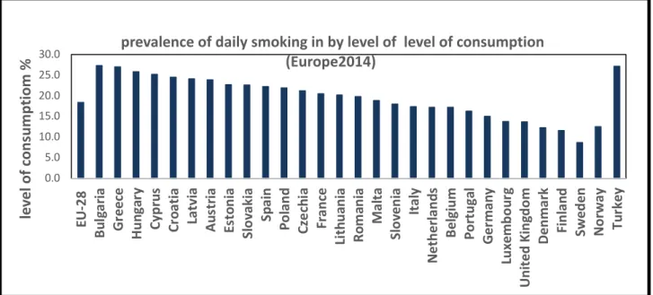
A European survey conducted from 2013 to 2015 with participants over the age of 15 showed that the proportion of daily smokers ranged from 8.7 to 27.3 % (Eurostat, 2018) showed that the proportion of daily smokers ranged from 8.7 to 27.3 %. The lowest number of smokers was recorded in Sweden, and the highest level in Bulgaria (Figure1.4). Among the 27 EU Member States, men were more prone to smoking than were women, except for Sweden, where 7.5 % men compared to 9.8 % women were smokers during the survey (Eurostat, 2018). The highest number of smokers was in the 25–54-year range, dropping thereafter at over 65 years of age. As for smoking frequency, close to 6 % of the European population over 15 years of age smoked at least 20 cigarettes per day and about 12 % smoked less than 20 cigarettes per day. The greatest numbers of heavy smokers were recorded in Greece and Turkey (Eurostat, 2018).
Figure 1.4: 2014 Europe smoking prevalence. Statistical results showing adults daily
consumption of cigarettes in Europe in 2014. Lowest smoke level was registered in Sweden 8.7%, and the highest level was registered in Bulgaria 27.3%. This figure is adapted from Eurostat Statistics Explained:
https://ec.europa.eu/eurostat/statistics-0.0 5.0 10.0 15.0 20.0 25.0 30.0 EU -2 8 Bu lg ar ia Gr ee ce Hu ng ar y Cy pru s Cr oa tia La tv ia Au st ria Es to ni a Sl ov ak ia Sp ai n Po la nd Cz ec hi a Fr an ce Li th ua ni a Ro m an ia Ma lta Sl ov en ia Ita ly Ne th erla nd s Be lgi um Po rt ug al Ge rm an y Lu xe m bo ur g Un ite d Ki ng do m De nm ar k Fi nl an d Sw ed en No rw ay Tu rk ey le ve l of c onsum ptiom %
prevalence of daily smoking in by level of level of consumption (Europe2014)
In Canada, tobacco smoking is also a major health concern. From a 2012 Statistics Canada survey, we learned that between 2001 and 2011, the percentage of light daily male smokers increased from approximately 51 to 62 % compared to an increase of 36 to 43 % in females (Statistics Canada, Canadian Community Health Survey, (2012). Of interest, is that the number of cigarettes smoked per day by light smokers dropped from 17 in 2001 to 15 in 2011. During the same period (2001 to 2011), the percentage of heavy male smokers decreased from 31 to 23 %, while heavy female smokers dropped from 20 to 14 %. These heavy smokers consumed approximately 28 cigarettes per day during this study period (Statistics Canada, Canadian Community Health Survey 2012). Similar to other world populations, Canadians start smoking at an early age. In 2011, approximately 19 % of Canadian smokers were 15 to 17 years old and close to 12 % of young smokers were living in lower-income households (Health Canada, 2012; Reid et al., 2017).
The most recent surveys report that in 2015, nearly 13 % of Canadians were smokers. Most of these were daily smokers (9.4 %) (Figure 1.5). It should be noted that during 2015, smoking prevalence was higher in males (15.6 %) than in females (10.4 %). In terms of distribution, 9.7 % of smokers were 15–19 years old, 18.5 % were 20–24 years old, and 10.6 % were over 55 (https://uwaterloo.ca/tobacco-use-canada/highlights).
Figure1.5: 2015 Canadian adults smoking prevalence:
3.7% were non-daily smoking and 9.4% were daily smoking. This figure is adapted from Reid and Hammond (2017)
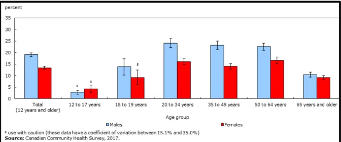
One Canadian report (Statistics Canada, Canadian Community Health Survey 2018) has shown that in 2017, daily or occasional smokers were close to 5 million (almost 16 %) in number, from the age of 12 years old. More smokers were males (19.1 %), compared to females (13.4 %) (Figure 1.6).
Globally, populations record a high number of smokers. According to the World Health Organization, tobacco is the largest public health threat worldwide, as tobacco kills over 7 million people each year, with 6 million of these deaths attributed directly to cigarette smoking (World Health Organization, 2013). There are an estimated 1 billion smokers worldwide (Eriksen et al., 2013), involving close to 30 % of men and 7 % of women (Gowing et al., 2015), with 80 % of these users in the low-to-mid income bracket. The most frustrating situation is that in several countries, children from poor families are frequently employed in tobacco farming to provide family income, which contributes to their early exposure to various tobacco chemicals (WHO, 2018). The worldwide prevalence of smokers was estimated to be 39 % in men in 2007 compared to 35 % in 2015. In women, the global prevalence estimation was approximately 6 % in 2015 and 8 % in 2017. Smoking prevalence was modulated by the countries’ income (Figure 1.7, WHO report, 2017). The global concern regarding cigarette smoking is the risk of contracting a wide range of long-term morbidities, and ultimately mortality.
1.3. Impact of smoking on health
Tobacco use has significant direct and indirect effects on life expectancy (Manuel et al., 2016), as unhealthy behaviours tend to cluster with tobacco use (Schuit et al., 2002; Alamian et al., 2009). Indeed, the WHO estimated that smoking was responsible for over 6 million premature deaths worldwide each year (WHO, 2013). Several of these premature deaths occur in people who have stopped smoking but whose health has already been harmed by this habit (Jha & Peto, 2014).
Figure 1.6 2017 : Canadian smoking prevalence proportion 2017: adult group aged 12
and older, higher smoking prevalence among males by (19.1%) compared to females (13.4%). This figure is adapted from Statistics Canada, Canadian Community Health Survey,2018 https://www150.statcan.gc.ca/n1/pub/82-625-x/2018001/article/54974-eng.htm
Figure 1.7: 2007-2017 Global tobacco smoking prevalence by countries’ income level. : The highest adult smoking prevalence was registered in high-income countries with 27% in 2007. The lowest one was reported in low-income countries with 11.2% in 2017. This figure is adapted from (WHO, 9 March 2018) (Tobacco Free Initiative (TFI) ; WHO report
Indeed, smokers are prone to various chronic diseases such as cardiovascular diseases (CVDs), respiratory diseases, diabetes and cancer, to name only these (Public Health Agency of Canada, 2017). In addition, smoking has created a significant economic consequences by imposing a substantial and unnecessary economic strain to healthcare systems (Krueger et al., 2016).
1.3.1. Tobacco and cardiovascular diseases
Multiple experimental and clinical studies have reported that tobacco remains an important risk in the development and progression of coronary and peripheral vascular diseases (Mainali et al., 2015). Cigarette smoking has long been suspected of increasing the risk of atherosclerosis and coronary diseases (Leone, 2003; Ngu et al., 2017). Indeed, cigarette smoke has been shown to increase the development and progression of atherosclerosis by increasing endothelial inflammation and vasomotor dysregulation through an oxidative stress process (Ross, 1999). Tobacco smoke can induce acute cardiovascular events by increasing heart rate, cardiac output, and blood pressure (Rahman et al., 2007). These effects have been frequently attributed to nicotine, despite the presence of other toxic and chemically active ingredients in tobacco which may be toxic to the vessel wall (Tracy et al., 1997; Kawada, 2016).
It is also important to note the link between cigarette smoke and coronary artery disease. Multiple studies have shown that over 30 % of coronary artery disease-associated mortality is due to tobacco consumption (Gepner et al., 2011). One of the mechanisms leading to coronary artery disease by tobacco is that smoking increases the oxidation of lipids which are toxic to endothelial cells, resulting in endothelium dysfunction (Wiest et al., 2017; Messner et al., 2014). In smokers, arterial plaque contains high levels of triglycerides and low-density lipoproteins. Such changes in plaque biology have been found to increase arterial thrombogenicity (Gać et al., 2017). Smokers also display low levels of secreted plasminogen activator which can lead to an altered fibrinolytic state (Newby et al., 1999).
A positive correlation between tobacco use and hypertension has also been reported. The effects of smoking on hypertension were indeed shown to be prevalent, particularly in mid and heavy smokers (Gać et al., 2014). Smokers are predisposed to increased blood pressure readings and hypertension diagnoses (Al-Safi et al., 2005; US Centers for Disease Control and Prevention, 2012). In addition, experimental research has shown that tobacco exposure decreases the synthesis of nitric oxide leading to the endothelial dysfunction of the endothelium and activation of the renin-angiotensin system (Abdelghany et al., 2018; Milara et al., 2010). Furthermore, the activation of the renin-angiotensin system by cigarette smoke is shown to lead to an increased conversion of angiotensin I to II (Stolle et al., 2010; Xiao-Ling et al., 1992), which correlates with blood pressure increases (Bennett et al., 1984).
1.3.2. Cigarette smoke and pulmonary diseases
It is well documented that smoking is a major lung disease-related factor responsible for morbidity and mortality. Unfortunately, the adverse effects of smoking on lung disease are not limited in time. They can begin at an early age and progress throughout the smoker’s life, with increasing deterioration in terms of quality of life. Smoking is a global epidemic problem among young people, as many users begin at an early age (Ribeiro et al., 2016; Reubi et al., 2016). It is estimated that 80,000 to 100,000 children worldwide start smoking every day (Ankola et al., 2007). By their middle age and old age, smokers frequently suffer chronic lung diseases such as COPD and pulmonary fibrosis (Gometz, 2011)
Cigarette smoke can affect both the upper and lower respiratory tracts. It has been demonstrated that children exposed to parental smoking are more likely to suffer recurrent otitis media which leads to an increasing need for tympanostomy tube placement (Csakanyi et al., 2012; Yilmaz et al., 2012). Furthermore, preventing their exposure to parental smoke improves children’s health quality and decreases health-care facility visits (Spangler et al., 2014).
The lower respiratory tract is also affected by smoking, as exposure to smoke can lead to a significant increase in lower respiratory tract infections, such as pneumonia (Miyahara et al., 2017; Ho et al., 2017; Jones et al., 2011). In addition, environmental tobacco smoke is an important asthma aggravator. Among children with asthma, chronic exposure to smoke has been associated with increased frequency of nighttime asthma symptoms (Morkjaroenpong et al., 2002). Smoke may also interfere with asthma medications by reducing the efficacy of inhaled corticosteroids, which may contribute to steroid resistance in asthmatics (Lazarus et al., 2007; Sheehan et al., 2015).
Deregulating lung functions could lead to the development of chronic obstructive pulmonary disease (COPD), which includes chronic obstructive bronchitis and emphysema responsible for air flow limitations (Verhamme et al., 2014). COPD, an increasing global health concern, was in fact predicted to become the third public cause of death and the fifth cause of disability in the world by 2020 (Lopez et al., 1998). Smoking was indeed found to promote COPD (Daher et al., 2019; Golpe et al., 2017) through the initiation and promotion of an abnormal inflammatory condition (Yao et al., 2009; Barnes et al., 2016). The most common pathological feature of COPD is emphysema caused by the reduction of the small airways and breakdown of lung tissue (Edwards et al., 2015). Most COPD cases can be prevented by reducing exposure to risk factors such as cigarette smoke (Rosenberg et al., 2015; Salvi, 2014). Cigarette smoke activates macrophages and epithelial cells in the airways to produce a variety of chemokines. These chemokines are involved in the recruitment of neutrophils, monocytes, and lymphocytes for damaged tissues (Costa et al., 2016; Blidberg et al., 2012). It has also been suggested that COPD is linked to an imbalance of T cells, increased inflammatory cells, and the release of inflammatory mediators (Sales et al., 2017; Kalathil et al., 2014). Thus, exposure to cigarette smoke is associated with an expanding list of serious diseases, including respiratory diseases, contributing to smokers’ overall health degradation.
1.3.3. Cigarette smoking and cancer
It is well known that cancers are major causes of morbidity and mortality worldwide. Cancer incidence is increasing, with an estimation of over 20 million people affected by 2030. Cancers affect different organs/tissues including the lungs, breasts, stomach, mouth, and skin, to name only a few. As a pathology, cancer is found in high- and low-income societies, with two-thirds of cancer-related deaths occurring in low- and middle-income countries (WHO, 2014; International Agency for Research on Cancer, 2011).
Different stressful toxic agents including cigarette smoke are cancer promoters. Indeed, it is well documented that tobacco is an important cause of cancer all over the world (International Agency for Research on Cancer, 2004). It is estimated that smoking causes over 30 % of all cancer mortalities (International Agency for Research on Cancer, 2004). Among the organs most affected by tobacco smoke are the lungs. Before cigarettes were commercialized, the incidence of lung cancer was extremely low. Immediately after cigarettes became available to smokers, a potential causative relationship between tobacco exposure and lung cancer cases was suggested (White et al., 1990). The rapid increase in lung cancers and lung cancer-related deaths was confirmed during the 20th century and this increase was first observed in male smokers (Thun et al., 2013). Lung cancer is not the only one promoted by smoking, as tobacco use is also associated with breast cancer risks (Anderson et al., 2012; Reynolds, 2013).
Pancreatic cancer is also highly attributed to smoking (Patel et al., 2005; Fuchs et al., 1996). Cigarette smoke was estimated to be responsible for 25 % of pancreatic cancer case, with the risk increasing with smoking period and frequency using cigarette smoke (Lowenfels et al., 2006). Smoking has also been shown to increase the risk of patients with a familial predisposition to pancreatic cancer (Lowenfels et al., 2000). Bladder cancer has also been intimately linked to smoking (International Agency for Research on Cancer, 2004; US Surgeon General, 2004). One group reported an increase of bladder cancer in men (Chen et al., 2005). One
year later, the association between smoking and bladder cancer in women in workplace exposure was reported. An estimated 50 % of bladder cancers has been attributed to smoking (Jin et al., 2017). Furthermore, current or past smokers have been shown to have a threefold higher risk of developing bladder cancer (Pitard et al., 2001).
Before reaching the different organs in the body, smoke comes in contact with oral tissues, which may lead to oral cancer. Oral cancer is responsible for millions of deaths around the world, with oral squamous cell carcinoma (OSCC) representing the most common histologic type of oral cancer. However, malignant tumors may originate from any tissue in the oral cavity (Warnakulasuriya, 2009). Cigarette smoke was reported as playing a major role in the occurrence of oral cancers (Warnakulasuriya et al., 2005), and smokers were shown to have an almost 3.5 -fold increased risk of developing oral cancer compared to non-smokers (Gandini et al., 2008). Thus, almost every tissue in the body can be subjected to the adverse effects of smoking which may lead to cancer development.
1.4. Cigarette smoke and oral health
Tobacco smoke enters into the human body through the oral cavity. Such contact with smoke products may therefore lead to physical and physiological damage to the oral cavity constituents. The first adverse effect of smoking on the oral cavity could be the increased temperature inside the mouth which burns oral mucosa and changes tooth color (Zhao et al., 2017). Several clinical investigations have in fact reported hyper-pigmentosis, black hairy tongue, superficial glossitis, periodontitis, leucoedema, nicotinic stomatitis, leukoplakia, and neoplasm in smokers (Taybos, 2003). Tobacco has also been shown to deregulate the innate immune system in the oral cavity, which may explain the frequent oral infections observed in smokers (Semlali et al., 2012).
1.4.1. Dental caries promotion
Smoking has indeed been associated with caries, as it decreases the buffering effect and pH of saliva. Smokers harbour a high density of bacteria, such as Lactobacilli sp. and Streptococcus mutans that are responsible for caries (Johnson et al., 2000; Kassirer, 1994). The association of smoking with dental caries is well documented in senior groups (Locker, 1992; Jette et al., 1993). Non-smokers have been shown to demonstrate more frequent healthy oral health behaviours compared to daily smokers. Daily smoking has been also associated with an increased use of sugar in tea or coffee (Telivuo et al., 1995), thereby promoting bacterial growth and tooth damage. Furthermore, brushing habits have been found to be less effective in smokers than in non-smokers (Kelbauskas et al., 2005).
The WHO estimates that nearly 700 million children are exposed to tobacco smoke through the second-hand smoke in their living environment (parents) (World Health Organization, 1999). The most damaging cigarette is the non-filtered one, as harmful substances come in direct contact with the teeth and bacteria to promote tooth decay (Zhou et al., 2014; Vellappally et al., 2007).
1.4.2. Periodontal disease promotion
Periodontitis, an oral infection disease, is also promoted by smoking. One survey was conducted with participants aged 30 years and older using periodontal assessments between 2009 and 2012 (Eke et al., 2015). This survey showed that 46.3 % of participants had periodontal disease (PD): 19.1 % had severe, 67.8 % had moderate, and 13.1 % had mild PD. Of note is that the PD prevalence ranged from 32.1 % in non-smokers to 62.4 % in cigarette smokers (Vogtmann et al., 2017). Comparative results were also obtained in Swedish (Bergstrom, 2003), Brazilian (Susin et al., 2004), and Thai (Torrungruang et al., 2005) populations. Clinical studies revealed smokers to have greater probing depth and attachment loss, compared to non-smokers (Grossi et al., 1995). PD severity was also shown to be
dependent on smoking length and frequency (Bergstrom et al., 2000; Tomar et al., 2000).
Smoking may also promote PD through the prevalence of periodontal pathogens, such as Porphyromonas gingivalis, Aggregatibacter actinomycetencomitans, Bacteroides forsythus, Prevotella intermedia, and Fusobacterium nucleatum (Zambon et al., 1996; Gomes et al., 2006; Teixeira et al., 2009). Smoke can either promote the virulence of these bacteria or decrease the host innate immune defense (Rouabhia et al., 2011; Semlali et al., 2012). Indeed, gingival epithelial cells exposed to cigarette smoke showed a different Toll -like receptors (TLR) expression and produced various levels of pro-inflammatory cytokines, compared to that observed in non-exposed cells (Mahanonda et al., 2009; Rouabhia et al., 2011). Smoking was also found to lead to PD by up-regulating RAGE receptors on gingival cells (Katz et al., 2005), promoting a pro-inflammatory response.
1.4.3. Oropharyngeal candidiasis promotion
Candida is the most common yeast found in the oral cavity in approximately 70 % of healthy individuals (Arendorf et al., 1980). Multiple endogenous (deregulation of the host immune system) and exogenous (smoking) factors promote Candida colonization and opportunistic infection, which give rise to candidosis (McCullough et al., 2002). Oliver and Shillitoe (1984) reported that the prevalence (35 %) of C. albicans was comparable in smokers and non-smokers but that the concentration of C. albicans colony-forming units in the saliva of smokers was twice that observed in the saliva of non-smokers (Oliver et al., 1984). Similar observations were reported by others (Darwazeh et al., 2010).
The growth promotion of C. albicans in smokers can be due to the deleterious effects of smoking on the immune, inflammatory, and healing systems in periodontal tissues. Smoking may impair innate defenses against pathogens and negatively modulate the adaptive immune response (Lee et al., 2012). In addition, cigarette
smoking has been shown to decrease the surface expression of selectin in neutrophils, reducing their capacity to phagocyte the microbes (Ryder et al., 1998). Smoking may also impair the innate immunity through a deregulation of TLR and defensin expression. Indeed, exposure to cigarette smoke was shown to decrease the expression of certain key TLRs (2, 4, and 6) as well as hBD-2. However, pro-inflammatory cytokines, such as IL-6 and IL-8, were found to increase (Mahanonda et al., 2009; Semlali et al., 2012).
1.5. Smoking cessation and health improvement
Smoking cessation is associated with considerable health benefits, particularly for smokers (Rigotti, 2002). Indeed, smoking abstinence increases smokers’ life expectancy and reduces the development of tobacco-related diseases. Strong evidence has shown that cessation following an acute coronary event results in a prevalent reduction of patient morbidity and mortality. With cessation, even a reduction of 5 smoked cigarettes/day was found to lead to an 18 % coronary mortality decrease in smokers (Gerber et al., 2009). In another literature review, smoking cessation was shown to contribute to reducing the risk of developing coronary heart disease by 36 % (Critchley et al., 2003).
Smoke abstinence reduces and reverses asthma-related symptoms in asthmatic smokers, confirming that smoking cessation has a positive impact on this group of asthmatics. Indeed, asthmatic smokers who stopped smoking were shown to have a better quality of life, with decreased hyperreactivity and a reduced amount of asthma medication (Tonnensen et al., 2005). Smoking cessation also reportedly improved asthma control, recovery to corticosteroid response (Chaudury et al., 2006), and airway hyperresponsiveness (Piccillo et al., 2008). In addition, smoking cessation was found to reduce the risk of developing a primary tumour in all major histological types of lung carcinoma (Khuder et al., 2001). Some evidence suggests that smoking cessation following diagnosis of early stage lung cancer can improve prognosis outcomes (Parsons et al., 2010).
Oral health is also affected by smoking cessation. For a long time, smoking cessation was reported to improve periodontal conditions (Haber et al., 1993; Bergstrom et al., 2000) and have a positive effect on periodontal treatment outcomes (Grossi et al., 1997). The link between smoking cessation and oral health improvement was later confirmed. Indeed, several studies have reported a reduction of probing pocket depth and improvement of clinical attachment in patients who had stopped smoking (Rosa et al., 2011; Preshaw et al., 2005). Overall research indicates that smoke abstinence improves the oral and general health of smokers.
1.6. Tobacco cessation strategies
Realistically, it is very difficult to analyse the addictive habit that is cigarette smoking. A cigarette smoker goes through different symptoms during the smoking cessation period. Early symptoms may include confusion, fatigue, sleep disturbance, moodiness, depression, and aggression, among others (Jarvis, 2004). These symptoms may disappear after one month. Late symptoms include cravings and increased appetite over time (Jarvis, 2004).
When this cessation is too difficult, it may require hospitalization for health management and to help smokers to effectively adopt a smoking cessation programme. For example, heart disease patients should monitor their smoking cessation with the help of physicians and appropriate medications (Gometz et al., 2011).
Any smoking cessation programme must involve active collaboration between the smoker and the health care system. Such programmes must be based on open discussion between the smokers and their health care professionals by clarifying the risks of continuing smoking and the smokers’ ability to quit and to maintain their cessation goals. One approach called the “Five A” (Table 1) has been developed to help patients stop smoking (Coleman et al., 2004). To achieve smoking cessation, other coercive strategies can be proposed, such as a raising cigarette prices and