SHORT COMMUNICATION
Clonal expansions of CD8
+
T cells in latently HSV-1-infected
human trigeminal ganglia
Kathrin Held&Ingrid Eiglmeier&Susanne Himmelein&
Inga Sinicina&Thomas Brandt&Diethilde Theil&
Klaus Dornmair&Tobias Derfuss
Received: 7 October 2011 / Revised: 23 November 2011 / Accepted: 28 November 2011 / Published online: 14 December 2011 # Journal of NeuroVirology, Inc. 2011
Abstract Herpes simplex virus type 1 latency in trigeminal ganglia (TG) is accompanied by a chronic immune cell infiltration. The aim of this study was to analyse the T-cell receptor β-chain repertoire in latently HSV-1 infected hu-man TG. Using complementarity-determining region 3 spectratyping, 74 expandedβ-chain sequences were identi-fied in five TG. No clone appeared in more than one subject. Similar clones were present in the right and the left TG of two subjects. This indicates that these T cells are primed in the periphery and recognise the same antigen in the TG of both sides.
Keywords HSV-1 . Latency . CD8+T cells . Human . Trigeminal ganglia
Introduction
Herpes simplex virus type 1 (HSV-1) is a double-stranded DNA virus, which, after productive infection of the mucosa, enters the local nerve endings and establishes lifelong latency in the sensory neurons of the trigeminal ganglia (TG) (Baringer and Swoveland 1973). The only prominent viral transcript during latency is the latency-associated transcript (LAT) (Stevens et al.1987). Other viral transcripts are only expressed on a very low level during latency in human TG (Derfuss et al.2007). Nevertheless, HSV-1 latency in human TG is accompanied by a prominent immune response (Theil et al.2003). Most of the infiltrating immune cells are CD8+T cells which are key players in the control of viral infections.
Electronic supplementary material The online version of this article (doi:10.1007/s13365-011-0067-9) contains supplementary material, which is available to authorized users.
T. Derfuss (*)
Department of Neurology/Department of Biomedicine, University of Basel,
Petersgraben 4, 4031 Basel, Switzerland e-mail: TDerfuss@uhbs.ch
K. Held
:
I. Eiglmeier:
K. DornmairInstitute of Clinical Neuroimmunology, Ludwig Maximilian University, 81377 Munich, Germany
K. Held
:
I. Eiglmeier:
K. DornmairInstitute of Neuroimmunology, Max-Planck-Institute of Neurobiology, 82152 Martinsried, Germany
K. Held
:
T. Brandt:
D. TheilDepartment of Clinical Neurosciences, Ludwig Maximilian University, 81377 Munich, Germany
S. Himmelein
:
T. BrandtIFB, Ludwig Maximilian University, 81377 Munich, Germany
I. Sinicina
Department of Legal Medicine, Ludwig Maximilian University, 80336 Munich, Germany
Present Address: D. Theil
Novartis Institute of Biomedical Research, Basel, Switzerland
These CD8+T cells are believed to control the latency state of HSV-1 in an antigen-specific, T-cell receptor (TCR)-mediated and non-cytolytic manner as shown in the mouse model (Knickelbein et al. 2008). In mice, most TG infiltrating CD8+T cells are specific for HSV-1 with about 50% recog-nising a specific epitope on HSV-1 glycoprotein B (Khanna et al. 2003; St Leger et al. 2011). The specificity of T cells infiltrating human TG remains to be elucidated.
The TCR is a heterodimeric cell surface protein, consist-ing of one α and one β chain. Each of these chains is composed of a variable (V), a joining (J) and a constant (C) region. The β chain further contains a diversity (D) region in between the V and the J region. During rearrange-ment of the TCRα and β chains, random nucleotides are inserted or deleted by the nucleotide transferase at the V-(D)-J junctions, thereby generating a hypervariable region, termed complementarity-determining region 3 (CDR3). Recognition of antigenic peptides bound to major histocom-patibility complex (MHC) molecules is mediated by three CDRs. Two CDRs are germline-encoded. The main contri-bution to recognition of the antigenic peptide, however, comes from the hypervariable, non-germline-encoded CDR3 loop. Because random nucleotides are inserted at the CDR3, CDR3 lengths of different T-cell clones may vary from zero to more than ten amino acids. CDR3 lengths of polyclonal T-cell populations follow a Gaussian distribu-tion, whereas the outgrowth of particular clones is indicated by a preferred CDR3 length. CDR3 spectratyping is a PCR-based method that measures the length distributions of theβ chains. It allows the analysis of TCR repertoire diversity and the identification of prominent clones (Pannetier et al.
1995). Here, we applied CDR3 spectratyping of TCR β chains to T-cell infiltrates in several human TG to analyse the local T-cell repertoire and to search for public T-cell clones, shared between individuals.
Results
Prominent T-cell infiltrates in latently HSV-1 infected human TG
To verify the correlation between HSV-1 latency and the infiltration of T cells into the TG, ganglia sections were stained for CD3 and the HSV-1 or VZV infection state was determined by nested PCR. Immunohistochemical staining of the human TG sections revealed higher numbers of infiltrating T cells in TG positive for HSV-1 by nested LAT RT-PCR. The two LAT negative cases showed signif-icantly lower numbers of T cells in immunohistochemistry (Fig. 1a, b). On average, the number of CD3+ T cells in latently HSV-1 infected TG was 39.54 cells per 0.1 mm2, whereas uninfected TG only contained 11.64 cells per
0.1 mm2(Fig. 1c and Table 1). There was no statistically significant difference in the T-cell counts between VZV infected vs. uninfected TG (p>0.05 Mann–Whitney U test) (Fig.1a–c).
Clonally expanded T-cell populations in latently infected human TG
TG of four subjects were analysed by CDR3 spectratyping (subjects 01 to 04). For two individuals, TG of both sides were assessed (subjects 01 and 03). These TG had well-preserved RNA, were positive for LAT and showed T-cell infiltrates. An HSV-1 non-infected TG from subject 04 was used as a control sample.
CDR3 spectratyping was carried out for each TG. We used 26 Vβ-specific primers combined with 13 Jβ-specific primers, which resulted in 338 PCR products per TG. PCR products were separated on a polyacrylamide gel to obtain the overall distribution of CDR3 lengths. Mono- or oligo-clonal expansions are indicated by single peaks over a polyclonal Gaussian background (Fig. S1 in the Electronic supplementary material). CDR3 spectratyping revealed 22 single peaks from the right TG and 26 peaks from the left TG of subject 01. In the one TG analysed from subject 02, 20 peaks were found. In subject 03, 22 peaks from the right TG and 18 peaks from the left TG were identified. From the negative control, subject 04, 14 peaks were obtained. PCR products from all obtained peaks were sequenced. Only if readable sequences were acquired, clones were considered as expanded.
In all TG, several clonally expanded TCR β chains were detected. In total, we identified 74 β-chain sequen-ces of clonal expansions in the latently infected ganglia and 10 expanded β chains in the negative control (Table S1 in the Electronic supplementary material). In subject 01, 34 out of 338 possible Vβ/Jβ combinations showed clonal expansions. For the other subjects 02 and 03, 15 and 25 out of 338 possible Vβ/Jβ combinations were clonally expanded. Table 2 shows identical or homolo-gous amino acid sequences of TCR β chains that were found in the left and the right TG of subjects 01 and 03. The clones with homologous CDR3 sequences which were identified in both ganglia belonged to identical Vβ families. The CDR 1 and 2 regions are germline-encoded within the V gene segments of the β chain and mainly mediate contact to the MHC. TCRs featuring the same Vβ segment already share two of three CDRs and are therefore more homologous than TCRs with different Vβ segments. The CDR3, which is most relevant for peptide binding, is either identical or homologous in the clonally expanded TCRs listed in Table 2. The amino acids at positions 3 to 5 after the conserved cystein are considered to play a major role in recognition of the
antigen presented by MHC class I (Rudolph et al.2006). Therefore, TCRs with common motifs in these amino acids recognise similar antigens. Interestingly, in subject 03 for each of the two identical clones occurring in both TG, homologous clones sharing the same Vβ segment and amino acids with similar properties in their CDR3 were identified.
An inter-individual comparison of the TCR repertoires obtained from TG showed that no clone appeared in more than one subject. Hence, all clones were private to each subject. This difference between individuals reflects the heterogeneous HLA background, although subjects shared some HLA class I alleles (Table1). However, in each of the two individuals where both TG were analysed, two match-ing clonally expanded TCRβ chains were found on each side. These TCRs not only displayed identical amino acid sequences, but also identical nucleotide sequences. The absence of clones shared by other analysed subjects excludes the possibility that the identical clones in both TG were detected due to contaminations.
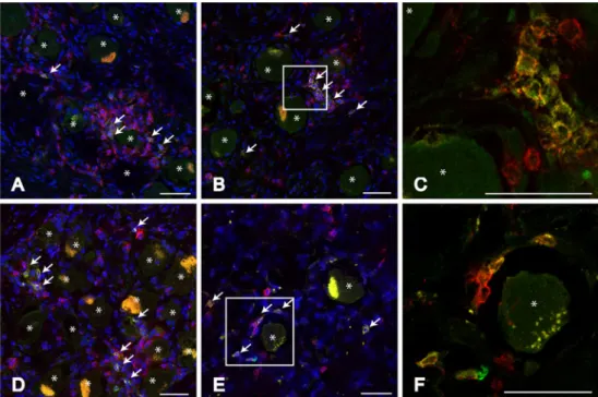
Localisation of clonally expanded TCRs in human TG CDR3 spectratyping of RNA isolated from TG analyses all TCR molecules with no differentiation of CD8+ or CD4+ T cells. However, in latently HSV-1 infected TG CD8+ T cells dominate the immune cell infiltrate (Theil et al.2003). To morphologically identify expanded T-cell clones, sections of TG showing clonal expansions of T cells within the Vβ1 family were double-stained for Vβ1 and CD8. Clones featuring the Vβ1 chain were present quite frequently in the TG of subjects 02 and 03. Among the CD8+ T-cell infiltrates surrounding neurons, several cells expressing the Vβ1 chain could be identified, whilst not all T cells surrounding one neuron expressed the same Vβ chain. Figure2shows representative micro-graphs of subjects 02 and 03 with clusters of CD8+ T cells. Some of these T cells also express the Vβ1 chain. The vast majority of cells that stained positive for Vβ1 also stained positive for CD8. Moreover, all identified Vβ1+
T cells were located adjacent to neurons.
Fig. 1 Prominent T-cell infiltrates in latently HSV-1 infected human TG. a, b Representative micrographs of latently HSV-1 infected (a) and uninfected (b) human TG sections stained for CD3 (green). Scale
bar 50μm. The red signal is autofluorescence of lipofuscin. Neurons
are indicated by asterisks. c Numbers of CD3+T cells per 0.1 mm2in
non-infected (blue; n02) vs. HSV-1 infected (red; n05) TG. Grey bars
show T-cell counts for those TG used for CDR3 spectratyping (01 to 03). Bars depict the range and mean of the T-cell numbers. The grey line indicates the mean T-cell count of HSV-1 negative TG
Table 1 Overview of tissue samples used in the present study
The HSV-1 and VZV infection state plus the average T-cell count per 0.1 mm² are listed. The HLA class I alleles expressed by the three HSV-1 infected sub-jects used for CDR3 spectratyp-ing are also stated
nd not done, m male, f female
Subject Gender Age HSV-1 VZV CD3+T cells HLA-A HLA-B HLA-C
01 m 56 + – 23.6 0101 0801 0304 1501 0701 02 m 41 + + 62.7 0101 0801 0602 0301 3701 0701 03 m 78 + + 29.2 2402 0801 0701 3001 4006 1502 04 m 62 – + 12.5 nd. nd. nd. 05 m 36 – + 10.7 nd. nd. nd. 06 f 17 + + 76.9 nd. nd. nd. 07 f 61 + – 24.0 nd. nd. nd.
Discussion
We show here, that latently HSV-1 infected human TG show increased numbers of infiltrating T cells compared to HSV-1 uninfected TG. CDR3 spectratyping of the TCR β chain revealed several clonally expanded T-cell clones with cer-tainβ chains. These expansions were private to the assessed individuals, and no public T-cell responses could be identi-fied, even though some HLA alleles were shared between individuals. However, it might be possible that particular TCR chains from expanded clones that were detected in one patient were present in other patients, but escaped detection because they were hidden in the polyclonal background. For several infections or autoimmune diseases as well as tumours (Dong et al.2010; Junker et al. 2007; Schwab et al.2009; Skulina et al.2004; Pellkofer et al.2009; Puisieux et al.1994; Miles et al.2011) biases in the T-cell receptor repertoire were identified. Some of these studies showed several T-cell clones appearing in different anatomical sites.
A former study examining the TCR usage in HSV-2-specific CD8+T cells derived from blood showed a strong bias in the TCR repertoire with public TCR usage (Dong et al.2010). Such pervasive clonal expansions are usually interpreted as being driven by sustained activation through a persisting antigen. One could speculate that lifelong exposure to HSV-1 antigens may shape the T-cell receptor repertoire in human TG. Infiltrating T cells in latently HSV-1-infected TG do not seem to exhibit public TCR usage. However, clones that had homologous or even identical amino acid sequences could be identified in both TG of two individuals. T cells with homologous TCRs are likely to respond to similar antigens. In subject 01 as well as subject 03, two clones with identical amino acid sequences were detected in both TG. These were even identical in their nucleotide sequence, which may suggest that these clones originated from the same T cell. This T cell probably encountered its respective antigen in the periphery, proliferated and after-wards migrated into both TG. HSV-1-specific T cells present
Table 2 Similar and identicalβ chains in the right and left TG of subjects 01 and 03
Subject side Vβ NDN Jβ FU 01 right 1 C A S T L T G G A G Y N E Q F F G P G 2.1 198 01 right 1 C A S S V A V N T D T Q Y F G P G 2.3 261 01 left 1 C A S S V G G P N Q P Q H F G D G 1.5 185 01 left 11 C A S S L S R T G V N Y G Y T F G S G 1.2 723 01 right 11 C A S S L S R T G V N Y G Y T F G S G 1.2 540 01 right 13.2 C A S S P S Q G G H Q P Q H F G D G 1.5 239 01 left 13.2 C A S T W S G R S Y G Y T F G S G 1.2 250 01 right 23 C A S S L R Q S Y E Q Y F G P G 2.7 303 01 left 23 C A S S L R Q S Y E Q Y F G P G 2.7 953 03 left 11 C A S S E W V S G S E Q Y F G P G 2.7 798 03 right 11 C A S S E W V S G S E Q Y F G P G 2.7 1138 03 left 11 C A S S E Y W G T G T G E L F F G E G 2.2 191 03 right 17 C A S S P D R A G G Y T F G S G 1.2 326 03 left 17 C A S S P D R A G G Y T F G S G 1.2 466 03 right 17 C A S S P G H L Y E Q Y F G P G 2.7 744
The amino acid sequence of the CDR3 is listed. Peak height is given in fluorescent units (FUs). CDR3 sharing amino acids with similar properties
in positions 3 to 5 after the conserved cysteine are surrounded by dashed lines (i.e. subject 01 Vβ1 position 3: T and S are both amino acids with
polar, neutral side chains, sharing similar hydropathy and L and V in position 2 both possess hydrophobic non-polar, neutral side chains). TCRs identical in their amino acid structure are surrounded by double-lined boxes
in human TG could be produced in the periphery during primary infection, may be also a reactivation event, and subsequently migrated to the latently infected tissue.
Staining for expanded Vβ chains revealed that the ex-panded T cells mostly belong to the CD8+subset, and that only some T cells in one cluster surrounding a neuron share the same Vβ chain. The presence of different clones in T-cell clusters around neurons could indicate that not all T cells react to the same antigen. Therefore, the TCR reper-toire present in human TG seems to be more heterogeneous than in mice. This further suggests that some of the infil-trating T cells might represent unspecific bystander T cells, which were attracted by the inflammatory milieu, like al-ready proposed by Verjans et al. in2007. This phenomenon has also been seen in the mouse model (van Lint et al.2005) where both specific and non-specific T cells persist in gan-glia harbouring latent HSV-1.
Taken together, our data show that the TCR repertoire of infiltrating T cells in human TG is complex and differs between individuals. However, single identical T-cell clones can be found in both TG of the same individual, indicating presence of the same antigen in both TG. The fact that neuron surrounding T cells are clonally expanded adds further evidence to the significance of these immune cells in latency and reactivation.
Material and methods
The Ethics Committee of the Medical Faculty of the Ludwig Maximilian University of Munich approved the use of hu-man TG autopsy samples. TG of both sides were removed
from seven subjects 6 to 24 h after death. None of the subjects had any history of neurological disorders or an active orolabial herpes infection. Table1 lists gender, age, HSV-1 infection state, T-cell counts and HLA-type of the TG used in this study. Ganglia were embedded directly after removal in Jung Tissue freezing medium (Leica Microsys-tems, Nussloch, Germany) and were stored at −80°C. Fro-zen sections of 10μm were cut for immunohistochemistry and mounted on positively charged slides (Superfrost Plus, Menzel, Braunschweig, Germany). Slides were subsequently stored at −20°C. RNA and DNA were isolated from ten 30-μm sections.
RNA extraction and HSV-1/VZV PCRs
RNA was extracted using Qiazol (Qiagen, Hilden, Ger-many) and the miRNeasy Mini Kit (Qiagen). The quality of the isolated RNA was analysed with an Agilent 2100 Bioanalyzer (Agilent Technologies, Waldbronn, Germany) combined with the Agilent RNA 6000p Kit (Agilent Tech-nologies). Subsequently, DNA was extracted from the or-ganic phase. After addition of back extraction buffer and phase separation by centrifugation, DNA present in the aqueous phase was precipitated by adding isopropanol. The DNA pellet was then washed three times with 75% ethanol and solubilised with water.
To assess the HSV-1 infection state of the TG, a nested LAT RT-PCR was done as described before (Derfuss et al.
2007). Furthermore, the VZV infection state was specified by nested PCR for the ORF 63 (Outer: 5′-CGCACTGGAATGT-GACGTAT, 3′-TCCCCGTCTCGATAACAATC; inner: 5′-TGAA-GACGATAGCGACGATG, 3′-CCCGTCTGGTTCACAAGAAT).
Fig. 2 Clusters of expanded T cells in human TG. Representa-tive micrographs of human TG sections stained for CD8 (red)
and Vβ1 (green) from S02 (a, b,
c) and S03 (d, e, f). C and F are enlargements of b and e,
re-spectively. CD8+Vβ1+T cells
are indicated by white arrows in micrographs a, b, d and e. Scale
bars 50μm. The yellow signal
within the neurons originates from lipofuscin. Neurons are indicated by asterisks
Immunohistochemistry
Immunohistochemistry was performed for CD3, CD8 and Vβ1. CD3 was detected using the polyclonal rabbit anti-human CD3 antibody (1:500; Dako Cytomation, Glostrup, Denmark) and visualised with a biotinylated goat anti-rabbit immunoglobulin antibody (1:100; Dako Cytomation), followed by Cy2-labelled streptavidin (1:100; Dianova, Hamburg, Germany) or HRP-conjugated streptavidin (1:100; Dako Cytomation) and incubation in DAB (Dako Cytomation). A dual staining for CD8 and Vβ1 was conducted using the LT8 mouse anti-human CD8α antibody (1:50; AbD Serotec, Düsseldorf, Germany) labelled with Cy3 (FluoroLinkTM MAb Cy3 labelling kit, Amersham Biosciences, Buckinghamshire, England) and the FITC-labelled Vβ1 BL37-2 antibody (1:25; Immunotech, Marseille, France). The Vβ1 signal was enhanced with an anti-FITC Alexa Fluor 488 labelled secondary antibody (1:100; Invitrogen, Karlsruhe, Germany). Antibodies to other Vβ families prominently expanded in the assessed TG were either not available or did not give reliable staining results. Stained sections were analysed by confocal imaging.
To obtain numbers of infiltrating CD3+ T cells, five randomly selected fields of view (0.123 mm2) were counted with an objective of ×20. Data were statistically analysed using Microsoft Excel 2003 and GraphPad Prism 5.
CDR3 spectratyping
The CDR3 lengths of TCR β chains were analysed by CDR3 spectratyping as described before (Junker et al.
2007; Schwab et al. 2009). In brief, cDNA was reverse-transcribed using a Cβ-specific primer and SuperScript II reverse transcriptase (Seitz et al.2006). For increased sen-sitivity, a semi-nested TCR Vβ gene family-specific PCR with 25 Vβ-specific (Monteiro et al.1996) and two different Cβ-specific primers (SpTy-β-out (Junker et al. 2007) and Cβ-reverse (Monteiro et al.1996)) was carried out followed by a subsequent runoff reaction for every product using fluorescence-labelled Jβ-specific primers (Puisieux et al.
1994). The obtained PCR products were separated by elec-trophoresis according to their CDR3 length on an ABI377 DNA sequencer (Applied Biosystems, Darmstadt, Ger-many). Mono- or oligoclonal expansions appear as peaks of certain CDR3 length over a Gaussian background of polyclonal T cells (Fig. S1in the Electronic supplementary material). Peak height is measured in fluorescent units (FU), the relative signal intensity. Higher numbers indicate more prominent clonal expansions. Peaks over 180 FU were considered as an actual aberration from the polyclonal back-ground. Finally, Vβ-Jβ amplification products of expanded clones were reamplified and directly sequenced. Only
clones for which we obtained a readable sequence were considered as clonally expanded.
The Vβ nomenclature described in Arden et al. (1995) is used throughout this manuscript. The subjects chosen for spectratyping were HLA typed by the Labor für Immunge-netik und Molekulare Diagnostik, Munich, Germany.
Acknowledgements This work was supported by the Deutsche
Forschungsgemeinschaft grant TH 894 (to DT, TD, KD).
Conflict of interest The authors declare that they have no conflict of
interest.
References
Arden B, Clark SP, Kabelitz D, Mak TW (1995) Human T-cell receptor variable gene segment families. Immunogenetics 42:455–500 Baringer JR, Swoveland P (1973) Recovery of herpes-simplex virus
from human trigeminal ganglions. N Engl J Med 288:648–650
Derfuss T, Segerer S, Herberger S, Sinicina I, Hufner K, Ebelt K, Knaus HG, Steiner I, Meinl E, Dornmair K, Arbusow V, Strupp M, Brandt T, Theil D (2007) Presence of HSV-1 immediate early genes and clonally expanded T-cells with a memory effector
phenotype in human trigeminal ganglia. Brain Pathol 17:389–398
Dong L, Li P, Oenema T, McClurkan CL, Koelle DM (2010) Public TCR use by herpes simplex virus-2-specific human CD8 CTLs. J
Immunol 184:3063–3071
Junker A, Ivanidze J, Malotka J, Eiglmeier I, Lassmann H, Wekerle H, Meinl E, Hohlfeld R, Dornmair K (2007) Multiple sclerosis:
T-cell receptor expression in distinct brain regions. Brain 130:2789–
2799
Khanna KM, Bonneau RH, Kinchington PR, Hendricks RL (2003) Herpes simplex virus-specific memory CD8+ T cells are selec-tively activated and retained in latently infected sensory ganglia.
Immunity 18:593–603
Knickelbein JE, Khanna KM, Yee MB, Baty CJ, Kinchington PR, Hendricks RL (2008) Noncytotoxic lytic granule-mediated CD8 + T cell inhibition of HSV-1 reactivation from neuronal latency.
Science 322:268–271
Miles JJ, Douek DC, Price DA (2011) Bias in the alphabeta T-cell repertoire: implications for disease pathogenesis and vaccination.
Immunol Cell Biol 89:375–387
Monteiro J, Hingorani R, Peroglizzi R, Apatoff B, Gregersen PK (1996) Oligoclonality of CD8+ T cells in multiple sclerosis.
Autoimmunity 23:127–138
Pannetier C, Even J, Kourilsky P (1995) T-cell repertoire diversity and clonal expansions in normal and clinical samples. Immunol Today
16:176–181
Pellkofer HL, Voltz R, Goebels N, Hohlfeld R, Dornmair K (2009) Cross-reactive T-cell receptors in tumor and paraneoplastic target tissue. Arch Neurol 66:655–658
Puisieux I, Even J, Pannetier C, Jotereau F, Favrot M, Kourilsky P (1994) Oligoclonality of tumor-infiltrating lymphocytes from
hu-man melanomas. J Immunol 153:2807–2818
Rudolph MG, Stanfield RL, Wilson IA (2006) How TCRs bind MHCs,
peptides, and coreceptors. Annu Rev Immunol 24:419–466
Schwab N, Bien ChG, Waschbisch A, Becker A, Vince GH, Dornmair K, Wiendl H (2009) CD8+ T-cell clones dominate brain infiltrates in Rasmussen encephalitis and persist in the periphery. Brain
132:1236–1246
Seitz S, Schneider CK, Malotka J, Nong X, Engel AG, Wekerle H, Hohlfeld R, Dornmair K (2006) Reconstitution of paired T cell
receptor alpha- and beta-chains from microdissected single cells of human inflammatory tissues. Proc Natl Acad Sci U S A
103:12057–12062
Skulina C, Schmidt S, Dornmair K, Babbe H, Roers A, Rajewsky K, Wekerle H, Hohlfeld R, Goebels N (2004) Multiple sclerosis: brain-infiltrating CD8+ T cells persist as clonal expansions in the cerebro-spinal fluid and blood. Proc Natl Acad Sci U S A 101:2428–2433 St Leger AJ, Peters B, Sidney J, Sette A, Hendricks RL (2011)
Defining the herpes simplex virus-specific CD8+ T cell repertoire
in C57BL/6 mice. J Immunol 186:3927–3933
Stevens JG, Wagner EK, vi-Rao GB, Cook ML, Feldman LT (1987) RNA complementary to a herpesvirus alpha gene mRNA is
prominent in latently infected neurons. Science 235:1056–1059
Theil D, Derfuss T, Paripovic I, Herberger S, Meinl E, Schueler O, Strupp M, Arbusow V, Brandt T (2003) Latent herpesvirus infection in human trigeminal ganglia causes chronic immune response. Am J Pathol 163:2179–2184
van Lint AL, Kleinert L, Clarke SR, Stock A, Heath WR, Carbone FR (2005) Latent infection with herpes simplex virus is associated with ongoing CD8+ T-cell stimulation by parenchymal cells with-in sensory ganglia. J Virol 79:14843–14851
Verjans GM, Hintzen RQ, van Dun JM, Poot A, Milikan JC, Laman JD, Langerak AW, Kinchington PR, Osterhaus AD (2007) Selec-tive retention of herpes simplex virus-specific T cells in latently infected human trigeminal ganglia. Proc Natl Acad Sci U S A