DEVELOPMENT OF A FRACTIONATION PROCESS
FOR THE PREPARATION OF A
FOLATE-ENRICHED PROTEIN EXTRACT FROM HEN EGG
YOLKS
Thèse
NASSIM NADERI
Doctorat en sciences et technologie des aliments
Philosophiæ doctor (Ph.D.)
Québec, Canada
iii RÉSUMÉ
Le fractionnement du jaune d’œuf est une façon judicieuse d’étendre les domaines d’application de cet ingrédient dans les industries alimentaire et nutraceutique. Le but de ce projet a été de mettre au point un fractionnement non-toxique du jaune d’œuf, par centrifugation, dans l’objectif d’obtenir un produit enrichi en extrait naturel de folate. De par sa teneur élevée en cholestérol, le jaune d’œuf a été considéré comme un sous-produit du procédé de séparation des œufs. Cependant, le jaune d'œuf contient aussi une forme précieuse et biodisponible d'acide folique. La dilution du jaune d’œuf et son fractionnement en granules et plasma par une technique de centrifugation (centrifugeuse de laboratoire et à échelle pilote) a permis l’obtention de granules d’une richesse en folate trois fois plus importante que celle du jaune d’œuf natif. Aussi, les granules obtenus ont présenté une concentration protéique deux fois plus élevée que celle du jaune d'œuf natif, et une concentration de lipides et cholestérol trois fois inférieure à celle du jaune d'œuf natif. Les granules sont apparus non solubles et de structure très compacte. Afin d’augmenter encore la concentration en folate, nous avons tenté de remettre les granules en suspension et de les séparer par centrifugation en utilisant des prétraitements tels qu'augmentation de la force ionique et traitements mécaniques (ultrasons puissants et pression hydrostatique élevée). L’application d’ultrasons puissants et l’augmentation de la force ionique n’ont que peu amélioré la concentration en folate. Les granules soumis à une force ionique de 0.15 M de NaCl et un traitement par ultrasons puissants de 10 minutes ont présenté des concentrations de 21 μg de folate / g de granulés. Une augmentation de la force ionique au delà de 0.15 M de NaCl a conduit à une concentration plus faible en folate, du fait de la déformation de la structure des granules et de la séparation de leur fraction soluble. Les observations ont indiqué qu'il pourrait y avoir une association entre la structure protéique des granules et leur contenu en folate. Les variations de solubilité et la modification du réseau structurel des granules par augmentation de la force ionique ont affecté la teneur en folate des structures granulaires. Les granules ont cependant présenté une structure difficilement modifiable sous ultrasons puissants et après augmentation de la force ionique. Le traitement à pression hydrostatique élevée (HHP), technique puissante, a été utilisé pour étudier l'effet des hautes pressions sur la concentration en folate des granules. Après 5 minutes de
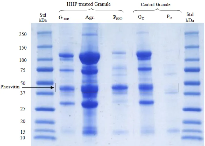
iv traitement à 600 MPa, la concentration en folate a été mesurée dans les granules et le plasma, séparé des granules par centrifugation. Le plasma issu des granules contenait des concentrations en folate plus importantes que celles des granules précipités. Une analyse SDS-PAGE a permis de vérifier le profil protéique des granules sous l'effet du traitement HHP. Il est intéressant de noter que pour le plasma séparé des granules après traitement à haute pression, les migrations sur gel SDS-PAGE présentaient une bande protéique principale correspondant à la phosvitine. Les résultats de notre étude nous ont encouragés à proposer un modèle schématique de la structure des granules. Ces granules contiennent des HDL, LDL et phosvitine, et conservent le contenu total en folate du jaune d’œuf. Les protéines des granules sont principalement phosphorylées et il existe une forte liaison entre les apoprotéines de HDL et la phosvitine du fait de ponts de phosphate de calcium. Des traitements mécaniques ont libérés des particules de LDL qui pourraient être piégées dans le réseau structurel des granules. Les interconnexions entre les apoprotéines de HDL, la phosvitine et le folate pourraient se faire par des ions calcium. Nos résultats mettent en évidence le potentiel du procédé afin de produire un concentré riche en folate à partir des jaune d’œuf. Cependant, d’autres travaux seront nécessaires afin de trouver des utilisations aux co-produits (plasma) et rendre le procédé viable à l’échelle commerciale.
v ABSTRACT
Fractionation of egg yolk is a smart way to expand the application of egg yolk ingredient in food and nutraceutical industry. The goal of this project was to develop non-toxic fractionation process of egg yolk by using centrifugation in order to prepare a natural folate-enriched extract. The egg yolk has been considered as a by-product of egg separation process due to its high cholesterol content. However, egg yolk contains valuable and bioavailable form of folate. Dilution of egg yolk and its fractionation into granule and plasma by centrifugation technique (lab- and pilot-scale centrifuge) resulted in separation of a granule fraction being rich in folate which was 3 fold higher than native egg yolk. This granule fraction was also characterized by high protein concentration (2 fold higher than protein content of yolk) and lower lipid and cholesterol (3 fold) content compared to non-treated egg yolk. The granule fraction appeared to be non-soluble with a very compact structure. By using the pre-treatments techniques such as increasing ionic strength and mechanical treatments (ultrasound and high hydrostatic processes), we attempted to re-suspend granules and separate them by centrifugation in order to further increase folate concentration. Results demonstrated that ultrasound and increased ionic strength did not largely change folate concentration. At ionic strength 0.15 M NaCl and after 10 min of ultrasound treatment granule contained 21 μg folate/g granules. By increasing ionic strength higher than 0.15 M NaCl the folate concentration was lower in granule due to the disruption of granule structure and separation of soluble fraction of granule. The observations denoted that there might be association between granular protein structure and folate content. Changes in solubility and disruption of granule network structure by increasing ionic strength affected folate content of granule structure. However, granules appeared to have very stable structure under the ultrasound and after increasing ionic strength, and their modifications were not easily possible. The high hydrostatic pressure processing (HHP) was used as an innovative and powerful technique in order to study the effect of high and drastic pressure on the concentration of folate in granules. After 5 min of 600 MPa HHP treatments, the folate concentration was measured in granule and separated plasma from granule after centrifugation. Plasma from granule contained higher concentration of folate compared to the precipitated granule. SDS-PAGE analysis was used
vi in order to verify the granule protein profiles as a function of HHP treatment. Interestingly, the plasma separated from granule after HHP treatment contained phosvitin as a leading protein band separate in SDS-PAGE gel. The results of our study allowed proposing a schematic model for the granule structure which contains HDLs, phosvitin and LDLs. Beside; granule contains large amount of folate. Proteins of granule are mostly phosphorylated and strong connection between apoproteins of HDLs and phosvitin exists through calcium phosphate bridges. LDL particles were liberated through mechanical treatments and might be entrapped in the granular network. The interconnection between the apoproteins of HDLs, phosvitin and folate could be through calcium ions. Our results provided highly promising evidences concerning the recovery of high-concentration folate extract from hen egg yolk. Our fractionation technique is also clean but it generates plasma as co-product that is still usable in food formulation. Such applications still need to be developed before the technology can be viable at commercial scale.
vii TABLE OF CONTENT
RÉSUMÉ ... iii
ABSTRACT ... v
TABLE OF CONTENT ... vii
TABLE LIST ... xii
FIGURE LIST ... xiii
EQUATION LIST ... xiv
ABREVIATIONS ... xv
ACKNOWLEDGMENTS ... xvi
FOREWORD ... xviii
CHAPTER 1: INTRODUCTION ... 1
CHAPTER 2: LITERATURE REVIEW ... 3
2.1. HEN EGG YOLK ... 3
2.1.1. Overall composition and structure ... 3
2.1.2. Nutritional and nutraceutical properties of egg yolk ... 11
2.1.3. Improving nutritional properties of egg yolk ... 16
2.2. FOLATE AND FOLIC ACID ... 21
2.2.1. Historical perspective ... 21
2.2.2. Structure and biochemistry ... 23
2.2.3. Importance of folate in human diet ... 24
2.3. FRACTIONATION METHODS FOR DEVELOPMENT OF FOOD-DERIVED BIOACTIVES ... 27
2.3.1. Overview of the separation methods in egg processing ... 28
2.4. FRACTIONATION CHALLENGE FOR PRODUCING FOLATE-ENRICHED PROTEIN EXTRACT FROM HEN EGG YOLK ... 45
viii
CHAPTER 3: PROBLEMATIC, HYPOTHESISAND OBJECTIVES ... 46
3.1.PROBLEMATIC ... 46
3.2.HYPOTHESIS ... 46
3.3. MAIN AND SPECIFIC OBJETIVES ... 46
CHAPTER 4: SCALING-UP A PROCESS FOR THE PREPARATION OF FOLATE-ENRICHED PROTEIN EXTRACTS FROM HEN EGG YOLKS ... 48
4.1.CONTEXTUALTRANSITION ... 48
4.2. RÉSUMÉ ... 49
4.3. ABSTRACT ... 50
4.4. INTRODUCTION ... 51
4.5. MATERIALS AND METHODS ... 53
4.5.1. Hen eggs ... 53
4.5.2. Chemicals ... 53
4.5.3. Fractionation of egg yolk ... 53
4.5.4. Pilot scale process ... 54
4.5.5. Chemical analyses ... 57
4.5.6. Extraction and analysis of folate content ... 57
4.5.7. Performance parameters of egg yolk fractionation ... 59
4.5.8. Sodium Dodecyl Sulfate–Polyacrylamide Gel Electrophoresis (SDS-PAGE) ... 60
4.5.9. Two dimensional electrophoresis ... 60
4.5.10. Statistical analysis ... 61
4.6. RESULTS AND DISCUSSION ... 62
4.6.1. Overall composition of the fractions ... 62
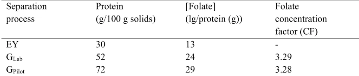
4.6.2. Folate enrichment ... 63
ix
4.6.4. Mass balance and centrifugation performance ... 71
4.7. CONCLUSION ... 74
CHAPTER 5: EFFECT OF SELECTED PRETREATMENTS TO INCREASE THE FOLATE CONTENT OF GRANULE SUSPENSIONS PREPARED FROM HEN EGG YOLK ... 75
5.1. CONTEXTUAL TRANSITION ... 75
5.2. RÉSUMÉ ... 76
5.3. ABSTRACT ... 77
5.4. Introduction ... 78
5.5. MATERIALS AND METHODS ... 80
5.5.1. Materials and chemicals ... 80
5.5.2. Preparation of yolk and granules ... 80
5.5.3. Application of pre-treatments on granule suspensions ... 81
5.5.4. Chemical analyses ... 82
5.5.5. Determination of cholesterol ... 82
5.5.6. Fatty acid analysis ... 83
5.5.7. Determination of folate content ... 83
5.5.8. SDS–PAGE protein profiles ... 83
5.5.9. Statistical analysis ... 84
5.6. Results and discussion ... 84
5.6.1. Effect of ionic strenght ... 86
5.6.1. Effect of ultrasound treatment on granule and plasma ... 97
5.7. Conclusion ... 105
CHAPTER 6: UNDERSTANDING THE EFFECT OF IONIC STRENGTH AND MECHANICAL TREATMENTS ON THE COMPOSITION AND MICROSTRUCTURE OF GRANULE SEPARATED FROM HEN EGG YOLK ... 106
x
6.1. CONTEXTUAL TRANSITION ... 106
6.2. RÉSUMÉ ... 107
6.3. ABSTRACT ... 108
6.4. INTRODUCTION ... 109
6.5. MATERIALS AND METHODS ... 111
6.5.1. Materials and chemicals ... 111
6.5.2. Preparation of granules ... 111
6.5.3. Ionic strength modification ... 112
6.5.4. Mechanical treatments ... 112
6.5.5. Compositional analyses ... 114
6.5.6. Sodium Dodecyl Sulphate–Polyacrylamide Gel Electrophoresis ... 114
6.5.7. Two-dimensional polyacrylamide gel electrophoresis (2D-PAGE) ... 115
6.5.8. Microstructure Determination by Confocal Laser Scanning Microscopy ... 115
6.5.9. Statistical analysis ... 116
6.6. RESULTS ... 116
6.6.1. Overall compositional changes induced in granules composition ... 116
6.6.2. Effect of treatments on protein distribution (2D-PAGE) ... 117
6.6.3. Microstructural changes following treatments ... 122
6.7. Discussion ... 126
6.8. Conclusion ... 129
CHAPTER 7: GENERAL CONCLUSION ... 130
7.1. Achievements and original contributions ... 130
7.2. Significance of the results ... 134
7.2.1. New knowledge on egg yolk granule structure ... 134
xi 7.1. Research perspectives ... 137 References ... 139
xii TABLE LIST
Table 2.1. Composition of whole egg, yolk and white. ... 12
Table 2.2. Egg compounds with summary of their biological activities ... 15
Table 2.3. The chronology of folic acid discovery ... 22
Table 2.4. Technological approaches and processes to fractionate peptides and proteins from egg. ... 32
Table 2.5. Fractionation of egg yolk into granule and plasma based on centrifugation methods ... 38
Table 2.6. Summary of granule and plasma composition after fractionation by centrifugation ... 40
Table 2.7. Protein bands observed in the SDS-PAGE analysis of granule and plasma fractions ... 42
Table 2.8. Protein species in egg yolk and plasma determined by 2D SDS-PAGE ... 44
Table 4.1. Composition of egg yolk and fractions from lab- and pilot-scale processes. ... 65
Table 4.2. Mass balance upon centrifugation at lab-, pilot-scale and recovery (%) of folate, protein and fat after fractionation process. ... 72
Table 4.3. Compositional characteristics of granule extracts compared to egg yolk. ... 73
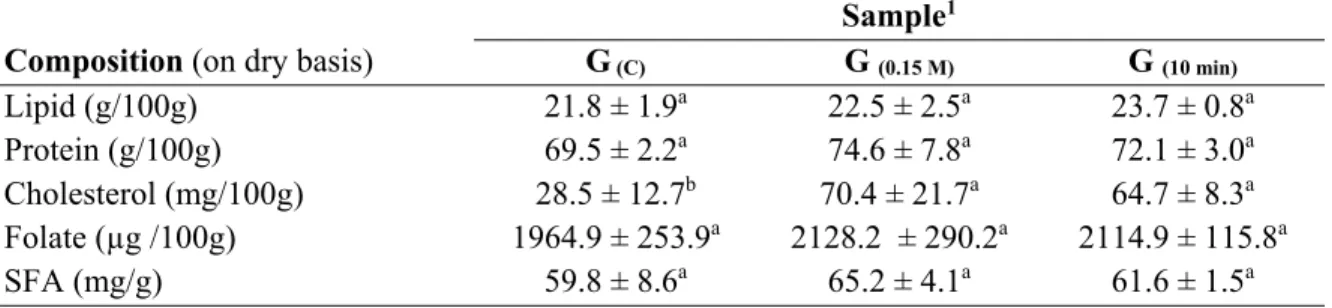
Table 5.1. Composition of granule fraction separated from yolk on dry basis. ... 85
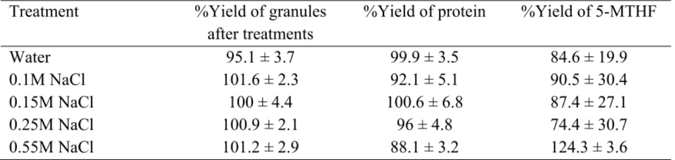
Table 5.2. Recovery of granules and major components: protein and folate ... 86
Table 5.3. Composition of granules and plasma as a function of ionic strength variation ... 89
Table 5.4. Fatty acid composition of granules and plasma as the function of ionic strength variation ... 92
Table 5.5. Recovery of granules and major components: protein and folate ... 98
Table 5.6. Effect of ultrasonic pre-treatment on composition of granule and separated-plasma from granules after treatments ... 100
Table 5.7. Fatty acid composition of granule and plasma separated from granule as a function of ultrasonic treatment ... 102
Table 6.1. Composition of granule at the optimal pre-treatment conditions ... 117
xiii FIGURE LIST
Figure 2.1. Components of the micro structure of egg yolk and Granule ... 5
Figure 2.2. Schematic presentation of egg yolk fractions and their composition ... 6
Figure 2.3. Schematic model proposed for egg yolk granule at pH 6.5. ... 9
Figure 2.4. Structure of native food folates and their substituent groups and positions. ... 23
Figure 4.1. Schematic of the tubular bowl vertical centrifuge ... 56
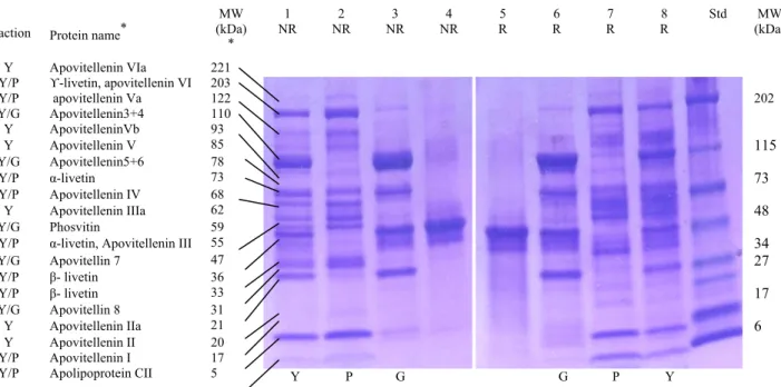
Figure 4.2. SDS-PAGE profile of proteins of EY fractions in NR and R conditions ... 68
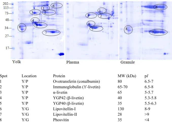
Figure 4.3. Two-dimensional polyacrylamide gel of whole egg yolk, plasma and granule fraction of proteins. ... 70
Figure 5.1. Electrophoretic profile of total granule proteins after changing ionic strength at 0, 0.1, 0.15, 0.25, 0.55 M NaCl and soluble proteins in plasma fraction separated from granules ... 95
Figure 5.2. Electrophoretic profile of the total proteins of granules after ultrasonic treatment and soluble proteins in plasma fraction separated from granules ... 104
Figure 6.1. Two-dimensional polyacrylamide gels of native yolk and granules of native egg yolk. ... 121
Figure 6.2. Confocal micrograph of egg yolk and granules, granules treated as the effect of increasing ionic strength, ultrasonic treatment and HHP ... 123
Figure 6.3. Electrophoretic profile of the total proteins of granules after HHP treatment . 125 Figure 6.4. Schematic model of a native and disrupted granule ... 128
xiv EQUATION LIST
% Recovery (R) of folate: Equation 4.1 58
Recovery (%) at lab- and pilot scale based on mass balance: Equation 4.2 ... 59
Recovery Ri (%) of ech component: Equation 4.3 ... 59
xv ABREVIATIONS
5-MTHF: 5-methyltetrahydrofolate
CLSM: Confocal laser scanning microscope DHA: Docosahexaenoic acid
DRI: Dietary reference intake FA: folic acid
GPC: Gel-permeation chromatography HDL: High density lipoproteins
IEC: Ion exchange chromatography IgY: Immunoglobulin Y
IOM: Institute of Medicine
IUPAC-IUB: International Union of Pure and Applied Chemistry-International Union of Biochemistry
LDL: Low density lipoproteins MUFA: Monounsaturated fatty acids NTD: Neural tube defects
PC: Phosphatidylcholine PE: Phosphatidylethanolamines PI: Phosphatidylinositol PS: Phosphatidylserine
PUFA: Polyunsaturated fatty acids
SDS-PADE: sodium dodecyl polyacrylamide gel electrophoresis SFA: Saturated fatty acids
SM: Sphingomyelin
UFA: Unsaturated fatty acids
xvi ACKNOWLEDGMENTS
A great many people have contributed irreplaceable to this thesis and because of whom my graduate experience has been one that I will treasure forever. I therefore wish to greatly acknowledge:
This thesis is the result of a perfect working relationship with my supervisor Professor Yves Pouliot, whose generous guidance, insightful vision, and steadfast support have paved the way for my development as a research scientist. My three years Ph. D. program was truly enjoyable and very much interesting being graduate student of Yves who was a true friend, like a father and very much supportive. I am grateful for the time that Yves has spent discussing ideas, revising papers, commenting on talks and funding to make my Ph.D. experience productive and exciting. I am deeply grateful and am very much benefitted from your insight and guidance. Thank you!
I would like to express my deepest appreciation to my co-supervisor Professor James D. House in Winnipeg for serving on my supervisory committee, his concern, inspiring discussions, reading and commenting on drafts and sharing his expertise in folate analysis. Thank you indeed for being always ready to help and guide!
I would like to acknowledge Dr. Alain Doyen for pre-reading of my dissertation. I appreciate for his time, interest, and helpful comments. I am indeed thankful for his support, continuous encouragement and help. I very much benefitted from his expertise and guidance.
I would like to thank my oral and reading committee members: Professor Ulrich Kulozik, Dr. Alain Doyen and Dr. Lucie Beaulieu for their time, interest, and helpful comments. I would like to express my gratitude to Professor Sylvie Gauthier for her advices, support and conversations during my education in Laval.
I would like to extend a special thanks to Diane Gagnon for her unconditional support and guidance in the lab. She is definitely loves science and it was a great pleasure working and
xvii discussing the technical matters with her. Moreover, she has such a great personality, so kind and friendly indeed. I will never forget her!
I would like to extend my sincere appreciation to Pascal Dubé and Véronique Richard for their support and assisting me with the folate and cholesterol HPLC analysis. They were always supportive and very interested in what I was doing. I have learned a lot from both of you and I am very much happy to have you by my side following up my interesting project. I would like to thank Jason Neufeld in Winnipeg in University of Manitoba for giving me the training with the HPLC machine and for his help during our inter-laboratory studies for the egg folate analysis.
I am also very thankful to my lab group for their support and friendship. Many thanks go to Frederic Lehance for his support and the positive energy that he gave me over the years I worked in the lab.
I have had the pleasure to work with the summer students: Stéphanie, Mélanie and Mélanie who have come through the lab.
My life at Laval has been significantly more gratifying, interesting and fun, because it has been filled with many smart, enthusiastic, and supportive friends. The companionship and personal support of my very best friend, Mathieu Persico, ensures that I can only think back upon the last few years with feelings of fondness and reminisce. I thank Mathieu Persico and Scott Benoit for their comments and corrections on my French translations.
I am particularly grateful to my family for their endless love, their support in all my pursuits and their encouragement. My parents, Homa and Hossein, who raised me with a love of science and without pressuring me, always encouraged me to further my education and follow my dreams. The unconditional love and encouragement provided by my sister, Nona, served as a secure anchor during the hard and easy times.
I gratefully acknowledge the funding sources that made my Ph.D. work possible. I was funded in part by Natural Science and Engineering Research Council of Canada (NSERC) for 3 years.
xviii FOREWORD
The work carried out in this thesis was aimed to develop egg yolk fractionation process to prepare a natural-folate enriched extract. This thesis contains all the results obtained during the course of realization of the project. Results are presented in the form of 3 research articles.
The first article “Scaling-up a process for the preparation of folate-enriched protein extracts from hen egg yolks” has been submitted and published in “Journal of Food Engineering” (2014, 141: 85-92). Authors are Nassim Naderi, James D. House and Yves Pouliot.
The second article “Effect of selected pretreatments to increase the folate content of granule suspensions prepared from hen egg yolk” is in preparation for a submission in “Journal of Food Engineering”. Authors are Nassim Naderi, James D. House and Yves Pouliot.
The third article “Understanding the effect of ionic strength and mechanical treatments on the composition and microstructure of granule separated from hen egg yolk” is in preparation for a submission in “Journal of Agriculture and Food Chemistry”. Authors are Nassim Naderi, Alain Doyen, James D. House and Yves Pouliot.
For each article, Nassim Naderi1 planned and achieved the experiments, presented the results and wrote articles. In consequence, she is the first author of all articles. Dr. James D. House1,2 is co-supervisor of this project and he participated to the planning, discussion of results and the revision of the writing of all articles. Dr. Alain Doyen1 is second co-author of third article and he participated in discussion of results and revision of article. Dr. Yves Pouliot1 is supervisor of this project and he participated to the planning, the discussion of the results and the revision of the writing of all articles. He is the last author of all articles. 1. Institute of Nutrition and Functional Foods (INAF), Faculty of Food and Agricultural
science, Université Laval, Québec, QC, Canada, G1V 0A6.
2. Department of Human Nutritional Sciences, University of Manitoba, Winnipeg, MB, Canada, R3T 2N2.
To who loves folate and the people who lived with Spina bifida!
Life consists in penetrating the unknown, and fashioning our actions in accord with the new knowledge thus acquired. ~Leo Tolstoy
It may not be possible to win the future but if we’re going to encourage more innovation, it’s not enough for us to just dig in and work harder. We also need to encourage surprise and serendipity.
We need to play each other’s instruments ~ Steven Johnson
1
1. CHAPTER 1: INTRODUCTION
The importance and consumption of the eggs by human dated back to antiquity. Eggs are cheapest source of animal protein that is used throughout the world.
The worldwide consumption of egg over the past twenty years has faced two different trends. The egg consumption declined by 14% until the mid-1990s. Since that time, the consumption of eggs has remained fairly constant. The decline of egg consumption by consumers in the first part of this period was partially due to concerns about eggs contributing to cholesterol levels. The other factors that affected consumption of egg were the lack of convenient egg products on the market. Therefore, the research and development of new processes and new value-added egg products will expand egg industry and add more benefit compare to simple processing for the egg breaking industry (Sim and Nakai, 1994).
Eggs are largely composed of the protein and lipids. Protein fraction is highly digestible and contains the most important essential amino acids in a profile that is not dissimilar to the ideal balance of amino acids needed by men and women. Egg yolk lipids are composed of triglycerides, phospholipids, cholesterol and free fatty acids. Based on the diet of hen the fatty acid composition of yolk corresponds to saturated fatty acids (SFA), polyunsaturated fatty acids (PUFA) and monounsaturated fatty acids (MUFA). Some of yolk lipids are particularly suitable for nutrition or health such as omega 3 fatty acids. Furthermore, eggs contain major vitamins and minerals. Some of those nutrients (i.e. folate, vitamin E, vitamin D, manganese, selenium) in egg can be manipulated by dietary means in order to improve the human diet (Surai and Sparks, 2001). Eggs are considered as an excellent source of folate and research showed that increasing the folate content of eggs will position the egg as an important source of dietary folate (Dickson et al., 2010; Hebert et al., 2005). Folate is essential water soluble B vitamin that acts as cofactors and co-substrates for different biological methylation reactions such as those involved in amino acid and nucleic acid synthesis. However, humans are devoid of folate biosynthesis, therefore, they are
2 completely dependent on dietary sources to meet their daily folate requirements. Sufficient folate status in humans is reported to lower the risk of neural tube defects (NTD) in babies (Czeizel and Dudas, 1992). Folic acid is a synthetic form of folate and is the major supplemental form used in food fortification. In the late 1990s, the governments of Canada and the USA introduced the mandatory fortification of folic acid in cereal grain products to reduce the number of NTD occurrences in newborn babies. However, while cases of NTDs were reduced significantly, an emerging body of literature suggests that consuming high levels of folic acid may have several negative consequences. As a result, attention has been directed into other strategies that may help to increase the population’s intake of natural folates.
Eggs naturally contain about 22 μg of folate (USDA, 2001), which is equivalent to 6% of the established adult daily requirements for folate. Based on recent research studies, synthetic folic acid added to laying hen diets was deposited in the egg yolk as natural folate (House et al., 2002; Sherwood et al., 1993) with high bioavailability. One large folate-enriched egg provides approximately 45 to 50 μg of folate, or roughly 12% of the current recommended nutrient intakes for adults that is 400 μg/day (DRIs, 1998). However, because one large egg contains about 210 mg of cholesterol, public has been recommended to limit their egg consumption (Krauss et al., 2000).
The current fractionation techniques available at industrial scales have enabled the development of bioactive-enriched food derived extracts. Since egg is widely used for its nutritional and functional properties, it is necessary to develop processes for the production of folate-enriched extract from egg yolk in order to improve health benefits of egg base products.
The goal of this work was to develop a non-toxic egg yolk fractionation process which is easily scalable for industrial application at the prospect of produce a proteinaceous extract from egg yolk enrich with natural folate with lower concentration of cholesterol.
3
2. CHAPTER 2: LITERATURE REVIEW
2.1. HEN EGG YOLK
Eggs produced for human consumption are infertile from hens classified as Gallus
domesticus. The ovary of female chick contains numerous (over 3000) small ova at the time
of hatching. The egg yolk is formed in three steps within the final 10-12 days prior to laying of egg: (1) the part that is formed during embryonic development of the female chick, (2) the normal slow development of the ovum from the time of hatching of the chick to the point in sexual maturity (some ten days before ovulation), and (3) the accelerated growth period during the last ten days before ovulation (release of the ovum or yolk) into the oviduct, a part of the female reproductive system. The egg white (albumen) is secreted in oviduct to surround the yolk and finally shell and its membranes are deposit to complete the formation process (Stadelman et al., 1995).
The average weight of eggs produced by selectively hen bred strains is 60 g. About 10% of egg is composed of shell, 59% albumen and 31% yolk. From the edible part of egg, 74.4% is water, 12.3% protein and 11.6% lipids. Egg contains all vitamins except vitamin C (Seuss-Baum and Nau, 2011).
2.1.1. Overall composition and structure
Egg yolk material is surrounded by vitelline membranes. It prevents mixing of yolk and egg white (albumen) and forms the last barrier to microbial infection. A thin continuous layer is located between two fibrous layers, the inner layer facing the oocyte (formed in the ovary), and the outer layer facing the albumen (deposited in the oviduct).
A freshly laid yolk contains 50 to 52% dry matter based on the age of the laying hen and the length of storage. Egg yolk is mainly composed of lipids (about 65% on dry basis) with a lipid to protein ratio of about 2:1. The lipid composition of egg yolk is made up of 62% triglycerides, 33% phospholipids, 5% cholesterol and less than 1% carotenoids which give
4 it its color. Based on a standardized feed of hens, the fatty acid composition of lipids in egg yolk consist of 30–35% saturated fatty acids (SFA), 40–45% monounsaturated fatty acids (MUFA), and 20–25% polyunsaturated fatty acids (PUFA). Oleic acid (C18:1, 40-45%), palmitic acid (C16:0, 20-25%), and linoleic acid (C18:2, 15-20%) are the main fatty acids in egg yolk (Wang et al., 2000). However, the fatty acid composition of egg yolk is highly reflective of the diet of laying hen.
The triglyceride contains most of the total fatty acid. The glycerol of the triglycerides is generally esterified by palmitic acid in position 1, by oleic and linoleic acids in position 2, and by oleic, palmitic, and stearic acids in position 3 (Kuksis, 1992). Egg yolk also contains great amount of phospholipids. Phospholipids are amphiphilic molecules which contain one hydrophilic head group (phosphoric acid + alcohol and amino acids), and one hydrophobic group (two fatty acids). Phospholipids in egg yolk are mainly composed of phosphatidylcholine (PC) which is 76% of total phospholipids. From other phospholipids, yolk contains 22% phosphatidylethanolamine and very low amount of phosphatidylinositol (PI), phosphatidylserine (PS), and sphingomyelin (SM). Fatty acids in phospholipids composed of 45% saturated fatty acids (SFA), 30–40% polyunsaturated fatty acids (PUFA), and 20–25% monounsaturated fatty acids (Kuksis, 1992).
Egg yolk also contains 5% cholesterol. About 85–90% of cholesterol is in free form which involves in the structure of low density lipoproteins (LDLs) and 10-15% in esterified form. The cholesterol esters are located in the lipid core of LDLs which contains oleic (35%), palmitic (33%), linoleic (12%) and stearic acid (11%) (Anton, 2007a).
Proteins in egg yolk are present as free proteins or apoproteins which contribute in lipoprotein assemblies. Lipoproteins (low and high density lipoproteins) are main constituents of egg yolk that formed due to the interactions between lipids and proteins. The major egg yolk proteins components are 68% low density lipoproteins (LDL), 16% high density lipoproteins (HDL), 4% phosvitin, and 10% livetins (Mine, 2010).
Egg yolk is a complex system and it has been considered as a natural oil-in-water emulsion. It is a pseudoplastic non-Newtonian fluid with viscosity which depends on the shear forces applied. It has a pH around 6.0 that only slightly increases (pH 6.4-6.9) after prolonged
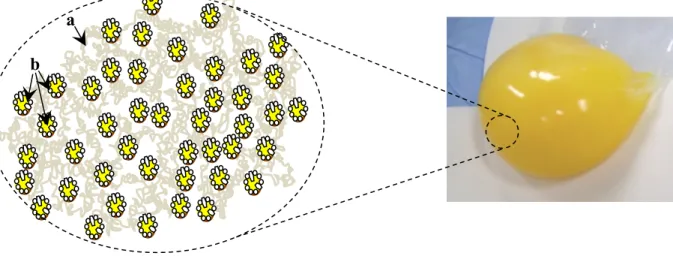
5 storage. Anton (2013) provided one of the latest description for the micro structure of egg yolk which consist of non-soluble protein aggregates (granules) in suspension in a clear yellow fluid (plasma) that contains LDLs and soluble proteins (Figure 2.1). Therefore, yolk can be separated into its two main fractions of plasma and granules by mild centrifugation, a technique largely used for protein analysis in both fractions. Figure 2.2 presents a schematic representation of fractionation of the egg yolk and major composition of each fraction. Since 1960s up to now, a wide range of centrifugation methods with different parameters (i.e. dilution agents and dilution factor, centrifugation speed and time, pre-treatments.) have been used to separate plasma and granule and investigate their lipoprotein structure (Anton and Gandemer, 1997; Burley and Cook, 1961; Cook and Martin, 1969a; Laca et al., 2010; Martin et al., 1964; Mc Bee and Cotterill, 1979; Strixner and Kulozik, 2013).
Figure 2.1. Components of the micro structure of egg yolk- a: Granule, b: LDL in plasma a
6 Figure 2.2. Schematic presentation of egg yolk fractions and their composition; Adapted from Li-Chan et al. (1995).
Egg Yolk (100%)
Solids (52%) Moisture (48%)
Lipoproteins and proteins Minor solids (2% neutral lipids,
carbohydrates, minerals, amino acids
Granules (12%) Very low density fraction LDL fraction (1-2%) HDL fraction (8%) Phosvitins (2%) α-HDL β-HDL Plasma (38%) Water-soluble fraction (5%) LDL fraction (33%) LDL1 LDL2 Livetins Lipoflavoproteins
7 The egg yolk particles have been classified based on their spherical sizes (4-150 µm in diameter), and granules (0.84-4.87 µm). The dilution and centrifugation of egg yolk result into two distinct factions: plasma, a dark orange supernatant fraction and granule, a precipitated pellet fraction with pale color. The granules contain 12% LDL but mainly consist of HDLs (60%) and phosvitin (16%) linked together through phosphocalcic bridges. Due to the high protein content, granules have a density of 1.190 g/ml. The plasma is extremely rich in LDLs (85%) with 15% livetins (Anton, 2007a). The density of LDL micelles is reported between 0.908 g/ml and 1.063 g/ml (Hevonoja et al., 2000; Li-Chan et al., 1995). The low density of LDLs has been attributed to their much higher lipid:protein content ratio which makes them more soluble in aqueous solution (Anton, 2007a; Anton et al., 2003).
The micro structure of egg yolk has been investigated in several studies. In earlier works, the transmission electron microscopy has been used to investigate the structure of particles in egg yolk (Chang et al., 1977). In their micrographs, the HDLs were presented as high electron-dense microparticles which were seemed to be attached to hair like strands (phosvitin molecules) even in 1.71M NaCl. On the surface of plasma LDLs, numerous electron-dense particles were observed. In another study, the researchers used transmission electron microscope to study the microstructure of fresh egg yolk. In their observations histological differences were observed in the yolk spheres in each yolk layer. The yolk spheres were closely packed in the vitelline membrane, with various sizes and shapes. The protein granules with high electron density were observed in the yolk sphere and granules were distributed between the outer and inner layers in different amount and with different shape ( Kobayashi, 1997).
LDLs (lipovitellenins) micelles are spherical particles of about 20–60 nm diameter with a neutral lipid core (triglycerides and cholesterol esters) surrounded by apoproteins and phospholipids. About six apoproteins have been reported in LDL structure which was characterized by their high hydrophobicity and flexibility. These apoproteins, named apovitellenins I-VI, represent about two third of yolk solids (Li-Chan and Kim, 2008). Very-low-density lipoproteins (VLDL), the precursors of LDL micelles, have a size range of 80 to 350 nm and are detectable in the plasma fraction of egg yolk even after transferring
8 some egg yolk compounds (Huopalahti et al., 2007; Sirvente et al., 2007). The plasma fraction of yolk is mainly composed of 85% LDL and 15% livetin. Livetins are water soluble globular proteins which are composed of α-livetin (serum albumin), β-livetin (α2 -glycoprotein), and -livetin (-globulin/immunoglobulin Y: IgY) (Li-Chan and Kim, 2008). Unlike LDLs, high density lipoproteins (HDL) or lipovitellin structure is similar to globular proteins. In HDL, the protein can be loaded with varying amount of non-covalently bound lipid from which phospholipid holds two third of lipids. Vitellogenin is the precursor of HDLs and it is synthesized in the liver and loaded with phosphorylated, glycosylated lipids and there it interacts with at least two Ca2+ ions and one Zn2+ ion (Anderson et al., 1998). HDLs, which represent 70% of the granule’s composition, consist of α- and β-HDL which are different in terms of amino acid composition, particle-bound phosphorus and carbohydrate content. Moreover, α-HDL has more acidic nature compare to β-HDL due to its higher sialic acid content. About five apoproteins have been recognized in the HDL structure (Guerrero-Legarreta, 2010). Phospholipid is the principal component in HDL while triglycerides and other neutral lipids make up the majority of the lipid in LDL and VLDL. The lipid contained in the HDL is bound in a noncovalent manner and is extractable with organic solvents (Anderson et al., 1998). The HDLs are structured through five disulfide bridges as well as several ionic and hydrophobic interactions. HDLs make complexes with phosvitin through phosphocalcic bridges which is a basic element of egg yolk granules.
At low ionic strength the HDL-phosvitin form non-soluble complexes which cause the granule structure to be very compact and weekly accessible to enzymatic digestion and also resistant to thermal denaturation or even heat gelation (Anton, 2007a). In the granules, HDLs are organized in a multilevel structure (Strixner et al., 2014) as presented in Figure 2.3. The smallest parts are the HDL proteins with a molecular weight of about 400 kDa and diameters between 7 and 20 nm (Burley and Cook, 1961).
9 Figure 2.3. Schematic model proposed for egg yolk granule at pH 6.5. HDL-submicelles (c) linked to phosvitin (b) through calcium phosphate bridges and embedded LDL vesicles (a). Adapted from Strixner et al. (2014).
The atomic force microscopy studies indicated the pH-dependent modification of HDL-granule structure. At pH 4.0, LDLs were strongly associated to the HDL-HDL-granules. LDLs were clearly identified based on their spherical shape and smooth surface. At pH 6.5, HDL granules form a protein network and the HDL-subunits were formed by loop like coiled strands (Strixner et al., 2014).
The electron microscopy images of native granules also revealed a satin-like rough surface for granules which were closely packed and intact with average size between 0.5 and 2 μm in diameter (Bäckermann et al., 2008). In earlier investigations of Chang et al. (1977), the granular structure consists in circular complexes ranging in diameter from 0.3–2 μm. In their recent investigations, Strixner et al. (2014) used different centrifugal force and determined that the granule size was ranged 0.8 to10 μm. However, there is still a gap of knowledge concerning the molecular structure of egg yolk HDL granule under different process conditions (i.e. in presence of salt, mechanical treatments) and further work is necessary to establish a link between the nano structure and composition of granules from hen egg yolks (Strixner et al., 2014).
10 Phosvitin, a phosphoglycoprotein, represents 11% of total egg yolk protein and 16% of granule proteins. Phosvitin is a glycoprotein with an exceptionally high amount of phosphoric acid bound to serine residues. Phosvitin consists of two polypeptides (α- and phosvitin). Alpha-phosvitin (160 kDa) contains three to four subunits of 35-40 kDa and β-phosvitin (190 KDa) contains four to five subunits of 45 kDa (Anton et al., 2007) . Amino acids of these subunits consist of about 50% serine. There is no or little sulfur containing amino acid in their composition (Anton et al., 2007). Phosvitin is extremely hydrophilic, and it has numerous negative charges and unusually low percentage of nonpolar hydrophobic side chains (Dickinson et al., 1997) with isoelectric point of pH 4.0 (Ternes, 1989). Phosvitins are highly phosphorylated proteins and strongest metal-binding biomolecules found in nature. Indeed, about 95% of iron in egg yolk is bound to phosvitin and more than 50% of its residues are serine of which more than 90% are phosphorylated. Similarly to HDLs, phosvitin is synthesized from vitellogenin but the two polypeptides (phosvitin and HDL) are liberated after cleavage of vitellogenin and interact through phosphocalcic bridges to form granular structure (Anton, 2007a).
Thermal (90ºC) or high pressure methods (600 MPa) did not resulted in aggregation and stronger or weaker iron binding capacity of phosvitin compared to that of untreated. These results indicated that unordered and high negatively charged structure of phosvitin prevents unfolding or modification by heat or pressure treatment (Castellani et al., 2005).
Minerals account for 1% of the egg yolk content. The major mineral in yolk is phosphorus which is primarily in bound form in phospholipids. Other minerals such as calcium, chloride, potassium, sodium, sulfur, magnesium and manganese are represented in lesser quantities and can be altered in egg yolk if hens diet is modified (Mine, 2010). Finally, higher vitamin concentration, represented by fat-soluble vitamins of A, D, E and also water-soluble vitamins such as folate, riboflavin, niacin and B12, was found in egg yolk compared to albumen.
11
2.1.2. Nutritional and nutraceutical properties of egg yolk
Eggs are excellent source of several important nutrients naming protein, monounsaturated fatty acids, polyunsaturated fatty acids, cholesterol, antioxidant carotenoids, choline, folate, iron, calcium, phosphorus, selenium, zinc and vitamins A, B2, B6, B12, D, E and K. Egg proteins have unique biological activities besides their excellent nutritional value with a good balance of essential amino acids (Hasler, 2000; Kerver et al., 2002; Watkins, 1995). Nutritional compositions of eggs (whole egg, egg yolk and egg white) are presented in Table 2.1. Nowadays, many research studies relating to utilization of the egg as a carrier of special nutrients or using the egg as the source of specific chemicals or pharmaceuticals.
12 Table 2.1. Composition of whole egg, yolk and white.
Composition (in 100 g) Whole egg Egg white Egg yolk Water (g) 74.4 88.6 49 Protein (g) 12.3 10.6 16.1 Lipid (g) 11.9 0.1 34.5 Triglycerides (g) 7.7 - 22.9 Phospholipids (g) 3.4 - 10.0 Cholesterol (g) 0.42 0 1.2 Lecithin (g) 2.3 0 1.2 Saturated fatty acids (g) 4.4 - 13.0 Palmitic acid (C16:1) 2.5 - 7.3 Stearic acid (C18:0) 0.86 - 2.5 Unsaturated fatty acids (g) 7 - 21.7 Palmitoleic acid (C16:1) 0.4 - 1.1 Oleic acid (C18:1) 4.1 12 - Linoleic acid (C18:2) 1.25 - 3.6 Linolenic acid (C18:3 n-3) 0.04 - 0.12 Arachidonic acid (C20:4 n-6) 0.2 - 0.6 EPA (C20:5 n-3) 0 - 0 DHA (C22:6 n-3) 0.15 - 0.4 Essential amino acids (mg)
Histidine - - - Isoleucine 290 240 410 Leucine 660 560 870 Lysine 1040 880 1390 Methionine+Cystine 820 660 1170 Phenylalanine+Tyrosine 640 670 660 Threonine 1150 1020 1420 Tryptophane 590 470 850 Valine 190 170 240 Carbohydrate (g) 0.7 0.8 0.5 Ash (g) 0.9 0.5 1.6 Sodium (mg) 120 155 50 Chlorine (mg) 172 175 162 Potassium (mg) 125 140 100 Calcium (mg) 50 8 133 Phosphorus (mg) 193 18 530 Iron (mg) 1.7 0.1 4.8 Magnesium (mg) 12 10 15 Sulphur (mg) 164 163 165 Zinc (mg) 1.4 0.12 3.9 Copper (mg) 0.06 0.02 0.14 Manganese (mg) 0.04 0.007 0.11 Iodine (mg) 0.05 0.003 0.14 Ascorbic acid (μg) 0 0 0 Vitamin A (μg) 150 0 450 Vitamin D (μg) 1.5 0 4.5 Vitamin E (μg) 1200 0 3600 Vitamin B1(μg) 913 10 250 Vitamin B2(μg) 447 430 480 Vitamin B6(μg) 133 10 370 Folate (μg) 56 12 140 Niacin (μg) 79 90 60 Biotin (μg) 25 7 60 Pantothenic acid (μg) 1700 250 4500 Adapted from: (Nys and Sauveur, 2004; Seuss-baum, 2007)
13 The egg yolk is a reservoir of antibodies (called IgY) with many proven uses as well as many theoretical applications. The main problem in isolating IgY from egg yolk is separating the lipoproteins from the egg yolk before purification of the IgY. Thereby, several purification methods of IgY have been reported, including lipoprotein separation by ultracentrifugation (Mc Bee and Cotterill, 1979), delipidation by organic solvents (Horikoshi et al., 1993), and lipoprotein precipitation by polyethylene glycol (Polson et al., 1980), sodium dextran sulfate (Jensenius et al., 1981), sodium alginate (Hatta et al., 1990), and ultrafiltration (Kim and Nakai, 1998, 1996). The use of egg antibodies (IgY) to prevent or treat gastrointestinal (GI) pathogens in both humans and animals is a recent and exciting application of egg technology (Mine and Kovacs-Nolan, 2002).
Lysozyme from egg is also used extensively as a food preservative. It shows high antimicrobial activities against mesophilic and thermophilic spore-forming bacteria. Consequently, lysozyme prevents the growth of pathogenic bacteria on refrigerated foods (Johnson, 1994). For example, this compound is used in cheese making process since it has no inhibitory effect on starter and secondary cultures required for the ripening of the cheeses but prevents contamination by spoilage and pathogenic bacteria.
The capacity of phosvitin from yolk for metal chelation makes it a very important natural food antioxidant. Phosvitin contains high proportion of phosphoserine residues responsible for its iron binding capacity. Some works demonstrated that iron fixation is pH and ionic strength dependent under the conditions employed. Indeed, at pH value of 3.5, the chelation of iron is inhibited by protonation of the phosphate groups and by conformational changes in the protein (Castellani et al., 2004). However, acidification of the solution after iron fixation does not promote the release of iron. This was explained by the presence of partially protonated phosphates that could inhibit the formation of iron/phosvitin complex. Thus, Castellani et al. (2004) have established that a pH value of 6.5 and ionic strength of 0.15 M NaCl represented the optimal conditions for iron fixation by phosvitin to an optimized concentration of 115 μg iron/mg protein. The antioxidant activity of phosvitin can be improved by chemical modification by conjugation with galactomannan through a controlled Maillard reaction at 60 °C in 79% relative humidity for one week. It was observed that the conjugation reaction significantly enhances the antioxidant activity of
14 phosvitin, and even improves emulsifying activity, emulsion, and heat stability (Nakamura et al., 1998).
Egg yolk lecithin contains great quantity of phosphatidylcholine. Phosphatidylcholine plays an important role in intestinal lipid absorption by enhancing micellar lipid solubility and formation of chylomicrons. Research showed that the intestinal absorption of egg cholesterol may be reduced by the presence of phosphatidylcholine and sphingomyelin from egg yolk (Jiang and Mine, 2001). Moreover, phosphatidylcholine is particularly valuable nutritionally because it is the source of two important nutrients of choline and polyunsaturated fatty acids (PUFA) from the n-3 and n-6 PL family. Consequently, interest for the production of phosphatidylcholine or lecithin from egg yolk has increased, especially if the production of polyunsaturated fatty acids enriched phospholipids may be increased through feeding modifications of laying hens (Gładkowski et al., 2012). A summary of egg components and corresponding biological activities are presented in Table 2.2.
15 Table 2.2. Egg compounds with summary of their biological activities
Applications Egg compounds References
Antibacterial activity Lysozyme (Baron and Rehault,
2007)
Ovotransferrin ″
Proteinase inhibitors: Ovostatin ″
Serine protease inhibitors: Ovomucoid and ovoinhibitor ″
cystatin inhibitors ″
Vitamin-binding proteins: riboflavin-binding protein, avidin, thiamin-binding protein.
″ Antihypertensive Activity:
Egg-Protein-Derived Peptides with
Peptic digest of ovalbumin (sequence: ERKIKVYL) (Iroyukifujita et al.,
2000)
Peptic digest of ovalbumin (sequence: FFGRCVSP) ″
Peptic digest of ovalbumin (sequence: LW) ″
Peptic digest of ovalbumin (sequence: SALAM) (Pellegrini et al., 2004)
Peptic digest of ovalbumin (sequence: FRADHPFL) (Fujita et al., 1995)
Peptic digest of ovalbumin (sequence: RADHPFL) (Miguel et al., 2004)
Chymotryptic digest of ovalbumin (sequence: RADHPF) (Matoba et al., 1999)
Pancreatic digest of FRADHPFL, RADHPFL (sequence: RADHP)
(Miguel et al., 2006)
Peptic digest of ovalbumin (sequence: YAEERYPIL) (Davalos et al., 2004;
Miguel et al., 2005)
Pancreatic digest of YAEERYPIL (sequence: YPI) (Davalos et al., 2004)
Immunoglobulin (IgY) antibodies in therapeutic or prophylactic applications
Gamma-Livetin (IgY):
In veterinary medicine (treatment of intestinal infections, IgY application in aquafarming)
In Human Medicine (treatment of intestinal infections in children, treatment of helicobacter pylori, use of IgY for treatment of colitis and celiac disease, treatment of cystic fibrosis, prophylactic use of IgY in dental caries, use of IgY for treatment of poisonings, in Proteomics)
(Huopalahti et al., 2007b)
Metal-binding capacity and biological activities
Ovotransferrin (Ion-binding capacity, metal-binding capacity and biological activities)
(Guérin-Dubiard et al., 2007)
Phosvitin (ion-binding capacity, nutraceutical applications, antibacterial and emulsifying properties, antioxidant activity in medical and food applications)
(Chay Pak Ting et al., 2011; Jiang and Mine, 2000; Jiang et al., 2001; Vijeeta et al., 2004) Lecithin and lecithin
fractions
Purified phospholipid (PL) fractions (Food, cosmetic, medical/pharmaceutical.)
(De Ferra et al., 1997) Lysophospholipids, glycerophosphorylcholine, PLs with
different acyl groups (Food, medical/pharmaceutical, laboratory/diagnostic.
(Kim et al., 2001; Pearce et al., 2002; Vijeeta et al., 2004)
16
2.1.3. Improving nutritional properties of egg yolk
Hen eggs have been used as food by human beings all over the world in such a variety of ways. Eggs obtained by conventional production represent important contributions to our diet. Indeed, the findings of a recent survey in the USA (Song and Kerver, 2000) showed that eggs contributed to almost 10% of the daily intake of energy and vitamin B6, 10-20% of folate and total saturated and polyunsaturated fat, 20-30% of the vitamins A, E and B12. However, recent investigations showed that it is possible to increase the amounts of certain nutrients in eggs and making them more like “free range” eggs (Bourre and Galea, 2006) or even further increasing these concentrations to produce “designer” eggs (Surai et al., 2000). The term designer eggs were assigned to those in which the content has been modified from the standard eggs as a new type of functional food (Surai and Sparks, 2001). Designer eggs represent over 5% of all shell eggs sold in Canada and United States (Leeson and Caston, 2003).
Consequently, modification or enrichment of eggs can be considered as an innovative approach in egg industry since improvement of egg composition by dietary means represent excellent vehicles for incorporation of several health promoting components in human diet.
2.1.3.1. Altering fatty acid content of egg
The total lipid of the egg yolk cannot be changed by manipulating hens’ feed formulation. However the composition of fatty acids can be altered. Indeed, eggs are consumed all over the world and studies have shown that the fatty acid profile of egg is fairly easy to manipulate (Cachaldora et al., 2008; Lewis et al., 2000; Surai and Sparks, 2001). Commercial table eggs contains high amount of omega-6 polyunsaturated fatty acids (PUFA) but they are poor source of omega-3 fatty acids. Consequently, the production of omega-3 enriched eggs was practiced in two ways. The first consists to increase the level of linolenic acid in egg which is the precursor of docosahexaenoic acid (DHA). To achieve this goal, the hen’s diet is usually fortified with flaxseeds or linseeds and subsequently the resulted eggs will be enriched with alpha-linolenic acid (ALA) (Ferrier et al., 1995; Van
17 Elswyk, 2007). The other alternative approach in enhancing the level of omega-3 levels in egg was done by including pre-formed DHA in hen’s diet usually in the form of fish oil (Leskanich and Noble, 1997).
The content of omega-3 low chain PUFA in eggs has been successfully increased by offering fish oil to laying hens, but its addition to the diet at levels above 1.5% affects the sensory quality of eggs since “fishy” off-flavor were reported (Van Elswyk, 2007; Woods and Fearon, 2009). Moreover, flaxseed has been a popular source of omega-3 PUFA for animal feeds but there have been reports that eggs from hens offered flaxseed also have a fishy odor or taste, similar to that found in eggs from hens fed with fish oil (Woods and Fearon, 2009). Moreover, an increase in the content of omega-3 PUFA in the egg may increase its susceptibility to lipid oxidation, and although this is not a problematic issue in shell eggs (Marshall et al., 1994), it is a problem which arises in the processing of eggs by spray drying for use in the food industry.
Consequently, incorporation of new feeds in hen died is necessary to improve egg composition while limiting organoleptic disadvantages. In this context, novel feeds such as naked oats, camelina, hemp, chia and the daisy plant are investigated in order to increase levels of beneficial (unsaturated fatty acids) UFA in eggs (Woods and Fearon, 2009). Including these fatty acid sources in the diet of hen improved the fatty acid profile of eggs by increasing the ratio of UFA:SFA and decreasing the ratio of n-6:n-3 fatty acids.
2.1.3.2. Cholesterol-reduced eggs
Consumption of eggs has been reduced by the public as eggs are a rich source of dietary cholesterol and experimental evidence showed that dietary cholesterol increased serum cholesterol which is associated with the risk of coronary heart disease (Weggemans et al., 2001). The cholesterol content of the yolk is difficult to manipulate. However, different strategies have been practiced in order to produce low-cholesterol eggs. Some studies aimed in reducing the amount of cholesterol per egg by decreasing the size of the yolk or altering the lipid profile of the yolk. Cobos et al. (1995a) found no significant difference in cholesterol content of egg yolk obtained from two distinct strains of laying hens fed with
18 four diets containing different fatty acid profiles. Consequently, these results suggested that the fatty acid composition of the diet does not affect the cholesterol level in yolk. Sarker et al. (2005) conducted a study to evaluate the effect of dietary tamarind on serum and egg yolk cholesterol concentration modifications and overall performance in different layer strains. The result of their attempts showed that, although serum cholesterol concentrations decreased quadratically with increasing dietary levels of tamarind, the concentrations of cholesterol in the yolk were unaffected by these treatments (Sarker et al., 2005).
2.1.3.3. Vitamin- and mineral-enriched eggs
The cardio protective benefits of vitamin E encouraged researchers to investigate the possibility of enriching eggs with vitamin E through a targeted manipulation of hen diets (Meluzzi et al., 2000). Addition of vitamin E in the hen's diet at 200 mg/kg of feed has been found to have a positive effect on oxidative stability of shell eggs storage (Galobart et al., 2001). A substantial increase in the amount of vitamin E in the yolk was achieved linearly as dietary supplemented vitamin-E increased in hen’s diet (Meluzzi et al., 2000).
Vitamin D plays an important role in the formation and maintenance of bone. Vitamin D is a generic term that refers to cholecalciferol (vitamin D3) and ergocalciferol (vitamin D2). The two main sources that supply vitamin D are the action of sunlight on the skin and through the diet. In many cases the dietary fortification is requirement in people with restricted exposure to ultraviolet light. Eggs are among the limited number of natural foods that contain both vitamin D. Different studies shows that the cholecalciferol content of eggs is proportional to the level of added cholecalciferol in hen feed. The studies on the vitamin D deposition pattern in eggs showed that 8 to 13 days of high dietary cholecalciferol supplementation (11,200 and 12,000 IU/kg feed) is enough to reach the top cholecalciferol content in egg (30 μg/100 g egg yolk) (Mattila et al., 2004). Besides, it was observed that high doses of vitamin D (>15,000 IU/kg feed) were not toxic for hens and did not affect the sensory or functional properties, fatty acid composition, or the egg quality parameters such as egg shell strength (Mattila et al., 2004).
19 Carotenoids are known to provide a range of health benefits. Studies were conducted to observe the effectiveness of supplementation of hen's diet with a carotenoid-rich extract of alfalfa on egg composition. The result showed that eggs contained up to 22 times more carotenoids than the non-enriched eggs (Karadas et al., 2005).
Selenium is incorporated into selenoproteins that have a wide range of pleiotropic effects, ranging from antioxidant and anti-inflammatory effects to the production of active thyroid hormone. Selenium (Se) deficiency is recognized as a global problem which urgently needs resolution. In particular, developments and commercialization of organic forms of selenium have initiated a new era in the availability of selenium-enriched products. It has been shown that egg selenium content can easily be manipulated to give increased levels, especially when organic selenium is included in hen’s diet at levels that provide 0.3-0.5 mg/kg selenium in the feed (Fisinin et al., 2009). As a result, the technology for the production of eggs delivering ~50% (30-35µg) of selenium recommended daily allowance (RDA) has been developed and successfully tested (Yaroshenko et al., 2003).
In order to solve the iodine deficiency problem, several studies were conducted to investigate the possible use of laying hen feed enriched with iodine for the production of iodine-enriched eggs. Iodine was included in feed formulation as KIO3 or in the form of seaweed (Kaufmann et al., 1998). The findings showed significantly increased in egg iodine concentration depending on iodine intake after a 2 week period. Although the bioavailability of iodine from seaweed is less compared to KIO3 (50-60%), feeding seaweed increased iodine concentrations in egg yolk and egg white significantly. The mean feed conversion rate was comparable (feed intake per produced egg mass) independent of iodine supplementation (KIO3 or seaweed) (Kaufmann et al., 1998). Iodine-enriched eggs were also produced by chickens fed a diet containing kelp (Garber et al., 1993). These eggs contained an average of 711 μg iodine/egg and they were effective in reducing plasma cholesterol in humans and laboratory animals.
Other attempts were also done to investigate the efficiency of transfer of dietary iron sources to eggs of laying hens (Park et al., 2004). Average iron enrichment of 18% was achieved after feeding hens with iron fortified feed for 15 days.
20
2.1.3.4. Folate-enriched eggs
Following the growing concern about the possible health risks of high doses of synthetic folic acid attempts has been directed to other strategies in order to increase the consumption of natural folates (Ulrich and Potter, 2006). One of the effective solutions in enhancing natural folate in food was the enrichment of folate in eggs (Dickson et al., 2010; Hebert et al., 2005; House et al., 2002; Sherwood et al., 1993). The finding of the pre-mentioned studies have shown that the folate content of eggs can be increased significantly through supplementation of the laying hen diet with synthetic crystalline folic acid (FA). By feeding hens for at least 3 weeks through supplemented diet with 4 mg FA/kg, the level of folate concentration in the egg can be increased by about 2 to 2.5 fold compared to birds fed the basal diet (Dickson et al., 2010; Hebert et al., 2005; House et al., 2002). The majority of the supplemented FA was converted to the natural form of folate (5-methyltetrahydrofolate or 5-MTHF). Because 5-MTHF is the biologically active form of folate, the enzymatic reduction and methylation processes required for the metabolic utilization of FA are not pre-requisites for its consumption. The Food and Nutrition Board (1998) places an upper limit on synthetic FA in fortified foods conversely the 5-MTHF is considered with no health risk. The introduction of folate-enriched eggs can improve the population’s intake of natural folates without having the same safety concerns associated with synthetic FA. In previous studies, FA addition to hen’s diet increased egg folate concentrations and resulted into an enriched egg containing 50-60 μg dietary folate which is equivalents or approximately 12.5-15% of the recommended dietary allowance for adults (Food and Nutrition Board, 1998). The relative bioavailability of folate in eggs is considered to be higher than or equal to 100% compared to FA (House et al., 2003a). The supplementation of 5-MTHF was compared to FA in order to characterize the biochemical changes in egg folate concentration and indices of folate status (Tactacan, 2011a). Experiments performed by Tactacan (2011) demonstrated that FA and 5-MTHF feed supplementation have equivalent effects in enhancing egg folate concentrations, improving folate status, and the overall activity of the different folate-dependent enzymes.
21 Attempts to further increase the level of folate in eggs have proven unsuccessful since folate levels reach a maximum plateau due to the presence of saturable processes during intestinal folate absorption (Said, 2004; Said et al., 2000).
2.2. FOLATE AND FOLIC ACID
2.2.1. Historical perspective
Folate, discovered in 1931, is a water-soluble B vitamin which is called vitamin B9. Folic acid received its name in 1941 when it was isolated from spinach (Table 2.3) and its structure was determined in the mid-1940s. The compound was consequently synthesized in pure crystalline form in 1943 and this finding proved that folic acid was composed of a pteridine ring, paraminobenzoic acid and glutamic acid. Afterward, it became evident that natural folates usually differed from pteroylglutamic acid. Today, folic acid refers to the fully oxidized chemical compound which does not exist in natural foods. The term ‘folate’ is designated to the large group of compounds having the same vitamin activity and includes natural folates and folic acid (Hoffbrand and Weir, 2001).
22 Table 2.3. The chronology of folic acid discovery-Adapted from Hoffbrand and Weir (2001) 1930 Wills & Mehta Yeast extract prevented the dietary anaemia in rats
1931 Wills Yeast or Marmite prevents macrocytic anaemia of pregnancy 1932 Vaughan & Turnbull Marmite corrects anaemia of coeliac disease
1938 Wills & Evans Purified liver extracts do not correct nutritional, pregnancy or macrocytic anaemia
1938 Day et al. Vitamin M corrects nutritional anaemia in monkeys
1940 Snell and Peterson Norit eluate factor-factor absorbed from yeast or liver is growth factor for Lactobacillus casei
1941 Mitchell et al. The term Folic acid coined and shown to be a growth factor for
Streptococcus lactis R (S. faecalis)
1943 Fullerton; Watson & Castle
Idiopathic steatorrhoea megalobastic anaemia responds to crude liver extracts or yeast extract
1943 Wright & Welch Enzyme hydrolysing folate polyglutamates to monoglutamates-folate conjugase
1944 Binkley et al. Yeast extracts effective as a source of vitamin BC only 2–5% being active for L. casei. Required enzymatic digestion to balance activity 1945 Angier et al. Synthesis of folic acid and using the term pteroylglutamic acid 1945 Day et al. Purified L.casei factor is vitamin M
23
2.2.2. Structure and biochemistry
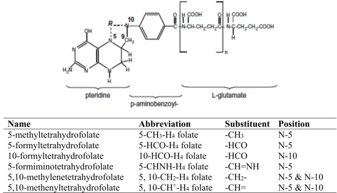
Folate is the generic term for folic acid (pteroylmonoglutamic acid) and related compounds exhibiting the biological activity of folic acid. The terms folacin, folic acids, and folates are used only as general terms for this group of heterocyclic compounds based on the N-[(6-pteridinyl)methyl]-p-aminobenzoic acid skeleton conjugated with one or more L-glutamic acid residues. Folates can consist of a mono- or polyglutamyl conjugate. The folates from most natural sources usually have a single carbon unit at N-5 and/or N-10; these forms participate in the metabolism of the single-carbon pool. The compound called folic acid is not present in living cells, being rather an artificial and manmade form of the vitamin. Folic acid (pteroylmonoglutamic acid) is an orange-yellow crystalline substance that is soluble in water but insoluble in ethanol or less polar organic solvents. It is unstable to light, acidic or alkaline conditions, reducing agents, and to heat (except in dry form). The UV absorption spectra of the folates are characterized by the independent contributions of the pterin and 4-aminobenzoyl moieties; most have absorption maxima in the region of 280–300 nm (Lavoisier, 2008).
Figure 2.4. Structure of native food folates and their substituent groups and positions. n:number of glutamates. Adapted from Witthöft (2011)
Name Abbreviation Substituent Position
5-methyltetrahydrofolate 5-CH3-H4 folate -CH3 N-5 5-formyltetrahydrofolate 5-HCO-H4 folate -HCO N-5
10-formyltetrahydrofolate 10-HCO-H4 folate -HCO N-10 5-formiminotetrahydrofolate 5-CHNH-H4 folate -CH=NH N-5
5,10-methylenetetrahydrofolate 5, 10-CH2-H4 folate -CH2- N-5 & N-10 5,10-methenyltetrahydrofolate 5, 10-CH+-H
24 Naturally occurring folates, such as those found in foods and body tissues, exist in different forms. They contain a fully-reduced pteridine ring together with additional glutamic acid molecules (Figure 2.4). They are usually substituted by different one-carbon units at the N-5 or N-10 positions, or have a single-carbon bridge spanning these positions. These one-carbon units can be at the oxidation level of methanol (5-methyltetrahydrofolate), or formaldehyde (5, methylenetetrahydrofolate), or formate (5, 10-methenyltetrahydrofolate) (Scott and Weir, 1993).
The 5-methyltetrahydrofolate (5-MTHF) is the predominant natural form of folate in fruits and vegetables (Vahteristo et al., 1997). In animal products the 5-MTHF and tetrahydrofolate are predominate (Vahteristo et al., 1997) while cereal products contain 5-MTHF, 5-formyltrahydrofolate, and 10-formlytrahydro folate (Pfeiffer et al., 1997).
2.2.3. Importance of folate in human diet
The naturally occurring form of folate lacks stability during food storage and preparation and thereby the stable folic acid (Eitenmiller et al., 2007) was used for supplements and food fortification. Folate has a crucial role as a one-carbon source for DNA, RNA synthesis and protein methylation (Stover, 2009). A number of genetic polymorphisms affect critical components of folate pathways and metabolism, and have been associated with an increased risk for neural tube defects (NTD) (Molloy et al., 2009). However, the exact mechanism by which folic acid reduces the risk of NTDs is not known and remains an active area of research (Crider et al., 2011a). NTDs occur when the neural tube fails to close early in embryonic development, resulting in damage to the exposed underlying neural tissue. These birth defects can result in significant morbidity and mortality depending on the location and severity of the lesion (Crider et al., 2011a). The most severe lesions observed with spina bifida cause a range of morbidities, including urinary and fecal incontinence and paralysis of the lower limbs (Sutton et al., 2008).
The relationship between NTD occurrence and folate deficiency encouraged scientist to demonstrate the link between folic acid intake and the risk of birth defects. A randomized control trial study found that the risk of NTD occurrence can be reduced by 70% when
25 women consume 400 µg of folic acid daily (MRC, 1991). In 1991, the Centers for Disease Control and Prevention recommended that women with a history of NTD affected pregnancy should consume 400 µg of folic acid daily before conception (CDC, 1991). Subsequently, in 1992, the U.S. Public Health Service recommended that all women of childbearing age consume 400 µg of folic acid daily through fortification, supplementation, and diet to prevent NTDs (CDC, 1991). In 1998, the Institute of Medicine (IOM) recommended that women at the age of becoming pregnant should consume additional 400 µg of folic acid daily from fortified foods or supplements to that obtained through a normal diet (1998). In order to increase the folic acid level in the diet of all women of childbearing age, the regulations for mandatory fortification of wheat flour with folic acid has begun. The mandatory folic acid fortification of enriched cereal grain products was authorized in 1996 in U.S. and fully implemented in 1998 (FDA, 1996). This program aimed to add 140 µg of folic acid/100 g of enriched cereal grain product in order to provide nearly 100–200 µg of folic acid/day for women of childbearing age (Quinlivan and Gregory, 2007).
Studies showed a significant decrease of 19%–32% in the prevalence of NTDs and spina bifida since the implementation of folic acid fortification in 1998 (Boulet et al., 2008). However, there are concerns about potential adverse consequences e.g. masking of B12 deficiency anemia, cancer and epigenetic changes, and circulating unmetabolized folic acid in the blood (Crider et al., 2011b).
Folic acid normally is reduced to tetrahydrofolate following uptake by the liver. If the folic acid is exceeded from tolerable upper intake level (1000 μg/day) (IOM, 1998), the non-metabolized folic acid will be found circulating in the blood. Synthetic folic acid is absorbed by passive diffusion and it should be reduced and then metabolized to 5-MTHF in the human mucosal cell and/or liver. Because the ability of conversion is limited, unmetabolized folic acid can appear in the systemic circulation, even after low-dose application (Pfeiffer et al., 1997). The debate addresses two main risk of long term consumption of high folic acid intake which could promote the formation of colorectal tumors (Kim, 2004) and masking the appearance of vitamin B12 deficiency anemia (Morris et al., 2010).