miRNAs as therapeutic agents in neurodegeneration,
a pilot study
Thèse
SEPIDEH PARSI
Doctorat en neurobiologie
Philosophiae doctor (Ph.D.)
Québec, Canada
© Sepideh Parsi, 2016
miRNAs as therapeutic agents in neurodegeneration,
a pilot study
Thèse
SEPIDEH PARSI
Sous la direction de :
RÉSUMÉ
L’échec des différents essais cliniques souligne la nécessité de développer des nouvelles thérapies pour la maladie d'Alzheimer (MA), la cause la plus commune de démence. Les microARNs (miARNs) sont les ARNs non-codants les plus étudiés et ils jouent un rôle important dans la modulation de l'expression des gènes et de multiples voies de signalisation. Des études antérieures, dont celles de mon laboratoire d’accueil, ont permis de développer l’hypothèse que certains membres de la famille miR-15/107 (c.-à-d. miR-15ab, miR-16, miR-195, miR-424, and miR-497) pourraient être utilisés comme agents thérapeutiques dans MA. En effet, cette famille avait le potentiel de réguler de multiples gènes associés à MA, tels que la protéine précurseur de l'amyloïde (APP), la β-secrétase (BACE1), et la protéine Tau.
Tel que démontré dans ce projet de thèse, j’ai choisi miR-16 comme cible thérapeutique potentielle pour MA parmi tous les membres de la famille. L’essai luciférase dans ce projet confirme que miR-16 peut réguler simultanément APP et BACE1, directement par une interaction avec la région non-codante en 3’ de l’ARNm). Notamment, nous observons aussi une réduction de la production des peptides amyloïdes et de la phosphorylation de Tau après une augmentation de miR-16 en cellule. J’ai ensuite validé mes résultats in vivo dans la souris en utilisant une méthode de livraison de miR-16 via une pompe osmotique implanté dans le cerveau. Dans ce cas, l'expression des protéines d’intérêts (APP, BACE1, Tau) a été mesurée par immunobuvardage et PCR à temps réel. Après validation, ces résultats ont été complémentés par une étude protéomique (iTRAQ) du tronc cérébral et de l'hippocampe, deux régions associées à la maladie. Ces données m’ont permis d’identifier d'autres protéines régulées par miR-16 in vivo, incluant α-Synucléine, Transferrine receptor1, et SRm300. Une autre observation intéressante : les voies régulées par miR-16 in vivo sont directement en lien avec le stress oxydatif et la neurodégénération.
En résumé, ce travail démontre l’efficacité et la faisabilité d’utiliser un miARN comme outil thérapeutique pour la maladie d’Alzheimer. Ces résultats rentrent dans un cadre plus vaste de découvrir de nouvelles cibles pour MA, et en
particulier la forme sporadique de la maladie qui représente plus de 95% de tous les cas. Évidemment, la découverte d’une molécule pouvant cibler simultanément les deux pathologies de la maladie (plaques amyloïdes et hyper phosphorylation de tau) est nouvelle et intéressante, et ce domaine de recherche ouvre la porte aux autres petits ARNs non-codants dans MA et les maladies neurodégénératives connexes.
ABSTRACT
Failure at different clinical trials emphasizes the need for developing new therapeutics for Alzheimer disease (AD) as the most common cause of dementia. MicroRNAs (miRNA) are the most studied groups of non-coding RNAs and have a critical role in modulating multiple signaling pathways and fine-tuning gene expression. Supporting evidence from other studies, including host lab, suggest that multiple members of the miR-15/107 family (miR-15ab, miR-16, miR-195, miR-424, and miR-497) could be used as therapeutic agents in AD. The potential ability of this miRNA family to modify disease pathway by multiple targeting of AD-associated genes such as Amyloid precursor protein (APP), β-site amyloid-β precursor protein cleaving enzyme (BACE1) and microtubule-associated protein Tau is of attention.
Based on documented results in this study I chose miR-16 as candidate therapeutic miRNA in AD. This choice is based on data obtain from cells and in vitro luciferase assay indicating the role of this miRNA in the simultaneous regulation of APP, BACE1 (directly by targeting 3’UTR of these genes). Decrease in Tau phosphorylation and amyloid beta peptides were further observed following increased miR-16 levels. Furthermore, I validated these results in vivo by delivering miR-16 oligos using Osmotic pumps implanted subcutaneously to deliver oligos to lateral ventricles of mouse brain also providing a wide distribution of these oligos. Expression of desired protein targets was measured by western blot and qPCR in different brain regions. Results demonstrated a context-dependent action of delivered miR-16 increase on the potential AD involved targets in mouse brain. These results were complemented by proteomics study of Brainstem and Hippocampus regions. Data indicated the potential regulation of other proteins by miR-16 in vivo such as α-Synuclein in Brainstem and Transferrin receptor1 and SRm300 in Hippocampus. The increase in miR-16 levels in vivo and in vitro was sufficient to downregulate the protein product of these genes confirmed by western blot. Enrichment study predicted oxidative stress and neurodegeneration as top terms in close connection with miR-16.
This work provided a proof-of-principle for possibility and efficiency of miRNA replacement based therapeutics delivered to CNS using miR-16 a member of the miR-15/107 family. Understanding the molecular mechanisms involved in the regulation of AD-related genes could have important implications for sporadic AD, which accounts for more than 95% of all cases with no effective therapy available. Multi-target therapy by non-coding RNA in AD is an emerging concept that would have the potential to change the way that therapeutics is developed for AD and other neurodegenerative diseases with complex nature and no effective therapy available.
TABLE OF CONTENTS
RÉSUMÉ ... iii
ABSTRACT ... v
TABLE OF CONTENTS ...vii
LIST OF TABLES ... x
LIST OF FIGURES ... xi
ABBREVIATIONS ...xii
ACKNOWLEDGEMENTS ... xiv
PREFACE ... xv
1. CHAPTER I: NTRODUCTION ... 1
1.1. Alzheimer’s Disease (AD) ... 1
1.2. Diagnosis ... 2 1.3. Microscopic features ... 2 Plaques ... 2 Neurofibrillary tangles (NFT) ... 3 Neural loss ... 4 1.4. Macroscopic features ... 5
1.5. Familial versus sporadic AD ... 5
1.6. Molecular targets in AD Therapeutics ... 8
Amyloid Precursor Protein (APP) ... 8
Proteolytic processing of APP ... 9
β Secretase... 11
γ-Secretase ... 13
Tau ... 14
Phosphorylation of Tau ... 16
Pathology of Tau in AD ... 17
Therapeutic targeting of Tau ... 18
1.7. microRNA ... 19
Biogenesis ... 20
Gene regulation ... 23
miRNA stability and turnover ... 24
miR-15/107 family members ... 31
Expression and regulation of miR-15a/16-1: a cluster in a loop with cell cycle proteins ... 33
miR-15/107 family potential regulator of APP and BACE1 in AD ... 36
1.8. Multitarget therapy in AD ... 38
1.9. Non-coding RNA-based Therapeutics ... 40
Oligonucleotide therapies from bench to Clinique ... 45
1.10. OBJECTIVES OF RESEARCH: ... 47
Technical note ... 48
2. CHAPTER II: Preclinical evaluation of miR-15/107 family members as multifactorial drug targets for Alzheimer’s disease ... 51
Résumé ... 52
Abstract ... 53
2.1. Introduction ... 54
2.2. Results ... 56
Comparative analysis of miR-15/107 family members in vitro and in cells ... 56
Loss of the DLEU2/miR-15a/16-1 in sporadic AD ... 59
Effective delivery of miR-16 mimics into the mammalian brain ... 60
Assessment of potential side effects related to miR-16 mimic brain delivery ... 62
2.3. Discussion ... 65
2.4. Materials and Methods ... 70
Cell culture ... 70 Cell transfection ... 70 Western blotting ... 70 qRT-PCR ... 71 Luciferase assays ... 72 ELISA ... 72
In vivo administration of mimics ... 72
Proteomics ... 73
RNA Immunoprecipitation (RIP-Chip) ... 74
Statistics ... 74 2.5. References ... 76 2.6. Supplementary data ... 84 2.7. Supplementary Methods ... 85 2.8. Supplementary Figures ... 86 2.9. Supplementary References ... 96
2.10. Supplementary Tables ... 97
3. CHAPTER III: MiR-16 and cellular stress ... 104
3.1. Introduction ... 104 3.2. Results ... 107 3.3. Discussion ... 109 3.4. Methods ... 111 HT22 cell culture ... 111 Viability assay ... 111
UPR stress measurement in vitro ... 111
4. CHAPTER IV: GENERAL DISCUSSION ... 113
4.1. miRNA-based Therapeutics challenges ... 113
4.2. miRNA gain of function study in vitro ... 114
4.3. MiR-16 non-modified mimics are functional in vivo ... 115
4.4. MiR-16 loss of expression could play potential key role in AD ... 117
4.5. Perspectives ... 120
BIBLIOGRAPHY ... 122
LIST OF TABLES
Table 1 Small-molecule BACE1 inhibitors in clinical trials. ... 12
Table 2 Highlighted protein factors affecting miRNA biogenesis ... 26
Table 3 Main miRNAs identified to be associated with AD ... 30
Table 4 Selected clinical trials with RNA-based therapy in neurodegenerative diseases ... 44
Table 5 A summary of miRNA therapeutics in clinical trials ... 44
Supplementary Table 1 Protein changes in the brainstem and hippocampus of treated mice versus controls. ... 97
Supplementary Table 2 DAVID gene enrichment analysis of top ranked targets identified in brainstem using Homo sapiens background. ... 99
LIST OF FIGURES
CHAPTER I
Figure 1 Alois Alzheimer. (b) Neurofibrillary lesions shown by Gallyas silver-iodide staining
technique. (c) Aβ plaques visualized by Campbell-Switzer silver-pyridine staining technique. ... 1
Figure 2 Neurofibrillary pathology in the entorhinal layer of cortex ... 4
Figure 3 A healthy brain contrasted with the effects of Alzheimer’s ... 5
Figure 4 Generation of Aβ from amyloid precursor protein ... 6
Figure 5 Schematic domain structure of APP695 ... 8
Figure 6 Schematic drawing of the Amyloid Precursor Protein (APP) and its processing pathways 10 Figure 7 Subunits of the γ -secretase complex. ... 13
Figure 8 MAPT, the gene encoding human tau, contains 16 exons ... 15
Figure 9 Location of tau phosphorylation sites and epitopes for tau antibodies ... 16
Figure 10 Schematic representation of tau-related processes that are potential targets for therapeutic intervention. ... 18
Figure 11 Comparison of general properties between siRNA and miRNA ... 21
Figure 12 Genomic location of miRNAs ... 22
Figure 13 Target scan analysis of human BACE1 UTR ... 29
Figure 14 Highly expressed miR-15/107 miRNAs with overlapping targets ... 32
Figure 15 Location of dleu2- miR-15a/16 and smc4-miR-15b/16 genes ... 34
Figure 16 miR-16 inhibits amyloid protein precursor (APP) in SAM mice in vivo ... 37
Figure 17 The main current targets in AD research ... 40
Figure 18 Structures of nucleic acid analogs and modifications ... 43
Figure 19 Implantable infusion's pump for a continuous intrathecal administration ... 46
CAHPTER II Figure 1 Comparative analysis of miR-15/107 family members in vitro. ... 58
Figure 2 Effects of miR-16 overexpression on APP, BACE1, and Tau in neuronal cells ... 59
Figure 3 In vivo regulation of AD genes by miR-16 mimics. ... 62
Figure 4 Analysis of inflammation markers following miR-16 mimic treatment. ... 64
Figure 5 Proteomics validation in vivo and in neuronal cells. ... 65
CHAPTER III Figure 1 The unfolded protein response (UPR) stress sensors ... 106
Figure 2 Induction of stress in HT22 reduces the expression level of miR-16. ... 108
Figure 3 miR-16 modifies ATF6a expression in cells under stress. ... 109
CHAPTER IV Figure 1 miR-16 and AD pathways regulation-a hypothesis. ... 117
ABBREVIATIONS
AChE Acetylcholinesterase AGO Argonaute protein
ALS Amyotrophic lateral sclerosis AD Alzheimer's disease
Aph1 Anterior pharynx-defective 1 protein ATF6 Activating transcription factor 6 Aβ Amyloid-β
BACE β-site amyloid-β precursor protein cleaving enzyme CB Calbindin
CamKII Calcium/Calmodulin-dependent kinase II CD86 T-Lymphocyte Activation Antigen
cDNA Complementary DNA CDS Coding sequence
CLL Chronic lymphocytic leukemia CSF Cerebrospinal fluid
CTFs C-terminal fragments DGCR8 DiGeorge critical region 8
ELISA Enzyme-linked immunosorbent assay ERK Extracellular signal-regulated kinase FAD Familial Alzheimer disease
FBS Fetal bovine serum
GAPVD1 GTPase Activating Protein and VPS9 Domains 1 GFAP Glial fibrillary acidic protein
GSK-3β Glycogen Synthase Kinase-3 LOAD Late-onset Alzheimer disease MAPT microtubule-associated protein tau miRNA microRNA
NMDA N-methyl-D-aspartate receptor NFT Neurofibrillary tangles
ORF Open reading frame
PARIS Protein and RNA isolation system PHF Paired helical filament
PS Presenilin
RNU48 Small Nucleolar RNA, C/D Box 48
RT-qPCR Reverse transcription quantitative polymerase chain reaction SAD Sporadic Alzheimer disease
SAMP8 Senescence-accelerated prone mouse Scr Scramble
SNCA α-synuclein
SNPs Single nucleotide polymorphisms UPR Unfolded protein response UTR Untranslated region
To my beloved parents those are always inspirational to me and to memory of my dear grandmother with whom I had great memories of my childhood
ACKNOWLEDGEMENTS
I must first thank my research advisor Dr. Sébastien Hébert for the continuous support of my study and related research, for his patience, motivation, and guidance. I appreciate the time and resources he has provided me to thrive as a researcher that I am today. Without his professional support, my research training and the work described in this dissertation would not have been possible.
I must also thank the other members of my research committee Dr. Emmanuel Planel, Dr. Georges Lévesque and Dr. Pascale Legault. They have been generous in providing their time and expertise and scientific advice. Their feedbacks have continually had a positive impact on the direction of my research. I truly appreciate their input.
I thank my fellow lab mates in for the stimulating discussions, for the beautiful moments we were working together even at nights, and for all the fun we have had in the last three years which remains the best memories of my stay in Canada. I especially thank our very hardworking and knowledgeable lab assistant, Claudia Goupil that contributed significantly to this work and for being willing to help with animal experiments. I should also thank Pascal smith, Veronique Dorval, my dear colleagues for their valuable contribution to this project. I would like to thank Julia Hernandes-Rapp, Ana Sofia Correia for their valuable friendship and scientific supports and our ex-members Francis Jolivette, Marie-José Dupras and Charlotte Delay.
I would also like to thank all members of Axe de Neuroscience who have been instrumental in helping me survive the vagaries of research. Inspiring people that were more than colleagues to me.
Last but not the least, I thank my family and friends for their continual support and bearing all this hardship of being away from me thousands of miles but the never let me feel they are not with me even for a second. I recognize that this research would not have been possible without the financial assistance of Le Fonds Ven-Huguette-Anil-Murthy.
PREFACE
The present thesis is written in four main chapters. Chapter I provides an introduction to the work, in which a general review of main therapeutic targets in AD such as APP, BACE1, and Tau is presented. The literature review on miRNA topic continues with a focused concentration on miR-15/107 family and introducing the concept of miRNA-based therapeutics at the end of the chapter. Chapter II delivers the main results of the current thesis which are already presented as a research article entitled: “Preclinical Evaluation of miR-15/107 Family Members as Multifactorial Drug Targets for Alzheimer’s Disease” published in the peer-reviewed journal of Molecular Therapy-Nucleic Acids (2015) 4, e256 in which I am the first author. The main goal of the project was an assessment of an AD-related miRNA in the context of noncoding RNA therapeutics. To achieve this aim, miR-16, as a candidate, was chosen from multiple members of miR-15/107 according to the in vitro result presented in this work. The following in vivo experiments tested miR-16 mimics action compared to the controls in mice. This paper was written and executed by me and majorly corrected by my supervisor Dr. Hébert. The first preliminary result indeed was obtained from the study of increased miR-16 levels and the reduction of amyloid beta in vitro and in mutant HEK cells performed by Pascal Smith. Claudia Goupil as our expert technician had the main role in optimization and performing osmotic pump implantation and Véronique Dorval contributed to this work by her data obtained from Rip experiment. Reviewer’s comments were also fundamental to shape the final work before its publication. The main revision was adding more supplementary data and information that was not included in the initial submission. The complete supplementary is presented at the end of chapter II of this work.
Chapter III presents the results obtained through parallel investigations of miR-16 upregulation in vitro and investigating the possible role of miR-miR-16 itself in cellular stress. The findings in this chapter complement the results of chapter II. The highlighted results in this chapter indicate that loss of miR-16 expression could potentially affect the cellular stress in neurodegenerative context.
Chapter IV conveys the general discussion of the work and attempts to cover the topics that have been less explored in previous chapters. Specifically, a more straightforward hypothesis of miR-16 in neurodegenerative context is presented.
1. CHAPTER I: NTRODUCTION
1.1. Alzheimer’s
Disease (AD)
“A peculiar severe disease process of the cerebral cortex” appears as the first report of neuropathological findings of AD in the brain by Alois Alzheimer in 1907. He described a 50 years old woman whom he had followed for paranoia, progressive sleep, memory disturbance, aggression, and confusion, until her death 5 years later (Hippius & Neundörfer, 2003). AD is the most common cause of dementia in the elderly and is characterized by the progressive memory loss leading to a gradual and irreversible deterioration of cognitive function. Two major hallmarks of this disease are senile plaques and neurofibrillary tangles (NFT) (Figure 1). Senile plaques are extracellular depositions of β-amyloid (Aβ) peptide while NFT are intraneuronal aggregations composed of hyperphosphorylated TAU (Hardy & Selkoe, 2002).
(D) The lesions (red) increase in severity and extent without remission during a long presymptomatic disease phase that reaches a shorter and final symptomatic phase. ’’With Permission of Springer’’(Heiko Braak & Del Tredici, 2015) Advances in Anatomy, Embryology, and Cell Biology V 215 2015 p4 © Springer International Publishing Switzerland
2015
It is expected that by 2050, one new case of AD is to develop every 33 seconds or nearly a million new cases per year, and the total estimated prevalence is expected to be 13.8 million (Alzheimer’s association, 2014)(Prince et al., 2013). According to Alzheimer Society of Canada in 2011 only, 747,000 Canadians were living with cognitive impairment, including dementia -including 14.9 percent of Canadians 65 and older.
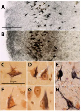
Figure 1 (A) Alois Alzheimer. (B)
Neurofibrillary lesions shown by Gallyas silver-iodide staining technique. (C) Aβ plaques visualized by Campbell-Switzer silver-pyridine staining technique.
1.2. Diagnosis
Early diagnosis of AD has come to attention because the disease manifests its earliest pathology years or decades prior to the onset of dementia. The diagnosis of AD generally resides in observation of neuropsychiatric features such as cognitive impairment that manifests itself at least by minimum two of these symptoms: inability of patients in new learning, disturbances of language function, impairment in reasoning and handling of complex tasks and changes in personality and behaviour (McKhann et al., 2011). Ante-mortem diagnosis still remains a challenge today due to lack of strong biomarkers. For instance, the CSF elevation of Tau could also be implicated pathologically in other diseases such as Parkinson disease (PD), Progressive supranuclear palsy (PSP), and Corticobasal degeneration (CBD) (Maruyama et al., 2013). With advancement in imaging techniques such as Magnetic resonance imaging (MRI), it has been proposed that gray matter atrophy patterns are capable of differentiating between subtypes of mild cognitive impairment (MCI) (H. Zhang et al., 2012). MCI is a syndrome characterized by cognitive impairments that are distinct compared to the decline in cognition by the normal aging process. MCI is often considered as a precursor to dementia or a transitional state between healthy cognitive aging and dementia (Sperling et al., 2011). The imaging study also implies a decrease in Hippocampal volume preceding the transition to AD can also be detected by Structural MRI scans (McEvoy & Brewer, 2010; van der Flier, 2005).
1.3. Microscopic
features
Plaques
In mild AD cases, pathological lesions are typically restricted to the medial temporal lobe, including the hippocampus, with a spread to the temporal, the parietal, and eventually the occipital and frontal lobes in severe cases (H Braak & Braak, 1991). Aβ derived from APP is the main component of plaques found in AD brain. Aβ plaques are different in shapes and sizes and with the advancement of the disease, become dense and sensitive to silver staining (argyrophilic). Aβ deposits in AD rarely develop in the white substance; instead, they mainly occur in the gray matter. Neurite plaques (NPs) appear in later stages of the disease.
NPs consist of abnormal astrocytes, microglial cells, dystrophic neuronal processes and Tau aggregates (Heiko Braak & Del Tredici, 2015).
Neurofibrillary tangles (NFT)
Ultra-structurally, NFTs are composed of dense accumulations of structures known as paired helical filaments (PHFs) (KIDD, 1963), which are mainly distributed in the perinuclear area of the neuron and in proximal processes. Neurofibrillary degeneration and phosphorylated tau accumulation are closely aligned with neurological signs, neuropathological stage, and clinical and pathological disease severity as compared to accumulations of Aβ (H Braak & Braak, 1991). Braak staging of AD pathology is based upon the level of progression of NFT pathology, which spans from stage1 or mild to stage 6 that is very late or severe dementia in which patients are not able to do functions of daily living and need constant support. Braak method applies immunoreaction (AT8) for hyperphosphorylated tau protein (Heiko Braak, Alafuzoff, Arzberger, Kretzschmar, & Tredici, 2006) by which six stages of disease propagation can be distinguished with respect to the location of the tangle-bearing neurons and the severity of changes (Heiko Braak et al., 2006). Abnormal tau aggregates appear for the first time in the cerebral cortex generally in the transentorhinal region progressing to the limbic area and neocortical as well as primary sensory areas. Gradually, NFTs fill large portions of the soma of neurons (Figure 2) (Heiko Braak & Del Tredici, 2015).
(A) The mildly involved entorhinal layer at NFT stage II. (B) A dense network of NTs at NFT stage V. (C–H) Development of neurofibrillary changes using the Gallyas silver-iodide technique combined with Pigment-Nissl staining. (C, D) NFTs stage I) (e, f) Consolidation of a sturdy NFT. A comparison with uninvolved neighboring neurons does not reveal obvious reactive changes (G, H) Dying NFT-bearing nerve cell (G) at the left side (H) NFT stage VI in a 60-yrs male AD patient, ’’With Permission of Springer’’(Heiko Braak & Del Tredici, 2015) Advances in Anatomy, Embryology and Cell Biology V 215 2015 p102 © Springer International Publishing Switzerland 2015
Neural loss
AD clinical symptoms develop subtly leading to a gradual loss of fundamental functions by cellular impairments that first appear after a given threshold is exceeded (Figure 1) (Heiko Braak & Del Tredici, 2015). In term of neural loss, degeneration of basal forebrain is more prominent in the advancement of the disease. The basal forebrain plays an important role in acetylcholine production and associated loss of cholinergic neurotransmission contribute significantly to the deterioration in cognitive function seen in patients with Alzheimer’s disease (Bartus, Dean, Beer, & Lippa, 1982). Acetylcholine (ACh) was the first neurotransmitter discovered with involvement in memory and cognition in central nervous system (Todman, 2008). In advanced AD, the relative loss of cholinergic connections by region shows different pattern; with the temporal lobe showing most loss of neurons contrary to other neuronal populations such as sensory and motor neurons (Geula & Mesulam, 1989). The mechanism behind this selective vulnerability of different neural populations of the brain in AD is not well understood. Interestingly it has been proposed that morphology also comes to attention where the neuronal types involved in AD-process are found among
Figure 2 Neurofibrillary pathology in
projecting neurons with a disproportionately long axon (Heiko Braak & Del Tredici, 2015).
1.4. Macroscopic
features
The decrease in brain volume is very prominent in AD, rather diffuse and uniform in cerebral white matter. The gray matter of frontal and parietal cortex and striatum is more affected compared to temporal cortex, cerebellar vermis, and hippocampus; and the occipital cortex is least affected (Mrak, Griffin, & Graham, 1997). Other pathologies include increased in lipofuscin content of neurons (insoluble, autofluorescent glycol lipoprotein), the ventricular and subarachnoid space expansion followed by shrinkage of brain and volume loss (Figure 3)(Shankar, 2010).
Figure 3 A healthy brain contrasted with the effects of Alzheimer’s1
1.5.
Familial versus sporadic AD
APP coding gene is located on the long arm of chromosome 21 with an extra copy only present in individuals with Down’s syndrome or trisomy 21 (Yoshikai, Sasaki, Doh-ura, Furuya, & Sakaki, 1990). APP and its proteolytic signaling play an important role in the onset and progression of familial and non-familial AD (sporadic). Several mutations in APP is reported in hereditary early-onset AD summarized in Figure 4. Mutations mainly located close to the major APP processing sites (β and γ secretase sites) or within the Aβ sequence (the α -secretase site). The mechanisms by which mutation causes disease are diverse
and complex. For example, Swedish mutation (K670NM671NL) (Mullan et al., 1992) makes APP mutant a better BACE-1 substrate, increasing β-secretase cleavage and enhancing Aβ production both Aβ42 and Aβ40 in plasma and fibroblasts from mutation carriers (Citron et al., 1992; Haass et al., 1995). Only one mutation (A673T) has been reported to protect against AD with high penetrance providing provides a proof of principle for the hypothesis that reducing the β-cleavage of APP may protect against the disease (Jonsson et al., 2012).
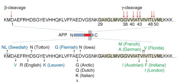
Figure 4 Generation of Aβ from amyloid precursor protein
The sites of β- and γ-secretase–mediated cleavage are indicated by arrows, and the transmembrane domain of APP is highlighted in gray. γ-cleavage produces a pool of Aβ fragments that vary in length and hydrophobicity. The mutations in Aβ region of APP either increase the total Aβ production (marked in blue), alter Aβ biophysical properties (in black) or affect the Aβ spectrum in both quantitative and qualitative ways (in green) Nature Publishing Group Permission (Benilova, Karran, & De Strooper, 2012).
The other important mutation reported is located in the presenilin 1 and presenilin 2 genes ( PSEN1, PSEN2 ) part of γ –secretase components (Strooper et al., 2013). Presenilin cause early-onset (<60 years) autosomal dominant AD (Bertram et al., 2010), which accounts for less than 1% of AD cases (Campion et al., 1999). PS mutations alter PS processing and So far, more than 150 familial AD-causing mutations in PSEN1 only have been identified, for instance, the ∆9 mutation completely inhibits PS processing (Crook et al., 1998; Mercken et al., 1996). Further studies demonstrated that transgenic mice carrying both mutant
amyloid precursor protein and presenilin 1 transgenes showed accelerated Alzheimer-type phenotype(Holcomb et al., 1998).
Unlike the early-onset familial form of AD as the result of main mutations at least in one of the three amyloid processing genes (APP, PSEN1 or PSEN2), the mechanisms underlying late-onset or sporadic form of the disease (LOAD/sAD) remains unknown. It is important to note that a single pathway does not explain the more common non-familial form (Medway & Morgan, 2014). It has been suggested that advancing age and a family history of dementia may contribute to decreasing threshold of AD appearance; however, it vastly remains to clarify the association of other factors i.e. oxidative stress, diabetes and hypertension with AD and whether they mainly contribute to clinical manifestations or they intervene with amyloidosis and neurofibrillary degeneration processes themselves (David
Knopman, 2011).
Studies suggest that both increased Aβ production and decreased Aβ clearance could play an important role in overall Aβ changes in sporadic AD (Carare, Hawkes, Jeffrey, Kalaria, & Weller, 2013). Recently, Aβ accumulation, tau accumulation, and molecules related to Aβ metabolism across 12 brain regions in postmortem tissue from sporadic AD, familial, controls have been studied. This study clearly indicates the difference between the Aβ pattern in both forms of the disease. In sporadic AD, Aβ42 was found to disproportionately accumulate in cortical regions, compared with familial AD, and to strongly correlate with the normal regional distribution of Synaptic markers. Although an increased production of Aβ42 or increased ratio of Aβ42 to Aβ40 is clearly implicated in the pathogenesis of familial type (Karran et al., 2011), it remains unclear how Aβ metabolism is disturbed and thus involved in sporadic AD where there is no autosomal dominant mutation (Shinohara et al., 2014). To clarify how Aβ is involved in sporadic AD some anatomical studies provide the evidence that some nerve cells in the brainstem nuclei are involved in Aβ deposition, those already have undergone cytoskeletal tau changes (Heiko Braak & Del Tredici, 2013).
1.6.
Molecular targets in AD Therapeutics
Amyloid Precursor Protein (APP)
APP is a well-conserved member of the greater APP family and the only member with an Aβ peptide domain. First cloned APP isoform is APP695, expressed mainly in the brain without exon 7 and 8 (Kang et al., 1987). An important characteristic of this protein family is having E1 and E2 domains, Kunitz protease inhibitor (KPI) region, and an OX-2 domain (encoded by exons 7 and 8). The latter two do not exist in brain isoform or APP695 (Figure 5) (Coburger et al., 2013) . APP is expressed highly in fetal tissues and has the highest expression levels in brain, kidney, heart and spleen while weak expression in liver. In adult brain, highest expression is found in the frontal cortex specifically (K. Tang et al., 2003).
Figure 5 Schematic domain structure of APP695
The different domains of APP brain isoform are schematically demonstrated in the picture. N-terminal growth factor-like domain (GFLD) copper-binding domain (CuBD). central APP domain (CAPPD). and the intracellular domain (AICD) (Coburger et al., 2013).
In neurons, APP can be found in both presynaptic and postsynaptic compartments (Yamazaki, Selkoe, & Koo, 1995). Although the physiological functions of APP are not clear, several possibilities have been proposed (Nakayama, Nagase, Koh, & Ohkawara, 2013). The most considerable functions
are synapse formation and repair (Priller et al., 2006). It has also been suggested that APP acts as a cell adhesion molecule and plays a role in cell-cell interaction (Soba et al., 2005).
Proteolytic processing of APP
The proteolytic process of APP follows the regulated intramembrane proteolysis or simply RIP mechanism. RIP follows a two-step pattern. An initial cut at a site outside the membrane is followed by a second cut inside. Cleavage of APP by α- secretase or β-secretase results in shedding of the entire extracellular domain and α- or β-carboxy terminal fragments (αCTF/C83 or βCTF/C99) (Esch et al., 1990) (Vassar et al., 1999). Several zinc-metalloproteinases, such as TACE and ADAM (Buxbaum et al., 1998; Lammich et al., 1999), and the aspartyl protease BACE2 (Farzan, Schnitzler, Vasilieva, Leung, & Choe, 2000) can cleave APP at the α-site, while BACE1 (β-site APP cleaving enzyme) cleaves APP at the β-site (Figure 6) (Vassar et al., 1999). After α- or β-cleavage, the resulting membrane associated C-terminal fragments are subsequently cleaved in the Transmembrane domain by a γ-secretase complex releasing the P3 or Aβ fragment and an APP intracellular domain (AICD). Generated Aβ peptides might differ in size slightly from 34–50 amino acids since the exact cut generated by the γ-secretase complex varies (Kummer & Heneka, 2014). Most of Aβ fragments are Aβ40, but a smaller fraction is the more prone to oligomerization which is Aβ42 and Aβ43 peptides found in amyloid plaques (Sisodia & St George-Hyslop, 2002). Several enzymes can degrade Aβ: Neprilysin (NEP) and insulin-degrading enzyme (IDE) are expressed in neurons as well as within the vasculature, and the levels of both these enzymes are reduced in AD. Post-translational regulation of NEP and IDE proteins by multiple factors i.e APP intracellular domain AICD leads to increased Aβ clearance (Kerridge, Belyaev, Nalivaeva, & Turner, 2014).
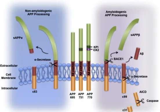
Figure 6 Schematic drawing of the Amyloid Precursor Protein (APP) and its processing
pathways
APP can be processed by α-secretase in a non- amyloidogenic pathway, which precludes the formation of Aβ and generates soluble APPα (sAPPα) and a C-terminal fragment (c83). Alternatively, in the amyloidogenic pathway, cleavage by the β-secretase BACE1 generates soluble APPβ (sAPPβ), which is secreted, and a C-terminal fragment (β-CTF or c99). Subsequent cleavage of c99 by the γ-secretase complex generates Aβ and the APP intracellular domain (AICD) which can be further cleaved by caspases to produce a c31 fragment Copyright © 2011 Elsevier Inc. with permission from Elsevier (Schonrock, Matamales, Ittner, & Götz, 2012)
As Aβ plays a role in the pathogenesis of AD the research on targeted therapy focused solely on treatments directed against Aβ. Anti-amyloid approaches intervene with Aβ production by immunotherapy, an increase of amyloid clearance and decrease of its aggregation, modulation of its transport or modulation of secretase (Kumar, Singh, & Ekavali, 2015). The term immunotherapy defies the manipulation of the immune system by inducing, enhancing, or suppressing immune responses in vivo (Lambracht-Washington & Rosenberg, 2013). Vaccination was the first treatment to have the significant effect on the AD in animal models. Vaccination of transgenic mice with Aβ prevented Aβ deposition and improved behavioral impairments related to Aβ deposition (Janus et al., 2000; Schenk et al., 1999).
Furthermore, the promising results of vaccination in preclinical and the lack of serious side-effects in transgenic mice encouraged other trials and it was followed by the launch of AN 1792 trial which strongly induces an immune response (T. Li et al., 2007). This trial was eventually stopped since 6% of participants developed symptoms of acute inflammation. Overall, failure of these drugs suggests that targeting Aβ alone might not be enough to prevent or slow AD. One very recent example is the most advanced trial study on monoclonal antibodies Solanezumab and Bapineuzumab, which have not been proven promising in clinical trials phase III (Boutajangout & Wisniewski, 2014).
β Secretase
The β-secretase enzyme that initiates the production of Aβ, BACE1 is a key therapeutic target for AD. The biochemical properties of the β-secretase enzyme had been established well before BACE1 was identified by independent groups as the β-secretase enzyme in 1999 (Vassar et al., 1999). BACE1 is a typeI transmembrane aspartyl protease with optimal protease activity in a low pH environment. As a result, BACE1 tends to localize in acidic compartments, such as endosome and trans-Golgi network (Vassar, Kovacs, Yan, & Wong, 2009). Immunohistochemical studies have indicated BACE1 is expressed both in neuronal and glial cells in the mammalian brain as well (Laird et al., 2005; Rossner, Lange-Dohna, Zeitschel, & Perez-Polo, 2005).
In sporadic AD brain, there is a strong correlation between Aβ loads and BACE elevation and enhanced deposition of amyloid plaques (R. Li et al., 2004) and AD progression (Che et al., 2014; Fukumoto, 2002; H. Harada et al., 2006). To study the role BACE1 in AD, several groups used gene targeting strategies to generate BACE1 knockout mice without gross abnormal phenotype implicating BACE1 inhibition as an interesting drug target (Cole & Vassar, 2007; Roberds et al., 2001). These genetics models further validated BACE1 as the major β secretase in the brain, making BACE1 inhibition a viable treatment strategy for AD (Y. Luo et al., 2003; Ohno et al., 2004). APP Overexpressing mice having a heterozygous BACE1 gene knockout were evaluated for Aβ generation and for the development of pathology. It was demonstrated that Although the 50% reduction in BACE1 enzyme levels caused only a 12% decrease in Aβ levels in young mice, it resulted
in a dramatic reduction in Aβ plaques, neuritic burden, and synaptic deficits in older mice (McConlogue et al., 2007). BACE1 is enriched in neuronal presynaptic terminals suggesting an important role for BACE1 at the synapse as well (Rajapaksha, Eimer, Bozza, & Vassar, 2011). Moreover, the protective A673T APP mutation in humans decreases Aβ generation via reduced β-secretase processing of APP, providing strong proof of concept that BACE1 inhibition should be efficacious for AD (Ohno et al., 2004; Vassar et al., 2009; R. Yan & Vassar, 2014). However, the design of small-molecule BACE1 inhibitors (i.e.LY2811376 a non-peptide molecule) has also shown no success to its extreme side effects in the very recent study (Yan and Vassar, 2014). It would be important to mention that complete suppression of BACE1 as the therapeutic approach is not ideal since recent studies of BACE1−/− mice have shown complex neurological phenotypes (R. Yan & Vassar, 2014). Recently, small-molecule BACE1 inhibitors have been tested that exhibit satisfactory pharmacokinetics and optimal effect on cerebral Aβ reduction in preclinical
animal models (R. Yan & Vassar, 2014).Most trials are in early phases and little
data about them have been published.The side effects of drugs inhibiting BACE1
still not examined but the possibility exists that BACE1 inhibitor drugs might also cause BACE2 mechanism-based side effects in addition to those of BACE1 (Table 1) (Dominguez et al., 2005).
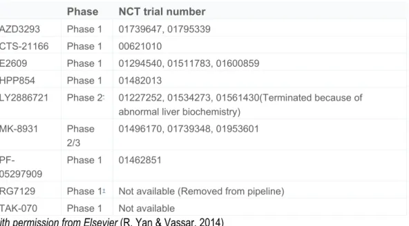
Table 1Small-molecule BACE1 inhibitors in clinical trials.
Phase NCT trial number
AZD3293 Phase 1 01739647, 01795339 CTS-21166 Phase 1 00621010
E2609 Phase 1 01294540, 01511783, 01600859 HPP854 Phase 1 01482013
LY2886721 Phase 2* 01227252, 01534273, 01561430(Terminated because of
abnormal liver biochemistry) MK-8931 Phase 2/3 01496170, 01739348, 01953601 PF-05297909 Phase 1 01462851
RG7129 Phase 1† Not available (Removed from pipeline)
TAK-070 Phase 1 Not available
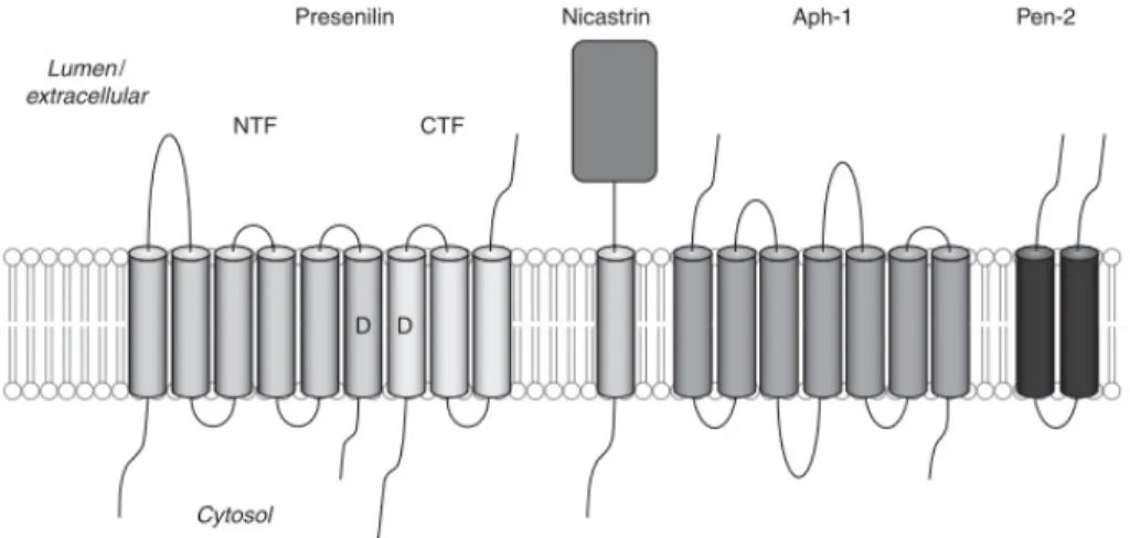
γ-Secretase
γ-Secretase is a complicated complex composed of Presenilin(PS), Nicastrin (NCT), anterior pharynx defective-1 (Aph-1), and PS enhancer-2 protein (Pen-2) (Figure 7). The first evidence for the role of presenilins in APP processing came from observations that AD-causing mutations in PSEN1 and PSEN2 affect the generation of Aβ peptides, changing the relative amount of Aβ peptide versus the shorter Aβ40 (Borchelt et al., 1996). PSEN1 mutations show the earliest age of onset more commonly affected by seizures and cerebellar signs, whereas PSEN2 mutations have a delayed onset with longest disease duration and significant
disorientation (Shea et al., 2015).PSEN1 and PSEN2, catalytic component of
γ-Secretase, both synthesized as precursor proteins of 50 kDa processed into a 30kD domain and a 20 kDa fragment during maturation (Thinakaran et al. 1996).
Figure 7 Subunits of the γ -secretase complex.
Presenilin is proteolytically processed into two fragments during maturation of the complex, an amino-terminal fragment (NTF) and a carboxy-terminal fragment (CTF). Other subunits are Nicastrin, APH-1, and PEN-2 Copyright © 2012 Cold Spring Harbor Laboratory Press (Strooper et al., 2013).
Aph-1 act as a scaffold during the process of γ-secretase complex assembly, and Pen-2 as a trigger for the cleavage of PS in order to activate it (Smolarkiewicz, Skrzypczak, & Wojtaszek, 2013). Human Pen-2 overexpression in transgenic mice has been shown to induce all the AD-like phenotypes, including behavioral deficits, motor activity, and feeding behavior dysfunction, Aβ-42 peptide deposition and chronic disease induction (Nam et al., 2011). A direct role of
PEN-2 in cleavage of PS1 and a regulatory function of APH-1, in coordination with PEN-2, in the biogenesis of the PS1 complex has been revealed (W. J. Luo et al., 2003). Unlike PS1, overexpression of APH1, PEN2, and Nicastrin proteins can increase the levels of Aβ, suggested that these proteins are limiting for γ-secretase activity (Marlow et al., 2003).
At 709 amino acids, Nicastrin is the largest component of γ-secretase, with the majority of its mass located to its large, heavily glycosylated ectodomain. Structural studies have been suggested a very comprehensive model of Nicastrin more favoring the hypothesis that Nicastrin may indeed serve as a receptor of γ-secretase substrates (Xie et al., 2014)
Development of therapeutic against secretase faces the specific challenge γ-secretase itself is also an essential component of the Notch signaling (Geling, Steiner, Willem, Bally-Cuif, & Haass, 2002; Godin et al., 2003; Kopan & Ilagan, 2009; Lundkvist & Näslund, 2007; Wolfe, 2008). Strong inhibition of γ-secretase leads to severe adverse effects, as it interferes with signaling by notch proteins and other cell surface receptors. A recent study concluded that a γ-secretase reduction of 30% was sufficient to attenuate Aβ deposition with little or no adverse side effects (Bard et al., 2000).
Heterogeneity of γ-secretase complex suggests that selective targeting of one particular subunit might be a more effective treatment strategy than non-selective γ- secretase inhibition (Serneels et al., 2009). For instance, deletions of APH-1B and APH-1C isoforms in a mouse model of AD decreased Aβ plaque formation and improved behavioral deficits (Serneels et al., 2005). They are current studies on developing more specific inhibitors (D’Onofrio et al., 2012).
Tau
Three types of filaments form the cytoskeleton: microfilaments, intermediate filaments, and microtubules. The cytoskeleton provides a dynamic scaffold to proteins, vesicles, and organelles, essential for cell function and changes in the state of its polymerization, play an important role in the neuronal process such as polarization, axonal transport, maintenance of neuronal extensions, synaptic plasticity and protein sorting (Morris, Maeda, Vossel, & Mucke, 2011). Tau protein
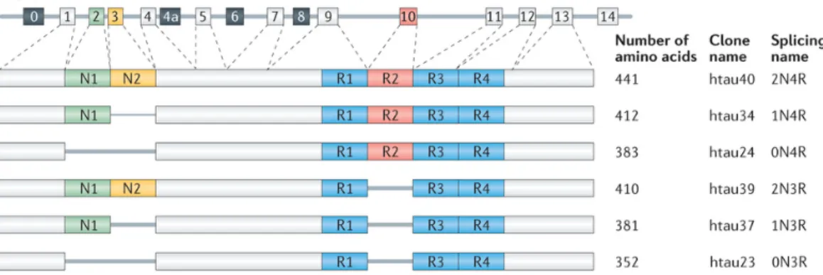
exists as a family of microtubule-associated protein (MAPs) that is found predominantly in axons with an important role in axonal transport (Dixit, Ross, Goldman, & Holzbaur, 2008; Weingarten, Lockwood, Hwo, & Kirschner, 1975; Witman, Cleveland, Weingarten, & Kirschner, 1976). The gene that encodes for tau consists of 16 exons and is located at the chromosomal locus 17q21 (Neve, Harris, Kosik, Kurnit, & Donlon, 1986). Through alternative splicing, six tau isoforms are generated in the CNS-isoforms (Figure 8). These tau isoforms differ according to the presence of 0, 1 or 2 near-amino-terminal inserts (0N, 1N or 2N) and the presence of R2 repeat, yielding 3 or 4 carboxy-terminal repeat domain (3R or 4R) tau species. The expression of human tau is developmentally regulated. In the adult brain, six isoforms of tau are expressed, whereas in the fetal brain only the shortest tau is expressed. In the adult human brain, levels of the 3R and 4R forms are roughly equal and the 2N isoform is underrepresented compared with the others: the 0N, 1N, and 2N tau isoforms comprise ~37%, ~54% and ~9% of total tau (Y. Wang & Mandelkow, 2015).
Through repeated domains located at the c-terminal of the protein and provides stability to the microtubule. This process can be regulated through a balance in the phosphorylation and dephosphorylation process of tau protein (Luna-Muñoz & Harrington, 2013).
Figure 8 MAPT, the gene encoding human tau, contains 16 exons
Exon 1 (E1), E4, E5, E7, E9, E11, E12, and E13 are constitutive, whereas the others are subject to alternative splicing. E0 and E1 encode the 5′ untranslated sequences of MAPT mRNA, whereas E14 is part of the 3′ untranslated region. E0 is part of the promoter, which is transcribed but not translated. The translation initiation codon ATG is in E1. E4a, E6, and E8 are transcribed only in peripheral tissue. The six human brain tau isoforms are generated through alternative splicing of
E2, E3, and E10.For more detail refer to the original Ref. Nature Publishing Group's permission (Y. Wang & Mandelkow, 2015).
Phosphorylation of Tau
Phosphorylation of Tau is the most studied modification of this protein to date. Protein phosphorylation is the addition of a phosphate group, by esterification, to one of three different amino acids: serine, threonine, and tyrosine. An increased tau phosphorylation reduces its affinity for microtubules leading to cytoskeletal destabilization. Phosphorylation is thought to be a critical event in both normal regulations of tau function and the pathogenesis of tau-related neuronal degeneration (Kawakami & Ichikawa, 2015).
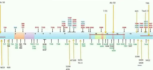
In AD, PHFs are characterized with abnormally phosphorylated Tau. Eighty-five putative phosphorylation sites on tau protein have been described in AD brain tissue (Hanger, Anderton, & Noble, 2009) (Figure 9). The phosphorylation of tau protein affects its solubility, localization, function, interaction with partners and susceptibility to other posttranslational modifications. However, the role of specific sites of tau phosphorylation in early neurodegenerative mechanisms is yet not fully understood(Luna-Muñoz & Harrington, 2013).
Figure 9Location of tau phosphorylation sites and epitopes for tau antibodies
Multiple amino acids are phosphorylated with some those observed in AD brain, normal brain (green) and both normal and AD brains (blue)(Luna-Muñoz & Harrington, 2013).
Phosphatases and kinases act in a concert to balance Tau phosphorylation. The imbalance between kinase and phosphatase acting on tau contributes to its abnormal phosphorylation and aggregation (Martin, Latypova, & Terro, 2011). The most implicated kinases for tau phosphorylation are Glycogen synthase kinase (GSK)- 3β, cyclin-dependent kinase 5 (cdk5), camp-dependent protein kinase (PKA), and calcium/calmodulin-dependent kinase II (CaMK-II). (Gong and Iqbal, 2008). Other tau kinases studied later like extracellular signal-regulated kinases 1 and 2- (ERKs 1 and 2) has been also marked in tau phosphorylation (Hébert et al., 2010). ERK1/2 is reported over activated in degenerating neurons in Alzheimer’s disease (Pei et al., 2002). Protein phosphatase 1 (PP1), PP2A, PP2B, PP2C, and PP5 have all been implicated in the dephosphorylation of tau (Hanger et al., 2009). Phosphatases downregulation plays an important role in the abnormal tau phosphorylation and aggregation linked to AD (Iqbal et al., 2005). In AD brains, total phosphatase activity is reduced around 50% (F. Liu, Grundke-Iqbal, Iqbal, & Gong, 2005). It has been recently studied this crosstalk between PP2A and Glycogen synthase kinase-3β (GSK-3β). Reduction of PP2A has been suggested to increase Ser9 phosphorylation of GSK-3β leading to its activity decrease, which may further promote a decrease PP2A activity, favoring PP2A against GSK-3β as a therapeutic target (Y. Y. Wang et al., 2014).
Pathology of Tau in AD
Hyperphosphorylation of Tau may also be a connecting bridge between other pathological events found in AD-like synaptic failure, mitochondrial dysfunction and oxidative stress (Mondragón-Rodríguez et al., 2013). Loss of function of Tau, induced by its hyperphosphorylation not only affects interaction with microtubules but also affects synaptic function and neural signaling (Bramblett et al., 1993; Pooler et al., 2012; Souter & Lee, 2010). Synaptic dysfunction is induced by tau missorting from axons to the somatodendritic compartment (Hoover et al., 2010; Thies & Mandelkow, 2007). It remains to clarify how Tau phosphorylation contributes to aggregation of this protein in AD (Y. Wang & Mandelkow, 2015). It is also hypothesized that Tau hyperphosphorylation also could be a contributor factor to cell cycle re-entry of post-mitotic neurons in AD (Jian Zhi Wang, Wang, & Tian, 2014). Tau is also subject to other post-translational modifications(Martin
et al., 2011). For instance, In human AD brains, but not in normal brains, tau is modified by N-glycosylation, which is proposed to help to maintain and stabilize PHF structure (J Z Wang, Grundke-Iqbal, & Iqbal, 1996).
Therapeutic targeting of Tau
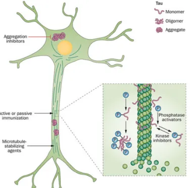
The interaction of Tau and Aβ is a determining factor in the pathogenesis of AD and the therapeutics failures focusing solely on one component of disease strengthen this idea; However, the mechanisms linking Aβ toxicity and Tau hyperphosphorylation remains still obscure (Lloret, Fuchsberger, Giraldo, & Viña, 2015). Considering the important role of Tau, blocking or reducing the pathological effects of this protein may be protective against amyloid pathology (Giacobini & Gold, 2013) different common approaches reducing tau pathology are tau-targeted immunotherapy, modulating axonal transport and microtubule function and promotion of tau degradation (Figure 10).
Figure 10Schematic representation of tau-related processes that are potential targets for therapeutic intervention.
Tau protein exists in neurons in soluble monomeric and oligomeric forms, and as insoluble aggregates (neurofibrillary tangles). Inhibitors of kinases and activators of the phosphatases that mediate this process, therefore, represent possible targets for therapeutic interventions for the treatment of dementia. In addition, anti-aggregation agents could reduce tau aggregation, which is an important step in tau-mediated neuronal damage. Microtubule-stabilizing agents might also have therapeutic potential. Nature Publishing Group Permission (Giacobini & Gold, 2013)
Anti-tau antibodies might block the spread of tau pathology from one neuron to another. As the mechanism of intercellular transmission of tau pathology is not yet fully understood; however, consideration of the process as a valid target for therapy is unanticipated (Giacobini & Gold, 2013).
Findings in transgenic animals carrying tau mutations (LaFerla & Green, 2012) supporting the concept that tau might be a stronger target for therapy tau-targeted interventions particularly anti-tau immunotherapy could reduce tau pathology, facilitate clearance of p-tau, improve cognitive deficits, and delay disease progression (Giacobini & Gold, 2013). TAU knock-out mice established by Harada et al in 1994 does not manifest macroscopic changes (A. Harada et al., 1994). It seems that presence of other MAPs that share several biological roles compensate Tau absence (Dehmelt & Halpain, 2005; Sontag, Nunbhakdi-Craig, White, Halpain, & Sontag, 2012). In term of a therapeutic advantage, it does not seem a complete reduction of Tau be considered as a therapeutic approach because of the compensatory effect of other MAPs (Götz, Xia, Leinenga, Chew, & Nicholas, 2013). Instead, reducing only TAU expression after the development of a pathology seems sufficient to alleviate the pathology (Hochgräfe, Sydow, & Mandelkow, 2013; Van Der Jeugd et al., 2012). Most recent pharmaceutics in test phase is aimed to reduce tau pathology by inhibition of GSK-3 (Tideglusib in phase II) and tau aggregation (TAURx phase II)(Del Ser et al., 2013) (Wischik & Wischik, 2012). It is yet to determine how these therapeutic strategies will work in later clinical phases. It is logical to assume that phosphatase can be more potent targets as it already mentioned earlier, but their activation is yet unlikely to become a therapeutic target for pharmacological intervention due to insufficient data available (Hanger et al., 2009).
1.7. microRNA
MicroRNAs (miRNAs) are short endogenous single-stranded RNA molecules that regulate gene expression (Bartel, 2004). Mature miRNAs and Argonaute (Ago) proteins form the RNA-induced silencing complex or RISC, a ribonucleoprotein complex mediating post-transcriptional gene silencing. Complementary
base-pairing of the miRNA guides RISC to target messenger RNAs, which are degraded, destabilized or translationally inhibited by the Ago protein (Filipowicz, Bhattacharyya, & Sonenberg, 2008).
The first miRNA, lin-4, was reported by Ambros and Ruvkun’s labs in 1993. In the nematode, Caenorhabditis elegans lin-4 activity is required for proper larva development and mutations in lin-14 gene causes an opposite phenotype of the null-lin-4 mutations indicating a negative regulation of lin-14 by lin-4 (R. Lee, Feinbaum, & Ambros, 2004). Ambros found two very small lin-4 transcripts of only 61 nt and 22 nt in length (R. C. Lee, Feinbaum, & Ambros, 1993). Another group at the same time found that lin-14 was downregulated at a posttranscriptional level and that the lin-14 3’UTR region was sufficient for the temporal regulation (Wightman, Ha, & Ruvkun, 1993) which finally resulted in the discovery that lin-4 interacts with 3’UTR of lin-14 transcripts. Since the discovery of these small RNAs thousands of them have been identified in different organisms reported in mirbase database (Griffiths-Jones, Grocock, van Dongen, Bateman, & Enright, 2006).
Biogenesis
Mainly, three categories of small RNAs have been previously described microRNAs (miRNAs), siRNAs and PIWI-interacting RNAs (piRNAs) (Aravin et al., 2006; Friedman, Farh, Burge, & Bartel, 2009; Ghildiyal & Zamore, 2009; Huntzinger & Izaurralde, 2011; Ishizu, Siomi, & Siomi, 2012)
miRNA are generated from a short hairpin RNA by the sequential action of two RNase III-type proteins called Drosha and Dicer (Bernstein, Caudy, Hammond, & Hannon, 2001) piRNA production is not RNaseIII-dependent and is shown to be produced from single-stranded precursors by an endonuclease called Zucchini (mitochondrial cardiolipin hydrolase in humans) and as yet uncharacterized trimming enzymes (M. C. Siomi, Sato, Pezic, & Aravin, 2011). siRNA has 21–23 nucleotides with 3′ two nucleotide overhangs.
The siRNA activates the RNA-induced silencing complex (RISC). The AGO2 component of the RISC cleaves the passenger strand while the guide strand remains associated with the RISC. Subsequently, the guide strand guides the active RISC to its target mRNA for cleavage by AGO2. As the guide strand only
binds to mRNA that is fully complementary to it, siRNA causes specific gene silencing (Agrawal et al., 2003; Pecot et al., 2011). The gene silencing effect of siRNAs and miRNAs is distinct. siRNA inhibit the expression of one specific target mRNA but miRNAs regulate the expression of multiple mRNAs (Figure 11) (Lam et al., 2015).
Figure 11 Comparison of general properties between siRNA and miRNA (Lam et al., 2015) Currently, several thousands of miRNA genes have been characterized in different species available from the miRbase database. miRNA sequences are located within various genomic regions (Figure 12), in general, they can be found clustered or not in both coding and non-coding areas of genome; However, the majority of described miRNAs to date are encoded by introns of noncoding or coding transcripts (Y. Lee, Jeon, Lee, Kim, & Kim, 2002; Olena & Patton, 2010). miRNA genes are transcribed by RNA polymerase II (Pol II)(X. Cai, Hagedorn, & Cullen, 2004), and the long primary transcript has a local hairpin structure where miRNA sequences are embedded. Some endogenous miRNA-like small RNAs are derived from tRNAs that are transcribed by RNA Pol III exceptionally (Babiarz, Ruby, Wang, Bartel, & Blelloch, 2008). In a majority of intronic miRNA cases, the promoter of host genes derives the miRNA expression, but it has been demonstrated in some cases they have multiple transcription start sites and that the promoters of intronic miRNAs are sometimes distinct from the promoters of their host genes (Marsico et al., 2013; Monteys et al., 2010).
Figure 12 Genomic location of miRNAs
(A) Intergenic miRNA. These miRNAs can be monocistronic (top part) with their own promoters
(black arrowhead), or polycistronic, where several miRNAs are transcribed as a cluster of primary transcripts (bottom part) with a shared promoter (black arrowhead). (B) Intronic miRNAs are found in the introns of annotated genes, both protein-coding and noncoding. These miRNAs can be present as a single miRNA (top part) or as a cluster of miRNAs (bottom part). Intronic miRNAs are thought to be transcribed from the same promoter as their host genes (black arrowhead, all parts) and processed from the introns of host gene transcripts. In the special case of mirtrons (middle part), the intron is the exact sequence of the pre-miRNA with splicing sites on either side (denoted by white asterisks). (C) Exonic miRNAs are far rarer than either of the types above and often overlaps an exon and an intron of a noncoding gene. Copyright © 2009 Wiley-Liss, Inc, with permission from Wiley.(Olena & Patton, 2010).
Drosha a nuclear protein of ~160kDa, belongs to a family of RNase III-type endonucleases that act specifically on double-stranded RNA (dsRNA) and initiate the first step of miRNA processing (Y. Lee et al., 2003; Provost et al., 2002). Several labs discovered that the double-stranded RNA-binding protein known as Pasha in flies, or its ortholog DGCR8 in Caenorhabditis elegans and mammals, acts together with Drosha to convert pri-miRNA to pre-miRNA (Han et al., 2006; Yeom, Lee, Han, Suh, & Kim, 2006) these two proteins form microprocessor complex. miRNA stem–loop is cut by Drosha to release a small hairpin structure of ~65 nucleotides in length (pre-miRNA). Additionally, sequence elements such as motifs of UG motif and the CNNC in pri-miRNA sequence affect the processing of human pri-miRNAs (Auyeung, Ulitsky, McGeary, & Bartel, 2013). CNNC motif is required for DEAD-box RNA helicase p72 (also known as DDX17) binding, which increases processing done by Drosha (Mori et al., 2014). After exportation
to the cytoplasm by protein exportin 5 (EXP5; encoded by XPO5) which is GTP-hydrolysis-dependent, Pre-miRNA is cleaved by Dicer (an RNase IIItype endonuclease of ~200kDa) near the terminal loop, liberating a small RNA duplex (Hutvágner et al., 2001). Although most alternative miRNA pathways depend on Dicer, biogenesis of miR-451 does not require Dicer and instead involves the catalytic activity of AGO2 (Cheloufi, Dos Santos, Chong, & Hannon, 2010).
Gene regulation
A small RNA duplex generated by Dicer is loaded onto an AGO protein to form RISC(Kawamata & Tomari, 2010). RNA duplex subsequently is unwinded with the involvement of AGO proteins then the passenger strand is removed. The less abundant passenger strand (or miRNA*) could be also active in silencing. For instance, miR-142-5p is a dominant isoform in ovaries, testes, and the brain, whereas miR-142-3p was found more frequently in embryonic and newborn tissue samples (Chiang et al., 2010).
miRNAs finely regulate translation by binding not only the 3′UTR but also the 5′UTR or both coding regions of target mRNA. While the binding with 3′UTR has been thoroughly investigated in the last decade, the binding mechanism to 5′UTR and the specificity of such a regulation is still not completely understood (da Sacco & Masotti, 2013). A miRNA can inhibit the initiation of translation by limiting the access of mRNA to the translation machine. At the level of post-initiation, miRNA can intervene by premature termination of translation and degradation of the incomplete forming protein and ribosome drop-off (Pillai, Bhattacharyya, & Filipowicz, 2007). mRNA can be consequent, degraded by deadenylation, 5′-terminal cap removal by decapping enzymes DCP1/2 and hydrolysis of the remaining portion of mRNA by 5′→3′ exonucleases (Parker & Song, 2004). The binding of miRNAs to the 5′UTR of target genes has been reported to repress or activate translation (I. Lee et al., 2009; Lytle, Yario, & Steitz, 2007). One classical example of this category is miR-10a that can alleviate the translational repression induced upon amino acid starvation (Meyuhas, 2000). Numerous miRNA’s targets also have been identified being regulated by the interaction of miRNA-coding sequence. One example of this is let-7 miRNA, which directly
targets the miRNA-processing enzyme Dicer within its coding sequence (Forman, Legesse-Miller, & Coller, 2008). The impact of miRNA on coding sequences is not yet fully understood. Multiple pieces of evidence suggest from literature that coding regions of genes can contain additional information besides the amino acid sequence of the encoded protein, including functional microRNA binding sites (Forman & Coller, 2010). miRNAs can target extensively the amino acid coding region of animal mRNAs at locations not necessarily conserved across organisms (Rigoutsos, 2009). A recent study also suggests that mRNA and protein expression of genes containing target sites both in coding regions and 3′UTRs are in general mildly but significantly more regulated than those containing target sites in 3′UTRs only (Fang & Rajewsky, 2011; Schnall-Levin et al., 2011).
miRNA stability and turnover
There are various mechanisms recognized that influence miRNA stability that is reviewed very briefly here but as a general principle, miRNAs is highly stable once they enter RISC because both ends are protected by AGO proteins (Ha & Kim, 2014). For miRNA decay to occur it has been proposed that miRNAs may need to be unloaded first so that exonucleases can access their termini. It remains unclear if and how miRNA unloading takes place or what controls the specificity (Ha & Kim, 2014)
One of the important aspects of miRNA stability resides in the secondary structure of hairpin that may explain variations in the stability of miRNAs (Belter et al., 2014). It has been shown that bases 2-7 of 5′ end of the miRNA are crucial to initiate mRNA binding. For example, 5'-end of miR-296 is knotted in the hairpin stem may explain the high stability of this miRNA (Belter et al., 2014).
Another factor important in miRNA stability is nucleotide polymorphisms that affect miRNA production. Single nucleotide polymorphisms (SNPs) are found in miRNA genes and sometimes affect their biogenesis or change their target specificity (Ryan, Robles, & Harris, 2010). For example, reduced miR-16 expression has been associated with chronic lymphocytic leukemia (CLL due to deletions spanning the intron-containing miR-15a and miR-16-1 (Calin et al.,
2002). Later, a germline C>T single-nucleotide polymorphism (SNP) downstream of the miR-16-1 hairpin was identified that lowered the expression of this miRNA cluster in vitro. This SNP lowers miR-16 accumulation by affecting Drosha-mediated processing and SRp20 splicing factor recruitment (Calin et al., 2005a). There are also numerous processing mechanisms recognized that regulate miRNA biogenesis in nuclear and cytoplasmic level by influencing the miRNA production. The binding of RNA-binding proteins for instance to miRNA precursors, particularly to the stem-loop structures, blocks or enhances further
processing by competing with or recruiting miRNA processing complexes (H.
Siomi & Siomi, 2010). In Table 2 some of the known protein factors that affect miRNA biogenesis have been summarized. The protein factors affect the miRNA at different levels of transcription, processing, maturation and as well direct modulation of Dicer-DGCR8.
Adenylation is another type of RNA tailing that affects the stability of miRNAs (e.g miR-122 in the liver) and occurs mainly after Dicer processing (Katoh et al., 2009). For example, the let-7 family members are post-transcriptionally suppressed in embryonic stages by Uridylation that recruits terminal uridylyl transferases (Heo et al., 2008, 2009). Multiple nucleases have been proposed to cleave and degrade miRNAs. Active degradation of miRNA was initially reported in Arabidopsis thaliana by exoribonucleases that are known as small-RNA-degrading nucleases removes miRNAs (Ramachandran & Chen, 2008). In C. elegans XRN-1 and XRN-2 were shown to degrade mature miRNAs with their 5’ to 3’ RNA degradation (Chatterjee & Grosshans, 2009). In mammals, rapid turnover of several neural miRNAs (miR-183, miR-96, miR-192, miR-204 and miR-211) in the retina has been reported but the mechanism remains unknown (Krol et al., 2010). Recent studies also suggest that highly complementary targets induce miRNA degradation accompanied by tailing and trimming (Ameres et al., 2010).
Table 2 Highlighted protein factors affecting miRNA biogenesis
p53, MYC and myoblast determination protein 1 (MYOD1)
Regulation of the miR-34, miR-17 and miR-1 clusters, respectively. miR-15a cluster. Suppression by myc.
(Yamakuchi & Lowenstein,
2009)(X. Zhang et al., 2012)
ZEB1-2 miR-200 cluster suppression (Brabletz &
Brabletz, 2010) p68, p72, KH-type splicing
regulatory protein (KSRP), heterogeneous nuclear ribonucleoprotein A1 (HNRNPA1) and LIN28
Regulation of pri-miRNA processing (Fukuda et al., 2007)
TAR DNA-binding protein 43 (TDP43)
Promote Drosha and Dicer processing (Di Carlo et al., 2013)(Kawahara & Mieda-Sato, 2012)
ADAR1-2 RNA editing, which interferes with
Drosha processing (Kawahara, Zinshteyn, Chendrimada, Shiekhattar, & Nishikura, 2007)
GSK3β Phosphorylation of Drosha and
nuclear localization of Drosha
(X. Tang, Zhang, Tucker, & Ramratnam,
2010)
ERK Phosphorylation of DGCR8 and
increase in its stability
(Herbert, Pimienta, DeGregorio, Alexandrov, & Steitz, 2013)
MECP2 Dephosphorylation of MECP2
releases DGCR8, which in turn leads to miRNA production and dendritic growth
(Cheng et al., 2014)
SMADs and p53 Promote Microprocessor activity through their interaction with p68.
(Davis, Hilyard, Nguyen, Lagna, & Hata, 2010)(Suzuki et al., 2009) Heterogeneous nuclear ribonucleoprotein A1 (HNRNPA1) and KSRP
Bind to the terminal loop of pri-mir-18a and pri-let-7, respectively, and facilitate Drosha-mediated processing
(Guil & Cáceres, 2007)(Michlewski et al 2008)(Gherzi et al., 2007) miRNA deregulation in AD
miRNA profiling of human cortical tissue identified thirteen miRNAs that are down-regulated in human AD brain (Cogswell et al., 2008; Hébert et al., 2008). The subsequent generation of mice with a neuronal knockout of Dicer is a proof-of-principle that miRNAs could play a role in neurodegeneration (Hébert et al., 2010). This mouse model expresses Cre recombinase mainly in excitatory neurons of the forebrain and demonstrates a neurodegeneration and inflammation with highlighted Tau hyperphosphorylation. Gene expression study in this experiment pinpointed elevated levels of one of the kinases acting on Tau- mitogen-activated protein kinase 3 (MAPK3/ERK1)- which further could be regulated by miR15/16. Importantly, the expression levels of these miRNAs were significantly reduced in 9-week-old Dicer ko (Hébert et al., 2010).
The miRNA network surrounding APP has both upstream and downstream components which directly or indirectly affect APP (Schonrock et al., 2012). A summary of some examples of miRNAs implication in AD has been summarized in Table 3. Several miRNAs have been identified in vitro to directly regulate APP. They include 106a, -520c (Patel et al., 2008) as well as members of the miR-20a family (Hebert et al., 2009), miR-16 and -101 (Long and Lahiri, 2011; Vilardo et al., 2010) and 147, -655, -323-3p and -153 (Delay et al., 2011). While miR-106b and miR-101 have been shown to be down-regulated in AD brain, therefore potentially contributing to increased APP expression and Aβ generation (Hebert