i
Micro-engineered substrates as bone extracellular
matrix mimics
Thèse en cotutelle
Doctorat en génie des matériaux et de la métallurgie
Ibrahim Bilem
Université Laval
Québec, Canada
Philosophiae doctor (Ph.D.)
et
Université de Bordeaux
Bordeaux, France
Docteur
© Ibrahim Bilem, 2016
ii
Micro-engineered substrates as bone extracellular
ma-trix mimics
Thèse en cotutelle
Doctorat en génie des matériaux et de la métallurgie
Ibrahim Bilem
Sous la direction de :
Gaétan Laroche, directeur de recherche
Marie-christine Durrieu, directrice de cotutelle
iii
Résumé
Il est de plus en plus évident que la matrice extracellulaire (MEC), au-delà de sa fonction d’échafaudage cellulaire, génère des signaux de nature biochimique et biophysique jouant un rôle primordial au cours du processus de différenciation des cellules souches. A l’heure actuelle, plus de 15 différents facteurs extrinsèques (environnementaux), incluant l’organi-sation spatiale de la MEC, sa topographie, rigidité, porosité, biodégradabilité et chimie ont été identifiés comme modulateurs potentiels de la différenciation des cellules souches en lignées cellulaires spécialisées. Ainsi, il est plausible que l’intégration d’un biomatériau au sein de l’organisme dépendra largement de sa capacité à mimer les propriétés de la MEC du tissu à remplacer.
Récemment, les techniques de micro-ingénierie ont émergé comme outil innovant pour dé-coupler les différentes propriétés de la MEC et étudier l’impact individuel ou combiné de ces facteurs sur le comportement des cellules souches. De plus, ces techniques de micro-fabrication ont un intérêt particulier dans une perspective de reconstruction de la MEC dans tous ses aspects, in vitro.
Dans ce projet de thèse, le concept de déconstruction/reconstruction de la complexité de la MEC a été appliqué pour récapituler, in vitro, plusieurs aspects inhérents à la MEC osseuse et explorer leurs effets individuels ou combinés sur la différenciation ostéoblastique des cel-lules souches mésenchymateuses (CSMs) humaines.
Trois principales composantes ont été utilisées tout au long du projet : un matériau modèle (verre borosilicate), des séquences peptidiques mimétiques dérivées de la MEC naturelle, favorisant à la fois l’adhérence cellulaire (peptide RGD) et la différenciation ostéoblastique (peptide BMP-2) des CSMs prélevées de la moelle osseuse des patients.
La première étude du projet consiste à greffer, d’une manière aléatoire, les peptides RGD et/ou BMP-2 sur la surface du matériau. Brièvement, nous avons développé trois types de matériaux bioactifs : matériaux fonctionnalisés avec le peptide RGD, matériaux fonctionna-lisés avec le peptide BMP-2 et matériaux bi-fonctionnafonctionna-lisés avec les peptides RGD/BMP-2. La caractérisation physicochimique de ces matériaux a été réalisée en utilisant la spectromé-trie photoélectrique à rayons X (XPS) pour évaluer la composition chimique de la surface, la microscopie à force atomique (AFM) pour évaluer la topographie de la surface et la mi-croscopie à fluorescence pour confirmer la présence des peptides sur la surface et évaluer leur densité. L’objectif de cette étude est d’évaluer le potentiel individuel et synergétique de ces peptides à induire et contrôler la différentiation ostéoblastique des CSMs. Dans un pre-mier temps, la caractérisation physicochimique nous a permis de confirmer l’immobilisation
iv
covalente des peptides sur la surface et de mesurer leur densité. En effet, la densité des pep-tides, mesurée sur les surfaces greffées uniquement avec le peptide RGD ou BMP-2, était de 1.8 ± 0.2 pmol/mm² et 2.2 ± 0.3 pmol/mm², respectivement. Cependant, sur les surfaces bi-fonctionnalisées, la densité de chaque peptide a diminué de presque la moitié, atteignant 0.7 ± 0.1 pmol/mm² pour le peptide RGD et 1 ± 0.1 pmol/mm² pour le peptide BMP-2. Ensuite, l’évaluation biologique des différents matériaux fonctionnalisés a clairement révélé que con-trairement au peptide RGD, le peptide BMP-2 induit la différenciation ostéoblastique des CSMs. Cependant, le greffage simultané des peptides RGD/BMP-2 améliore significative-ment la différenciation des CSMs en ostéoblastes et cela malgré la diminution significative de la densité de chaque peptide sur les surfaces bi-fonctionnalisées, comparativement aux surfaces contenant qu’un seul peptide. Ces résultats montrent que les peptides RGD et BMP-2 peuvent engendrer un effet synergétique pour améliorer la différenciation ostéoblastique des CSMs.
Le second chapitre de thèse vise à déterminer si la microstructuration de la surface des ma-tériaux avec des ligands bioactifs améliore la différenciation ostéoblastique des CSMs. En effet, les peptides RGD et BMP-2 ont été greffés séparément sur la surface du matériau sous forme de micro-motifs de différentes formes mais de taille similaire. En se basant sur des précédents travaux de littérature – discutés dans le chapitre II – nous avons sélectionné trois différentes formes de motifs peptidiques (triangle, carré et rectangle) dont la surface est de 50 µm². Ces micromotifs ont été créées grâce à une technique assez répondue et facile à utiliser qui est la photolithographie. Les surfaces microstructurées ont été caractérisées avec l’interférométrie optique et la microscopie à fluorescence. Les résultats montrent que les micromotifs peptidiques ont à la fois la forme et les dimensions prédéfinies. In vitro, les résultats de différenciation cellulaire ont révélé que la distribution spatiale des ligands à l’échelle micrométrique joue un rôle très important dans l’engagement et la différenciation des CSMs en ostéoblastes. En effet, contrairement aux micromotifs peptidiques en forme de rectangles, les micromotifs triangulaires et carrés améliorent significativement l’expression des marqueurs ostéogéniques (Runx-2 et Ostéopontine) comparativement à la distribution aléatoire des peptides. Il est important de noter que ce profile d’expression des marqueurs biologiques a été observé que sur les matériaux fonctionnalisés avec le peptide BMP-2, tant dis que les matériaux fonctionnalisés avec le peptide RGD n’ont induit aucun effet spécifique sur la différenciation des CSMs et cela peu importe la forme des micromotifs peptidiques. En conclusion, cette étude a permis d’identifier un nouveau facteur extracellulaire capable de contrôler la différenciation des CSMs. De plus, nous avons démontré que la distribution
v
spatiale des ligands à l’échelle micrométrique affecte le devenir des CSMs, dépendamment de la nature du principe actif.
Finalement, la troisième étude de ce projet de thèse est une suite logique de l’étude 1 et 2, puisqu’elle consiste à greffer simultanément les peptides RGD et BMP-2 sous forme de mi-cromotifs. En effet, ces surfaces ont été développées afin de bénéficier à la fois de l’effet synergétique des peptides RGD/BMP-2, observé dans l’étude 1 (facteur 1), et de l’effet de la distribution spatiale contrôlée des ligands, observé dans l’étude 2 (facteur 2). Les diffé-rents types de matériaux ont été caractérisés avec les mêmes techniques de caractérisation de surface mentionnées dans l’étude 2. Les résultats montrent clairement que les surfaces microstructurées sont très bien définies et correspondent à un damier de micromotifs RGD, intercalé par un damier de micromotifs BMP-2. L’évaluation de la différenciation des CSMs sur ces matériaux a révélé que la combinaison des facteurs 1 et 2 améliore significativement la différenciation des CSMs vers le lignage ostéoblastique, comparativement à l’exposition des CSMs à un seul facteur extracellulaire (1 ou 2). De plus, cette étude confirme les résultats obtenus dans l’étude 2, puisque les micromotifs triangulaires et carrés ont permis une meil-leure différenciation cellulaire, comparativement aux micromotifs rectangulaires. Il est important de noter également que l’évaluation biologique des différentes surfaces bio-mimétiques a été réalisée dans un milieu de culture basal qui ne contient pas de facteurs ostéogéniques solubles, afin d’étudier d’une manière assez précise et fiable les interactions des CSMs avec les différents microenvironnements in vitro développés dans ce projet. En conclusion générale, les travaux effectués jusqu’à présent ont permis d’identifier deux aspects de la MEC qui influencent considérablement la différenciation ostéoblastique des CSMs. De plus, nous avons démontré que ces deux facteurs peuvent coopérer pour induire une meilleure différenciation cellulaire. Cela révèle clairement l’intérêt des techniques de micro-ingénierie pour une meilleure et plus profonde compréhension des mécanismes d’in-teractions des cellules souches avec leurs niches, ce qui permettra éventuellement de conce-voir des produits d’ingénierie tissulaire sur-mesure.
Mots clés :
Microstructuration de la surface des matériaux, matrice extracellulaire bio-mimétique, peptides mimétiques, BMP-2, cellules souches, ostéogenèse.vi
Abstract
It is becoming increasingly appreciated that the role of extracellular matrix (ECM) extends beyond acting as scaffolds to providing biochemical and biophysical cues, which are criti-cally important in regulating stem cell self-renewal and differentiation. To date, more than 15 different extrinsic (environmental) factors, including the matrix spatial organization, to-pography, stiffness, porosity, biodegradability and chemistry have been identified as potent regulators of stem cells specification into lineage-specific progenies. Thus, it is plausible that the behavior of biomaterials inside the human body will depend to a large extent on their ability to mimic ECM properties of the tissue to be replaced. Recently, nano- and microen-gineering methods have emerged as an innovative tool to dissect the individual role of ECM features and understand how each element regulates stem cell fate. In addition, such tools are believed to be useful in reconstructing complex tissue-like structures resembling the na-tive ECM to better predict and control cellular functions.
In the thesis project presented here, the concept of deconstructing and reconstructing the ECM complexity was applied to reproduce several aspects inherent to the bone ECM and harness their individual or combinatorial effect on directing human mesenchymal stem cells (hMSCs) differentiation towards the osteoblastic lineage.
Three main components were used throughout this project: a model material (borosilicate glass), ECM derived peptides (adhesive RGD and osteoinductive BMP-2 mimetic peptides) and bone marrow derived hMSCs. All cell differentiation experiments were performed in the absence of soluble osteogenic factors in the medium in order to precisely assess the in-terplay between hMSCs and the different artificial matrices developed in the current study. First, RGD and/or BMP-2 peptides were covalently immobilized and randomly distributed on glass surfaces. The objective here was to investigate the effect of each peptide as well as their combination on regulating hMSCs osteogenic differentiation. The most important fund-ing was that RGD and BMP-2 peptides can act synergistically to enhance hMSCs osteogen-esis.
Then, micropatterning technique (photolithography) was introduced to control the spatial distribution of RGD and BMP-2 at the micrometer scale. The peptides were grafted individ-ually onto glass substrates, as specific micropatterns of varied shapes (triangular, square and rectangle geometries) but constant size (50 µm² per pattern). In this second part of the
pro-vii
ject, the focus was made on investigating the role of ligands presentation in a spatially con-trolled manner in directing hMSCs differentiation into osteoblasts. Herein, we demonstrated that the effect of microscale geometric cues on stem cell differentiation is peptide dependent. Finally, glass surfaces modified with combined and spatially distributed peptides were used as in vitro cell culture models to evaluate the interplay between RGD/BMP-2 crosstalk and microscale geometric cues in regulating stem cell fate. In this study, we revealed that the combination of several ECM cues (ligand crosstalk and geometric cues), instead of the action of individual cues further enhances hMSCs osteogenesis.
Overall, our findings provide new insights into the role of single ECM features as well their cooperation in regulating hMSCs fate. Such studies would allow the reconstruction of stem cell microenvironment in all the aspects ex vivo, which may pave the way towards the de-velopment of clinically relevant tissue-engineered constructs.
Keywords:
Chemical micropatterning, bioactive surfaces, mimetic peptides, BMP-2, mesenchymal stem cells, stem-cell differentiation, stem-cell niche, osteogenesis.viii
Table of contents
Abstract ... vi
Table of contents ... viii
List of tables ... x List of figures ... x Abbreviations ... xii Acknowledgement ... xv FOREWORD ... xviii INTRODUCTION ... 1 I. Physiology of bone tissue ... 2 1. Bone tissue functions ... 2 2. Bone components ... 2
2.1 Cellular components of bone tissue ... 2
2.2 Extracellular matrix of bone tissue ... 4
3. Bone tissue structure: From the macro‐ to nanometer scale ... 4 II. Bone development and repair ... 7 1. Physiology of bone development... 7 1.1. Osteogenesis ... 7 1.2. Calcification ... 9 1.3. Remodeling ... 9 2. Physiology of bone fracture healing ... 10 III. Clinical needs in the bone replacement/regeneration ...11 IV. Strategies for bone regeneration and replacement ...13 1. Bone grafts (auto‐, allo‐, xeno‐grafts) ... 13 2. Biomaterials ... 14 2.1. Biomaterials market ... 14
2.2. Load-bearing biomaterials and their limitations ... 14
2.2.1. Metallic materials ... 15
2.2.2. Bio-inert ceramics ... 16
2.2.3. Polymeric materials ... 17
3. Bone tissue engineering ... 19
3.1. Cell sources for bone tissue engineering ... 22
3.1.1. Mesenchymal stem cells ... 22
ix
3.2. Osteoinductive growth factors ... 24
3.3. Materials used as scaffolds for BTE and their limitations ... 25
3.3.1. Bioactive ceramics ... 26
3.3.2. Biodegradable polymers ... 27
V. Recent advances in biomaterial design to enhance stem cells/progenitors osteogenesis ...29
1. Surface modification strategies for enhanced osteogenesis ... 30
1.1. Physicochemical surface modification ... 31
1.2. Chemical surface modification ... 33
1.3. Biochemical surface modification ... 37
1.3.1. Extracellular matrix derived proteins/growth factors and mimetic peptides ... 38
1.3.2. Integrin ligands and growth factors crosstalk ... 45
1.4. Physical surface modification ... 47
1.5. Topographical surface modification ... 51
1.5. Chemical surface patterning ... 59
RESEARCH PROJECT & OBJECTIVES ... 69
RESULTS & DISCUSSION ... 73
I. Study 1: RGD/ BMP‐2 mimetic peptides act synergistically to enhance hMSCs osteogenic differentiation ... 74 II. Study 2: Microscale geometric cues enhance osteogenic differentiation on BMP‐2‐, but not RGD‐modified surfaces. ... 101 III. Study 3: Microscale geometric cues enhance RGD/BMP‐2 crosstalk‐mediated hMSCs osteogenesis. ... 129
x
List of tables
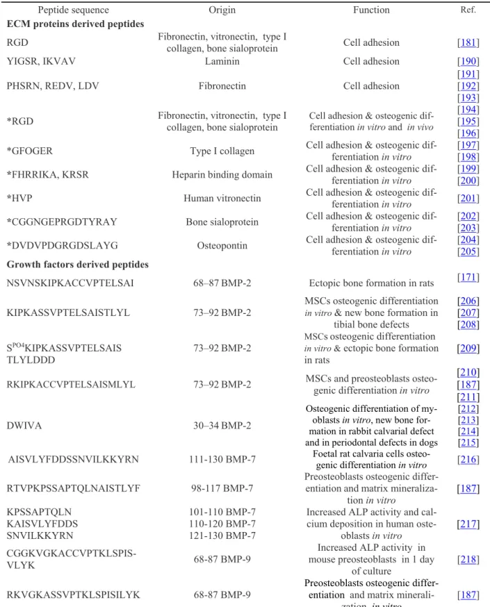
Table 1: Biological effect of peptides derived from ECM proteins and growth factors
covalently immobilized on biomaterials... 41
Table 2: XPS survey analyses of glass surfaces at each step of peptide grafting. ... 91
Table 3: Surface roughness measurements after each step of surface modification. ... 93
Table 4: Measurements of pattern dimensions obtained on X and Y interferometry profiles and compared to the pattern features defined by the photomask. ... 109
List of figures
Figure 1: Schematic summary of the main guidelines of the thesis project ... xxivFigure 2: Bone tissue remodeling. ... 3
Figure 3: Hierarchical structure of human cortical/compact bone ... 6
Figure 4: Endochondral ossification process ... 8
Figure 5: Intramembranous ossification process ... 8
Figure 6: Schema showing evolution of osteoblasts and osteoclasts during bone formation ... 9
Figure 7: Physiological wound healing in bone ... 11
Figure 8: Global potential market of therapeutics and biomaterials for musculoskeletal disease, 2009 and 2014. ... 12
Figure 9: Factors of risk of implants failure that may require revision surgery ... 18
Figure 10: Principle of bone tissue engineering. ... 21
Figure 11: Effect of surface features on cell behavior at the interface bone/ implant ... 31
Figure 12: Light micrographs showing bone ingrowth at (A) uncoated titanium implant and (B) plasma-sprayed hydroxyapatite coating, gaps 1 mm ... 36
Figure 13: Time-line showing few of the most important findings on BMP-2 in biology and in material sciences. ... 44
Figure 14: Hierarchical structure of human thick skin dermis surface over different length scales, from millimeter to micron range. ... 52
Figure 15: Precise replication of pig small intestinal basement membrane using plasma enhanced chemical vapor deposition (CVD) of poly(2-hydroxyethyl methacrylate) p(HEMA) ... 54
Figure 16: Schematic presentation of two typical micropatterning techniques. (a) Microcontact printing (μCP). (b) Microtransfer lithography ... 60
Figure 17: Effect of ligand micropatterning on cell behaviors... 62
Figure 18: CYTOOplates™for High Content Screening assays. ... 68
Figure 19: The potential of micropatterning techniques in reconstituting the in vivo situation ... 71
Figure 20: Schematic of the different steps involved in the grafting of each peptide alone or in combination. ... 84
Figure 21: C1s XPS spectra obtained at each step of RGD-TAMRA and BMP-2-FITC peptide grafting. ... 92
xi
Figure 22: AFM images of the surface topography on different modified glass surfaces. 93 Figure 23: Fluorescent measurements of peptide surface density on different peptide modified surfaces using a standard curve with well-known peptide concentrations ... 94 Figure 24: Osteogenic commitment of hBMSCs after 4 weeks of culture on Bare glass surfaces. ... 96 Figure 25: Fluorescence microscopy images of resist micropatterned surfaces showing three different pattern geometries (Triangle, rectangle, square) with a constant surface area. ... 109 Figure 26: X and Y surface profiles obtained on different resist micropatterned surfaces using optical interferometry. ... 110 Figure 27: Fluorescence images of the different patterned and unpatterned surfaces with RGD-TAMRA (labeled in red) and BMP-2-FITC (labeled in green). ... 111 Figure 28: Fluorescent images of hMSCs cultured for 4 weeks on homogeneous and micropatterned surfaces with peptides. ... 113 Figure 29: Fluorescence images of hMSCs cultured for 4 weeks on glass surfaces
containing spatially distributed BMP-2-FITC peptide ... 115 Figure 30: Quantitative analysis of the total cellular immunofluorescence intensity of STRO-1, Runx-2, and OPN in hMSCs cultured on patterned and unpatterned BMP-2-FITC surfaces. ... 116 Figure 31: Schematic representation of peptide micropatterning onto glass surfaces using photolithography technique. ... 124 Figure 32-S1: Quantitative analysis of the total cellular immunofluorescence intensity of STRO-1, Runx-2, and OPN in hMSCs cultured on patterned and unpatterned TAMRA surfaces. ALP activity measured by colorimetric assay on the different RGD-TAMRA modified materials ... 127 Figure 33-S2: ALP activity measurements on patterned and unpatterned surfaces
containing BMP-2-TAMRA mimetic peptide. ... 128 Figure 34: Schematic representation of peptide micropatterning onto glass surfaces using photo-lithography. ... 138 Figure 35: Optical interferometry 2D maps of the resist micropatterns of varied geometries created onto glass substrates. ... 141 Figure 36: Fluorescence photomicrographs of triangular (a), square (b), rectangular (c) micropatterned glass surfaces containing combined fluoro-tagged RGD-TAMRA (red) and BMP-2-FITC (green) peptides. ... 141 Figure 37: hMSCs cultured for 4 weeks on micropatterned and unpatterned surfaces containing combined RGD/BMP-2 peptides.. ... 142 Figure 38: hMSCs cultured for 4 weeks on micropatterned and unpatterned surfaces containing combined RGD/BMP-2 peptides. ... 143 Figure 39: hMSCs cultured for 4 weeks on micropatterned and unpatterned surfaces containing combined RGD/BMP-2 peptides. ... 144 Figure 40: Some expected mechanisms involved in triggering integrin and growth factor receptors interactions-mediated signaling pathways. ... 153
xii
Abbreviations
ADSCs AFM ALP Al2O3 AR BCP BMPCs BMPR BMSCs BSP CAGR CaP Co-Cr Cr ECM ERK FAK FDA FGF GVHD HA HA hESCs hMSCs HSC HUVEC IGF iPS JNK MAPCs MAPK Ni Nb OCN ON OPG OPN PAA PCL PDGF PDMS PEEK PEG PEO PET PGA PLGA PLAAdipose Derived Stem Cells Atomic Force Microscopy Alkaline Phosphatase Alumina
Aspect Ratio
Biphasic Calcium Phosphate
Blood Mesenchymal Precursor Cells Bone Morphogenetic Protein Receptor Bone Marrow Mesenchymal Stem Cells Bone Sialoprotein
Compound Annual Growth Rate Calcium Phosphate
Cobalt/Chromium Chromium
Extracellular Matrix
Extracellular Signal Regulated Kinase Focal Adhesion Kinase
Food and Drug Administration Fibroblast Growth Factor Graft Versus Host Disease Hydroxyapatite
Hyaluronic Acid
human Embryonic Stem Cells Human Mesenchymal Stem Cells Hematopoietic Stem Cells
Human Umbilical Vein Endothelial Cells Insulin-like Growth Factor
Induced Pluripotent Stem Cells c-Jun N terminal kinase
Multipotent Adult Progenitor Cells Mitogen Activated Protein Kinase Nickel Niobium Osteocalcin Osteonectin Osteoprotegerin Osteopontin polyacrylic Acid Polycaprolactone
Platelet Derived Growth Factor Poldimethylsiloxane Polyetheretherketone Polyethylene Glycol Polyethylene Oxide Polyethylene Terephthalate Polyglycol Acid
Polyglycol Lactic Acid Poly Lactic Acid
xiii PMMA Poly-HEMA PPAR-γ (P(PF-co-EG)) PS PVA PTFE PTH RANK rhBMP RhoA ROCK Runx-2 SAMs α-SMA SS TCP TGF-β Ti TiO2 THR TKA TRAP UHMWPE VEGF WHO XPS ZrO2 Polymethyl Methacylate Poly-hydroxyethylmethacrylate
Peroxisome Proliferator Activated Receptor gamma Poly(propylene furmarate-co-ethylene glycol) Polystyrene
Polyvinyl Alcohol Polytetrafluoroethylene Parathyroïd Hormone
Receptor Activator of Nuclear factor Kappa-B Bone Morphogenetic Protein
Ras homolog gene family, member A Rho-Associated Protein Kinase Runt Related Transcription Factor 2 Self-assembled Monolayers
Alpha-Smooth Muscle Actin Stainless Steel
Tricalcium Phosphate
Transforming Growth Factor-beta Titanium
Titanium Oxide Total Hip Replacement Total Knee Arthroplasty
Tartrate Resistant Acid Phosphatase Ultra High Molecular Weight Polyethylene Vascular Endothelial Growth Factor World Health Organization
X-ray photoelectron spectroscopy Zirconia
xiv
« L’esprit d’équipe ? C’est des mecs qui sont une équipe, ils ont un esprit ! Alors, ils partagent ! » Michel Colucci "Coluche"
«Mice predict the effect on humans with about 43 % efficiency, so sometimes it would seem that tossing a coin would give a better result ! »Dr. Thomas Hartung.
«The more you know, the more you realize how much you don’t know- The less you
know, the more you think you know» David T. Freeman
A ma famille, A mes amis…
xv
Acknowledgement
Je tiens tout d’abord à remercier les membres du jury, Madame Nathalie Faucheux, Monsieur Emmanuel Pauthe et Monsieur Jean-Christophe Fricain d’avoir accepté d’évaluer ce travail à titre de rapporteurs et d’examinateurs et Monsieur Diego Mantovani qui a gentiment ac-cepté d’effectuer la prélecture de ce document. Je vous remercie pour vos appréciations et vos commentaires pertinents, particulièrement Madame Nathalie Faucheux pour vos ques-tions et remarques très instructives sur ce manuscrit de thèse, lors de mon examen predocto-ral et également au congrès mondial WBC 2016. Je remercie également Monsieur René Therrien pour avoir eu la gentillesse de présider cette soutenance.
Je remercie très sincèrement mon directeur de thèse Gaétan Laroche pour m’avoir accueilli si chaleureusement au sein de son laboratoire et m’avoir accordé une très grande liberté quant à l’organisation et l’amélioration du projet au fil des années. Merci pour m’avoir confié la responsabilité sur ce projet à la fois original et passionnant, pour m’avoir laissé proposer des idées et de les appliquer, tout en gardant un œil attentif sur l’avancement des travaux. Je te remercie également pour m’avoir donné l’opportunité de faire des activités très enrichis-santes en parallèle à mon projet de thèse, y compris l’enseignement, la participation à la rédaction d’un chapitre de livre et l’organisation et la participation à des congrès nationaux et internationaux.
Je remercie également ma directrice de thèse Marie-Christine Durrieu pour son accueil au sein de son laboratoire, pour m’avoir donné la chance de travailler sur un projet innovant et d’avoir dirigé de près ce travail. Je te remercie aussi pour ta disponibilité et tout ce que tu m’avais appris.
Thanks Eli Sone for your contribution to the project as a co-supervisor. I really appreciated your very insightful comments and recommendations throughout my thesis. I hope working with you on further challenging projects.
Je remercie les co-auteurs qui ont contribué à la réalisation de ce projet ; Pascale Chevallier et Laurent Plawinski. Je remercie également Andrée-Anne Guay-Begin, Christian Sarra-Bournet et Cedric Ayela pour leurs précieux conseils et pour m’avoir aidé à me familiariser avec des techniques que je n’avais pas utilisées auparavant, XPS, AFM, photolithographie. Je remercie particulièrement et du fond du cœur Pascale Chevallier d’avoir cru en moi de début à la fin, pour son grand soutien et sa grande générosité. Je te remercie énormément
xvi
pour tout ce que tu m’as apporté durant ces années de thèse, professionnellement et person-nellement parlant. T’avais toujours les bons mots et les bons gestes pour me remonter le moral et me redonner le sourire quand ça n’allait pas. Tu fais partie des plus belles rencontres que j’ai faites au Canada et je révérais travailler avec toi dans le future ou du moins travailler avec des personnes avec autant de qualités que toi !!!
Je remercie les organismes subventionnaires, les programmes CRSNG et NCPRM qui ont assumé généreusement les couts de ce projet. Merci également à Karine Fortin, Ginette Ca-dieux, Andrée Lord, Martine Demers et Geneviève Bruneau pour leur travail irréprochable, leur accueil très chaleureux et pour m’avoir beaucoup facilité les démarches administratives et l’établissement de mon statut au Canada.
Je remercie tous mes collègues du coté Canadien qui étaient une deuxième famille pour moi et qui m’ont accueilli les bras grands ouverts. Je m’estime très chanceux d’avoir eu l’oppor-tunité de travailler durant toutes ces années dans un environnement multiculturel où j’ai pu rencontrer des personnes aussi chaleureuses que généreuses de tous les coins du monde. Je dois dire que c’était une expérience humaine extraordinaire qui m’a fait beaucoup gagner en humanité, maturité et ouverture d’esprit. Je remercie Stéphane, Morgane, Marie, Gad, Co-rinne, Michael (encore une fois Pascale et Andrée-Anne; elles méritent !), Farid, Ivan, Au-drey, Stéphanie, Jessie, Max, Sébastien, Carlo, Lucie, Vanessa, Eléonore, Nina, Livia, Ca-roline, Essowè, Ranna, Juliana, Mahrokh, Dimitria, Clayton, Maxime, Olexandr, Francesco et tous les autres sans exception.
Un merci aussi sincère à mes collègues de France ; Laurence, Caroline, Guillaume, Gregor, Marie-Christine, L’équipe de Madame Reiko Oda, Emilie, Sylvain et Marion.
Un merci particulier à Mathieu (le mentor !), Laurence (Roomie), Morgane et Marie (col-lègues de bureau) et Ludivine (et ses histoires super drôles !) avec qui j’ai partagé des mo-ments inoubliables au travail et en dehors du travail.
Je remercie chaleureusement mes amis en France et au Canada et tous ceux qui ont contribué d’une manière ou d’une autre à cet étape de ma vie ; Manal, Akli, Nesrine, Marie-Hélène, Kamal, Enora, Wiame, Sid Ali, Alexandra, Basma, Victoria, Yamanda, Tanu et tous ceux que j’oublie.
Mille merci à Nesrine….. Sans toi j’aurais eu beaucoup plus de difficultés à surmonter les périodes difficiles de ma thèse. T’es tout simplement une personne en or et même si j’écris autant de pages que ce que j’ai écrit dans mon manuscrit de thèse je manquerai certaines de tes qualités. T’es la plus belle rencontre que j’ai faite depuis que je suis à l’étranger et j’es-père que notre amitié durera pour la vie !!!
xvii
J’adresse mes remerciements à mes meilleurs amis d’enfance qui sont toujours restés dans mon cœur et mes pensées malgré les milliers de kilomètres qui nous séparent ; Amine, Ab-dou, Aminou, Mhamed, Nour eddine et Ala. Merci pour votre fidélité, disponibilité, généro-sité et d’être là dans le meilleur et le pire. Pour rien au monde je vous oublierai.
A ma famille…. Ma mère Djamila et mon père Abdelmajid… Je vous dédie ce travail en témoignage de ma profonde reconnaissance, mon grand amour et ma gratitude pour vos sa-crifices. Vous êtes ma fierté, mon exemple dans la vie et ma source d’inspiration et de mo-tivation. Je suis tellement reconnaissant envers vous que j’ai pris la décision en étant encore très jeune de quitter la maison en ayant un seul objectif ; c’est de revenir vous revoir un jour avec un stent qui stabilisera à vie la maladie de mon père…. J’ai travaillé dur et je travaillerai encore plus dur pour y arriver... Je vous aime plus que tout, que dieu vous vous accorde une longue vie. Merci de m’avoir, en plus, donné 3 formidables frère et sœurs ; Amel, Ayoub et la petite merveille Ines. Mon amour pour vous est inconditionnel et je serai toujours là pour vous. Merci ma petite princesse Ines pour le beau gâteau que tu as fait à cette occasion que je n’ai pas eu la chance de le gouter ; tu me manques tellement…
xviii
xix
The use of orthopaedic biomaterials has expanded dramatically during the past decades ow-ing to increased life-expectancy and musculoskeletal diseases, changes in lifestyle and pro-gress in implantology. Several facts reflect the rapid growing of orthopaedic biomaterials prevalence. Babies born today will live almost 3 decades longer than those born in 1900 [1] and almost 7 years than those born in 1960 [2], which reflects an increase in life-expectancy and worldwide population. It was estimated that 90 % of the population over the age of 40 years suffers from a degenerative joint disease [3]. Chronic disability among the elderly has decreased by almost one-third, showing that medical devices have improved both the quality and the length of person’s life [4]. The Bone and Joint Decade organization has been launched in 2000, remandated in 2010 and endorsed by the United Nations and the World Health Organization (OMS) [5]. Last decade’s estimates showed that orthopaedic implants are the bulk of all implanted devices worldwide and will remain the largest segment in bio-materials market [6] [7].
Although these facts and statistical data highlight the upsurge in the demand of orthopedic biomaterials as well as their substantial contribution in improving the life and comfort of patients, the acceptance of these implants by the human body is far from being trivial. In addition, the consequences of implant rejection are sometimes more dramatic than what un-treated damaged bone do.
Currently, there are several clinical needs that are not satisfactory filled in orthopaedic, spi-nal, dental, cranial and maxillofacial surgery. The work presented here, although being in its infancy, addresses two main issues. The reader will be introduced to the thesis project chal-lenges by means of a set of questions that span from the current clinical needs in orthopaedics to the approach proposed in this research project as a promising way toward the resolution of the addressed clinical issues.
xx
What are the main unmet clinical needs in orthopaedic surgery?
Clinical need 1: The long term performance of orthopaedic biomaterials, especially used in load bearing parts, given that their lifespan is still limited to 10-15 years after implantation [8].
Clinical need 2: The reconstruction of large bone defects caused by diseases, non-union fractures or tumor resection.
Why is the long term performance of commercially available orthopaedic bio-materials limited (Clinical need 1)?
One key raison for their limited performance is the lack of a robust integration of implanted biomaterial with the host bone tissue, often due to the formation of a fibrous layer at the interface bone/implant.
Why large bone defects reconstruction is still a challenging task (Clinical need 2)? This is due to the low potential of currently used biomaterials in clinics to induce mesenchy-mal stem cells (MSCs) and progenitor cells migration to the defect site and ensure their differentiation into mature bone cells. Consequently, the implant is poorly colonized by bone cells, resulting in a weak host bone-to-implant contact.
One promising approach to enhance the biological compatibility of biomaterials is to com-bine them with the patient’s MSCs, differentiated ex-vivo, prior to implantation. Neverthe-less, this strategy has limited impact nowadays because MSCs can be harvested from the human body only in few amounts [9]. In addition, ex-vivo expansion and differentiation of stem cells are not only time-consuming protocols but also fail to produce clinically relevant amounts of osteogenic cells.
How the underlying clinical needs could be met?
From a rational point of view, the most effective and reliable approach to circumvent the underlying concerns is to mask the bio-inertness of biomaterials used as cell culture plat-forms or orthopaedic implants by creating on their surfaces an artificial extracellular matrix (ECM). This ECM should mimic the in vivo microenvironment features that mediate the switch of MSCs from their stemness state into an osteoblast lineage.
xxi
While creating a biomimetic microenvironment on biomaterial surfaces seems to be a good idea to overcome the above-mentioned issues, translating this idea to bone tissue-engineered product is extremely complex, owing to the complexity of the native ECM. Just 40 years ago, the ECM was considered as an inert scaffold, providing only a supportive environment on which cells can attach [10]. To date, thanks to increased investments, funding and grants in biomaterials and stem cell research [4], the ECM is recognized to do more than just sup-port the cells, as over 15 different factors have been shown to influence MSC fate. Among these ECM features, physical properties, including stiffness, topography and porosity, and biochemical cues, including ligands density, spatial distribution and combinatorial effects have been extensively studied during the last decade [11] [12] [10]. All these ECM aspects are interlinked and can act independently or in concert to control MSCs fate decision in vivo, hence the difficulty of mimicking ECM features in vitro. Therefore, deconstructing the com-plexity of the native ECM and harnessing the interplay between MSCs and individual ECM cues will undoubtedly pave the way towards the reconstruction of finely-tuned artificial MSC microenvironments for bone tissue engineering applications.
In this regard, the studies illustrated in this manuscript provide new insights into the potential role of the native ECM features, especially biochemical cues, in directing human MSCs (hMSCs) osteogenesis. Two aspects innate to the natural microenvironment and expected to drive hMSCs osteogenic differentiation in vivo will be addressed: (1) the cooperation be-tween integrin ligands and growth factors and (2) the spatial distribution of ECM adhesive ligands and growth factors at the micrometer scale. A schema providing an overview of the general methodology of the thesis project is depicted in Figure 1.
In the first stage, cell adhesive RGD and osteoinductive BMP-2 mimetic peptides were co-valently immobilized onto a model material (glass substrates). Three different conditions were prepared; glass surfaces homogeneously functionalized with RGD, BMP-2 or com-bined RGD/BMP-2 peptides (Paper I). The objective here was to investigate the effect of each peptide on hMSCs fate and whether or not these peptides can act synergistically to enhance osteogenesis, when combined. Also, the stimulatory effect of BMP-2 was correlated to its surface density. The peptide grafting was ascertained using complementary physical-chemical techniques, X-ray photoelectron spectroscopy (XPS), atomic force microscopy (AFM) and fluorescence microscopy. Cell differentiation analyses revealed that BMP-2 pep-tide induced hMSCs osteogenic differentiation and the presence of RGD peppep-tide improved
xxii
its osteoinductive capacity, even in the absence of soluble osteogenic factors in the cell cul-ture medium. Thereby, the study described in the paper 1 highlighted the existence of a cer-tain crosstalk between ECM derived adhesive ligands and growth factors in regulating stem cell fate.
Subsequently, by seeking to harness the potential presentation of ECM derived ligands in a spatially controlled manner, which somewhat mimics the in vivo situation, RGD and BMP-2 peptides were finely distributed over glass surfaces at the micrometer scale, using micro-patterning strategy (photolithography) (Paper II). To this end, the peptides were grafted as specific micropatterns of varied shapes (triangle, rectangle and square) but constant overall area (50 µm²) in order to evaluate the effect of geometric cues on hMSC fate decision. Pep-tide micropatterns were assessed for their shape, size and reproducibility by optical micros-copy, interferometry and fluorescence microscopy. hMSCs cultured on the different micro-patterned surfaces exhibited different cell behaviors in relationship with the pattern shape and the type of patterned ligand. In fact, on RGD micropatterned surfaces, geometric cues did not affect hMSCs osteogenic differentiation as the expression of osteogenic markers was very low and similar between homogenous and micropatterned surfaces functionalized with RGD peptide. In contrast, the effect of geometric cues was clearly visible on BMP-2 surfaces containing BMP-2 peptide. That is, osteogenesis was significantly enhanced on triangular and square BMP-2 micropatterns as compared to the rectangular ones. Thus, we evidenced throughout this set of experiments (Paper II) that micro-scale geometric cues, when carefully selected, can effectively dictate hMSCs specification towards the osteoblastic lineage. The third challenge in this thesis has raised by considering the insightful knowledge gained from the studies 1 and 2. By seeking to partially recapitulate ECM cues that dictate the switch in lineage differentiation from MSCs into osteoblasts in vivo, we created artificial ECMs where both peptides crosstalk and geometric cues signaling could be triggered to regulate MSCs osteogenesis. Therefore, this biomimetic microenvironment consisted of a checker-board of juxtaposed RGD and BMP-2 micropatterns (Paper III). The patterns were similar in shape and size to those used in the study II and characterized using the same techniques. Quite consistent with the findings reported in the study II, in vitro analyses confirmed again that triangular and square micropatterns are of potential relevance in directing hMSCs fate towards the osteoblastic lineage.
By combining results from these three studies, we were able to evaluate to which extent triangular and square RGD/BMP-2 micropatterns affect hMSCs osteogenic differentiation. Study III VS study II revealed that the spatial distribution of combined RGD/BMP-2 peptides
xxiii
as triangular and square micro-sized geometries enhanced hMSCs osteogenesis as compared to their homogenous distribution on the material surface. Study III VS study II demonstrated that triangular and square RGD/BMP-2 micropatterns improved hMSCs osteogenesis as compared to triangular and square BMP-2, respectively. Taken together, these findings sug-gest that integrin ligands/growth factors crosstalk and geometric cues are not only potent modulators of hMSCs osteogenic differentiation but can also overlap to further enhance lin-eage-specific differentiation.
The studies achieved in this thesis project provide new insight into the stem cell-ECM inter-actions that are likely to contribute to the design of finely-tuned biomaterials capable to meet the current clinical demand in orthopaedic surgery.
xxiv
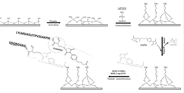
Axis 1.
Strategy of the covalent immobilization of RGD and BMP-2 peptides on glass sur-faces. For the homogenous peptide grafting, SMPB-NH2-glass surfaces were directly cov-ered with RGD and/or BMP-2 solution. However, the peptide micropatterning required the structuration of SMPB-NH2-glass surfaces using photolithography prior to peptide grafting.Axis 2.
Microscale patterning of RGD and/or BMP-2 peptides using photolithography.NH2 NH2 NH2 Piranha Activation NH2 (CH2)3 Si(OEt)3 Aminosilylation NH NH2 NH NH NH2 NH2 SMPB SM P B gr af tin g Peptide immobilization RGD-TAMRA BMP-2-mp-FITC APTES SMPB-Glass SMPB-Glass SMPB-Glass SMPB-Glass UV UV SMPB-Glass Photoresist Surface coated with photoresist
The photoresist coated surface after exposure
After development, the resist pattern is observed
Grafting of the first
peptide « BMP-2 » Grafting of the second peptide « RGD» RGDmicropatterns /BMP-2 peptides
1
2
3
Photoresist
xxv
Some details regarding the articles achieved during the three years of the thesis as well as the role of the co-authors in the accomplishment of this work are given bellow.
Paper 1:
Title: RGD and BMP-2 mimetic peptide crosstalk enhances osteogenic commitment of hu-man bone marrow stem cells.
Authors: I. Bilem, P. Chevallier, L. Plawinski, E.D. Sone, M.C. Durrieu, G. Laroche. Journal: Acta Biomaterialia, 36 (2016) 132-42.
State: Submitted 2015, Dec 8th. Published 2016, Mar 18th .
Author’s contribution: The first author designed the experiments based on the instructive discussion with all the authors. The first author performed all the experiments, analyzed and interpreted the data and drafted the manuscript. P. Chevallier characterized most of the sam-ples under XPS and significantly contributed to the interpretation of XPS data. All the au-thors reviewed the manuscript.
Paper 2:
Title: The spatial distribution of RGD and BMP-2 mimetic peptides at the subcellular scale modulates of human mesenchymal stem cells osteogenesis.
Authors: I. Bilem, L. Plawinski, P. Chevallier, E.D. Sone, G. Laroche, M.C. Durrieu. Journal: ACS Nano.
State: Submitted 2016, Mar 19th.
Author’s contribution: The first author designed the experiments based on the instructive discussion with all the authors. The first author performed all the experiments, analyzed and interpreted the data and drafted the manuscript. The difference between the submitted article and that inserted in the thesis is that ALP results are provided as supplementary data only in the thesis manuscript. L. Plawinski collected the raw ALP data and converted them into ALP values (pmole). All the authors reviewed the manuscript.
xxvi Paper 3:
Title: Interplay of geometric cues and RGD/BMP-2 crosstalk in directing stem cell fate. Authors: I. Bilem, P. Chevallier, L. Plawinski, E.D. Sone, M.C. Durrieu, G. Laroche. Journal: Biomaterials.
State: Submitted 2016, Apr 13th.
Author’s contribution: The first author designed the experiments based on the instructive discussions with all the authors. The first author performed all the experiments, analyzed and interpreted the data and drafted the manuscript. All the authors reviewed the manuscript.
1
2
I. Physiology of bone tissue
Bone is a complex organ composed of several tissues working together: osseous tissue, cartilage, dense connective tissues, epithelium, adipose tissue and nervous tissue. Bone tissue is a highly specialized and dynamic living tissue that contributes significantly to the homeostasis of the body. It continuously grows and repairs itself since it is subjected to a regular process of breaking down of old bone and building of new bone tissue. This turnover is called bone remodeling [13].
1. Bone tissue functions
Bone tissue plays an important role within the body, both biomechanically as metaboli-cally. It has three main functions:
Mechanical function. Since the bone has high mechanical properties, it plays an im-portant role in supporting the body and protecting organs, nervous system and stem cells in the marrow.
Metabolic function. Bone plays a vital role in maintaining mineral homeostasis. It stores and releases several minerals, especially calcium and phosphate, which contribute to the bone strength.
Hematopoietic function. Embryonic bone and some adult bones such as hip bones, ribs, breastbone, vertebrae (backbones) and skull contain hematopoietic stem cells which are responsible for generating blood cells (red and white blood cells and platelets).
2. Bone components
Bone is a connective tissue that comprises various cell types entrapped in a mineralized extracellular matrix (ECM), occupying about 90% of the tissue volume and conferring rigidity and strength to the skeleton while still maintaining some degree of elasticity.
2.1 Cellular components of bone tissue
Bone tissue is composed of four different cell types, osteoblasts, osteocytes, osteoclasts and bone-lining cells [13] [14].
Osteoblasts. They originate from the differentiation of MSCs that reside in the perios-teum, endosteum and bone marrow [15]. MSCs maintain their stemness until they are stimulated (due to injury or bone development) to gradually become mature osteoblasts. Osteoblasts express different osteogenic factors, including alkaline phosphatase (ALP), type I collagen, osteopontin (OPN), osteocalcin (OCN) and bone sialoprotein (BSP)
3
which are involved in the formation of an organic bone matrix (osteoid) and its mineral-ization (Figure 2).
Osteocytes. These cells are osteoblasts that have reached a high level of maturation. They are surrounded by a mineralized bone matrix and have the capacity not only to synthesize, but also to resorb the bone matrix to a limited extent, thus regulating bone remodeling process. Osteocytes are also responsible for the transmission of mechanical and biochem-ical stimuli, essential for bone metabolism.
Osteoclasts. They are giant multinucleated cells (diameter ≈ 100 microns) derived from the self-fusion of macrophages. These macrophages (posteoclasts) express RANK re-ceptor (Rere-ceptor Activator of Nuclear factor Kappa-B) and then merge under the activa-tion of (RANKL) ligand, which leads to their differentiaactiva-tion into multinucleated active cells (osteoclasts). The main function of osteoclasts is to digest the old mineralized bone matrix by releasing tartrate-resistant acid phosphatase, cathepsin K, matrix metallopro-teinase 9 and gelatinase [16] (Figure 2).
Bone-lining cells. These cells are flat, elongated and inactive. They cover bone surfaces that undergo neither bone formation nor resorption. Their principle function is to protect bone matrix against the osteoclast action. However, during bone remodeling, these cells degrade the osteoid by secreting collagenases. Bone-lining cells are also a source of os-teoblasts. Indeed, under the action of various stimuli, such as parathyroïd hormone (PTH), they can dedifferentiate into active osteoblasts.
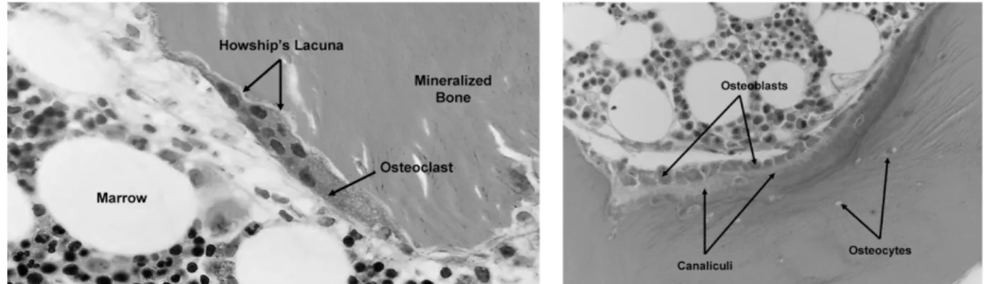
Figure 2: Bone tissue remodeling. Osteoclasts resorb bone to form resorption pits known as Howship's lacunae (left). Osteoblasts synthesize the inorganic matrix (osteoid), rich in type I collagen, to fill in resorption pits (right). The osteoid is gradually mineral-ized to form new bone [14].
4
2.2 Extracellular matrix of bone tissue
Bone ECM is made of non-mineralized organic components (predominantly type I colla-gen) and mineralized inorganic components (mineral crystals) [17].
Organic matrix. Type I collagen constitutes approximately 80-90% of the organic ma-trix. It confers bone tissue its hierarchical structure and ensures its viscoelasticity. The remaining 10-20 % correspond to other types of collagen (collagen type III, V, X) and over 200 different non-collagenous proteins such as proteoglycans, osteonectin (ON), BSP, OCN, OPN and ALP, all of them contribute to the maintenance of bone tissue. Other molecules have a direct action on the activity of bone cells, such as Bone Morphogenetic Proteins (BMPs), known to induce the differentiation of osteoblast precursors or osteo-protegerin (OPG) glycoprotein that inhibits the differentiation of osteoclast precursors. Organic matrix also comprises specific proteins involved in bone remodeling and vascu-lature, such as TRAP (tartrate-resistant acid phosphatase) and VEGF (Vascular Endothe-lial Growth Factor), respectively [16] [18].
Mineral matrix. About 50-70% of the weight of bone tissue corresponds to mineral ma-trix that ensures its rigidity. The mineral content in bone is mostly a crystalline calcium phosphate deposited in thin apatite platelets, with small amounts of carbonate, magne-sium, and acid phosphate. Bone mineral is initially deposited in the form of hexagonal apatite nano-crystals in small gaps generated by the regular stacking pattern of collagen molecules in a fiber [19]. As the hexagonal apatite nano-crystals mature they take on a plate shape. Although this process is mediated by bone cells and the organic phase, it may also be facilitated by ECM vesicles in bone in which calcium and phosphate concentra-tions can increase sufficiently to precipitate apatite crystals [20].
3. Bone tissue structure: From the macro- to nanometer scale
The squeleton is made up of more than 206 different bones that can be classified depend-ing on their shape into short, flat and long bones. As with all organs in the body, bone tissue is hierarchically organized over length scales that span several orders of magnitude from the macrometer scale to the nanometer scale [21] [22] [23] (Figure 3):
- At the macrometer scale, bone is made of 80% of cortical (compact) bone and 20% of trabecular (cancellous) bone. Cortical bone is much denser and less porous (5% to 30 %) than trabecular bone (30% to 90%). Cortical bone forms the outer shell (cortex) of most bones and provides mechanical and protective functions, while trabecular bone is typi-cally found at the ends of long bones (epiphysis) and provides metabolic functions.
5
- At the micrometer scale (10 to 100 µm), osteon and trabeculae are the anatomical and functional units of cortical and trabecular bone, respectively.
- At the sub-micrometer scale (1 to 10 µm), an osteon is organized in concentric flat sheets of mineralized collagen fibers, called lamellae. An osteon or Haversian system consists of a set of 8-15 lamellae surrounding the Haversian canal. In cortical bone, lamellae are highly organized and oriented in parallel to the longitudinal axis of bone, while in trabec-ular bone their arrangement is irregtrabec-ular.
- At the nanometer scale (~100 nm to 1 µm), mineralized collagen fibers of ~200 nm in diameter are the structural unit of bone tissue.
- At the sub-nanometer scale (˂100 nm), the main components are the apatite nano-crys-tals, type I collagen proteins and non-collagenous proteins. Apatite nano-crystals exhibit typical average dimensions of 50 x25x3 nm. Depending on their maturity, their dimen-sions can vary from 15 to 150 nm in length, 10 to 80 nm in width and 2 to 5 nm in thickness. On the other hand, triple helices collagen molecules have average dimensions of 200 nm in length and 2–3 nm in diameter.
6
7
II. Bone development and repair
1. Physiology of bone development
During life, bone undergoes processes of longitudinal and radial growth, modeling (re-shaping) and remodeling, as described below [25].
1.1. Osteogenesis
Normal bone develops through two mechanisms that can act independently or together during bone formation: endochondral and intramembranous ossification [26] [27] (Fig-ure 4) (Fig(Fig-ure 5).
The process of endochondral bone formation involves different phases of cell prolifera-tion, differentiaprolifera-tion, migration and ECM remodeling. This begins with the differentiation of MSCs into chondrocytes that synthesize a hyaline cartilage model, rich in type II col-lagen, whose shape resembles a small version of the bone to be formed [28]. Gradually, chondrocytes in the primary ossification center -in the middle of diaphysis- grow, differ-entiate into hypertrophic chondrocytes and begin secreting ALP, thus allowing for the calcification of the cartilaginous matrix. Simultaneously, a vascularized periosteal bone -rich in osteoprogenitor cells that later become osteoblasts- appears around the diaphysis of the hyaline cartilage model. At this moment, hypertrophic chondrocytes (before apop-tosis) secrete VEGF, leading to blood vessels sprouting from the periosteal bone to the primary ossification center. Blood vessels, forming the periosteal bud, invade cavities left by apoptotic chondrocytes, thus carrying hematopoietic cells, osteoprogenitor cells and other cells inside the cavities. While hematopoietic cells will later form the bone marrow, osteoprogenitor cells specialize into osteoblasts that form osteoid over the calcified car-tilage [26]. Subsequently, the primary ossification center progresses in the direction of the epiphysis, leading to a secondary ossification center. Osteoprogenitor cells invade epiphyseal cartilage, differentiate into osteoblasts and secrete osteoid onto the cartilage matrix -cartilage tissue remains in two places: articular cartilage and epiphyseal plate- [29]. Finally, a woven bone (immature bone) is formed which will be replaced by lamellar bone at the next steps of bone development [27].
The bone development via endochondral process occurs in initial bone formation in an embryo and fetus as well as in bone growth during infancy, childhood and adolescence. Indeed, it is involved in growth in length of the most bones in the body, mainly long bones such as femur, tibia, humerus and radius [26].
8
Unlike the endochondral ossification, the intramembranous ossification does not involve a cartilaginous tissue formation. This process begins by the aggregation of MSCs into layers at specific regions of a highly vascular connective tissue, called center of ossifica-tion. MSCs proliferate, condense around a profuse capillary network and differentiate into osteoblasts. The latter secrete organic matrix (osteoid), get surrounded by collagen fibers and transform into osteocytes. At this stage, the collagen fibers of osteoid form a woven bone that gradually thickens. Eventually, woven bone is remodeled and replaced by la-mellar bone [26] [27]. The intramembranous ossification mainly occurs in embryogenesis during formation of the flat bones such as skull, mandible, maxilla and clavicles [26].
Figure 4: Endochondral ossification process. (a) Aggregates of osteoprogenitor cells (b) Model of hyaline cartilage (c) Primary center of ossification (d) Secondary center of ossification (e) Bone with medullary cavity and epiphyseal ends (f) Highlighting feeding blood vessels [26].
Figure 5: Intramembranous ossification process. (a) Aggregates of mesenchymal stem cells (b) Amorphous ground substance and collagen network formed in the center and between the cells. (c) Mesenchymal stem cells transform to osteoblasts which synthesize organic matrix (osteoid) in the center of the aggregate. (d) Organic matrix mineralization and the transformation of some osteoblasts incorporated within the osteoid into osteocytes [26]. Secondary os-sification center Chondrocyte hypertrophy f a b c d e
9
1.2. Calcification
This stage of bone development involves the calcification of woven bone previously formed osteogenesis process. The mineralization of woven bone occurs 24-74 h after or-ganic matrix synthesis, through two main steps: nucleation of calcium phosphate crystals and crystal growth [20]. This results in the precipitation of calcium phosphate, the for-mation of small apatite nano-crystals and their growth along the collagen fiber axis under the effect of ALP and several non-collagenous proteins, including as OCN, OPN, ON and BSP [25] [30].
1.3. Remodeling
Bone remodeling ensures the transformation of woven bone to mature lamellar bone. This process is a lifelong phenomenon that permits the maintenance of bone tissue, the repair of damaged tissue and the homeostasis of the phosphocalcic metabolism. It is achieved by the combination of bone resorption/formation process (Figure 6). Bone resorption in-volves removal of an old bone while bone formation inin-volves the synthesis of a newly organic matrix and its subsequent mineralization to form new bone which replaces the removed one. Thus, approximately 5–10 % of total bone is renewed per year [13].
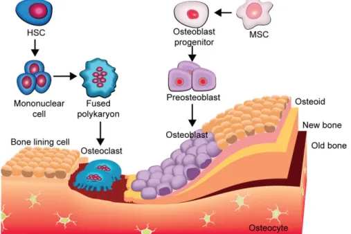
Figure 6: Schema showing evolution of osteoblasts and osteoclasts during bone for-mation. HSC: Hematopoietic Stem Cells [31].
10
2. Physiology of bone fracture healing
Bone fracture repair is similar in many ways to pre- and post-natal bone development. For example, fractures heal via the endochondral ossification when the fracture is not really stabilized, as in the case of fractures treated by cast immobilization [26] [27]. On the other hand, the intramembranous ossification is involved in the repair of bone frac-tures of size between 0.25 and 0.5 mm, stabilized by metal plate and screws [26] [32]. However, in contrast to the natural bone development, bone healing begins with an flammatory reaction due to bone injury caused by a trauma or surgical procedure to in-troduce an implant or bone graft (Figure 7) [33].
First, vascular lesions result in the formation of blood clot and granulation tissue as well as the accumulation of platelets. Platelets start then to secrete cytokines and growth fac-tors, thus triggering an inflammatory response that manifests itself by the migration of leucocytes, lymphocytes and monocytes to the site of injury. Among cytokines and growth factors that play an important role during fracture repair are Interleukins-1 and -6 (IL-1 and IL-6) and TNF-α as pro-inflammatory cytokines, BMPs -mainly 2, BMP-4 and BMP-6- as osteoinductive factors and VEGF as angiogenic factors. The involve-ment of the underlying regulators during the different stages of fracture healing is well reviewed in [34].
Then, the repair process begins when MSCs, from the periosteum, endosteum and bone marrow, migrate to the lesion area, proliferate and differentiate to cover the blood clot [35]. If the fracture is mechanically stable, MSCs differentiate into osteoblasts that even-tually ensure the production of organic matrix and its mineralization. If the fracture is unstable, MSCs differentiate into chondrocytes that secrete a cartilaginous matrix, called fracture callus, to temporarily stabilize the fracture [28] [36]. The cartilaginous matrix is then replaced by woven bone via endochondral ossification.
Finally, the vascularization of newly formed tissues permits the resorption of woven bone, which is gradually replaced with more resilient lamellar bone during the remodeling phase.
Given that fracture healing requires the migration of inflammatory cells and MSCs to the site of lesion, bone repair takes place only when the fracture gap is too small. However, in the case of critical-size bone defects, the recourse to bone grafts and biomaterials is usually advocated.
11
Figure 7: Physiological wound healing in bone [33]
III. Clinical needs in the bone replacement/regeneration
Trauma has been recognized as a major healthcare epidemic by the World Health Organ-ization (WHO), with over 16, 000 people die each day and injury accounting for 16 % of the global burden of disease [37]. Injuries affecting the musculoskeletal system are the most common and hence significantly contribute to the increase of musculoskeletal dis-ease prevalence, that is already affecting roughly 20% of the population [38]. This has been endorsed by the United Nations and WHO, as they recognized musculoskeletal dis-ease as a major burden on individuals, health systems and social care systems with a high financial impact [39]. For instance, osteoporosis is a major risk factor for fractures of the hip, vertebrae, and distal forearm [40], resulting in more than 8.9 million fractures around the world annually; i.e. an osteoporotic fracture every 3 seconds [41].
Among these skeletal fractures, many heal spontaneously in the first 6 to 8 weeks and require short-term and low-cost treatment. However, a fracture can be clinically consid-ered as a delayed union or nonunion if no bony healing is observed after 4 months and 6 months, respectively. Such fractures has been estimated at 100,000 annually in the United
12
States [42]. They are difficult and slow to heal and significantly harm the national econ-omy, with respect to medical resources (hospitalization, medical equipment, medical im-plants, diagnostic tests, outpatient follow-ups, therapies and drugs) [43].
Overall, bone injuries, aging population and lifestyle factors, such as obesity are unques-tionable risk factors that drastically accentuate musculoskeletal disease burden and con-sequently boost the demand for the orthopaedic devices market. In term of costs, the global market of therapeutics and orthopaedic biomaterials for musculoskeletal disease has approached $45 billion in 2010 with a compound annual growth rate (CAGR) fore-casted to be about 5 %, bringing the total market to more than $57 billion by 2014 (Figure 8) [38]. Therefore, medical approaches that help to accelerate bone healing in critical-size bone defects will not only improve medical outcomes for the patient, but they will also contribute to reduce the financial burden related with musculoskeletal disease.
Figure 8: Global potential market of therapeutics and biomaterials for musculoskeletal disease, 2009 and 2014 [38]. 2009 2014 Osteoarthritis Lupus Osteoporosis Synthetic bone grafts Rheumatoid arthritis Gout Psoriatic arthritis Ankylosing spondylitis Bone substitutes Juvenile idiopathic arthritis 20,000 18,000 16,000 14,000 12,000 10,000 8,000 6,000 4,000 2,000 0
$ Mi
ll
io
ns
13
IV. Strategies for bone regeneration and replacement
Among approaches currently employed for the treatments of bone injuries are the excision of the fibrous tissue formed at the bone defect site, the use of internal and external skeletal fixation devices for fractures stabilization or the recourse to bone grafts and orthopaedic biomaterials to replace or restore damaged bone.
1. Bone grafts (auto-, allo-, xeno-grafts)
As previously highlighted, the high regenerative capacity of bone tissue ensures a natural facture healing in small bone defects. Unfortunately, diseases such as osteogenesis im-perfecta, osteoarthritis, osteomyelitis, along with fractures and traumas as well as tumor resections may lead to critical-size defects (gap size beyond 2-2.5 times the radius of the affected bone) that require surgical intervention to restore or replace lost bone [44]. Cur-rently, the gold standard treatment is the use of autologous bone graft, taken from another part of the patient’s own body. These grafts integrate reliably with the host bone, lack the immune-related complications and provide osteogenic cells as well as osteoinductive fac-tors needed for bone healing and regeneration [44] [45]. Nevertheless, the use of this strategy is mainly hampered by the limited supply of autologous bone and the risk of necrosis at the donor site.Allograft (i.e. bone from a human cadaver) and xenograft (i.e. bone from an animal source) represent an alternativesince larger bone grafts could be provided. However, these grafts should be sterilized, which leads to the loose of osteoin-ductive factors and living cells. In addition, they present a potential risk of viral and bac-terial infections and immune rejection after implantation [45].
Although these grafts are of great interest in reconstructive orthopaedic surgery, their potential to repair large bone defects is limited and, as consequence, an incomplete graft/host tissue osseointegration was observed in several clinical cases [46] [47].
14
2. Biomaterials
2.1. Biomaterials market
Owing to the pressing clinical need in orthopaedics highlighted above, the market of bi-omaterials-based treatments is growing at a rapid rate. Predominantly based in North America, the biomaterials market is expected to be worth $88.4 by 2017 and is forecasted to increase at a CAGR of 16% to reach $130.57 Billion by 2020. Several factors contrib-ute to the growth of the overall market, including increased funds & grants by government bodies worldwide, technological advancements, population ageing and the growth of the implantable devices market (www.marketsandmarkets.com).
Specifically, the global orthopaedic devices market was valued at $34.9 billion in 2014 by Frost & Sullivan's research and at $57.9 billion in 2016 by marketsandmarkets. The North America is the largest orthopaedic market, especially the United States that stands as the leadership with its 60 % of contribution in the market. Indeed, orthopaedic bio-materials are the most implanted bio-materials, especially load-bearing implants such as arti-ficial hip and knee joints and fixation devices. These have been designated as “the ortho-pedic success story” by the American Academy of Orthopaedic Surgeons, representing 52% of all implantations [48].
2.2. Load-bearing biomaterials and their limitations
During the last decades, many bone substitute materials have been evaluated with the aim of resolving the need for autologous or allogenic grafts. The strength of implantable ma-terials is their large availability, safety (no potential diseases transmission), handling characteristics as well as the possibility to incorporate drugs and bioactive molecules in the bulk or the surface of materials.
The choice of a suitable material, exhibiting a high level of biocompatibility, is crucial to successfully replace or support bone repair. Actually, materials used in load-bearing parts are often made of metals or ceramics due to their resistant to the load and their high frac-ture toughness. The use of polymeric materials in such applications is more restricted due to the limited number of polymers exhibiting adequate mechanical properties.

![Figure 9: Factors of risk of implants failure that may require revision surgery (adapted from[8])](https://thumb-eu.123doks.com/thumbv2/123doknet/6440696.170988/44.918.71.850.120.681/figure-factors-implants-failure-require-revision-surgery-adapted.webp)

![Figure 11: Effect of surface features on cell behavior at the interface bone/ implant [133]](https://thumb-eu.123doks.com/thumbv2/123doknet/6440696.170988/57.918.196.776.98.644/figure-effect-surface-features-cell-behavior-interface-implant.webp)
![Figure 13: Time-line showing few of the most important findings on BMP-2 in biology (in red) and in material sciences (in blue) [223]](https://thumb-eu.123doks.com/thumbv2/123doknet/6440696.170988/70.918.229.753.304.669/figure-time-showing-important-findings-biology-material-sciences.webp)
![Figure 14: Hierarchical structure of human thick skin dermis surface over different length scales, from millimeter to micron range (Magnification: (A) 20x, (B) 130x, (C) 260x, (D) 1,040x, (E) 2,800x) [256]](https://thumb-eu.123doks.com/thumbv2/123doknet/6440696.170988/78.918.125.798.96.642/figure-hierarchical-structure-surface-different-scales-millimeter-magnification.webp)
