Human Reproduction vol.11 no.l pp.224-228, 1995
Stereometric evaluation of peritoneal endometriosis and
endometriotic nodules of the rectovaginal septum
J.Donnez
1, M.Nisolle, F.Casanas-Roux, P.Brion and
N.Da Costa Ferreira
Department of Gynaecology, University Catholique de Louvain, Cliniques Universitaires St Luc, Avenue Hippocrate 10, B-1200 Brussels, Belgium
'To whom correspondence should be addressed
A computerized morphometrical investigation was per-formed on endometriotic tissue from the peritoneum (n = 225) and rectovaginal nodules (n = 65) to compare histologically and stereologically the rectovaginal septum endometriotic nodule to peritoneal endometriosis. Mitotic activity, stromal vascularization and the epithelium/stroma ratio were found to be significantly different in peritoneal and rectovaginal endometriosis. The evaluation revealed a major role of glandular epithelium in rectovaginal nodules where the stroma sometimes appeared absent around glandular epithelium. The study demonstrated opposite effects of gonadotrophin-releasing hormone agonists (GnJRHa) and lynestrenol on the two lesions. Indeed, in peritoneal endometriosis, after GnRHa therapy, our study demonstrated a lower rate of mitosis and poor stromal vascularization. The same drug was unable to induce the same effects in the nodule although, in contrast, lynestrenol has a strong effect on nodule vascularization. In conclusion, it is suggested that the rectovaginal adenomyotic nodule is a specific disease, different from peritoneal endometriosis. It is not the consequence of 'deep infiltrating' endometriosis but can probably develop from Mtillerian rests present in the rectovaginal septum.
Key words:
adenomyosis/endometriosis/peritoneal/rectova-ginal nodule/stereometry
Introduction
The incidence of endometriosis has increased progressively over the years with the recognition and awareness of subtle lesions. During the last decade, attention has been focused on subtle lesions such as white and red lesions (Chatman, 1981; Jansen and Russel, 1986; Redwine, 1981; Stripling et al, 1988; Martin et al., 1989; Nisolle et al, 1990; Brosens, 1994). Typical and subtle endometriotic lesions are histologically characterized by both epithelium and stroma of the endometrial type. Recently, the three-dimensional evaluation of peritoneal endometriosis demonstrated two different types of peritoneal endometrial lesions, according to the degree of ramification (Donnez et al, 1992). The stereometric evaluation demon-strated a higher vascularization and mitotic activity in red 224
lesions and we suggested that such lesions were the first stage of implantation. The depth of infiltration of endometriotic lesions was also recognized as an important aspect. Indeed, deep endometriosis was found to be an active disease and often associated with pelvic pain (Koninckx and Martin, 1992). The authors have recently proposed three subtypes of deep endometriosis, but the rectovaginal septum endometriotic nodule (type HI according to Koninckx and Martin), a frequent form of the disease, has not yet been fully and precisely evaluated histologically. Recently, Brosens (1995) and our group (Donnez et al, 1994, 1995) have suggested that the rectovaginal endometriotic nodule was an adenomyotic nodule.
The aim of this study is to compare histologically and morphometrically the rectovaginal septum endometriotic nodule to peritoneal endometriosis, using recently advanced stereographic computer technology in order to differentiate between the diseases.
Materials and methods
In a first series of 225 women who were undergoing laparoscopy for infertility, peritoneal biopsies of 3—5 mm in size were taken from vesicular papular red peritonea] lesions (n = 43) and from typical black endometriotic implants (n = 182) with a biopsy punch forceps (26-175 DH; Store, Tuttlingen, Germany). Among the 182 black lesions, 133 were biopsied during the luteal phase, 36 after a 3 month gonadotrophin-releasing hormone agonist (GnRHa) therapy and 13 after a 3 month lynestrenol (5 mg daily) therapy. All red lesions were biopsied during the luteal phase.
In a second series of 65 women complaining of pelvic pain and/ or infertility, an endometriotic nodule of the rectovaginal septum was diagnosed by palpation. In this second series, 35 women underwent surgery during the luteal phase, 14 women were treated with GnRHa therapy and 16 with lynestrenol for >3 months before laparoscopy. A laparoscopy was carried out and the nodule removed using the surgical technique previously described (Donnez et al., 1994, 1995). The rectovaginal endometriotic nodule was defined, in our series, as a large and deep nodule (>2 cm in size) whose largest area was under the peritoneal surface. The lesions visible on the peritoneal surface through the laparoscope are often minimal.
Peritoneal endometriotic biopsies and the rectovaginal septum nodule were fixed in formaldehyde and embedded in paraffin. Serial sections (6 um) were stained with Gomori's trichrome and examined on a blind basis with a Leitz Orthoplan microscope (Leitz, Wetzlar, Germany). An endometriotic lesion was considered 'active' when typical glandular epithelium that appeared either proliferative or completely unresponsive to progestogens was found with typical stroma. The epithelium was not flattened, was >10 uxn thick and showed no signs of pyknosis (Nisolle et al., 1988, 1990). In all cases, the mitotic index was calculated, as previously described (Donnez et aL, 1985), by counting mitotic figures (prometaphase, metaphase, anaphase and telophase) for 2000 epithelial cells per biopsy.
at Ud Walthere Spring on November 9, 2011
http://humrep.oxfordjournals.org/
Evaluation of peritoneal endoraetriosis and endometriotic nodules
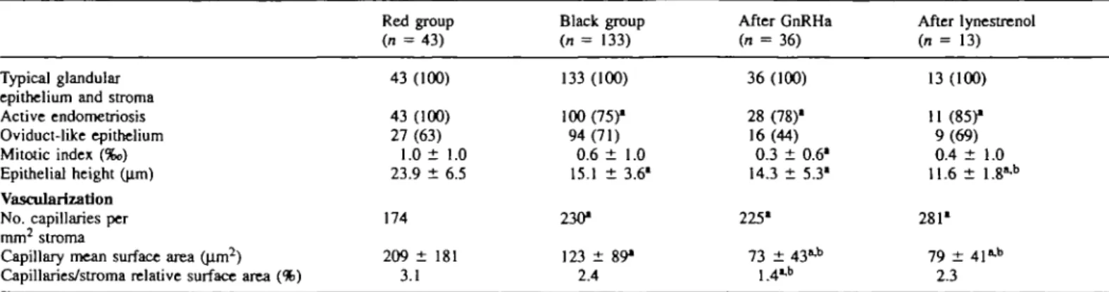
Table I. Typical peritonal lesions. Figures in
Typical glandular epithelium and stroma Active endometriosis Oviduct-like epithelium Mitotic index (%o) Epithelial height (pm) Vascularization No. capillaries per mm2 stroma
Capillary mean surface area (um2)
Capillaries/stroma relative surface area (%)
parentheses are percentages Red group (n = 43) 43 (100) 43 (100) 27 (63) 1.0 ± 1.0 23.9 ± 6.5 174 209 ± 181 3.1 Black group (n = 133) 133 (100) 100 (75)1 94(71) 0.6 ± 1.0 15.1 ± 3.6" 230* 123 ± 89" 2.4 After GnRHa (n = 36) 36 (100) 28 (78)1 16(44) 0.3 ± 0.61 14.3 ± 5.3" 225* 73 ± 43»-b 1.41* After lynestrenol (n = 13) 13 (100) 11 (85)1 9(69) 0.4 ± 1.0 11.6 ± 1.8"-b 281" 79 ± 41"-b 2.3 GnRHa = gonadotrophin-releasing hormone agonist.
•Significantly different from the red group. Significantly different from the black group.
A morphometrica] investigation of stroma] vascularization was carried out by image analysis programs set on a Vidas 21 computer (Kontron Bildanalyse GmBH, Eching, Germany). All samples were analysed using an Axioskop light microscope (Zeiss, Oberkochen, Germany) through a CCD 72 E camera (Dage-MTI, Michigan City, IL, USA). The image features were displayed on a Red/Green/Blue (RGB) monitor and stored for processing by the image analysis program. Data management and evaluation were checked according to specific search criteria on the Videoplan (Kontron Bildanalyse GmBH) and displayed on a Video Graphic Array (VGA) monitor and printed. All cases were analysed field by field using the objective X40 of the Axioskop light microscope. Histological structures of interest such as the stroma, the glandular epithelium and lumen and the capillaries, were drawn moving a cursor, discriminated, and the grey level images were transferred to binary images. The interactive measurements of the selected parameters (number of structures, area of the structures per field) were appended and stored at the end of an existing data base as previously described by Nisolle et al. (1993). The x2 test> the median test and Student's r-test were used for statistical analysis.
Results
Peritoneal biopsy
Biopsies taken from typical puckered black or bluish peritoneal lesions showed the presence of endometrial elements (glands and stroma) in all cases (100%). 'Active' endometriosis was found in 75% of cases in the non-treated group, in 78% after GnRHa therapy and in 85% after lynestrenol. The incidence of active endometriosis was significantly (P < 0.01) lower when compared to red lesions (100%). Areas of oviduct-like epithelium with ciliated cells were demonstrated in respectively 63, 71, 44 and 69% of cases. No significant differences were observed among the subgroups. The mitotic activity was calculated in glandular epithelium and its value was 1.0%o in red lesions, 0.6%o in the non-treated black lesions, 0.3% after 12 weeks of GnRHa therapy, and 0.4%o after lynestrenol. The value observed after GnRHa therapy was significantly (P < 0.01) lower than that observed in the red lesions. The epithelial height was respectively 23.9 ± 6.5, 15.1 ± 3.6, 14.3 ± 5.3 and 11.6 ± 1.8 |im. The value was significantly
(P < 0.001) higher in red lesions when compared to other
subgroups.
The results concerning the vascularization are shown in Table I. The number of capillaries per mm2 of stroma, then-mean surface area, and the surface area ratio (capillaries/ stroma) were calculated. In red lesions, the number of capillar-ies per mm2 of stroma was 174. Their mean surface area was 209 ± 1 8 1 |im and the ratio of capillaries/stroma surface area was 3.1%. The number of capillaries was respectively 230 in non-treated black lesions, 225 after GnRHa therapy and 281 after lynestrenol therapy. All values were significantly different
(P < 0.01) from those observed in red lesions. The capillary
mean surface area was significantly (P < 0.001) reduced in these three subgroups when compared to red lesions. The capillary mean surface areas after GnRHa therapy and after lynestrenol therapy were significantly (P < 0.001) lower when compared to black lesions. The capillaries/stroma relative surface area was significantly (P < 0.01) lower after GnRHa therapy when compared to all other subgroups. After GnRHa therapy, the number of capillaries per mm2 of stroma was similar to that observed without therapy, but the ratio of capillaries/stroma surface area was found to be significantly (P < 0.01) reduced after the administration of GnRHa. After lynestrenol, the number of capillaries, the capillary surface area and the ratio of capillaries/stroma surface area were similar to the values observed without therapy.
Nodules
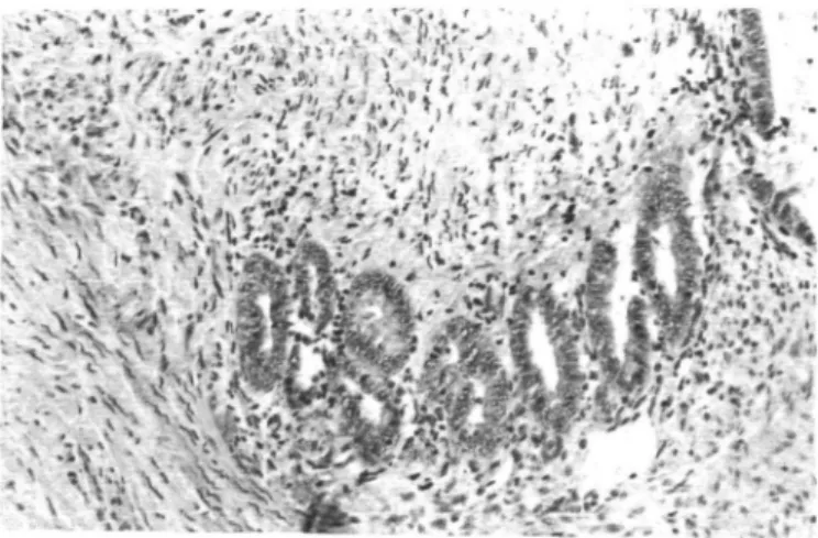
Biopsies taken from endometriotic nodules showed the pres-ence of endometrial elements in all cases (100%). Histologi-cally, scanty endometrial-type stroma and glandular epithelium are disseminated in muscular tissue (Figure 1). Cellular activity was found in 97% of cases and cellular differentiation in phase with the eutopic endometrium was never observed. Infiltration of surrounding fibromuscular tissue by endometrial glands with some signs of hyperplasia can be found (Figure 1). Very often, endometriotic glands and stroma were found by serial section up to the vaginal mucosa (Figure 2) which was sometimes replaced by endometrial epithelium.
It is obvious that the invasion process of the smooth muscle
225
at Ud Walthere Spring on November 9, 2011
http://humrep.oxfordjournals.org/
Donnez et aL
Figure 1. Rectovaginal adenomyosis (Gomori's trichome).
Endometrial glands with signs of hyperplasia in the smooth muscle. Original magnification X240.
/ •'"•
• •:• - i
- ; : - \
Figure 2. Rectovaginal adenomyosis (Gomori's trichome). Serial
sections reveal the presence of endometrial glands and stroma up to the vagina] mucosa. Original magnification X85.
Figure 3. Rectovaginal adenomyosis (Gomori's trichome). Invasion
of the smooth muscle by only the glandular epithelium (without stroma). Original magnification X240.
by glandular epithelium did not require the presence of stroma (Figure 3). Indeed, the glandular epithelium was often seen without any surrounding stroma very deep in the nodular
Table IL Rectovagina] endometriosis. Figures in parentheses are percentages
Control group After GnRHa After lynestrenol (n = 35) {n = 14) (n = 16) Typical glandular
epithelium and stroma Active endometriosis Oviduct-like epithelium Mitotic index (%o) Epithelial height (Jim) Vascularization No. capillaries
per mm2 stroma Capillary mean
surface area (u.m2)
Capillaries/stroma relative surface area
35 (100) 34(97) 27 (77) 0.09 ± 0.25 16.5 ± 4.1 161 140 ± 80 1.8 14 (100) 12 (86) 3(21) 0.03 ± 0.60 12.9 ± 3.3 208' 120 ± 60 if* 16(100) 13(81) 7(44) 0.03 ± 0 . 1 0 11.4 ± 2.9 139 76 ± 70b
GnRHa = gonadotrophin-releasing hormone agonist. "Significantly different from lynestrenol group. ''Significantly different from the control group.
fibromuscular tissue; it consisted of whorled anastomosing fascicles of uniform, fusiform, smooth muscle cells.
'Active' endometriosis was demonstrated in 97, 86 and 8 1 % of cases in the different subgroups (without therapy, after GnRHa therapy, after lynestrenol therapy) respectively. No difference was observed between the different subgroups. When compared to the mitotic index observed in peritoneal endometriosis (0.6%o), it was found to be significantly lower (P < 0.001) in glandular epithelium of nodules (0.09%o). No significant differences were noted after GnRHa and lynestrenol therapy. The results concerning the vascularization are shown in Table II. In the untreated group, the number of capillaries per mm2 of stroma was 161, their mean surface area was 140 ± 80 |im and the ratio of capillaries/stroma surface area was 1.8%.
After GnRHa therapy, the number of capillaries per mm2 of
stroma (208), their mean surface area (120 ± 60 u.m) and the ratio of capillaries/stroma surface area (2.2%) were found to be similar to the values observed in the non-treated group. After lynestrenol, the number of capillaries (139), the capillary/ surface area (76 ± 70 (im) and the capillaries/stroma surface ratio (0.9%) were found to be significantly (P < 0.01) different from the non-treated group and from the GnRHa group. Discussion
It is generally believed that endometriosis is caused by the implantation of retrograde menstrual endometrial cells, or by metaplasia. In the pelvis, three different forms of endometriosis must be considered: (i) peritoneal; (ii) ovarian; and (iii) rectovaginal septum (Donnez etal., 1995). The early manifesta-tions of the disease are believed to be subtle or non-coloured lesions such as white lesions (white opacification) (Jansen and Russel, 1986; Nisolle et aL, 1990; Donnez et aL, 1992; Brosens, 1994). The presence of a lower mitotic activity and poor stromal vascularization in white lesions suggests that this type of lesion is a quiescent form of the disease. Red lesions (red vesicles, polypoid lesions, flame-like lesions) (Jansen and
at Ud Walthere Spring on November 9, 2011
http://humrep.oxfordjournals.org/
Evaluation of peritoneal endometriosis and endometriotic nodules
Russel, 1986; Nisolle et al, 1993; Brosens et al, 1994) are more active forms of the disease. In women, our hypothesis is that red lesions are more aggressive and progress to the so-called typical or black 'lesions which must be considered as an enclosed implant surrounded by fibrosis. Another form of the disease is 'deep-infiltrating endometriosis of the rectovaginal septum'. Sampson (1922) defined cul-de-sac obliteration as 'extensive adhesions in the cul-de-sac, obliterating its lower portion and uniting the cervix or the lower portion of the uterus to the rectum; with adenoma of the endometrial type invading the cervical and the uterine tissue and probably also (but to a lesser degree) the anterior wall of the rectum.
In the present study, two different types of endometriosis (peritoneal endometriosis and rectovaginal endometriosis) were morphologically analysed in order to specify their individual characteristics and to evaluate the influence of hormonal therapy.
Histologically, typical peritoneal endometriosis is character-ized by both epithelium and stroma of the endometrial type. Nevertheless, the frequent occurrence of a variety of epithelial features, other than typical endometrium, lends further support to the coelomic epithelial origin theory on this entity. Oviduct-like epithelium was often found in our study in peritoneal lesions as well as in rectovaginal endometriotic nodules. As suggested by Czernobilsky and Morris (1979) and Nisolle
et al. (1988), oviduct-like epithelium signifies the same
patho-physiology as ectopic endometrial glands and stroma. Deep vaginal endometriosis associated with pelvic endo-metriosis can take the form of nodular or polypoid masses, involving the posterior vaginal fornix (Reica et al, 1991; Nezhat et al, 1992; Koninckx and Martin, 1992; Donnez et al, 1994). The differential diagnosis of vaginal endometriosis, particularly of the superficial type, includes vaginal adenosis of the tuboendometrial variety, but the latter lacks endometrial stroma and the characteristic inflammatory response of endo-metriosis (Zaloudek et al, 1981). In the uterus corpus, adeno-myosis is a common condition characterized pathologically by the presence of endometrial glands and stroma within the myometrium. Microscopically, there are endometrial glands and stroma within the myometrium. Adenomyosis exhibits a varied functional response to ovarian hormones. Proliferative glands and stroma are generally observed in the first half of the menstrual cycle.
Adenomyosis may not respond to physiological concentra-tions of progesterone, and secretory changes are frequently absent or incomplete during the second half of the cycle. Similar histological observations (no one case showed cellular differentiation in phase with eutopic endometrium) are made at the level of the 'endometriotic' rectovaginal nodule which is, like an adenomyoma, a circumscribed nodular aggregate of smooth muscle, endometrial glands, and usually, endometrial stroma. This lesion originates from the rectovaginal septum tissue and consists essentially of smooth muscle with active glandular epithelium and scanty stroma. The invasion of the smooth muscle by very active glandular epithelium widiout stroma proved that the stroma is not mandatory for invasion and that the nodule is different from peritoneal endometriosis in which epithelial glands are systematically surrounded by
endometrial-type stroma. The very similar histological descrip-tions to uterine adenomyosis lead us to suggest, like Brosens (1995) that the so-called endometriotic nodule of the recto-vaginal septum is, in fact, just like an adenomyoma or an adenomyotic nodule. In our study, whatever the hormonal treatment used, a high incidence of active endometriosis without signs of degeneration was found, but differences could be observed between peritoneal endometriosis and rectovaginal 'adenomyosis'. After GnRHa therapy, the incidence of active endometriosis and the mitotic activity were significantly lower in peritoneal endometriosis but not in rectovaginal endo-metriosis. The ectopic foci are more or less autonomous, not governed by the normal control mechanisms governing the uterine endometrial glands and stroma. The exact reason why a number of implants or cells do not respond to hormonal therapy is not known, but at least four hypotheses have been proposed: (i) the drug does not gain access to the endometriotic foci because fibrosis surrounding the foci prevents access locally; (ii) endometriotic cells may have their own genetic programming, while endocrine influence appears to be only secondary and dependent on the degree of differentiation of the individual cell; (iii) the lower oestrogen receptors in peritoneal ectopic endometrium and in rectovaginal nodule endometrium than in eutopic endometrium (Bergqvist et al, 1993; Bergqvist and Ferno, 1993; Nakamura et al, 1993; Howell et al, 1994; Nisolle et al, 1994; Bergqvist, 1995); and (iv) the different regulatory mechanisms of endometriotic steroid receptors may result in deficient endocrine dependency (Nisolle et al, 1994) because the receptors, although they are present, are biologically inactive (Metzger, 1992).
In peritoneal endometriosis, the stromal vascularization was found to be significantly lower after GnRHa therapy. This change was due to a decrease in the volume occupied by the vessels, as proved by both the mean capillary surface area and the ratio of capillaries/stroma surface area. This last parameter which is the stromal vascularization index (Nisolle et al, 1993) was not reduced after lynestrenol therapy. In rectovaginal endometriosis, lynestrenol, on the contrary, appeared more active in the reduction of vascularization than GnRHa.
In peritonea] endometriosis, after GnRHa administration, our study demonstrated a low rate of mitosis and poor stromal vascularization. Our hypothesis is that under GnRHa treatment, lesions are in latent stages. They are probably non-active lesions that could be quiescent as long as the oestrogen secretion is suppressed. But the fact that residual areas of active endometriosis are present in 75% of cases explains the quick recurrence observed after cessation of therapy.
When data from rectovaginal endometriosis are evaluated, we can see that GnRHa is unable to suppress the mitotic activity and to decrease the stromal vascularization. All these parameters were strongly in evidence in peritoneal endo-metriotic lesions. The absence of any lynestrenol effect on the peritoneum was in contrast to the strong effect of the same drug on rectovaginal endometriotic nodule vascularization. The completely different responses of peritoneal lesions and nodular rectovaginal lesions to GnRHa therapy or lynestrenol therapy suggest that peritoneal endometriosis and rectovaginal endometriosis are two different diseases and have a different
227
at Ud Walthere Spring on November 9, 2011
http://humrep.oxfordjournals.org/
Donnez et al
physiopathology. Peritoneal endometriosis is probably caused by the implantation of regurgitated menstrual cells. Rectova-ginal endometriosis is a lesion of adenomyosis and can possibly develop from MUllerian rests. The very low mitotic activity observed in this pathology can explain the relatively slow evolution of the adenomyoma.
In conclusion, it is suggested that rectovaginal adenomyosis is a specific disease, different from peritoneal endometriosis.
Acknowledgements
Supported by Fonds de la Recherche Scientifique, grant 3.4587.90, Brussels, Belgium and a grant from Ipsen Biotech, Paris, France. References
Bergqvist, A., Ljungberg, O. and Skoog, L. (1993) Immunohistochemical analysis of oestrogen and progesterone receptors in endometriotic tissue and endometrium. Hum. ReprxxL, 8, 1915-1922.
Bergqvist, A. and Ferno, M. (1993) Oestrogen and progesterone receptors in endometriotic tissue and endometrium: comparison of different cycle phases and ages. Hum. Reprod., 8, 2211-2217.
Bergqvist, A. (1995) Hormonal regulation of endometriosis and the rationales and effects of gonadotrophin-releasing hormone agonist treatment: a review. Hum. Reprod., 10, 446-^52.
Brosens, LA. (1994) New principles in the management of endometriosis. Acta Obstet. GynecoL Scand., 159, 18-21.
Brosens, I.A. (1995) Is mild endometriosis a progressive disease? Hum. Reprod., 9,2209-2211.
Chatman, D.L. (1981) Pelvic peritoneal defects and endometriosis: Allen-Masters syndrome revisited. FertiL SteriL, 36, 751.
Czemobilsky, B. and Morris, SJ. (1979) A histologic study of ovarian endometriosis with emphasis on hyperplastic and atypical changes. Obstet. GynecoL, 53, 318.
Donnez, J., Casanas-Roux, F., Caprasse, EJ., Ferin, J. and Thomas, K. (1985) Cyclic changes in ciliation, cell height, and mitotic activity in human tuba] epithelium during reproductive life. FertiL SteriL, 43, 554-559.
Donnez, J., Nisolle, M. and Casanas-Roux, F. (1992) Three-dimensional architectures of peritoneal endometriosis. FertiL SteriL, 57, 980-983. Donnez, J., Nisolle, M., Casanas-Roux, F., Anaf, V. and Smets, M. (1994)
Laparoscopic treatment of rectovaginal septum endometriosis. In Donnez, J. and Nisolle, M. (eds), An Atlas of Laser Operative Laparoscopy and Hysteroscopy. Parthenon Publishing, Camforth, UK, pp. 75-85.
Donnez, J., Nisolle, M., Casanas-Roux, F., Bassil, S. and Anaf, V. (1995) Rectovaginal septum, endometriosis or adenomyosis: laparoscopic management in a series of 231 patients. Hum. Reprod., 10, 630-635. Howell, RJ., Dowsett, M. and Edmonds, D.K. (1994) Oestrogen and
progesterone receptors in endometriosis: heterogeneity of different sites. Hum. Reprod., 9, 1752-1758.
Jansen, R.P.S. and Russel, P. (1986) Non-pigmented endometriosis: clinical laparoscopic and pathologic definition. Am. J. Obstet. GynecoL, 155, 1154-1158.
Koninckx, PR. and Martin, D. (1992) Deep endometriosis: a consequence of infiltration or retraction or possible adenomyosis externa? FertiL SteriL, 58,924-928.
Martin, D.C., Hubert, G.D., Van der Zwaag, R. and El-Zeky, F. (1989) Laparoscopic appearances of peritoneal endometriosis. FertiL SteriL, 51, 63-67.
Metzger, M. (1992) Cyclic changes in endometriosis implants. In Brosens, LA. and Donnez, J. (eds), Current Status of Endometriosis. Research and Management Parthenon Publishing, Camforth, UK, pp. 89.
Nakamura, M., Katabuchi, H., Tohya, T., Fukumatsu, Y., Matsuura, K. and Okamura, H. (1993) Scanning electron microscopic and immuno-histochemical studies of pelvic endometriosis. Hum. Reprod., 8,2218-2226. Nezhat, C , Nezhat, F. and Pennington, E. (1992) Laparoscopic treatment of lower colorectal and infiltrative rectovaginal septum endometriosis by the technique of video laparoscopy. Br. J. Obstet. GynaecoL, 99, 664—667. Nisolle, M., Casanas-Roux, F. and Donnez, J. (1988) Histologic study of
ovarian endometriosis after hormonal therapy. FertiL SteriL, 49, 423-426. Nisolle, M., Paindaveine, B., Bourdon, A., Berliere, M., Casanas-Roux, F.
and Donnez, J. (1990) Histologic study of peritoneal endometriosis in infertile women. FertiL SteriL, 53, 984-988.
Nisolle, M., Casanas-Roux, F., Anaf, V., Mine, J.M. and Donnez, J. (1993) Morphometric study of the stromal vascularization in peritoneal endometriosis. FertiL SteriL, 59, 681-684.
Nisolle, M., Casanas-Roux, F., Wyns, Ch., De Menten, Y., Mathieu, P.E. and Donnez, J. (1994) Immunohistochemical analysis of estrogen and progesterone receptors in endometrium and peritoneal endometriosis: a new quantitative method. FertiL SteriL, 62, 751-759.
Redwine, D.B. (1987) The distribution of endometriosis in the pelvis by age groups and fertility. FertiL SteriL, 47, 173-175.
Reich, H., McGlynn, F. and Salvat, J. (1991) Laparoscopic treatment of cul-de-sac obliteration secondary to rectocervical deep fibrotic endometriosis. Reprod. Med., 36, 516.
Sampson, J.A. (1922) Intestinal adenomas of endometrial type. Arch Surg., 5,21.
Stripling, M.C., Martin, D.C., Chatman, D.L., Van der Zwaag, R. and Poston, W.M. (1988) Subtle appearances of pelvic endometriosis. FertiL SteriL, 49, 42-31.
Zaloudek, C. and Norris, H.J. (1987) Mesenchymal tumors of the uterus. In Kurman, R. (ed.), Blaustein's Pathology of the Female Genital Tract. Springer-Verlag, pp. 373.
Received on June 14, 1995; accepted on October 4, 1995
228
at Ud Walthere Spring on November 9, 2011
http://humrep.oxfordjournals.org/