HAL Id: dumas-02507399
https://dumas.ccsd.cnrs.fr/dumas-02507399
Submitted on 13 Mar 2020
HAL is a multi-disciplinary open access archive for the deposit and dissemination of sci-entific research documents, whether they are pub-lished or not. The documents may come from teaching and research institutions in France or abroad, or from public or private research centers.
L’archive ouverte pluridisciplinaire HAL, est destinée au dépôt et à la diffusion de documents scientifiques de niveau recherche, publiés ou non, émanant des établissements d’enseignement et de recherche français ou étrangers, des laboratoires publics ou privés.
Quelle est la place de la curiethérapie préopératoire
suivie d’une hystérectomie par voie mini invasive dans la
prise en charge des cancers du col de stade précoce ?
Clémence Beyer
To cite this version:
Clémence Beyer. Quelle est la place de la curiethérapie préopératoire suivie d’une hystérectomie par voie mini invasive dans la prise en charge des cancers du col de stade précoce ?. Médecine humaine et pathologie. 2020. �dumas-02507399�
AVERTISSEMENT
Ce document est le fruit d'un long travail approuvé par le
jury de soutenance et mis à disposition de l'ensemble de la
communauté universitaire élargie.
Il n’a pas été réévalué depuis la date de soutenance.
Il est soumis à la propriété intellectuelle de l'auteur. Ceci
implique une obligation de citation et de référencement
lors de l’utilisation de ce document.
D’autre part, toute contrefaçon, plagiat, reproduction illicite
encourt une poursuite pénale.
Contact au SID de Grenoble :
bump-theses@univ-grenoble-alpes.fr
LIENS
LIENS
Code de la Propriété Intellectuelle. articles L 122. 4
UNIVERSITÉ GRENOBLE ALPES UFR DE MÉDECINE DE GRENOBLE
Année : 2020
Is there a place for preoperative brachytherapy followed by
Laparoscopic Hysterectomy
in less than IB3 cervical cancer after LACC trial?
Oncological Outcomes of a retrospective study
THÈSE
PRÉSENTÉE POUR L’OBTENTION DU TITRE DE DOCTEUR EN MÉDECINE
DIPLÔME D’ÉTAT
Mlle Clémence BEYER
THÈSE SOUTENUE PUBLIQUEMENT À LA FACULTÉ DE MÉDECINE DE GRENOBLE
Le 6 mars 2020
DEVANT LE JURY COMPOSÉ DE
Président du jury :
M. Le Professeur Didier RIETHMULLER
Membres :
M. Le Professeur Eric LAMBAUDIE, Directeur de thèse Mme Le Professeur Pascale HOFFMANN
Mme Le Docteur Anne Cécile PHILIPPE
L’UFR de Médecine de Grenoble n’entend donner aucune approbation ni improbation aux opinions émises dans les thèses ; ces opinions sont considérées comme propres à leurs auteurs.
REMERCIEMENTS
Je tiens tout d’abord à remercier les membres du jury :
Au Professeur Eric LAMBAUDIE, merci mille fois d’avoir accepté de diriger ma thèse. Si je dois
retenir deux choses qui font l’estime que j’ai pour vous, sans aucun doute votre talent chirurgical
inénarrable et la qualité de votre humour. Merci de m’avoir tant appris en 6 mois, puis d’avoir
managé mes idées disparates et mon niveau d’anglais.
Au Professeur Didier RIETHMULLER, merci d’avoir accepté d’être président du jury de ma thèse.
Beaucoup de respect pour votre parcours et votre ambition pour notre service. J’espère de
nombreux travaux conjoints dans le futur.
Au Professeur Pascale HOFFMANN, pour votre savoir physiopathologique, ces discussions
philosophico-sociétales et cette bienveillance constante qui ont jalonné mon parcours d’interne.
Merci pour vos remarques et commentaires pour ce travail.
Au Docteur Anne Cécile PHILIPPE, qui m’a transmis son engouement pour la cancérologie. Pour
l’exemplarité de son parcours et de son travail, son esprit affuté et sa sensibilité hors du commun.
Merci pour ton soutien, ces journées de blocs en osmose et ce sens de la vanne qui t’es propre.
A l’équipe du service de gynécologie du CH de Voiron : merci au Dr AMBLARD pour ces cours
d’anatomie sur un coin de table, au Dr LAZZARON et au DR GAILLARD pour leur détermination
et leur prévenance.
A l’équipe du service de Gynécologie du Centre Hospitalier de Chambéry :
Au Dr Victoire CABAUD, merci d’avoir été là pour mes débuts en chirurgie, et d’avoir cru en moi.
Merci pour tous vos conseils et votre force de caractère immuable. Au Dr DEYROLLE pour ses
leçons de rigueur inopinées et aux Dr Haller, Dr Decroisette et Dr Mirouse : merci pour votre
bienveillance dans mes débuts en obstétrique et cette année passée à vos côtés.
A l’équipe de l’Institut Paoli Calmettes, dans son intégralité, mais surtout :
Merci Au Dr Oona FRANKE, LA rencontre de ces 6 mois, tant professionnel que personnel, au Dr
Marie BANNIER pour son énergie et ses conseils précieux, au Dr Isabelle MASQUIN et au Dr
Emilie DEMARQUET, pour leur bonne humeur, vraiment un beau semestre passé à vos côtés, au
Dr HEINEMANN Mellie pour ce rire communicatif, et au Dr Sandrine RUA, pour l’apprentissage
de la chirurgie en situation de stress.
A l’équipe du CHUGA :
L’équipe des sages-femmes : toutes celles avec qui j’ai travaillé et qui m’ont tellement apporté et
appris : merci pour tous ces consensus sur les tracés en milieu de nuit, une ovation particulière pour
Audrey Lalanne et Sandrine Barbier et Big up à ma chère Solène, qui est une femme et une
A l’équipe de gynécologues et d’obstétriciens : Au Dr Anne Laure COSTON à qui je dois beaucoup
de ma formation obstétricale, au Dr EQUY pour ses cours d’anatomie de l’ERCF et son déhanché
légendaire, au Dr THONG VANH pour ses connaissances 2.0 sur les probiotiques, au Dr Virginie
GUIGUE pour cette classe résistante à toutes épreuves, au Dr Camille DUNAND FAURE pour
ces discussions improbables à 5h du matin en césarienne, au Dr Aurore GUENIFFEY, pour son
écoute et ses pintes réconfortantes et au Dr REBOUX, qui va nous manquer !
Au Dr Cécile GOUPIL et au Dr Sophie GOBILLOT, un vrai bonheur de travailler ensemble, vous
êtes des machines de guerre !
Au Dr François ISTASSE pour ces journées blocs dans la bonne humeur et ce petit côté gossip
derrière tant de discrétion et au Dr Thierry MICHY.
A l ‘équipe d’IBODE et d’IDE de l’HCE : en particulier à Sarah, Marie et Mauricette : merci pour
votre soutien !
Merci à toute l’équipe du centre de cancérologie de la femme : Marine, les Célines, Angélique…
A l’équipe du service d’oncologie médicale du CHUGA, pour leur accueil, spécialement au Dr
Emmanuelle JACQUET, pour son intelligence et son élégance, et au Dr Matthieu LARAMAS:
j’aimerais tellement que tu te mettes à l’onco-gynécologie !
A l’équipe du service de chirurgie plastique et maxillo-faciale du CHUGA : merci de m’avoir si
Mes 2 compères, Delphine et Anne pauline, pour ces nombreux moments tout en finesse et blagues
délicates.
A Mathilde, Nathan et Alexandra pour avoir rempli mon 10ème semestre de joies et de Quenelles!
(cuppower+).
A mon petit Lamotte d’amour, et à Stefan, mon binôme dans les galères de DIU /DESC.
A notre quatuor gagnant, Mac-Faisant, Chacha et Collarde !
A Lamercerette, Clémentine, Lucile, Buisson et Jannie, qui ont rendu mon dernier stage au CHU
si léger.
Et tout ceux que j’ai croisé et qui ont contribué à améliorer mes longues années internat : Mathilde,
Salomé, Julie, Charles, Marie L., Célia, Davy, Agathe, Théo, Marie B., Myriam.
A mes acolytes de l’externat : Salomé, Elsa, Pauline, Louis, David, Elodie et Célia, pour ces soirées
de folie et notre amitié indéfectible.
A ma famille, et en particulier à mes 2 sœurs Sarah et Claire, mes piliers, et mon fabuleux petit
frère David.
A ma grand-mère, Claire HOFFMANN - FAZI, qui a fait tant de bonheurs de ma vie.
Et avant tout à PG, pour notre vie merveilleuse ensemble, et ses trois princesses Thalia, Emma et
Is there a place for preoperative brachytherapy
followed by Laparoscopic Hysterectomy
in less than IB3 cervical cancer after LACC trial?
Oncological Outcomes of a retrospective study
RESUME
OBJECTIFS
L'objectif de cette étude est d’évaluer les résultats oncologiques et chirurgicaux d'une stratégie
thérapeutique consistant en une curiethérapie préopératoire suivie d'une hystérectomie abdominale
ou laparoscopique de type A, comme alternative à la chirurgie première dans le traitement des
cancers du col de stade précoce (Stade FIGO < IB3).
MATÉRIEL ET MÉTHODES
Il s'agit d'une étude rétrospective menée à l'Institut Paoli Calmettes, centre anticancéreux
(Marseille, France) entre 2001 et 2012, concernant des patientes prises en charge pour un cancer
du col de l'utérus de stade précoce (taille de la tumeur < 4 cm, stade FIGO IA1, IA2, IB1 et IB2 -
FIGO 2018) par une approche radiochirurgicale.
Une curiethérapie préopératoire à faible débit de dose a été réalisée chez des patientes N0
(chirurgicale et radiologique), suivie 6 à 8 semaines plus tard d'une hystérectomie de type A par
voie mini-invasive ou par laparotomie.
Les patientes présentant des métastases ganglionnaires pelviennes ou ayant bénéficié d’un
traitement adjuvant ont été exclues.
Le critère d'évaluation principal était la survie sans progression (SSP) et les critères d'évaluation
secondaires étaient la morbidité de la chirurgie et celle de la curiethérapie.
RÉSULTATS
Au total, 138 patientes ont été incluses. L'analyse histologique a montré une réponse complète chez
Avec un suivi médian de 132 mois (60 - 204 mois), la SSP était de 93,5 % et il y a eu 9 récidives
- 6 métastatiques et 3 récidives loco régionales (2 ganglionnaires pelviennes et 1 dans le muscle
pyramidal gauche)-.
En analyse univariée, un délai de plus de 52 jours entre la fin de la curiethérapie et la chirurgie était
associé à une SSP significativement plus faible (p = 0,004, OR = 8,5, IC95 {1,5 ; 48,7}), et une
réponse pathologique complète était associée à une meilleure SSP (p = 0,03, OR = 6,1, IC95
{1,8 ;55,3 }).
Nous avons rapporté un taux global de complications tardives de la curiethérapie (glossaire
Chassagne) de 17,3 % (n= 24) et un taux global de lésions urinaires chirurgicales de 6,5 % (n = 9),
avec seulement 2 patientes (1,5 %) présentant des complications de grade 3 (classification Clavien
et Dindo).
CONCLUSION
Avec un suivi médian de 132 mois, une stratégie thérapeutique radio chirurgicale des cancers du
col de l'utérus à un stade précoce (inférieur à IB3 FIGO 2018 ) incluant une curiethérapie
endocavitaire préopératoire suivie d’une hystérectomie totale par voie laparoscopique de type A
semble offrir un taux de récidive raisonnable par rapport aux résultats de l'essai LACC
(Laparoscopic approach to cervical cancer ). Cette stratégie peut être une alternative à
l’hystérectomie radicale initiale chez certaines patientes afin de maintenir une approche minimale
invasive.
MOTS CLES : cancer du col, stade précoce, curiethérapie préopératoire, hystérectomie type A,
ABSTRACT
OBJECTIVES
The aim of this study is to report the oncological and surgical outcomes concerning the combination
of a preoperative brachytherapy followed by Type A laparoscopic or abdominal hysterectomy as
an alternative to upfront surgery in early stage cervical cancer (<FIGO IB3).
MATERIAL AND METHODS
This is a retrospective study conducted at Paoli Calmettes Institute, comprehensive cancer center
(Marseille, France) between 2001 and 2012, concerning patients managed for early stage cervical
cancer (tumor size < 4 cm, FIGO stage IA1, IA2, IB1 et IB2 - FIGO 2018) by a radiosurgical
approach.
A preoperative low dose rate brachytherapy was performed in pN0 patients (surgical and
radiological staging), followed 6-8 weeks later by a type A hysterectomy by laparoscopic or open
surgical approach.
Patients with pelvic lymph node metastases or adjuvant treatment were excluded.
The primary endpoint was the Disease Free Survival (DFS) and the secondary endpoint was the
surgical and brachytherapy morbidities.
RESULTS
A total of 138 patients were included. Histological analysis showed a complete response in 68
patients (49,3 %) and a residual tumor < 1 cm in 36 patients (26 %).
With a median follow up of 132 months (60 - 204 months), DFS was 93,5% and 9 recurrences
In univariate analysis, a delay between the end of brachytherapy and surgery superior to 52 days is
associated with a significative poorer DFS (p = 0,004, OR = 8,5, IC95 {1,5 ; 48,7}), and a
pathological complete response is associated with a better DFS (p = 0,03 OR = 6,1 IC95 {1,8 ;
55,3}.
We have reported an overall late complication rate for brachytherapy of 17,3% (n = 24) (Chassagne
glossary) and an overall rate of surgical urinary injuries of 6,5% (n = 9), with only 2 patients (1,5%)
with grade 3 complications (Clavien and Dindo classification).
CONCLUSION
With a median follow up of 132 months, a radiosurgical management of early stage cervical cancers
(less than IB3) including brachytherapy and type A laparoscopic hysterectomy seems to offer a
reasonable rate of recurrences compared to LACC trial’s results. This strategy may be an
alternative to upfront radical hysterectomy in selected patients to maintain a minimal invasive
approach.
KEY WORDS: cervical cancer, early stage, preoperative brachytherapy, hysterectomy type A,
Abbreviations
ARH: Abdominal Radical Hysterectomy
DFS: Disease Free Survival
ESCC: Early Stage Cervical Cancer
FIGO : Fédération international de gynécologie et d’obstétrique
HT: Hysterectomy
LACC: Laparoscopic Approach to Cervical Cancer
LRH: Laparoscopic Radical Hysterectomy
LVSI: LymphoVascular Space Involvement
MIS: Minimally Invasive Surgery
MISRH: Minimally Invasive Surgical Radical Hysterectomy
OH: Open Hysterectomy
Remerçiements……… 6
RESUME……… 11
ABSTRACT………. … 13
Abbreviations……….. 15
1.INTRODUCTION……… 17
2.MATERIAL & METHODS………... 19
2.1 Characteristics of patients………... 19
2.2 Treatments……….. 20
2.2.1 Preoperative Brachytherapy (POBT)……… 20
2.2.2 Surgery ……… 20
2.3 Follow-up……… 21
2.4. Statistical analysis……… 21
3.RESULTS……… 22
3.1 Characteristics of patients and pathology……… 22
3.2 Surgical outcomes………... 22
3.3 Disease free survival and recurrences……… 23
3.4 Toxicities of brachytherapy and surgery……… 24
4. DISCUSSION………... 25 Preoperative Brachytherapy………... 29 5. LIMITATIONS……….. 32 6. CONCLUSION………. 33 BIBLIOGRAPHY………. 34 ANNEXES………. 41
Table 1 Patients and tumors characteristics……… 42
Table 2 Characteristics of recurrences……… 43
Table 3a/3b Brachytherapy toxicities and surgery complications………. 44
Table 4 Literature review of series evaluating different therapeutic strategy of ESCC…………. 46
1. INTRODUCTION
Early stage cervical cancer, defined as a tumor size less than 4 cm (< stage IB3 FIGO 2018)
confined to the pelvis, with clinically and radiologically negative pelvic nodes, have no standard
management and differs, according to the country.
Upfront surgery is the standard suggested by international guidelines (National Comprehensive
Cancer Network NCCN (1)), ESGO (European Society of gynecological oncology (2)) and ESMO
(European Society for medical Oncology (3)). The surgical procedure is represented by a radical
hysterectomy Type B1 for low risk, B2 for intermediate risk or C1 for high risk according to The
Querleu - Morlow classification (4), associated with pelvic lymph nodes staging.
According to ESGO and French guidelines (2, 5), an option might be a preoperative intracavitary
uterovaginal brachytherapy (POBT) followed by type A hysterectomy, even if this option is used
in a limited number of centers, mostly in France.
The superiority of one of the two methods has never been established, but the feasibility and safety
of the POBT has been already reported in several studies (6-14).
Furthermore, the minimally invasive approach (conventional laparoscopy or robotic assisted
laparoscopy) have been recently developed and is considered as the standard approach for early
stage cervical cancer treatment, since the demonstration, for several years, of its feasibility, surgical
and oncological safety compared to open surgery (17,18,19).
However, since 2018 and the LACC (Laparoscopic Approach to Cervical Cancer) trial’s
publication (20), a new question has emerged: is laparoscopic approach as safe as previously
stage < IB3 FIG0 2018, after upfront surgery, the author reported a higher rate of recurrence with
the minimal invasive approach compared to open surgery with a significative impairment of the
survival (8.8% vs 2,9% after a follow up of 3 years, and 14% vs 3.5 % for open surgery, after a
follow up of 4.5 years).
Questions remain concerning the methodology of this "real life" trial, but we must take into account
its results to inform our patients. Results of upfront surgery in FIGO IB1-IB2 cancers may be
compared to other options including minimal invasive surgical approach, particularly in the sub
group of patients with tumor size between 2 and 4 cm.
The objective of this study is to report our surgical and oncological results from an historical cohort
of patients managed with pre operative brachytherapy followed by laparoscopic hysterectomy for
2. MATERIAL AND METHODS
It is a retrospective single center study concerning patients managed between 2001 to 2012 at Paoli
Calmettes Institute, comprehensive cancer center (Marseille, France) for early stage cervical cancer
(< stage IB3 FIGO 2018).
2.1. Characteristics of Patients
Inclusion criteria were: histologically confirmed carcinoma of the uterine cervix, operable tumor
(size less than 4 cm of diameter, stages IA1 with lympho vascular space involvement (LVSI), IA2,
IB1, IB2 according to the new classification International Federation of Gynecology and Obstetrics
FIGO 2018) (21), with disease confined to the cervix. Systematic pre therapeutic pelvic
lymphadenectomy has been performed in all patients.
Exclusion criteria were lymph nodes metastasis and/or adjuvant treatment: radiotherapy or
chemotherapy.
For each patient, we collected data from their medical files: age, comorbidities, body mass index
(BMI), history of the oncologic disease, surgical procedure’s characteristics (surgical approach),
number of lymph nodes removed, post-operative outcomes (complications, readmissions),
definitive pathology, and tumor characteristics (tumor size, histology, Lymphovascular space
invasion LVSI).
Tumor size was assessed clinically and then adjusted based on MRI findings or by results of
2.2. Treatments
2.2.1 Preoperative Brachytherapy (POBT)
For all patients, pN0 status was proven by a systematic laparoscopic pelvic lymph node dissection,
before preoperative low dose rate (LDR) uterovaginal brachytherapy.
The brachytherapy procedure aimed at delivering 60 Gy (0,5-0,8 Gy/h) to the cervix, the proximal
part of the parametria and the upper third of vagina, according to the guidelines in Report No.38
of the International Commission on Radiation Units and Measurements (ICRU) (22).
Dosimetry was done from radiographs (2D) or from 3D acquisition with CT scanner, using Cesium
sources (137 Cesium), during an average time of 120 hours.
Brachytherapy complications were graduated according to the Chassagne glossary (16).
The complications examined are those that have persisted 2 months after the end of Brachytherapy.
2.2.2 Surgery
Six to eight weeks after brachytherapy, patients underwent type A laparoscopic or open
hysterectomy according to Querleu-Morrow classification (4), with bilateral
salpingectomy-oophorectomy. Per and post-operative complications were collected according to the
Clavien-Dindo classification (15), from grade 1 for lower toxicity (no treatment needed) to grade 5 (death).
2.3 Follow-up
The follow up was carried out as follows: a clinical monitoring including speculum and pelvic
examination was performed 3 – 4 weeks after Brachytherapy, then 4 weeks after surgery, then
every 4 months the following three years, then every 6 months the following two years then every
years.
MRI or CT scan has only been realized in the case of biological or clinical abnormalities.
Disease free survival (DFS) time was calculated from the date of diagnosis of cancer to the date of
diagnosis of recurrence.
2.4 Statistical analysis
All statistical analyses were performed at the significance level α=0.05 and using Xlstat software
(version 16.34). Patients’characteristics were summarized using counts (frequencies) for
categorical variables and means (standard deviations) or median (min-max) for quantitative
variables. Data were managed and analyzed with an Excel database.
The Kaplan Meier method was used to estimate the survival distribution.
The Cox proportional hazards model was used to account for the influence of multiple variables.
Intra-group differences in categorical variables were analyzed by independent Fisher’s exact test
and in continuous variables by the Student’s t-test.
This study was approved by our ethical committee (Paoli-Calmettes Institute’s review board), with
3. RESULTS
3.1 Characteristics of patients and pathology
From 2001 to 2012, 138 patients were included retrospectively, with early stage cervical cancer
strictly inferior to FIGO IB3 and negative pelvic nodes. All were treated by low dose intracavitary
uterovaginal brachytherapy followed by type A hysterectomy.
The baseline characteristics of patients are reported in Table 1.
Median age was 49 years (28 - 81). Concerning tumor stage, 64 patients (46,3 %) had IB1 stage
and 64 patients (46,3 %) had IB2 stage according to FIGO 2018 classification.
A total of 74 patients (53,6%) have tumor size less than 2 cm, and 64 patients (46,3%) have a tumor
size between 2 to 4 cm.
Median tumor size was 19 mm (3 - 39 mm).
The histopathology distribution showed a majority of squamous cell carcinoma (61.6%; n = 85).
The diagnostic has been confirmed with a conization in 92 patients (66,7%) or with a simple biopsy
for 43 patients (31,2%).
3.2 Surgical outcomes
Pelvic lymphadenectomy was performed in all patients, and all patients had negative pelvic lymph
nodes.
The median time between POBT and surgery was 53 days (32-145). Surgical approach was
At definitive pathology, LVSI was found in 2,2% of cases (0 for IA1 to IB1 stage and 3 for IB2
stage).
Positive margins were found in 1 patient (0,7%) with initial IB2 FIGO stage.
3.3 Disease free survival and recurrences
All patients received a radiation dose of 60 Gy. After low dose brachytherapy and surgery, a
pathological complete response (pCR) was observed in 68 patients (49,3%). A residual tumor less
than 1 cm was observed in 36 patients (26%) and a residual tumor superior to 1 cm was observed
in 6 patients (4,3%).
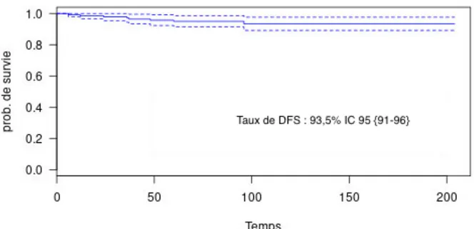
Median follow up was 132 (range 60-204) months. DFS was 93,5% and 9 recurrences occurred in
our cohort (6,5%): 6 metastatic diseases, 2 pelvic nodes recurrences and 1 local recurrence (left
pyramidalis muscle). We did not report any vaginal recurrence.
Figure 1 described the survival curve of the study population.
All the characteristics of recurrences are described in table 2.
The rate of recurrences was also examined according to surgical approach: in the subgroup of
patients with laparoscopic approach, recurrences occurred in 6 of 117 patients (5,1%).
In the subgroup of patients with IB2 stage according to FIGO 2018 classification, we reported 4
recurrences on 64 patients (6,2%).
In univariate analysis, a pathological complete response is associated with a better DFS (p = 0,03
OR = 6,1 IC95 {1,8 ; 55,3}).
The strongest prognostic factor of recurrence (local or metastatic) in our study is the time between
the end of brachytherapy and surgery. If this time is superior to 52 days, it is associated with a
Surgical approach (open surgery or mini invasive surgery) does not impaired the DFS (p=0,11
OR=0,33 IC 95 % { 1,62, 2,774 }).
Concerning initial tumor or patient characteristics, none of the others factors studied (tumor size >
2 cm, histological type or BMI) were statistically significant for DFS.
3.4 Toxicities of brachytherapy and surgery
Complications following therapeutic sequence (brachytherapy followed by surgery) were
encountered in 43 patients (31,1%) distributed as follows:
After brachytherapy, we reported 24 late complications (17,3%), most of them were grade 1 and 2
complications (15,9 %, n = 22) as urinary dysfunctions, dyspareunia and rectitis or proctitis.
We observed 2 grade III complications: 1 vaginal stenosis and 1 uretero vaginal fistula.
Concerning post operative morbidity, we reported 19 complications (13,7 %),
12 grade I - II complications (8,7%) : 3 hematoma, 1 lymphoedema , 7 dysuria, 1 dyspareunia.
7 grade III complications (5%) : 3 abscess to pelvic wall, 2 vaginal scare disunion, 1 ureteral
4. DISCUSSION
Surgical management of early stage cervical cancer using minimal invasive surgery is nowadays
strongly debated in relation with the recent publications of LACC trial (20).
We report in this study our oncological and surgical outcomes after a combined treatment for early
stage cervical cancer (IA1 to IB2 according to FIGO 2018 classification, (21)) using POBT and a
systematic simple hysterectomy, with a majority of laparoscopy (84,8%), and we compare our
results to the recent literature.
With one of the greatest follow up reported in the literature (132 months), we described a DFS of
93,5% (n = 9 recurrences in 138 patients). We observed 6 recurrences in 117 patients with
laparoscopic approach (DFS = 94,8%), in our cohort of patients with a tumor size less than 4 cm,
with a great tumor local control (TLC) (only 1 pelvic recurrence and 2 pelvic lymph nodes
recurrences).
However, recent studies open the debate concerning the oncological safety of the minimally
invasive approach for the management of early stage cervical cancer.
The LACC study (20) showed that Minimally invasive Surgery (MIS) is associated with a poorer
prognostic: after 4,5 years follow up, MIS arm had more recurrences (14%) than open surgery
(3,5%) and more deaths (6,2% versus 1%). Then, several studies have reported their own
retrospective cohort results in the literature that corroborate with LACC’s Trial. Melamed and al.
(23), with a retrospective cohort of 2461 patients diagnosed with IA2 to IB1 stage of cervical
approach compared to open surgery (90,9% vs 94,7%; p = 0.002) with an HR of 1,65 (95% CI,
1,22-2,22).
Cusimano and al (24), examined data from a retrospective cohort of 958 patients between 2006 to
2017, to compare outcomes of patients with stage IA1, IB1 and IIa who underwent upfront surgery
performed by open abdominal versus MIS approaches. Patients with stage IB1 who underwent MIS
had decreased 7 years survival and DFS compared with those undergoing open surgery (HR of 2,20
(95% CI, 1,15 - 4,19); HR of 1,97 (95% CI, 1,10 - 3,50)).
This level A of Evidence Based Medicine should not be ignored, and several centers have disrupted
their daily practices in the management of early stage cervical cancer.
NCCN's new 2019 recommendations (25) report that taking into account these recent studies who
showed poorer oncologic outcomes and survival with the MIS approach, “women should be
carefully counseled about the oncologic risks and potential short-term benefits of the different
surgical approaches”.
At the same time, preoperative brachytherapy is only suggested as an option in the European
(ESMO) and French (SFCO) recommendations (3,5).
However, our rate of DFS corroborate with those observed in the literature:
First, with the publications evaluating preoperative brachytherapy followed by surgery:
UZAN and al (8) in 2012, with a retrospective cohort of 162 patients who underwent POBT
followed by MISRH surgery, reported a DFS of 94,3% after a follow up of 39 months, and Escande
patients who underwent POBT followed by a majority of laparoscopic radical hysterectomy (Piver
type II or III).
Vizkelety and al. (13), in a randomized controlled trial, compared POBT followed by laparoscopic
surgery and upfront laparoscopic surgery, reported no difference, with a DFS of 95,7 % for TLC
and 93,2% for distance relapse for the POBT arm, and a DFS of 97,5 % for the upfront surgery
arm, after a median follow up of 29 months.
Second, several studies have evaluated upfront surgery, and DFS rates are lower than in our study:
For Kim et al. (26) with a median follow up of 113 months, a cohort of 207 Patients and stage IB1
and IB2, after upfront surgery, DFS was statistically different between MISRH approach (78,5%)
and ARH approach (89 %).
Ramirez et al. (20), with a follow up of 54 months presented a DFS of 86% for upfront MISRH
and 96,5% for upfront ARH with a strong RCT of 631 patients (LACC trial).
Even with all the safety procedures, (no uterine manipulator to limit spillage of tumor cells, creation
of a vaginal cuff to exclude the tumor and a minimal handling of the uterine cervix), Doo and al.
(27), described in a retrospective study concerning with a cohort of 105 patients who underwent
upfront surgery, a DFS of 76% for MISRH and 86% for ARH after 25 months. Kanao et al. (28)
published the results of a retrospective study with a cohort of 163 patients treated by upfront MIS
surgery or open surgery. After a median follow up of 30 months, no statistical difference was noted:
DFS of 90,9% for upfront ARH, and 94,4 % for upfront MISRH (p = 0,591).
A complete literature review of series of different treatments including more than 100 patients is
Looking at literature results, and to maintain the place of MIS in the management of early stage
cervical cancer, POBT before surgery may be an alternative to upfront surgery:
First, to optimize the rate of local tumor control before surgery compared to surgery alone.
The rate of pathological complete response (pCR) in our study is 49,3% (68 patients) and local
recurrences are very rare, one pelvic recurrence in our study.
In the literature, pCR rates in cohorts of patients treated with the radio surgical combination are
between 55% (Bruand-Charra (29)), 70% (Escande and al. (6)) and 71,6% (Resbeut and al.(11)).
It probably decreases the risk of tumor spillage during the surgery and reduces local recurrences.
Moreover, in our study, a residual tumor after POBT is a prognostic factor of recurrence.
Second, European and French Guidelines allow for a less radical hysterectomy, type A according
to Querleu and Morlow for the patients with early stage cervical cancer with intermediate or high
risk tumor (size less than 2 cm with LVSI or tumor size between 2 and 4 cm) treated by POBT
(2,3).
By reducing the radicality of the surgery, we minimize the rate of perioperative urinary injuries
with parametrial dissection.
After POBT followed by type A hysterectomy, we report in our study a rate of surgical urinary
injuries of 6,5% (n=9), 7 grade I - II complications (5%) and only 2 (1,5%) grade III complications.
With upfront surgery (Piver II or III hysterectomy), literature show a rate of grade III - IV
complications (stenosis or fistulas) more than 5% (Pellegrino and al. 10% (30), Lee and al. 5%
Moreover, Landoni and al. (32) has demonstrated that a reduction of the radicality for hysterectomy
offered similar oncological outcomes without differences concerning recurrences, but also a
decrease of urinary toxicities (28% for Piver III vs 13 % for Piver II).
Third, POBT minimize the need of adjuvant EBRT (External Beam Radiotherapy), by reducing
the histological factors of poor prognosis, such as LVSI, parametrial invasion (estimated at less
than 1% in several studies), marginal resection or tumor size.
Atlan and al. (14) compared POBT followed by surgery with upfront surgery followed par pelvic
radiotherapy. The author showed a significative higher rate of grade 3 - 4 late toxicities
complications in the group with adjuvant radiotherapy (22% versus 7% p = 0,0002).
In others retrospective studies evaluating radical upfront hysterectomy for the management of early
stage cervical cancer, the rate of adjuvant treatment for IB1 and IB2 patients are between 24%
(Pellegrino and al.30) to 39% (Yan and al. (18)).
Thus, in most patients treated by upfront surgery for cervical cancer with poor prognostic factors
(tumor size > 2 cm or LVSI), the need for postoperative radiotherapy could be avoided by using
preoperative brachytherapy.
POBT brachytherapy reduces poor prognostic factors: in our cohort, only 3 patients (2,2%) have
post operative LVSI after surgery, whereas in the literature, the rate of LVSI in patients with
early-stage cervical cancer (tumor size < 4 cm) is approximately 26% (Dabi and al.) or 35% (Wright et
al.).
POBT brachytherapy reduces the rate of positive margins after surgery. In our cohort, only one
These results corroborate those of the prospective randomized study of Vizkelety and al. (13)
comparing in a RCT, upfront surgery to POBT in which positive margins was observed for 1,5%
for patients with POBT followed by surgery, versus 11,5% of patients with upfront surgery.
So, we therefore report a benefit of preoperative brachytherapy in the management of early stage
cervical cancer (tumor size less than 4 cm).
Nevertheless, the current trend is to decrease the radicality of surgery, even in the case of exclusive
surgery, in selected patients (SHAPE trial, phase III, in progress, aiming to evaluate the oncological
safety of a simple hysterectomy compared to a radical hysterectomy in patients with early stage
cervical cancer < 2 cm, without risk factor of recurrence).
In addition, in the LACC trial, analysis of the subgroup of patients with tumors less than 2 cm in
size showed no statistically significant difference between the open and minimally invasive
approach, suggesting the possibility of maintaining the minimally invasive approach for this
subgroup of patients.
One may assume, for optimal management of these patients:
an exclusive first surgery by minimally invasive approach to patients with low-risk cervical cancer
(< 2 cm without LVSI or lymph node invasion) and offer preoperative brachytherapy to patients
with cervical tumors < 2 cm with risk factors of recurrence and tumors between 2 and 4 cm,
followed by a minimally invasive type A hysterectomy, as an alternative to a more radical
abdominal hysterectomy.
Then, a quality preoperative evaluation of tumor size and invasion of the parameters is necessary.
The new FIGO classification system (2018) (21) makes it possible to regain this precision, with
This new staging system also proposes a subdivision of the former stage IB, grouping patients with
a tumor size < 4 cm into IB1 (tumor size < 2 cm), IB2 (tumor size between 2 and 4 cm) and IB3
(>3 cm) and removes from this category patients with lymph node involvement, for an accurate
evaluation of a treatment plan adapted to each patient.
Brachytherapy
One of the most important point is the delay between POBT and surgery. Our study found that an
interval time more than 52 days between brachytherapy and surgery was a risk factor of recurrence.
This factor had been already suggested by Escande and al. in a study published in 2016 (p=0,03).
Concerning POBT morbidity, it has previously been investigated and appears to be very low,
particularly since the era of radio-guided BT (6-14).
In our study, long term morbidity of brachytherapy concerned 24 patients (17,3%), with a majority
of grade I and II complications (22 patients 15,9%), a majority of dyspareunia (11 patients (8%)).
Moreover, cervical cancer is a great indication for brachytherapy, since the source is easily
implanted by the vagina and the surrounding organs at risk (bladder, rectum, sigmoid) are
radioresistant. Finally, SCC (more than 90% of all cervical tumors) is commonly considered as
radiosensitive (35).
Lately, the main evolution in gynecological brachytherapy has been the development of
tridimensional (3D) dosimetry with optimization, realized on MRI or CT imaging and then the
The dosimetric scanner or MRI allows the delineation of target volumes and organs at risk (OAR)
according to the recommendations of the European group in Gynecological brachytherapy
(GYN-GEC-ESTRO) (36,37).
A French prospective multicenter study, conducted by Charra-Brunaud in 2012 (29), showed that
brachytherapy based on CT and MRI improved the rate of local control of the disease and reduced
morbidity.
Two-dimensional brachytherapy using the International Commission on Radiation Units and
Measurements (ICRU) (22) points for the bladder and rectum underestimates (or sometimes
overestimates) the dose delivered to organs at risk (Pelloski and al. (38)).
In our comprehensive center in Marseille, low dose rate intrauterine brachytherapy with 2D
treatment plan using guidelines of ICRU for the plan treatment, is no longer used since 2012, and
currently the main technique is high dose rate brachytherapy with tridimensional CT scanner
according to the GEC ESTRO guidelines (36,37).
This may account for the low rate of pathological complete response (pCR) in our study, compares
to the literature (49,3% for Marseille versus 75,3% for Uzan and al.).
5. LIMITATIONS
The retrospective nature of the study constitutes its most important limitation which impaired data
6. CONCLUSION
With a median follow up of 132 months, a radio surgical management of early stage cervical
cancers (less than IB3) including brachytherapy and type A laparoscopic hysterectomy seems to
offer a reasonable rate of recurrences compared to LACC trial’s results. This strategy may be an
alternative to upfront radical hysterectomy in selected patients to maintain a minimal invasive
Bibliography
1) NCCN GUIDELINES®: Cervical Cancer, Version 3.2019 12/17/18 © National
Comprehensive Cancer Network, Inc. 2019 Journal of the National Comprehensive Cancer
Network J Natl Compr Canc Netw 17, 1; 10.6004/jnccn.2019.0001
2) Cibula D, Pötter R, Planchamp F, Avall-Lundqvist E, Fischerova D, Haie-Meder C et al.
The European society of Gynaecological Oncology /European society for radiotherapy and
oncology/ European society of Pathology Guidelines for the management of patients with
cervical cancer. Airshows Arch 2018; 472: 919-36.
3) Marth C, Landoni F, Mahner S, McCormack M, Gonzalez-Martin A, Colombo N On behalf
of the ESMO Guidelines Committee. Cervical cancer: ESMO Clinical Practice Guidelines
for diagnosis, treatment and follow-up. Annals of Oncology 28 (Supplement 4): iv72–iv83,
2017 doi:10.1093/annonce/
4) Querleu D, Cibula D, Abu-rostum NR, 2017 Update on the Querleu morrow classification
of radical hysterectomy. Ann surgery oncol 2017; 24: 3406-12.
5) Azaïs H, Canlorbe G, Belghiti J, Maingon P « Treatment of early stage cervical cancer»
EMC -Gynecologie 2019;14(3):1-16 (Article 605:1:57).
6) Escande A, Gouy S, Mazeron R, Bentivegna E, Bacorro W, Maroun P, Schernberg A et al.
“Outcome of Early Stage Cervical Cancer Patients Treated According to a Radiosurgical
Approach: Clinical Results and Prognostic Factors” Gynecologic Oncology 144, nᵒ 3, mars
7) Haie-Meder C, Chargari C, Rey A, Dumas I, Morice P, Magné N “DVH Parameters and
Outcome for Patients with Early-Stage Cervical Cancer Treated with Preoperative
MRI-Based Low Dose Rate Brachytherapy Followed by Surgery” Radiotherapy and Oncology
93, nᵒ 2, novembre 2009; 316:21.
8) Uzan C, Nikpayam M, Merlot B, Gouy S, Belghiti J, Haie-Meder C, Nickers P, Narducci
F, Morice P, Leblanc E “Laparoscopic radical hysterectomy after preoperative
brachytherapy for stage IB1 cervical cancer: feasibility, results, and surgical implications
in a large dicentric study of 162 consecutive cases” Ann. Surg Oncol 2013; 20: 872-80.
9) Lamblin G, Rouffiac M, Mathevet P, Martin E “Surgery alone or in association of with
preoperative low dose rate brachytherapy for stage IB1 cervical cancer: toxicities profiles”
Gynecol obst fertiles 2015; 43: 485-90.
10) Ngo C, Alran S, Plancher C ,Fourchotte V , Petrow P, Campitelli M et al. “Outcome in
early cervical cancer following pre operative low dose rate brachytherapy: a ten year follow
up of 257 patients treated in a single institution” Gynecologic oncology 2011; 123:
248-52.
11) Resbeut M, Alzieu C, Gonzague-Casabianca L et al. “Combined brachytherapy and surgery
for early carcinoma of the uterine cervix: analysis of extent of surgery on outcome” Int.
Journal of Radiation Oncology.Biol.Phys. 50,2001; 873-881.
12) Lacorre A, Merlot B, Garabedian C et al. “Early stage cervical cancer: brachytherapy
followed by type a hysterectomy versus type B radical hysterectomy alone, a retrospective
evaluation” Eur. J. Surg. Oncol. 42,2016; 376-382.
13) Vízkeleti, J, Vereczkey, I, Fröhlich G et al. “ Pathologic Complete Remission after
Preliminary Results of a Prospective Randomized Multicenter Study” Pathol. Oncol. Res.
2015; 21: 247
14) Atlan D, Touboul E, Deniaud-Alexandre E et al. “Operable stages IB and II cervical
carcinomas: a retrospective study comparing preoperative uterovaginal brachytherapy and
postoperative radiotherapy” Int. Journal of Radiation Oncology.Biol.Phys. 2002; 54:
780-93.
15) Dindo D, Demartines N, Clavien PA “Classification of surgical complications: a new
proposal with evaluation in a cohort of 6336 patients and results of a survey” Ann Surg
2004; 240(2):205-213.
16) Chassagne D, Sismondi P, Horiot JC, Sinistrero G, Bey P, Zola P, Pernot M, Gerbaulet A,
Kunkler I, Michel G “A glossary for reporting complications of treatment in gynecological
cancers Author links open overlay panel” Radiotherapy and oncology, volume 26, Issue 3
, March 1993.
17) Geisler JP, Orr CJ, Khurshid N, Phibbs G, Manahan KJ “Robotically assisted laparoscopic
radical hysterectomy compared with open radical hysterectomy” Int J Gynecol Cancer
2010; 20: 438-42.
18) Yan X, Li G, Shang H et al. “Twelve-year experience with laparoscopic radical
hysterectomy and pelvic lymphadenectomy in cervical cancer” Gynecol Oncol. 2011; 120:
362.
19) Geetha P, Nair MK “Laparoscopic, robotic and open method of radical hysterectomy for
cervical cancer : a systematic review” J. min Access Surg 2012; 8:67-73.
20) Ramirez P, Pedro T, Frumovitz M, Pareja R, Lopez A, Vieira M, Ribeiro R, Buda A, et al.
“Minimally Invasive versus Abdominal Radical Hysterectomy for Cervical Cancer” New
21) Bhatla N, Aoki D, Sharma DN, Sankaranarayanan R “Cancer of the cervix uteri” Int. J.
Gynaecol. Obstet. 143, 2018; 22-36.
22) International Commission on Radiation Units and Measurements. ICRU report 38: dose
and volume specification for reporting intracavitary therapy in gynaecology. Bethesda,
MD: ICRU; 1985.
23) Melamed A, Margul J, Chen L, Keating N, Del Carmen MG, Yang J “Survival after
minimally Invasive Radical Hysterectomy for early stage cervical cancer” New England
Journal of Medicine 379, nᵒ 20, 15 nov. 2018.
24) Cusimano MC, Baxter NN, Gien LT and al. “Impact of surgical approach on oncologic
outcomes in women undergoing radical hysterectomy for cervical cancer” Am J Obstet
Gynecol 2019; 221: 619, e1-24.
25) Pennington KP, Urban RR, Gray HJ, “Revisiting Minimally Invasive Surgery in the
Management of Early-Stage Cervical Cancer” JNCCN vol.17, 1, jan. 2019.
26) Kim Se Ik, Jae Hyun Cho, Aeran Seol, Young Im Kim, Lee M, Hee Seung Kim, Hyun
Hoon Chung, Jae-Weon Kim, Noh Hyun Park and Yong-Sang Song “Comparison of
survival outcomes between minimally invasive surgery and conventional open surgery for
radical hysterectomy as primary treatment in patients with stage IB1–IIA2 cervical cancer”
Gynecologic Oncology 153, 1, 1 ap. 2019; 3-12.
27) Doo D, Tyler Kirkland C, Griswold LH, McGwin G, Huh WK, Leath CA Third, Kenneth
H. Ki8. “Comparative outcomes between robotic and abdominal radical hysterectomy for
IB1 cervical cancer: Results from a single high volume institution” Gynecologic Oncology
153, mar.2019; 242-247.
No-Touch Technique for FIGO IB1 Cervical Cancer” Journal of Gynecologic Oncology
30, nᵒ 3, 2019; e71.
29) Charra-Brunaud C, Harter V, Delannes M, Haie-Meder C, Quetin P, Kerr C et al. “Impact
of 3D image-based PDR brachytherapy on outcome of patients treated for cervix carcinoma
in France : results of the french stic prospective study” Radiother Oncol 2012; 103:
305-13.
30) Pellegrino A, Vizza E, Fruscio R et al. “Total laparoscopic radical hysterectomy and pelvic
lymphadenectomy in patients with Ib1 stage cervical cancer: analysis of surgical and
oncological out- come” Eur J Surg Oncol. 2009; 35: 98-103.
31) Lee CL, Wu KY, Huang KG, Lee PS, Yen CF “Long-term survival outcomes of
laparoscopically assisted radical hysterectomy in treating early-stage cervical cancer” Am
J Obstet Gynecol. 2010; 203:165.e1-7.
32) Landoni F, Maneo A, Zapardiel I, Zanagnolo V, Mangioni C “Class I versus class III radical
hysterectomy in stage IB1-IIA cervical cancer. A prospective randomized study” Eur J
Surg Oncol 2012; 38(3): 203-9.
33) Dabi Y, Willecocq C, Ballester M, Carcopino X, Bendifallah S, Ouldamer L, Lavoue V,
Canlorbe G, Raimond E, Coutant C, Graesslin O, Collinet P, Bricou A, Huchon C, Daraï
E, Haddad B, Touboul C “Identification of a low risk population for parametrial invasion
in patients with early-stage cervical cancer” Groupe de Recherche FRANCOGYN J Transl
Med. jun.14, 2018; 16(1):163. doi: 10.1186/s12967-018-1531-6.4
34) Wright JD, Grigsby PW, Brooks R, Powell MA, Gibb RK, Gao F, Rader JS, Mutch DG
“Utility of Parametrectomy for Early Stage Cervical Cancer Treated With Radical
35) Huertas A, Oldrini S, Nesseler JP, Courrech F, Retie P, Charra-Brunaud C, Peiffert D
“FIGO Stage IB1 cervical carcinoma : Place and principles of brachytherapy” Société
française de radiothérapie oncologique (SFRO), 2017; 1278- 3218.
36) Potter R, Haie-Meder C, Van Limbergen E, Barillot I, De Brabandere M, Dimopoulos J, et
al. “Recommendations from gynaecological (GYN) GEC-ESTRO working group (II):
concepts and terms in 3D image-based treatment planning in cervix cancer
brachytherapy-3D dose volume parameters and aspects of brachytherapy-3D image-based anatomy, radiation physics,
radiobiology” Radiother Oncol 2006; 78(1):67-77.
37) Haie-Meder C, Potter R, Van Limbergen E, Briot E, De Brabandere M, Dimo- poulos J et
al. “Recommendations from gynaecological (GYN) GEC-ESTRO working group (I):
concepts and terms in 3D image based 3D treatment planning in cervix cancer
brachytherapy with emphasis on MRI assessment of GTV and CTV” Radiother Oncol
2005; 74(3): 235–45.
38) Pelloski CE, Palmer M, Chronowski GM, Jhingran A, Horton J, Eifel PJ “Comparison
between CT-based volumetric calculations and ICRU reference- point estimates of
radiation doses delivered to bladder and rectum during intracavitary radiotherapy for
cervical cancer” Int J Radiat Oncol Biol Phys 2005; 62: 131-7.
39) Alfonzo E, Wallin E, Ekdahl L, Staf C, Radestad AF, Reynisson P, Stalberg K, Falconer
H, Persson J, Dahm-Kahler P “No survival difference between robotic and open radical
hysterectomy for women with early-stage cervical cancer: results from a nationwide
population-based cohort study” European Journal of Cancer 116, 2019; 169-177.
40) Matsuo K, Machida H, Mandelbaum RS, Konishi I, Mikami M “Validation of the 2018
87-41) Benedetti-Panici P, Maneschi F, D’Andrea G, Cutillo G, Rabitti C, Congiu Ferdinando Coronetta M, Capelli A “Early Cervical Carcinoma The Natural History of Lymph Node Involvement Redefined on the Basis of Thorough Parametrectomy and Giant Section Study” American cancer society, may 2000, vol.88, 10.
42) Sala E, Rockall AG, Freeman SJ, Mitchell DG, Reinhold C. “The added role of MR imaging
in treatment stratification of patients with gynecologic malignancies: what the radiologist
SERMENT D’HIPPOCRATE
En présence des Maîtres de cette Faculté, de mes chers condisciples et devant l’effigie d’HIPPOCRATE,
Je promets et je jure d’être fidèle aux lois de l’honneur et de la probité dans l’exercice de la Médecine.
Je donnerai mes soin s gratuitement à l’indigent et n’exigerai jamais un salaire au dessus de mon travail. Je ne participerai à aucun partage clandestin d’honoraires.
Admis dans l’intimité des maisons, mes yeux n’y verront pas ce qui s’y passe ; ma langue taira les secrets qui me seront confiés et mon état ne servira pas à corrompre les mœurs, ni à favoriser le crime.
Je ne permettrai pas que des considérations de religion, de nation, de race, de parti ou de classe sociale viennent s’interposer entre mon devoir et mon patient.
Je garderai le respect absolu de la vie humaine.
Même sous la menace, je n’admettrai pas de faire usage de mes connaissances médicales contre les lois de l’humanité.
Respectueux et reconnaissant envers mes Maîtres, je rendrai à leurs enfants l’instruction que j’ai reçue de leurs pères.
Que les hommes m’accordent leur estime si je suis fidèle à mes promesses. Que je sois couvert d’opprobre et méprisé de mes confrères si j’y manque.