Contents lists available atScienceDirect
Journal of Functional Foods
journal homepage:www.elsevier.com/locate/jffDifferential effects of inulin or its fermentation metabolites on gut barrier
and immune function of porcine intestinal epithelial cells
Julie Uerlings
a,c, Martine Schroyen
a, Els Willems
b, Sofie Tanghe
b, Geert Bruggeman
b,
Jérôme Bindelle
a, Nadia Everaert
a,⁎aPrecision Livestock and Nutrition Unit, TERRA Teaching and Research Centre, Gembloux Agro-Bio Tech, ULiège, Gembloux, Belgium bRoyal Agrifirm Group, Apeldoorn, the Netherlands
cResearch Foundation for Industry and Agriculture, National Scientific Research Foundation (FRIA-FNRS), Brussels, Belgium
A R T I C L E I N F O Keywords:
Prebiotic In vitro fermentation
Porcine intestinal epithelial cells Short-chain fatty acids Gut barrier Inflammatory response
A B S T R A C T
Prebiotics can modulate gut fermentation and improve intestinal barrier function in mammals. First, inulin fermentation profile was tested in a three-step in vitro model of the piglet’s gastro-intestinal tract combining a hydrolysis – dialysis step to a batch fermentation. Then, the differential effects of digested inulin (after the hydrolysis – dialysis steps) or fermented inulin (after the fermentation step) on the expression of gut barrier and immune-related genes of IPEC-J2 cells were investigated by high-throughput qPCR. Inulin was associated with elevated short-chain fatty acids and butyrate levels. Upregulated expressions of tight and adherens junction genes were observed in IPEC-J2 cells supplemented with inulin fermentation supernatant compared to control IPEC-J2 cells and digested inulin. Therefore, metabolites arising from the fermentation process, including bu-tyrate, could be responsible for the reinforcement of the barrier function.
1. Introduction
To date, prebiotics represent a wide-spread dietary approach to modulate intestinal fermentation and manipulate gut ecology for health purposes (Roberfroid, 2007). The most commonly used prebiotics, in humans (Roberfroid, Van Loo, & Gibson, 1998) and pigs (Samanta, Jayapal, Senani, Kolte, & Sridhar, 2013) diets, are fructo-oligo-saccharides such as inulin. Prebiotics help creating and maintaining an optimal environment in the host gastro-intestinal tract by selectively stimulating the proliferation and metabolic activity of health-associated microbiota communities and lowering the pathogenic bacteria load (Gibson & Roberfroid, 1995). The fermentation of prebiotics by the endogenous microbiota yields short-chain fatty acid (SCFA) end-pro-ducts, acetate, propionate, and butyrate. The latter is of special interest for its anti-inflammatory properties (Canani et al., 2011) and is used as energy source by the colonocytes (Roediger, 1982), leading to a re-inforcement of the intestinal barrier integrity (Peng, Li, Green, Holzman, & Lin, 2009). Moreover, a reduction of butyrate and butyrate-producing bacteria has been related to colonic-derived diseases such as inflammatory bowel disease (Canani et al., 2011) and Crohn’s disease (Sokol et al., 2008). Alternatively, acetate and propionate may influ-ence cholesterol metabolism (Chen, Anderson, & Jennings, 1984) and
appetite regulation (Chambers, Morrison, & Frost, 2015; Frost et al., 2014), respectively. The ratio between individual SCFA and the total amount of SCFAs produced vary with the source of prebiotic (Grootaert et al., 2009; Van den Abbeele, Venema, Van de Wiele, Verstraete, & Possemiers, 2013).
There is increasing evidence of a strong interaction between the intestinal barrier mucosa, the gut microbiota and the local immune system (Takiishi, Fenero, & Câmara, 2017). The intestinal mucosa, with lined-up enterocytes joined by tight and adherens junctions, together with a mucus layer, creates a physical barrier allowing the absorption of nutrients while preventing bacterial translocation (Ulluwishewa et al., 2011). This dynamic structure is constantly remodelled due to inter-actions with feed residues, metabolites, as well as with pathogenic and commensal bacteria. This way, a compromised barrier integrity could trigger an exacerbated inflammatory response. Alternatively, the use of prebiotics to preserve the intestinal mucosal barrier function has gained considerable interest in the recent years and inulin has been postulated to enhance gut barrier integrity in vitro (Van den Abbeele et al., 2018) and in vivo (Russo et al., 2012) in humans.
So far, many studies focused on the action of prebiotics on the in-testinal barrier mucosa via direct signalling routes initiated by the oligosaccharides themselves, and much less by their bacterial
https://doi.org/10.1016/j.jff.2020.103855
Received 5 November 2019; Received in revised form 22 January 2020; Accepted 13 February 2020 ⁎Corresponding author at: Gembloux Agro-Bio Tech, Passage des Déportés, 2, 5030, Gembloux, Belgium.
E-mail address:nadia.everaert@uliege.be(N. Everaert).
1756-4646/ © 2020 The Authors. Published by Elsevier Ltd. This is an open access article under the CC BY license (http://creativecommons.org/licenses/BY/4.0/).
metabolites. Several mechanisms have been attributed to this direct effect on gut health, such as interference with pathogenic attachment (Sun, Gänzle, & Wu, 2019), interaction with recognition molecules (Shoaf, Mulvey, Armstrong, & Hutkins, 2006) and influence on cytokine transduction pathways (Ortega-González et al., 2014; Zenhom et al., 2011). However, studies conducted with single bioactive compounds do not take into account the fermentation process that may affect the physiochemical properties of the ingredient and the possible synergistic activities between metabolites (Nielsen et al., 2018). Many in vitro models have been investigating the host-prebiotic or the host-metabo-lite interactions separately. Detailed evidence is still missing on which health-beneficial effects should be attributed to the direct and/or the prebiotic effects of dietary fibre ingredients in the intestine. Owing to the complexity of the intestinal chyme and lumen, it is important to implement a mechanistic, although holistic approach combining an in
vitro fermentation model of the gastro-intestinal tract with intestinal
epithelial cell cultures, studying the effect of bioactive compounds on cell lines. In this study, the intestinal epithelial cell line (IPEC-J2), of porcine origin, was chosen to represent the intestinal wall in the in vitro model of the young piglet’s gastro-intestinal tract. Although the IPEC-J2 cell line is derived from the small intestine of young piglets, no colonic cell line from porcine origin is available. Moreover, the in vitro batch fermentation was prepared with feces of young piglets which is a sui-table and representative inoculum to mimic the in vivo gut fermentation (Williams, Voigt, & Verstegen, 1998) and justifies the model chosen for the following research.
The aim of the study was to evaluate the prebiotic potential of inulin to modulate intestinal fermentation for young mammalian health pur-poses and to investigate if inulin digesta (inulin DI) or inulin fermen-tation metabolites (inulin FS) induced differential effects on intestinal immunity and barrier function using IPEC-J2 gene expression re-sponses.
2. Materials and methods
2.1. Analysis of inulin
Inulin (Fibruline Instant) was provided by Cosucra Warcoing SA (Belgium). It was analyzed for organic matter (AOAC 923.03), dry matter (AOAC 967.03), crude protein (Foss Kjeltec Analyzer Unit 2300, Hilleroed, Denmark; CP = N × 6.25), fat content (Soxhlet method; AOAC 920.29) and neutral (NDF) and acid (ADF) detergent fibre (Foss Fibrecap system, Hilleroed, Denmark;Van Soest, Robertson, and Lewis (1991)). Non-cellulosic total monosaccharides composition was de-termined according to the method of Englyst and Cummings (1984) adapted by Aguedo, Fougnies, Dermience, and Richel (2014). The fructan molecular mass distribution was assessed by size-exclusion high performance liquid chromatography (HPSEC) performed with a Waters 2690 Alliance chromatograph (Waters, Milford, USA) coupled to a re-fractometer Waters 2410 as previously described (Aguedo et al., 2014).
2.2. In vitro digestion and batch fermentation of inulin
Inulin was studied using a modified three-step in vitro model of the pig’s gastro-intestinal tract (Bindelle, Buldgen, Boudry, & Leterme, 2007) combining an enzymatic hydrolysis and dialysis to a batch fer-mentation with fecal microbiota (Uerlings et al., 2019a).
For the batch fermentation, a fecal inoculum was prepared from a buffer solution composed of salts and minerals (pH 6.8;Menke and Steingass (1988)) devoid of reducing agent (Poelaert et al., 2018) and frozen feces (2.5% [w/v]) from piglets, under anaerobic conditions (Invivo2, Led Techno, Heusden-Zolder, Belgium). Feces were previously collected from pre-weaned three week-old-piglets (male and female) by fecal stimulation with sterile swabs. All experimental procedures led on piglets (feces collection) were in accordance with European and Belgian regulations concerning the care and use of animals for research
purposes and were approved by the Animal Ethical Committee of Liège University, Belgium (protocol number: 1860). They were carried out in accordance with the U.K. Animals Act, 1986 and EU Directive 2010/ 63/EU for animal experiments.
Three mucin microcosms (Tran et al., 2016) were added to each fermentation vial to enhance the growth of communities found on mucus, with inulin digesta or not (blank vials). The vials (n = 3 for the inulin or blank treatments per time-point) were placed into an agitating water-bath at 39 °C with 50 rpm agitation and the fermentation su-pernatants were stored at −80 °C.
2.3. Fermentation kinetics profile of the in vitro batch fermentation
The released gas volumes (n = 3 vials for the inulin or blank treatments) were repeatedly recorded with a Tracker 200 manometer (Bailey & Mackey Ltd, Birmingham, UK) at following time points; 2, 5, 8, 12, 16, 20, 24, 48 and 72 h according to the model ofGroot, Cone, Williams, Debersaques, and Lantinga (1996) and gas production re-cordings were fitted to the mathematical monophasic model, with A (mL / g DM) as the maximum gas volume, G (mL / g DM) as the gas accumulation to time, B (h) as the time to half asymptote when G = A/ 2, RMAXas the maximum rate of gas production (mL / g DM * h) and TMAX, the time to reach RMAX.
2.4. Short-chain fatty acid profile of supernatants from in vitro batch fermentation
Fermentation broths sampled after 6, 12 and 24 h of fermentation (n = 3 vials for the inulin or blank treatments) were analyzed by iso-cratic high-performance liquid chromatography (HPLC) using the Alliance System e2695 (Waters, Milford, CT) with an Aminex HPx-87H column (BioRad, Hercules, CA) as previously described (Uerlings et al., 2019a).
2.5. Inulin digesta (DI) preparation
Inulin digesta (inulin DI), pooled from 8 experimental units that had undergone the in vitro hydrolysis and dialysis steps, was diluted using sterile PBS to reach 0.5% [w/v] as final concentration. Then, the sus-pension was sonicated in a water bath (3 times of 30 s each) and cen-trifuged (460 g, 5 min) as described byBecker, Galletti, Roubos-van den Hil, and Van Wikselaar (2007). Inulin DI was frozen at − 20 °C until further application on IPEC-J2 cells.
2.6. Fermentation supernatant (FS) preparation
Inulin fermentation broths (inulin FS) from 3 different fermentation vials as well as fermentation broth from 3 blank fermentation vials (blank FS) were pooled after 12 h of fermentation, were sterile-filtered with 0.22-µm pore filters and were stored at −80 °C until further ap-plication on IPEC-J2 cells.
2.7. IPEC-J2 cell line and culture conditions
IPEC-J2 cells were grown at 37 °C in a humidified atmosphere of 5% CO2 in complete Dulbecco’s Modified Eagle Medium DMEM/F-12, supplemented with 1% penicillin–streptomycin, 5% fetal bovine serum, 2 mM L-glutamine, 5 ng / mL epidermal growth factor, 5 μg / mL in-sulin, 5 μg / mL transferrin and 5 ng / mL selenium (all from Sigma, Saint Louis, MO). Culture medium was renewed once every two days, and cells were passaged when they reached confluence.
2.8. Measuring IPEC-J2 cell viability
The viability test (n = 6 culture well-replicates per treatment) was used to determine the concentrations of inulin DI, inulin FS and blank
FS suitable to use in the gene expression study. Cell proliferation was measured with a 3-(4,5-dimethylthiazol-2-yl)-2,5-diphenyltetrazolium bromide (MTT) assay. IPEC-J2 cells between passages 15 and 20 were seeded in 96-well flat bottomed plates at a density of 20 000 cells / 100 μL (100 µL per well). Cells were allowed to adhere for 24 h until confluence was reached and were re-fed with experimental media without antibiotics before being treated with different concentrations of blank FS and inulin FS (2.5, 5, 10, 15, 50% [v/v]) or inulin DI (0.25, 0.5, 0.75, 1, 1.25, 2.50% [w/v] (Becker et al., 2007)). After incubation with different concentrations of supernatants for 24 h, the culture medium was removed. Next, fresh antibiotic-free culture medium and 15 μL of MTT reagent (Promega, Madison, WI) were added to each well for another 4 h at 37 °C prior to measurement of cell viability. The absorbance was determined at 570 nm in a micro-plate reader (VICTOR plate reader, PerkinElmer, Waltham, MA). According to the cell viabi-lity test and to the literature using a similar methodology (Borowicki et al., 2010; Fässler, Gill, Arrigoni, Rowland, & Amadò, 2007; Stein, Borowicki, Scharlau, & Glei, 2010), blank FS, inulin FS and inulin DI were applied at 10% [v/v] for the FS and 0.5% [w/v] for the DI.
2.9. Expression of barrier function and immune-related genes in IPEC-J2
IPEC-J2 cells between passages 15 and 20 were seeded in 24-well plates at a density of 2.5 × 10⁵cells / mL (1 mL per well). Prior to the treatment, confluent monolayers of the IPEC-J2 cells were washed with plain medium without antibiotics. Blank FS, inulin FS and inulin DI were applied at 10% [v/v] for the FS and 0.5% [w/v] for the DI for 24 h (n = 3 culture well-replicates per treatment). For sham-stimulation, cells were maintained in the culture medium for 24 h (n = 3).
Total RNA from IPEC-J2 cells treated with the blank FS, inulin FS, inulin DI and control cells was extracted using the RNeasy Mini kit (RNeasy Mini Kit, Qiagen, Hilden, Germany) as previously described (Uerlings et al., 2019b). Extracted RNA was converted into cDNA by reverse transcription using Reverse Transcription Master Mix (Fluidigm Corporation, San Francisco, CA). High-throughput qPCR was performed as previously described (Uerlings et al., 2019b) with intron spanning primer pairs (Table 1). High-throughput qPCR was performed in 48x48 dynamic array integrated fluidic circuits (Fluidigm Corporation, San Francisco, CA). After loading, the dynamic array was placed in BioMark HD Real-Time PCR System (Fluidigm Corporation, South San Francisco, CA), and the following cycle parameters were used: 60 s at 95 °C, fol-lowed by 35 cycles (5 s at 96 °C and 20 s at 60 °C).
Quantification cycles (Cq) were acquired using the Fluidigm real-time PCR analysis software 3.0.2 (Fluidigm Corporation, San Francisco, CA). The geometric mean of four reference genes (ribosomal protein L13a (RPL13a), glyceraldehyde-3-phosphate dehydrogenase (GAPDH), peptidylprolyl isomerase A (PPIA) and tyrosine 3-monooxygenase/ tryptophan 5-monooxygenase activation protein zeta (YWHAZ)) stably expressed between treatments using NormFinder (Andersen, Jensen, & Ørntoft, 2004) was used to normalize samples. For each target, the relative expression level was calculated by the 2-ΔΔctmethod (Livak & Schmittgen, 2001).
2.10. Statistical analysis
Homogeneity between variances and normality among treatments was confirmed using Bartlett’s and Ryan-Joiner’s tests, respectively. The experimental unit for SCFA analysis was the fermentation vial and the one for the gene expression assay was the cell culture well. The ex-perimental data were subjected to GLM procedures and the comparison of means was evaluated by post-hoc Tukey’s multiple range HSD using SAS 9.4 software (SAS Institute) with one fixed criteria of classification for the gene expression assay (type of treatment). The analyses of SCFA were performed similarly. However, the procedure included two fixed criteria of classification (type of ingredient and sampling time) as well as their interaction. For SCFA profiles, when a significant interaction
was found, parameters were studied by one-way ANOVA per time point. Adjusted p-values for the gene expression assay were obtained using a false discovery rate (FDR) correction with the linear method of Benjamini and Hochberg. P-values < 0.05, < 0.01 and < 0.001 were considered as statistically significant, highly significant and very highly significant.
3. Results
3.1. Inulin and its fermentation kinetics
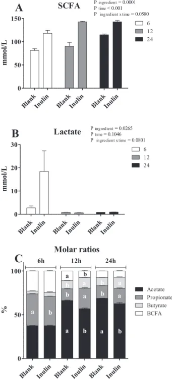
Inulin contained high amounts of fructans (89.4%;Table 2) and was characterized by a weight average molecular weight (Mw) of 5690 Da (Fig. 1). The ingredient induced a total gas production of 300 mL g−1 DM (A) and a maximal rate of fermentation of 27.4 mL g−1DM h−1 (RMAX). The time to reach RMAXwas 4.4 h (TMAX) and the half-time to asymptotic gas production was 7.3 h (B) as displayed infigure S1. The interaction between the treatments and the time of fermentation was significant for all the measured metabolites except for lactate and total SCFA amounts (Fig. 2A and B). After 12 and 24 h, fermentation of inulin induced greater molar ratio of propionate and butyrate in comparison to the blank vials (P < 0.001;Fig. 2C) which consequently showed higher acetate proportions.
3.2. IPEC-J2 cell viability
In order to choose the most appropriate concentrations of fermen-tation supernatants and digesta for the IPEC-J2 model, a cell viability assay was conducted (figure S2A and S2B). Inulin DI induced a reduc-tion of the cell viability under 50% with a concentrareduc-tion superior to 0.75% [w/v] (figure S2A) and 0.5% [w/v] was chosen as the con-centration for the immunomodulatory model. Inulin and blank FS col-lected after 12 h of fermentation were not toxic for IPEC-J2 at a con-centration lower than 25% [v/v] with a reduction of the cell viability around 50% at the cited concentration (EC50). A concentration of 10% [v/v] led to a reduction of approximately 30% of cell viability for both supernatants (figure S2B). According to these results, 10% [v/v] was chosen as FS concentration for the immunomodulatory model.
3.3. Expression of barrier function and immune-related genes in IPEC-J2
Beta-2-microglobulin (B2M), espin (ESPN), hydroxymethylbilane synthase (HBMS), interferon beta (IFNβ), interleukin 1-beta (IL1β) and proliferating cell nuclear antigen (PCNA) genes showed low expressions in the high-throughput qPCR and were therefore undetectable. Chemokine ligand 5 (CCL5) and monocyte chemoattractant protein 1 (MCP1) primers’ efficiencies did not range between 90% and 110% and their results were excluded from the study.
Concerning the inflammation signalling pathways and the pro-in-flammatory proteins (Fig. 3A and B), the mRNA levels of all 18 target genes were similar between inulin DI and the control cells except for the serine/threonine-protein kinase 1 (AKT1) and the C-X-C motif chemo-kine 10 (CXCL10) which were significantly higher and the nuclear factor-kappa B inhibitor alpha (NF-kBIα) and the tumor necrosis factor alpha (TNFα) which were significantly lower for inulin DI compared to the control cells (P < 0.001;Fig. 3A and B). The mitogen-activated protein kinase 14 (MAPK14), myeloid differentiation primary response 88 (MyD88), nucleotide-binding oligomerization domain-containing protein 1 (NOD1), defensin beta 1 (DEFβ1), defensin beta 4a (DEFβ4a), interleukin-18 (IL18), TNFα and the peroxisome proliferator-activated receptor gamma (PPARγ) levels were significantly higher for inulin FS in comparison to inulin DI and the opposite was observed for AKT1,
CXCL10 and the cyclooxygenase 2 (COX2). AKT1, COX2 and CXCL10
levels also differed between inulin DI and both the inulin FS and blank FS with the digesta displaying the highest levels.
Table 1
Nucleotide sequences of primers for the IPEC-J2 cell response.
Target family Target gene Primer sequence 5′ → 3′ Accession number
Housekeeping genes ACTB F CTACGTCGCCCTGGACTTC XM_003124280.5
R GCAGCTCGTAGCTCTTCTCC B2M F ACCACTTTTCACACCGCTC NM_213978.1 R GCTTTCCGTTTTCCGCTGG ESPN F CACTGGCAAAGTGAGAGTCCT XM_021095253.1 R TGTGGTCAGCCCCTTACTCT GAPDH F GATGGTGAAGGTCGGAGTGAA XM_021091114.1 R GTGGAGGTCAATGAAGGGGT HBMS F CCTTTAGCGGGGGAAATCAC XM_021102144.1 R CTGAAGCCCATCCCAGCTAA HPRT1 F AATTCTTTGCTGACCTGCTGGA XM_021079503.1 R TCCACCAATTACTTTTATATCGCCC PCNA F CTGCAAGTGGAGAACTCGGAA NM_001291925.1 R AAGTTCAGGTACCTCAGTGCAA PPIA F GGGACCTGGAAACCAAGAAGTG XM_013985800.2 R ACTTTGTCTGCAAACAGCTCCAATC RPL13a F ATTGTGGCCAAGCAGGTACT XM_013998640.2 R AATTGCCAGAAATGTTGATGC RPL32 F GCTTGAAGTGCTGCTAATGTG XM_021068582.1 R GGATTGGTGACCCTGATGGC RPL4 F GAGAAACCGTCGCCGAATCC XM_005659862.3 R CCCACCAGGAGCAAGTTTCAA SDHA F GTCGTCGGCCAAAGTTTCAG XM_021076930.1 R TGTGTTAAACCCGGCCTCAG TBP F CGGACCACCGCACTGATATT XM_021085483.1 R TTCTTCACTCTTGGCTCCCG YWHAZ F TTGTAGGAGCCCGTAGGTCA NM_001315726.1 R AGCACCTTCCGTCTTTTGCT
Inflammation signalling pathway genes AKT1 F CTAAGCCCAAACACCGCGT XM_021081499.1
R TCAGGATCTTCATGGCGTAGT MAPK14 F TACCCGAGCGTTACCAGAAC XM_001929490.6 R TTCACTGCAACACGTAACCCA MyD88 F GCATCACCATTCGAGATGACC NM_001099923.1 R TCCTGCACAAACTGGGTATCG NF-kB1 F AAGAAGTCCTACCCTCAGGTCA NM_001048232.1 R CAGTGACAGTGCAGATCCCA NF-kBIα F GAGGATGAGCTGCCCTATGAC NM_001005150.1 R CCATGGTCTTTTAGACACTTTCC NOD1 F GTCGTCAACACCGATCCAGT NM_001114277.1 R CCTCCTTCTGGGCATAGCAC PPARγ F ACAGCGACCTGGCGATATTTA XM_005669784.3 R GAGGACTCTGGGTGGTTCAA TLR2 F GTTTTACGGAAATTGTGAAACTG XM_005653576.3 R TCCACATTACCGAGGGATTT TLR4 F ATGATTCCTCGCATCCGCCT NM_001113039.2 R AATTCAGCTCCATGCATTGGTAA
Pro-inflammatory genes CCL5 F ACACCACACCCTGCTGTTTT NM_001129946.1
R TCTTCTCTGGGTTGGCACAC COX2 F TCGAGATGATCTACCCGCCT NM_214321.1 R ACATCATCAGACCAGGCACC CXCL10 F CCCACATGTTGAGATCATTGC NM_001008691.1 R GCTTCTCTCTGTGTTCGAGGA DEFβ1 F TTCCTCCTCATGGTCCTGTTAC MF925344.1 R CCACAGGTGCCGATCTGTTTC DEFβ4a F CAGGATTGAAGGGACCTGTT NM_214442.2 R CTTCACTTGGCCTGTGTGTC IFNβ F TTCGAGGTCCCTGAGGAGATT NM_001003923.1 R GCTGGAGCATCTCGTGGATAA IL1β F CCAAAGAGGGACATGGAGAA XM_021085847.1 R GGGCTTTTGTTCTGCTTGAG IL18 F CTGAAAACGATGAAGACCTGGA XM_005667326.2 R CCTCAAACACGGCTTGATGTC IL6 F TGGGTTCAATCAGGAGACCT NM_001252429.1 R CAGCCTCGACATTTCCCTTA IL8 F GACTTCCAAACTGGCTGTTGC JF906514.1 R ATTTGGGGTGGAAAGGTGTG ILRN1 F TGCCTGTCCTGTGTCAAGTC NM_214262.1 R GTCCTGCTCGCTGTTCTTTC MCP1 F CTCACTGCAGCCACCTTCT NM_214214.1 R CACTTGCTGCTGGTGACTCT TNFα F TCTGCCTACTGCACTTCGAG NM_214022.1 R GTTGATGCTCAAGGGGCCA
MyD88, PPARγ and DEFβ1 gene expressions and lower levels of AKT1
and CXCL10 compared to the IPEC-J2 control cells while the blank FS also induced lower levels of NF-kB1, NF-kBIα, interleukin-8 (IL8) and
TNFα and higher interleukin-1 receptor antagonist (ILRN1) mRNA
le-vels in comparison to the control (P < 0.05;Fig. 3A and B). In addition to the genes altered by both inulin FS and blank FS, inulin FS also had higher IL18 and DEFβ4a levels and lower COX2 levels in comparison to
the control cells. AKT1, MAPK14, MyD88, DEFβ1, DEFβ4a, NF-kBIα,
IL18, IL8, TNFα and PPARγ levels were significantly higher for the
in-ulin FS in comparison to the blank FS.
Inulin DI induced higher adherens (CDH1, i.e. e-cadherin) and tight junction gene expression levels (occludin, claudin-3 and zonula occlu-dens-1 (ZO-1)) in comparison to the control cells (P < 0.01;Fig. 3C) while CDH1, mucin 1 (MUC1) and claudin-3 mRNA levels of inulin DI were inferior to the ones of both inulin FS and blank FS. CDH1, claudin-1, −3, epidermal growth factor receptor (EGFR) and MUC1 expression levels were significantly higher for inulin FS in comparison to inulin DI and the opposite was observed for tricellulin (MARVELD2).
Inulin FS and blank FS displayed greater adherens (CDH1), tight junction gene expression levels (claudin-1, −3 and ZO-1), EGFR and
Table 1 (continued)
Target family Target gene Primer sequence 5′ → 3′ Accession number
Intestinal barrier integrity genes CASP3 F AAGCAAATCAATGGACTCTGGAA NM_214131.1
R TTGCAGCATCCACATCTGTACC CDH1 F AGCCCTGCAATCCTGGCTTT NM_001163060.1 R AGAAACATAGACCGTCCTTGGC Claudin-1 F GGTGACAACATTGTGACGGC NM_001244539.1 R TACCATCAAGGCACGGGTTG Claudin-3 F TATCACAGCGCGGATCACC NM_001160075.1 R CTCTGCACCACGCAGTTCAT Claudin-4 F CTTCATCGGCAGCAACATCG XM_013995522.2 R CGAGTCGTACACCTTGCACT EGFR F GCACAAGGACAACATCGGCTC NM_214007.1 R GATCTTGACATGCTGCGGTGT MARVELD2 F CTCAGCCCCGCCATTACCTG NM_001243948.1 R TAGAGGTGATGTGCTGTTGCC MUC1 F GGATTTCTGAATTGTTTTTGCAG XM_021089728.1 R ACTGTCTTGGAAGGCCAGAA Occludin F AACGTATTTATGACGAGCAGCCC NM_001163647.2 R CACTTTCCCGTTGGACGAGTA TGFβ1 F CATTCACGGCATGAACCGGC XM_021093503.1 R CGCACGCAGCAGTTCTTCTC VIL1 F ACAAAGGTCGCTGTCCTCCA XM_001925167.6 R TGACCTGGGCGTTCAGTTTG ZO-1 F AAGGTCTGCCGAGACAACAG XM_021098827.1 R TCACAGTGTGGTAAGCGCAG
Abbreviations: actin beta (ACTB); serine/threonine-protein kinase 1 (AKT1); beta-2-microglobulin (B2M); caspase 3 (CASP3); chemokine ligand 5 (CCL5); E-cadherin (CDH1); cyclooxygenase 2 (COX2); C-X-C motif chemokine 10 (CXCL10); defensin beta (DEFβ); epidermal growth factor receptor (EGFR); espin (ESPN); glycer-aldehyde-3-phosphate dehydrogenase (GAPDH); hydroxymethylbilane synthase (HBMS); hypoxanthine phosphoribosyltransferase 1 (HPRT1); interferon (IFN); in-terleukin (IL); inin-terleukin-1 receptor antagonist (ILRN1); mitogen-activated protein kinase 14 (MAPK14); tricellulin (MARVELD2); monocyte chemoattractant protein 1 (MCP1); mucin 1 (MUC1); myeloid differentiation primary response 88 (MyD88); nuclear factor-kappa B (NF-kB); nuclear factor-kappa B inhibitor alpha (NF-kBIα); nucleotide-binding oligomerization domain-containing protein 1 (NOD1); proliferating cell nuclear antigen (PCNA); peroxisome proliferator-activated receptor gamma (PPARγ); peptidylprolyl isomerase A (PPIA); ribosomal protein L (RPL); succinate dehydrogenase complex, subunit A (SDHA); TATA box binding protein (TBP); transforming growth factor beta 1 (TGFβ1); toll-like receptor (TLR); tumor necrosis factor alpha (TNFα); villin 1 (VIL1); tyrosine 3-monooxygenase/tryptophan 5-monooxygenase activation protein zeta (YWHAZ); zonula occludens-1 (ZO-1).
Table 2
Chemical composition (g/kg dry matter) of inulin constituent monosaccharides composition of the non-cellulosic polysaccharide fraction.
Inulin Dry matter (g/kg) 953 Fat (g/kg DM) – Protein (g/kg DM) – Ash (g/kg DM) 0.7 NDFa(g/kg DM) 4.3 ADFb(g/kg DM) 1.2 Fructan (g/kg DM) 894.2 CHO composition Rhamnose (g/kg DM) 4.8 Arabinose (g/kg DM) 2.3 Xylose (g/kg DM) 1.8 Mannose (g/kg DM) 95.4 Glucose (g/kg DM) 162.2 Galactose (g/kg DM) 7.7
Abbreviations: acid detergent fibre (ADF); carbohy-drates (CHO); dry matter (DM); neutral detergent fibre (NDF).
a NDF : hemicelluloses + cellulose + lignin b ADF : cellulose + lignin
Fig. 1. High performance liquid chromatography (HPSEC) profiles (n = 3) of inulin. Abbreviations: arbitrary unit (a.u.); number average molecular weight (Mn); weight average molecular weight (Mw); polydispersity index (PDI).
MUC1 mRNA levels in comparison to the control cells (P < 0.05;
Fig. 3C). Inulin FS also had higher caspase 3 (CASP3) levels compared to the control cells while blank FS levels showed greater occludin mRNA levels compared to the control cells. CDH1, claudin-1, claudin-3 and EGFR levels were significantly higher for inulin FS in comparison to blank FS.
4. Discussion
The fermentation capacities of inulin were investigated via the in
vitro batch fermentation model. Inulin, the high-molecular weight
polymer of fructose, mainly composed of soluble fibres, was rapidly and extensively fermented by porcine fecal microbiota which is in line with several studies highlighting the extensive gas capacities of inulin in comparison with other prebiotics using human fecal microbiota as in-oculum (Carlson, Erickson, Lloyd, & Slavin, 2018; Fehlbaum et al., 2018). Moreover, we demonstrated in a previous study that the rapidity and extensity of the fermentation were directly correlated to the fructan content of the ingredient (Uerlings et al., 2019a). According to the gas profile, the prebiotic is therefore more likely to be fermented at the end of the small intestine. Moreover, inulin fermentation induced elevated butyrate proportions in comparison to the blank treatment which is in agreement with in vitro human models (Grootaert et al., 2009; Van De Wiele, Boon, Possemiers, Jacobs, & Verstraete, 2007). The in vitro model using young piglets’ feces as inoculum hereby confirmed the ability of young mammals to ferment prebiotics in the early stage of life (Strube, Ravn, Ingerslev, Meyer, & Boye, 2015).
The research aimed to compare the direct (inulin DI) and indirect effects of inulin (inulin FS) on key gut barrier targets and on the im-mune system by application on IPEC-J2 cells. In addition, a blank FS (feces control) was added to this study, in order to show the added value of the fermentation metabolites of inulin (inulin FS) in compar-ison to the standard fermentation metabolites present in the fecal in-oculum (blank FS).
Decreased TNFα and NF-kBIα gene expressions and upregulated
CXCL10 and AKT1 targets were observed with inulin DI, differing from
the control cells levels, indicating an activation of the inflammatory response, while other pro-inflammatory targets or signalling pathway proteins remained unaltered. In human intestinal and immune cell cultures (Ortega-González et al., 2014; Zenhom et al., 2011), however, the direct immunomodulatory effect of inulin on cytokine signalling (PPARγ) and inflammation pathways (TLR4 and NF-kB) has been re-ported. The concentration of the prebiotic, the origin of the cell line or the time of exposure on the cultured cells might explain such dis-crepancies.
Application of FS (blank FS and inulin FS) induced conflicting re-sults in terms of immune processes with the upregulation of several pro-inflammatory targets such as MAPK14, NOD1, MyD88, PPARγ and
DEFβ1 and the inhibition of CXCL10 and AKT1 targets compared to
control cells. In addition, we also observed other immunological reac-tions such as increased IL18 and DEFβ4a gene expressions following the supplementation with inulin FS which is in contrast with the results of Pham et al. (2018) who did not observe any immune-related gene modification with fermented inulin in HT29-MTX and HT29 cell models. Therefore, so far, it is conceivable that unidentified metabolites dependent of the microbiota present in the fecal inoculum might be responsible for the immune cell-modulating effects of FS as hypothe-sized byBorowicki et al. (2010)andStein et al. (2010). In view of the results of the blank FS and the inulin FS, fermented inulin had a mild additional effect on immune-related genes compared with metabolites present in the blank FS as seen by upregulated levels of AKT1, MAPK14,
MyD88, DEFβ1, DEFβ4a, NF-kBIα, IL18, IL8, TNFα and PPARγ with
in-ulin FS.
In the current study, inulin DI upregulated the transcription of tight (claudin-3, occludin, ZO-1) and adherens (CDH1) junctions in com-parison to control cells. Such indicators may predict a beneficial impact of inulin directly on the gut epithelial barrier. Nevertheless, the blank FS and the inulin FS exerted an even greater effect on the barrier function with upregulated tight (claudin-1, -3, ZO-1) and adherens junction (CDH1) as well as EGFR and MUC1 gene expressions. These results suggest that the fermentation step promotes the production of beneficial metabolites, such as butyrate, which are enhancing intestinal barrier integrity.
Fig. 2. Fermentation products profile of inulin and blank supernatants (FS) after 6, 12, 24 h of fermentation. (A) SCFA amounts. (B) Lactate amounts. (C). Molar ratios. SCFA = total amount of short-chain fatty acids (acetic + pro-pionic + i-butyric + butyric + i-valeric + valeric; expressed as mM); acetic, propionic and butyric acid proportions (expressed as % of SCFA). BCFA = branched chain fatty acid proportion (i-butyric + i-valeric + valeric scaled to SCFA, expressed as %). Values are means of three measurements (n = 3 fermentation vials), with standard error of the mean. For one sampling time, different superscripts denote significant difference (P < 0.05). Mean values (n = 3) ± SEM.
Furthermore, our results demonstrate that inulin FS had an addi-tional effect on the expression of gut barrier genes compared to the blank FS, with upregulated CDH1, claudin-1, claudin-3 and EGFR transcription activities, which in is line withAllsopp et al. (2013)and Pham et al. (2018)who observed an increased transepithelial electrical resistance (TEER) using fermented inulin in human cell lines. One reason for this protective effect might be the production of beneficial SCFAs and in particular butyrate (Fässler et al., 2007; Lux, Scharlau,
Schlörmann, Birringer, & Glei, 2012; Schlörmann et al., 2012) which is of physiological significance as a primary source of energy for the co-lonic epithelial cells (Roediger, 1982). As the maintenance of the gut barrier tightness is an energy-requiring process, it could be postulated that tight and adherens junction strength is promoted via increased cellular energy levels, as hypothetised byCommane et al. (2005), due to butyrate production via the fermentation of inulin. Therefore, we de-monstrated the added value of the fermentation metabolites present in
Fig. 3. Impact of inulin DI and FS as well as blank FS collected after 12 h on gene expression in IPEC-J2 cells in comparison to the control cells. (A) Inflammation signalling pathway targets; (B) Pro-inflammatory targets; (C) Barrier integrity targets; Inulin digesta (DI): underwent the in vitro hydrolysis and dialysis steps; Inulin fermentation supernatant (inulin FS): subsequently underwent colonic fermentation for 12 h; Blank fermentation supernatant (blank FS): blank FS from the colonic fermentation after 12 h; Values are means of triplicate well-replicates, with standard error of the mean. Geometric mean of RPL13a, GAPDH, PPIA and YWHAZ was used to normalize samples. Figures display the % of difference in comparison to the control cells (sham-treated), considered as 1. Different superscripts letters denote significant difference between blank FS and inulin FS and inulin DI (P < 0.05). Superscript symbol (*) denotes significant difference between the treatments and control cells hereby considered as 1 (P < 0.05). Abbreviations: serine/threonine-protein kinase 1 (AKT1); caspase 3 (CASP3); E-cadherin (CDH1); cyclooxygenase 2 (COX2); C-X-C motif chemokine 10 (CXCL10); defensin beta (DEFβ); epidermal growth factor receptor (EGFR); interleukin (IL); interleukin-1 receptor antagonist (ILRN1); mitogen-activated protein kinase 14 (MAPK14); tricellulin (MARVELD2); mucin 1 (MUC1); myeloid differentiation primary response 88 (MyD88); nuclear factor-kappa B (NF-kB); nuclear factor-kappa B inhibitor alpha (NF-kBIα); nucleotide-binding oligomerization domain-containing protein 1 (NOD1); peroxisome proliferator-activated receptor gamma (PPARγ); transforming growth factor beta 1 (TGFβ1); toll-like receptor (TLR); tumor necrosis factor alpha (TNFα); villin 1 (VIL1); tyrosine 3-monooxygenase/tryptophan 5-monooxygenase activation protein zeta (YWHAZ); zonula occludens-1 (ZO-1).
inulin FS compared to the initial metabolites of the blank FS, especially concerning gut barrier integrity. Interestingly, CASP3, a protein in-volved in apoptosis, was upregulated upon treatment with inulin FS in comparison to control cells possibly due to butyrate as this key fer-mentation product is known to mediate pro-apoptotic effects and is involved in cell death (Munjal, Glei, Pool-Zobel, & Scharlau, 2009). Nevertheless, it is likely that synergistic effects of butyrate with other fermentation metabolites present in the inoculum may increase the health-promoting capacities of the FS and it would be of great interest to highlight potential unidentified metabolites through a metabolomics approach. Nonetheless, with this model, novel prebiotics may be tested for their bioactive properties for future inclusion in young human and pig diets.
In conclusion, a remarkable upregulation of genes related to the intestinal barrier integrity was observed following both blank FS and inulin FS application compared to inulin DI, with the additional effects therefore arising from the fermentation process. Moreover, this response was exacerbated with inulin FS in comparison to the blank FS, mediated by butyrate levels as key bioactive metabolite.
Declaration of Competing Interest
None.
Acknowledgments
The expertise about culturing IPEC-J2 cells was acquired in the laboratory of Animal Biology from the National Research and Development Institute for Biology and Animal Nutrition (IBNA, Romania) under the supervision of Ionelia Taranu and Gina C. Pistol with the support of Wallonia-Brussels International. The non-transformed porcine intestinal epithelial cell line (IPEC-J2), was a generous gift from the Laboratory of Dr. Ravallec at IUT « A » Génie Biologique, Polytech’ Lille (France).
The Research Foundation for Industry and Agriculture (FRIA-FNRS, Belgium) funded this research as a grant attributed to J.U., consisting in PhD financing (ID33848511).
Authors contributions to manuscript
J.U., M.S., J.B., E.W., S.T., G.B. and N.E for Conceptualization, Investigation, Methodology, Supervision and Validation; J.U. for Data curation and Formal analysis; E.W., S.T., G.B. and N.E for Resources and Funding acquisition; J.U. for Visualisation, Roles/Writing; original draft; J.U., M.S., J.B., E.W., S.T., G.B. and N.E Writing – review and editing.
Appendix A. Supplementary data
Supplementary data to this article can be found online athttps:// doi.org/10.1016/j.jff.2020.103855.
References
Aguedo, M., Fougnies, C., Dermience, M., & Richel, A. (2014). Extraction by three pro-cesses of arabinoxylans from wheat bran and characterization of the fractions ob-tained. Carbohydrate Polymers, 105, 317–324.https://doi.org/10.1016/j.carbpol. 2014.01.096.
Allsopp, P., Possemiers, S., Campbell, D., Oyarzábal, I. S., Gill, C., & Rowland, I. (2013). An exploratory study into the putative prebiotic activity of fructans isolated from Agave angustifolia and the associated anticancer activity. Anaerobe, 22, 38–44.
https://doi.org/10.1016/j.anaerobe.2013.05.006.
Andersen, C. L., Jensen, J. L., & Ørntoft, T. F. (2004). Normalization of Real-Time Quantitative Reverse Transcription-PCR Data: A Model-Based Variance Estimation Approach to Identify Genes Suited for Normalization, Applied to Bladder and Colon Cancer Data Sets. Cancer Research, 64(15), 5245–5250.http://cancerres.aacrjournals. org/content/64/15/5245.abstract.
Becker, P., Galletti, S., Roubos-van den Hil, P., & Van Wikselaar, P. (2007). Validation of growth as measurand for bacterial adhesion to food and feed ingredients. Journal of Applied Microbiology, 103(6), 2686–2696.https://doi.org/10.1111/j.1365-2672.
2007.03524.x.
Bindelle, J., Buldgen, A., Boudry, C., & Leterme, P. (2007). Effect of inoculum and pep-sin–pancreatin hydrolysis on fibre fermentation measured by the gas production technique in pigs. Animal Feed Science and Technology, 132(1), 111–122.https://doi. org/10.1016/j.anifeedsci.2006.03.009.
Borowicki, A., Stein, K., Scharlau, D., Scheu, K., Brenner-Weiss, G., Obst, U., ... Glei, M. (2010). Fermented wheat aleurone inhibits growth and induces apoptosis in human HT29 colon adenocarcinoma cells. British Journal of Nutrition, 103(3), 360–369.
https://doi.org/10.1017/S0007114509991899.
Canani, R. B., Costanzo, M. Di, Leone, L., Pedata, M., Meli, R., & Calignano, A. (2011). Potential beneficial effects of butyrate in intestinal and extraintestinal diseases. World Journal of Gastroenterology, 17(12), 1519–1528.https://doi.org/10.3748/wjg.v17. i12.1519.
Carlson, J. L., Erickson, J. M., Lloyd, B. B., & Slavin, J. L. (2018). Health Effects and Sources of Prebiotic Dietary Fiber. Current Developments in Nutrition, 2(3), nzy005.
https://doi.org/10.1093/cdn/nzy005.
Chambers, E. S., Morrison, D. J., & Frost, G. (2015). Control of appetite and energy intake by SCFA: What are the potential underlying mechanisms? Proceedings of the Nutrition Society, 74(3), 328–336.https://doi.org/10.1017/S0029665114001657.
Chen, W.-J. L., Anderson, J. W., & Jennings, D. (1984). Propionate May Mediate the Hypocholesterolemic Effects of Certain Soluble Plant Fibers in Cholesterol-Fed Rats. Proceedings of the Society for Experimental Biology and Medicine, 175(2), 215–218.
https://doi.org/10.3181/00379727-175-41791.
Commane, D., Shortt, C., Silvi, S., Cresci, A., Hughes, R. M., & Rowland, I. (2005). Effects of Fermentation Products of Pro- and Prebiotics on Trans-Epithelial Electrical Resistance in an In Vitro Model of the Colon. Nutrition and Cancer, 51, 102–109.
https://doi.org/10.1207/s15327914nc5101_14.
Fässler, C., Gill, C. I. R., Arrigoni, E., Rowland, I., & Amadò, R. (2007). Fermentation of Resistant Starches: Influence of In Vitro Models on Colon Carcinogenesis. Nutrition and Cancer, 58(1), 85–92.https://doi.org/10.1080/01635580701308232. Fehlbaum, S., Prudence, K., Kieboom, J., Heerikhuisen, M., van den Broek, T., Schuren, F.
H. J., ... Raederstorff, D. (2018). In Vitro Fermentation of Selected Prebiotics and Their Effects on the Composition and Activity of the Adult Gut Microbiota. International Journal of Molecular Sciences, 19(10), 3097.https://doi.org/10.3390/ ijms19103097.
Frost, G., Sleeth, M. L., Sahuri-Arisoylu, M., Lizarbe, B., Cerdan, S., Brody, L., ... Bell, J. D. (2014). The short-chain fatty acid acetate reduces appetite via a central homeostatic mechanism. Nature Communications, 5, 3611.https://doi.org/10.1038/ncomms4611. Gibson, G. R., & Roberfroid, M. B. (1995). Dietary Modulation of the Human Colonic
Microbiota: Introducing the Concept of Prebiotics. The Journal of Nutrition, 125(6), 1401–1412.http://jn.nutrition.org/content/125/6/1401.short.
Groot, J. C. J., Cone, J. W., Williams, B. A., Debersaques, F. M. A., & Lantinga, E. A. (1996). Multiphasic analysis of gas production kinetics on in vitro ruminal fermen-tation. Animal Feed Science and Technology, 64(April 2017), 77–89.https://doi.org/ 10.1016/S0377-8401(96)01012-7.
Grootaert, C., Marzorati, M., Van den Abbeele, P., Verstraete, W., Van de Wiele, T., Courtin, C. M., ... Broekaert, W. F. (2009). Comparison of prebiotic effects of arabi-noxylan oligosaccharides and inulin in a simulator of the human intestinal microbial ecosystem. FEMS Microbiology Ecology, 69(2), 231–242.https://doi.org/10.1111/j. 1574-6941.2009.00712.x.
Livak, K. J., & Schmittgen, T. D. (2001). Analysis of Relative Gene Expression Data Using Real-Time Quantitative PCR and the 2−ΔΔCT Method. Methods, 25(4), 402–408.
https://doi.org/10.1006/meth.2001.1262.
Lux, S., Scharlau, D., Schlörmann, W., Birringer, M., & Glei, M. (2012). In vitro fermented nuts exhibit chemopreventive effects in HT29 colon cancer cells. British Journal of Nutrition, 108(7), 1177–1186.https://doi.org/10.1017/S0007114511006647.
Menke, K., & Steingass, H. (1988). Estimation of the energetic feed value obtained from chemical analysis and in vitro gas production using rumen fluid. Anim. Res. Dev. 28, 7–55.
Munjal, U., Glei, M., Pool-Zobel, B. L., & Scharlau, D. (2009). Fermentation products of inulin-type fructans reduce proliferation and induce apoptosis in human colon tu-mour cells of different stages of carcinogenesis. British Journal of Nutrition, 102(5), 663–671.https://doi.org/10.1017/S0007114509274770.
Nielsen, D. S. G., Jensen, B. B., Theil, P. K., Nielsen, T. S., Knudsen, K. E. B., & Purup, S. (2018). Effect of butyrate and fermentation products on epithelial integrity in a mucus-secreting human colon cell line. Journal of Functional Foods, 40, 9–17.https:// doi.org/10.1016/j.jff.2017.10.023.
Ortega-González, M., Ocón, B., Romero-Calvo, I., Anzola, A., Guadix, E., Zarzuelo, A., ... Martínez-Augustin, O. (2014). Nondigestible oligosaccharides exert nonprebiotic ef-fects on intestinal epithelial cells enhancing the immune response via activation of TLR4-NFκB. Molecular Nutrition & Food Research, 58(2), 384–393.https://doi.org/10. 1002/mnfr.201300296.
Peng, L., Li, Z.-R., Green, R. S., Holzman, I. R., & Lin, J. (2009). Butyrate Enhances the Intestinal Barrier by Facilitating Tight Junction Assembly via Activation of AMP-Activated Protein Kinase in Caco-2 Cell Monolayers. The Journal of Nutrition, 139(9), 1619–1625.https://doi.org/10.3945/jn.109.104638.
Pham, V. T., Seifert, N., Richard, N., Raederstorff, D., Steinert, R., Prudence, K., & Mohajeri, M. H. (2018). The effects of fermentation products of prebiotic fibres on gut barrier and immune functions in vitro. PeerJ, 6, 5288–5291.https://doi.org/10. 7717/peerj.5288.
Poelaert, C., Nollevaux, G., Boudry, C., Taminiau, B., Nezer, C., Daube, G., ... Bindelle, J. (2018). Reducing agent can be omitted in the incubation medium of the batch in vitro fermentation model of the pig intestines. Animal, 12(6), 1154–1164.https://doi.org/ 10.1017/S1751731117002749.
Roberfroid, M. (2007). Prebiotics: The Concept Revisited. The Journal of Nutrition, 137(3), 830–837.https://doi.org/10.1093/jn/137.3.830S.
Roberfroid, M. B., Van Loo, J. A. E., & Gibson, G. R. (1998). The Bifidogenic Nature of Chicory Inulin and Its Hydrolysis Products. The Journal of Nutrition, 128(1), 11–19.
https://doi.org/10.1093/jn/128.1.11.
Roediger, W. E. W. (1982). Utilization of Nutrients by Isolated Epithelial Cells of the Rat Colon. Gastroenterology, 83(2), 424–429.https://doi.org/10.1016/S0016-5085(82) 80339-9.
Russo, F., Linsalata, M., Clemente, C., Chiloiro, M., Orlando, A., Marconi, E., ... Riezzo, G. (2012). Inulin-enriched pasta improves intestinal permeability and modifies the cir-culating levels of zonulin and glucagon-like peptide 2 in healthy young volunteers. Nutrition Research, 32(12), 940–946.https://doi.org/10.1016/j.nutres.2012.09.010. Samanta, A. K., Jayapal, N., Senani, S., Kolte, A. P., & Sridhar, M. (2013). Prebiotic inulin: Useful dietary adjuncts to manipulate the livestock gut microflora. Brazilian Journal of Microbiology, 44(1), 1–14.https://doi.org/10.1590/S1517-83822013005000023. Schlörmann, W., Hiller, B., Jahns, F., Zöger, R., Hennemeier, I., Wilhelm, A., ... Glei, M. (2012). Chemopreventive effects of in vitro digested and fermented bread in human colon cells. European Journal of Nutrition, 51(7), 827–839.https://doi.org/10.1007/ s00394-011-0262-8.
Shoaf, K., Mulvey, G. L., Armstrong, G. D., & Hutkins, R. W. (2006). Prebiotic galactoo-ligosaccharides reduce adherence of enteropathogenic Escherichia coli to tissue culture cells. Infection and Immunity, 74(12), 6920–6928.https://doi.org/10.1128/ IAI.01030-06.
Sokol, H., Pigneur, B., Watterlot, L., Lakhdari, O., Bermúdez-Humarán, L. G., Gratadoux, J.-J., ... Langella, P. (2008). Faecalibacterium prausnitzii is an anti-inflammatory commensal bacterium identified by gut microbiota analysis of Crohn disease patients. Proceedings of the National Academy of Sciences of the United States of America, 105(43), 16731–16736.https://doi.org/10.1073/pnas.0804812105.
Stein, K., Borowicki, A., Scharlau, D., & Glei, M. (2010). Fermented wheat aleurone in-duces enzymes involved in detoxification of carcinogens and in antioxidative defence in human colon cells. British Journal of Nutrition, 104(8), 1101–1111.https://doi.org/ 10.1017/S0007114510001881.
Strube, M. L., Ravn, H. C., Ingerslev, H.-C., Meyer, A. S., & Boye, M. (2015). In situ prebiotics for weaning piglets: In vitro production and fermentation of potato ga-lacto-rhamnogalacturonan. Applied and Environmental Microbiology, 81(5), 1668–1678.https://doi.org/10.1128/AEM.03582-14.
Sun, X., Gänzle, M., & Wu, J. (2019). Glycopeptides from egg white ovomucin inhibit K88ac enterotoxigenic Escherichia coli adhesion to porcine small intestinal epithelial cell-line. Journal of Functional Foods, 54, 320–328.https://doi.org/10.1016/j.jff. 2019.01.033.
Takiishi, T., Fenero, C. I. M., & Câmara, N. O. S. (2017). Intestinal barrier and gut mi-crobiota: Shaping our immune responses throughout life. Tissue Barriers, 5(4), e1373208.https://doi.org/10.1080/21688370.2017.1373208.
Tran, T. H. T., Boudry, C., Everaert, N., Théwis, A., Portetelle, D., Daube, G., ... Bindelle, J. (2016). Adding mucins to an in vitro batch fermentation model of the large in-testine induces changes in microbial population isolated from porcine feces de-pending on the substrate. FEMS Microbiology Ecology, 92(2), 1–13.http://femsec. oxfordjournals.org/content/92/2/fiv165.abstract.
Uerlings, J., Bindelle, J., Schroyen, M., Richel, A., Bruggeman, G., Willems, E., & Everaert, N. (2019a). Fermentation capacities of fructan- and pectin-rich by-products and purified fractions via an in vitro piglet faecal model. Journal of the Science of Food and Agriculture, 99(13), 5720–5733.https://doi.org/10.1002/jsfa.9837.
Uerlings, J., Schroyen, M., Bautil, A., Courtin, C., Richel, A., Sureda, E. A., ... Everaert, N. (2019b). In vitro prebiotic potential of agricultural by-products on intestinal fer-mentation, gut barrier and inflammatory status of piglets. British Journal of Nutrition, 1–37.https://doi.org/10.1017/S0007114519002873.
Ulluwishewa, D., Anderson, R. C., McNabb, W. C., Moughan, P. J., Wells, J. M., & Roy, N. C. (2011). Regulation of Tight Junction Permeability by Intestinal Bacteria and Dietary Components. The Journal of Nutrition, 141(5), 769–776.https://doi.org/10. 3945/jn.110.135657.
Van De Wiele, T., Boon, N., Possemiers, S., Jacobs, H., & Verstraete, W. (2007). Inulin-type fructans of longer degree of polymerization exert more pronounced in vitro prebiotic effects. Journal of Applied Microbiology, 102(2), 452–460.https://doi.org/ 10.1111/j.1365-2672.2006.03084.x.
Van den Abbeele, P., Taminiau, B., Pinheiro, I., Duysburgh, C., Jacobs, H., Pijls, L., & Marzorati, M. (2018). Arabinoxylo-Oligosaccharides and Inulin Impact Inter-Individual Variation on Microbial Metabolism and Composition, Which Immunomodulates Human Cells. Journal of Agricultural and Food Chemistry, 66(5), 1121–1130.https://doi.org/10.1021/acs.jafc.7b04611.
Van den Abbeele, P., Venema, K., Van de Wiele, T., Verstraete, W., & Possemiers, S. (2013). Different Human Gut Models Reveal the Distinct Fermentation Patterns of Arabinoxylan versus Inulin. Journal of Agricultural and Food Chemistry, 61(41), 9819–9827.https://doi.org/10.1021/jf4021784.
Van Soest, P. J., Robertson, J. B., & Lewis, B. A. (1991). Methods for Dietary Fiber, Neutral Detergent Fiber, and Nonstarch Polysaccharides in Relation to Animal Nutrition. Journal of Dairy Science, 74(10), 3583–3597.https://doi.org/10.3168/jds. S0022-0302(91)78551-2.
Williams, B. A., Voigt, C., & Verstegen, M. W. A. (1998). The faecal microbial population can be representative of large intestinal microfloral activity. In: Proc. Br. Soc. Anim. Sci, 165.
Zenhom, M., Hyder, A., Heller, K. J., de Vrese, M., Roeder, T., & Schrezenmeir, J. (2011). Prebiotic Oligosaccharides Reduce Proinflammatory Cytokines in Intestinal Caco-2 Cells via Activation of PPARγ and Peptidoglycan Recognition Protein 3. The Journal of Nutrition, 141(5), 971–977.https://doi.org/10.3945/jn.110.136176.