THÈSE
En vue de l’obtention du
DOCTORAT DE L’UNIVERSITÉ DE TOULOUSE
Délivré par l'Université Toulouse 3 - Paul Sabatier
Présentée et soutenue par
Thomas GENAIS
Le 28 novembre 2019
Caractérisation de la fonction et du mode d'action d'Enok, une
Histone Acétyltransférase de type MOZ, dans le contrôle de la
prolifération et de la di1érenciation des cellules sanguines de
drosophile
Ecole doctorale : BSB - Biologie, Santé, Biotechnologies Spécialité : GENETIQUE MOLECULAIRE
Unité de recherche :
CBD - Centre de Biologie du Développement
Thèse dirigée par
Marc HAENLIN et Vanessa GOBERT
Jury
Mme Allison Bardin, Rapporteure M. Elio Sucena, Rapporteur Mme Sarah Bray, Rapporteure
M. David Cribbs, Examinateur M. Marc HAENLIN, Directeur de thèse Mme Vanessa Gobert, Co-directrice de thèse
Résumé
L’hématopoïèse est un processus très finement régulé qui mène à la formation de chaque cellule sanguine d’un organisme. Chez les mammifères il existe un nombre important de types cellulaires qui participent à l’établissement des mécanismes de défense du corps. Toutes ces cellules proviennent de la différenciation terminale d’une cellule unique appelée Cellule Souche Hématopoïétique (CSH) qui, par le biais de différenciations successives donnant naissance à des progéniteurs de plus en plus spécifiés, va permettre l’établissement normal de tous les types de cellules sanguines. Les CSH sont parmi les cellules les plus finement contrôlées de l’organisme, car en effet une dérégulation de leur fonctionnement normal (prolifération excessive, différentiation prématurée…) peut entrainer de graves conséquences, à savoir des maladies du sang appelées leucémies.
De nombreux facteurs moléculaires sont impliqués dans la régulation des CSH, et certains d’entre eux sont les cibles de mutations ou réarrangements chromosomiques à l’origine de leucémies, tels que le facteur de transcription RUNX1 et la Lysine Acétyl-Transférase (KAT) Monocytic Leukemia Zinc-Finger (MOZ).
Chez la Drosophile, les cellules sanguines sont apparentées au lignage myéloïde des mammifères, et les acteurs moléculaires contrôlant leur formation sont très conservés. Ainsi, les cellules à cristaux (CC), qui sont les homologues fonctionnels des mégakaryocytes, sont formées suite à l’action conjointe de l’homologue de RUNX1, Lozenge (Lz) et de GATA1, Serpent (Srp). Un crible pan génomique mené par mon équipe d’accueil visant à trouver des modulateurs de l’activité transcriptionelle de Lz et Srp, a permis de d’identifier le gène enoki mushroom (enok) comme étant un régulateur négatif de cette activité. Enok est l’homologue chez la Drosophile de MOZ, et une étude préliminaire de son rôle in vivo a pu mettre en évidence une fonction essentielle dans le développement des CC, qui disparaissent quasiment en totalité dans un contexte mutant pour enok.
L’objectif de ma thèse a été de comprendre les mécanismes par lesquels Enok régule la formation des CC chez la larve de Drosophile.
Les CC sont générées au stade larvaire à partir de la transdifférenciation de macrophages après activation par la voie Notch, et l’initiation de Lz. Au contraire de données publiées récemment par un autre groupe, j’ai démontré que Lz est requis et suffisant pour induire l’expression de l’effecteur de la voie Hippo Yorkie, et non l’inverse. De plus, grâce à des expériences de perte de fonction et de sauvetage phénotypique, j’ai montré qu’Enok est requis pour l’expression de Lz de façon autonome cellulaire dans les précurseurs de CC.
Chez les mammifères, MOZ est connu pour faire partie d’une tétrade d’acétylation, et j’ai montré qu’ici seul un de ses partenaires est requis, le facteur de type BRPF Br140. Pour approfondir l’étude de la fonction d’Enok, j’ai généré grâce au système CRISPR/Cas9 un allèle catalytiquement inactif d’enok. J’ai ainsi montré que cette fonction n’est absolument pas requise durant la différenciation des CC.
Enfin, j’ai montré qu’Enok permet la régulation de l’expression de Lz en se fixant sur un enhancer dans le troisième intron du gène. De façon intéressante, sa délétion provoque le même phénotype de perte de CC qu’une perte de fonction d’enok, montrant qu’il est requis pour l’expression de lz dans le système hématopoiétique.
Ces résultats mettent en valeur un nouveau mode d’action d’Enok dans l’hématopoïèse chez la Drosophile. De façon intéressante, une étude récente propose qu’Enok et Br140 se fixent sur la chromatine pour réguler l’expression de gènes cibles. Mes résultats sur une fonction non-catalytique d’Enok viennent enrichir ce modèle, qui montre un mode d’action peu exploré dans le domaine. Dans le futur, ce travail pourrait ouvrir des portes sur une meilleure compréhension des fonctions normales et pathologiques de MOZ chez les mammifères.
Abstract
Hematopoiesis is a very tightly regulated process leading to the normal production of every blood cells in an organism. In mammals there is a lot of cell types that participate to the establishment of the defense mechanisms of the body. All those cells come from the terminal differentiation of a single cell called Hematopoietic Stem Cell (HSC) which will differentiate to give rise to committed progenitors that will eventually differentiate all blood cell types. HSCs are among the most controlled cells in the organism. Indeed, deregulation of their normal function (excessive proliferation, premature differentiation, …) can be at the onset of severe blood pathologies called leukemias.
Several molecular factors are involved in HSC regulation, and some of them, like RUNX1 and the Monocytic Leukemia Zinc-Finger protein (MOZ), are targets of mutations or chromosomal rearrangements that lead to a leukemic transformation.
In Drosophila, blood cells share functional homology with the mammalian myeloid lineage, and the molecular actors controlling their formation are well conserved. Indeed, differentiation of crystal cells (CC), which have similar functions than megakaryocytes, occurs following the interaction of RUNX1 homolog, Lozenge (Lz) and GATA1 Serpent (Srp). With the aim at identifying regulators of Srp/Lz transcriptional activity, a genome-wide screen led by my team allowed the identification of enoki mushroom (enok) as a strong negative regulator of this activity. Enok is the homolog of MOZ in Drosophila, and a preliminary study in vivo showed that Enok is essential for CC development during the larval stage, as CC almost completely disappear in enok loss of function context.
The objective of my PhD was to understand the mechanisms by which Enok regulates CC formation in the Drosophila larvae.
During the larval stage, CC are generated by the transdifferentiation of macrophages, after their activation by the Notch signaling pathway and onset of Lz expression. In contrast to data published by another group, I demonstrated that Lz is required and sufficient to initiate the expression of the Hippo signaling pathway effector Yorkie, and not the contrary. Furthermore, using loss of function and rescue experiments, I showed that Enok is absolutely required cell autonomously in CC precursors for Lz expression but not for the proper Notch signaling which appears normal in an enok mutant context.
In mammals, MOZ is known to be a part of an acetylation complex, and I showed that only one of its members, the BRPF factor Br140, is required alongside with Enok during CC differentiation. To go further into the characterization of Enok mode of action, I generated a catalytically inactive enok mutant using the CRISPR/Cas9 system and demonstrated that this function is completely dispensable for CC differentiation in the larvae.
Finally, I show that Enok regulates the maintenance of lz expression by binding to an enhancer located in its third intron. Hence, deletion of this enhancer provokes the same loss of CC phenotype as what is observed after enok loss of function showing that this enhancer is required for lz expression in the hematopoietic system.
These results highlight a new mode of action of Enok during Drosophila hematopoiesis. Interestingly, a recent study proposed that Enok and Br140 bind chromatin together in order to regulate gene expression. Therefore, my results on a non-catalytic activity of the drosophila Enok brings new insights into this new model, which uncovers a mode of action not much explored in the field. In the future, my work should open new avenues and might allow a better understanding of MOZ normal functions in mammals and thus give rise to more efficient ways to treat MOZ-associated pathologies.
1
Table of Contents
I. Preamble ... 3
II. Introduction ... 4
A. Normal Hematopoiesis in vertebrates and mammals ... 5
1. Blood cell types and functions ... 5
a) Myeloid lineage ... 5
b) Lymphoid lineage ... 6
2. Blood cell formation ... 7
a) First hematopoietic wave and primitive hematopoiesis ... 7
b) Second hematopoietic wave and emergence of the first definitive blood cells ... 8
c) Third hematopoietic wave, HSC emergence and bone marrow niche ... 9
d) A particular case of blood cell formation: the transdifferentiation process ... 10
B. Pathological hematopoiesis and its major actors ... 10
1. Leukemias: multifaceted malignant blood disorders ... 10
2. The highly conserved family of RUNX transcription factors and its links to leukemia ... 11
a) RUNX1 in Acute Myeloid Leukemia ... 13
b) RUNX1 during normal hematopoiesis ... 14
c) RUNX1 partners and regulators during normal hematopoiesis ... 15
3. MOZ/MORF and the MYST family of Lysine Acetyl-Transferases ... 17
a) MOZ and MORF in AML ... 18
b) MOZ during normal hematopoiesis ... 19
c) MOZ and the ING5 acetylation complex ... 20
C. Drosophila as a model to study hematopoiesis ... 21
1. Generalities on the model ... 21
2. Drosophila hematopoiesis ... 23
a) Drosophila blood cell types ... 23
b) Drosophila blood cell formation ... 24
c) Embryonic wave of hematopoiesis ... 25
d) Larval wave of hematopoiesis ... 25
e) Hematopoiesis in the adult fly ... 26
3. Molecular actors during Drosophila hematopoiesis ... 27
a) GATA transcription factors ... 27
2
4. Drosophila as a model for pathological hematopoiesis ... 31
a) RUNX1-ETO as a model for human leukemic transformation ... 31
b) Myeloid Leukemia Factor ... 32
5. Identification of enoki mushroom as a novel actor of Drosophila hematopoiesis ... 33
III. Objectives of my PhD work ... 37
IV. Results ... 38
A. Thesis article in preparation ... 39
1. Abstract ... 40 2. Introduction ... 41 3. Results ... 44 4. Discussion ... 49 5. Figure legends ... 52 6. Figures ... 57 7. Acknowledgments ... 58 8. References ... 59 B. Complementary results ... 63
1. Complements on the study of Enok function during crystal cell formation ... 63
2. Complements on the study of the regulatory regions controlling lz expression in the crystal cell precursors 65 V. Material and methods ... 68
VI. Discussion ... 73
A. General conclusions ... 74
B. A possible role for Yki in Hematopoietic Pockets ... 75
C. Study of Enok catalytic-independant activity during Drosophila development ... 76
1. enok KAT activity during embryogenesis ... 76
2. Conservation of Enok/MOZ roles in cell cycle regulation and progenitor proliferation ... 77
3. Mechanistic hypotheses accounting for catalytic-independent regulation of lz expression by Enok in larval Hematopoietic Pockets ... 78
4. Mechanistic hypothesis accounting for Enok-dependent negative regulation of Srp/Lz-transactivation in cultured cells ... 80
5. A possible conservation of KAT independent functions in the MYST family ... 80
D. Drosophila as a model to study MOZ-related diseases ... 80
3
I. Preamble
Hematopoiesis is the process that leads to the normal production of every mature blood cell type in an organism; by definition, maintenance of its homeostasis is crucial throughout development, as hematopoietic deregulation has dramatic consequences on the organism. Indeed, blood disorders are even nowadays among the deadliest pathologies. In order to maintain a normal blood content, the hematopoietic process is tightly regulated, and this regulation is extensively studied in biological research with the aim of understanding the molecular mechanisms that are deregulated in pathologies and of finding new efficient ways to cure them.
All mammalian blood cells come from the differentiation of Hematopoietic Stem Cells (HSCs). These HSCs have the essential property of being able to self-renew indefinitely; it has been shown that they can regenerate the entire hematopoietic system of irradiated mouse recipients through several successive graft experiments (Jacobson et al. 1951a; Spangrude, Heimfeld, et Weissman 1988). HSCs differentiate asymmetrically and give rise to more and more committed progenitors that ultimately generate all blood cell types.
HSC discovery led many groups to pursue the study of those cells during normal and pathologic hematopoiesis. Several models emerged over the years to study the tight regulation controlling the formation of every blood cell lineage, such as vertebrate models like mouse and zebrafish. However, the strong conservation of molecular factors controlling hematopoiesis across evolution shed light on the Drosophila model, which has been widely used for over fifteen years for the study of both normal and pathological hematopoiesis.
I will describe in a first part how mammalian blood cells are formed under normal and pathological conditions and which molecular actors are involved during those processes. In a second part, I will summarize what is known about Drosophila blood cell formation and show why it has emerged as an invaluable model to study normal and pathologic hematopoiesis. Finally, I will describe the discovery of enoki mushroom and how it was linked to a function in hematopoiesis, which is the basis of my thesis work.
4
5
A.
Normal Hematopoiesis in vertebrates and mammals
1. Blood cell types and functions
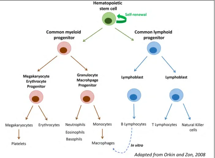
Mammalian hematopoiesis is characterized by a great number of cell types that are formed; ten different cell types and several subtypes are produced (reviewed in (Orkin et Zon 2008; Weiskopf 2016); (Figure 1). These highly specialized cells of the myeloid and lymphoid lineages ensure a large panel of functions.
a) Myeloid lineage
Myeloid cells are responsible for innate immunity, which allows a rapid and direct defense of the organism against invading pathogens, wound healing and transport of gases. Cells involved in the innate immunity are found in many organisms, and innate immunity is the major defense system in insects and plants.
• Granulocytes are distributed in three types: neutrophils, eosinophils and basophils that are categorized based on their ability to absorb neutral, acid or basic eosin-derived dyes. Neutrophils are phagocytes and can exocytose granules, thereby reducing inflammation. Eosinophils are responsible for the elimination of parasites, using cytotoxic granules, and basophils produce histamine during inflammatory and allergic reactions.
• Macrophages arise from the terminal differentiation of monocytes and are responsible for phagocytosis. They eliminate apoptotic or necrotic cells, and also cancer cells that express specific membrane markers. They also have a defense function during innate immunity, as they phagocytize pathogens like bacteria. Macrophages are furthermore found as resident cells in several adult tissues where they self-maintain (Hashimoto et al. 2013); in addition, they participate in maintaining the steady state of tissues. Indeed, macrophages are able to activate BMP signaling in enteric neurons in order to control gastrointestinal motility (Muller et al. 2014). It also has been shown that macrophages
Figure 1: Representation of the mammalian hematopoietic tree
At the top of the tree, hematopoietic stem cells are responsible for the formation of every type of blood cell. Asymetric divisions allow self-renewal and generate progenitors of the myeloid and lymphoid lineages. These progenitors further differentiate into more committed progenitors that will eventually give rise to all mature blood cells. The blue dashed line indicates the ability of B lymphocytes to transdifferentiate into macrophages. (Adapted from Orkin and Zon, 2008)
6
protect tissue against inflammatory response by inhibiting neutrophil recruitment (Uderhardt et al. 2019).
• Megakaryocytes are responsible for the production of platelets that are released in circulation after megakaryocyte fragmentation and, upon injury, they aggregate to allow wound healing by promoting coagulation.
• Erythrocytes are the most abundant blood cell type; they are enucleated cells mostly responsible for the transport of oxygen throughout the organism during embryonic and adult life. They have other functions such as regulating blood viscosity and maintaining the shear stress necessary for vascular development and remodeling (Baron 2013; Lucitti et al. 2007).
b) Lymphoid lineage
Lymphoid cells are responsible for adaptive immunity, a second, highly specific, wave of defense against invading pathogens. Adaptive immunity arose in gnathostomes, as all jawed vertebrates possess lymphocyte receptors.
• B lymphocytes are key effectors of the adaptive response. They are activated by T lymphocytes (see below) at the onset of the response to initiate the production of antibodies specific to the pathogen. A subset of B lymphocytes subsequently becomes memory B cells and allows an efficient response, if the organism has to fight the same pathogen later in life.
• T lymphocytes are classified in 3 main groups of cells: (i) T helper cells (positive for the CD4 marker) that are activated after the presentation of an antigen by Antigen Presenting Cells; the activated T cell then interacts with a B lymphocyte to trigger specific antibody production; (ii) cytotoxic T lymphocytes (positive for the CD8 marker) that directly eliminate the pathogen after their activation and (iii) memory T lymphocytes that have a long lifespan and participate in long-term immunity.
• Natural Killer cells, thanks to their cytotoxic granules, are responsible for direct destruction of cancer cells, viral-infected cells and foreign bodies.
7
Adult definitive blood cells all come from HSCs, localized in the bone marrow. This environment is a niche allowing HSCs to proliferate and differentiate normally (reviewed in (Asada, Takeishi, et Frenette 2017). However, these HSCs themselves emerge after successive events taking place during embryonic development that I will describe below.
2. Blood cell formation
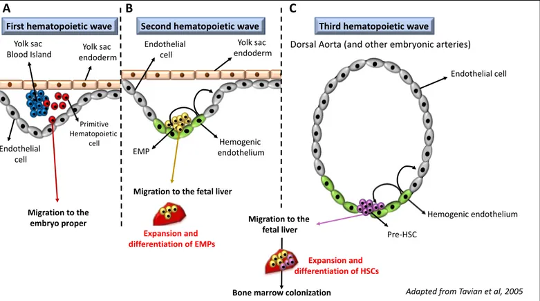
Blood cell formation is a process that initiates during early embryonic stages. Indeed, during its development, the embryo has to be supplied in oxygen and may also have to be able to fight off some pathogens that would be a threat to its survival. In order to meet those needs, three successive hematopoietic waves take place during development (Figure 2).
a) First hematopoietic wave and primitive hematopoiesis
The first hematopoietic wave consists of a transient myelopoiesis that takes place inside blood islands of the yolk sac during early embryogenesis (reviewed in Lacaud et Kouskoff 2017). Absence of primitive hematopoiesis in the mouse embryo, notably after loss of function of GATA-1, which is a major hematopoietic transcription factor, leads to early embryonic lethality presumably due to the absence of erythrocytes (Fujiwara et al. 1996); this phenotype underlines the importance of early hematopoiesis during development.
This first embryonic wave gives rise to primitive myeloid cells (erythrocytes, megakaryocytes and macrophages) that will migrate to the embryo proper. Some studies suggested that endothelial cells and primitive blood cells in blood islands of the yolk sac arise from a common progenitor called hemangioblast, but although it seems to be conserved from fly to mouse (Mandal, Banerjee, et Hartenstein 2004; Vogeli et al. 2006; Huber et al. 2004), this has not yet been demonstrated in humans and is a matter of debate. In mouse, primitive erythrocytes come from erythroblasts produced in the blood islands that enter the bloodstream, continue to divide (Bethlenfalvay et Block 1970) and finally differentiate into enucleated erythrocytes that express both embryonic and adult globins (Kingsley 2004, 2006;
Figure 2: Embryonic waves of hematopoiesis in mammals
A. During the first wave, primitive hematopoietic cells (in red) arise from cells inside blood islands (in blue) in the yolk sac. They migrate to the embryo proper, where they differentiate into primitive erythrocytes, macrophages and megakaryocytes. B. During the second wave, the first definitive blood cells arise. These cells are Erythromyeloid Myeloid Progenitors (EMPs, in yellow); they originate from the hemogenic endothelium (in green) of the yolk sac through the Endothelial to Hematopoietic Transition process and migrate to the fetal liver, where they proliferate and differentiate. C. The third wave gives rise to the hematopoietic stem cells (HSCs). They emerge from the hemogenic endothelium of major arteries in the embryo, and in particular in the dorsal aorta. The emerging pre-HSCs will then, like EMPs, migrate to the fetal liver to expand and differentiate. At the end of embryogenesis, newly formed hematopoietic cells will migrate again to colonize the bone marrow niche. (Adapted from
Tavian et al, 2005 Endothelial cell Hemogenic endothelium Pre-HSC Migration to the fetal liver
Dorsal Aorta (and other embryonic arteries)
Third hematopoietic wave First hematopoietic wave Second hematopoietic wave
Bone marrow colonization Expansion and differentiation of HSCs Yolk sac endoderm Migration to the embryo proper Endothelial cell Yolk sac Blood Island Primitive Hematopoietic cell Yolk sac endoderm Endothelial cell Hemogenic endothelium EMP
Migration to the fetal liver
Expansion and differentiation of EMPs
A B C
8
Qiu et al. 2008). It also has been shown that in the yolk sac, primitive megakaryocytes emerge from bipotent progenitors called megakaryocyte/erythroid progenitors (MEPs) that can give rise to both primitive lineages (Tober et al. 2007). These megakaryocytes participate in the formation of embryonic platelets that circulate in the embryo. Primitive macrophages in the yolk sac have two origins: a first population is composed of maternally-derived macrophages that do not express all adult macrophages markers, and a second population comes from monopotent progenitors that differentiate into macrophages exhibiting definitive adult features (Bertrand 2005).
Hematopoietic cells formed during the first embryonic wave are transient and thus are replaced later on during embryogenesis by definitive cells with adult features. Consequently, they are not found in post-natal stages and are not involved in definitive hematopoiesis. However, it has recently been shown that primitive macrophages play an essential role in colonizing hematopoietic organs, notably by remodeling the extra cellular matrix (Travnickova et al. 2015).
b) Second hematopoietic wave and emergence of the first definitive blood cells
In addition, the second hematopoietic wave occurs in the extra embryonic yolk sac, with the emergence of a myeloid progenitor called the Erythromyeloid Progenitor (EMP; reviewed in (Frame, McGrath, et Palis 2013). EMPs emerge from the hemogenic endothelium of the yolk sac (Frame et al. 2016) through a process called Endothelium to Hematopoietic Transition (EHT). EMPs migrate rapidly through the new circulatory system to colonize the fetal liver (Palis 1999), where they proliferate and differentiate into mature macrophages, megakaryocytes and erythrocytes. These cells are considered part of definitive hematopoiesis, since they have the same characteristics as cells arising later on from the differentiation of HSCs (Bertrand 2005). The cells produced by the differentiation of EMPs are mostly transient, but some of them can be found in the adult organism, where some resident macrophages in the adult tissues derive from EMPs (Schulz et al. 2012; Gomez Perdiguero et al. 2015).
9
Although EMPs lack any lymphoid potential, it has been shown that lymphocytes are present in the yolk sac during early embryogenesis, anterior to the emergence of HSCs (Yoshimoto et al. 2011, 2012), and that they come from independent lymphoid progenitors.
c) Third hematopoietic wave, HSC emergence and bone marrow niche
The third and last embryonic wave gives rise to the HSCs. They were first shown to emerge in the aorta-gonad-mesonephros (AGM) region of the mouse embryo, through the process of EHT from hemogenic endothelium in the dorsal aorta (Medvinsky et Dzierzak 1996; de Bruijn 2000; Tavian et Peault 2005). After this discovery it was shown that HSCs can arise from hemogenic endothelium in several regions of the embryo, including the yolk sac, the umbilical and vitelline arteries, and the placenta (de Bruijn 2000; Gekas et al. 2005; Chen et al. 2009). Then, newly formed hematopoietic cells called pre-HSCs delaminate from the endothelium and enter the circulation to colonize the fetal liver, probably via umbilical vessels. Once in the liver, they rapidly expand and mature to become fully competent (repopulating) HSCs (Kieusseian et al. 2012). They can then yield committed progenitors that in turn give rise to the complete hematopoietic system (Mikkola 2006). Finally, around the time of birth, HSCs colonize the bone marrow, where their proliferation and differentiation properties are tightly controlled by the microenvironment provided by the niche during adult life.
The bone marrow compartment constitutes a very complex microenvironment (reviewed in Morrison et Scadden 2014; Asada, Takeishi, et Frenette 2017) to protect HSCs against pathological transformation. In this niche HSCs remain mostly quiescent, with a limited rate of self-renewal activity that ensures continuous production of new hematopoietic cells in normal proportions (Akunuru et Geiger 2016).
10
d) A particular case of blood cell formation: the transdifferentiation process
Even though all cells come from the differentiation of HSCs, it has been shown that in
vitro, B lymphocytes can be reprogrammed by a process called transdifferentiation (H. Xie et
al. 2004); during this process committed B cells are induced to become another type of committed cell, macrophages, without passing through a progenitor step. They simultaneously dedifferentiate from their original identity and start to express macrophages markers, meaning that at some point these cells express genes of both lineages (Jopling, Boue, et Belmonte 2011; Cieślar-Pobuda et al. 2017). Although rarely observed, this phenomenon does happen in mammals, yet it has been more thoroughly characterized in Drosophila (see section C-2b).
The fine equilibrium between proliferation and differentiation of HSCs can sometimes be unbalanced (notably by the mutation of key molecular actors such as transcription factors of epigenetic regulators), and this deregulation leads to the development of severe pathologies such as leukemia, which I will describe below.
B.
Pathological hematopoiesis and its major actors
1. Leukemias: multifaceted malignant blood disorders
There are four major classified types of leukemia, depending on which cell lineage is affected and on the invasiveness of the disease: chronic, acute myeloid or lymphoid leukemia. Chronic leukemia impacts 1 out of 100 000 persons per year in France, and this condition can persist for several years. In this disease, progenitor cells are not affected but do not produce mature cells able to fight off infections efficiently. With time, immature cells replace normal cells in the bone marrow niche, which is deleterious for the organism, because it completely impairs the ability of the immune system to respond to infection.
Acute leukemia has an incidence of 4 out of 100 000 cases per year in France and has a much poorer prognosis, as it is very invasive. Contrary to chronic leukemia, acute leukemia
11
affects progenitor cells: it is characterized by the induction of immature blood cells and abnormal proliferation that rapidly inhibits production of differentiated cells in the niche.
Classically, leukemias appear as the consequence of somatic mutations accumulated during life. Those mutations have been classified into two categories (De Kouchkovsky et Abdul-Hay 2016). Class I mutations affect genes implicated in proliferative pathways (such as c-Kit and STAT3; The Cancer Genome Atlas Research Network 2013; Yamada et Kawauchi 2013), and class II mutations affect genes implicated in normal blood cell formation (such as NMP1 and CEBPA; The Cancer Genome Atlas Research Network 2013); according to the classical double hit model of leukemogenesis, class I and II mutations have to occur concomitantly to trigger the pathology. However, over the past few years, a third type of mutation has emerged, which corresponds to mutations targeting epigenetic regulators (such as DNMT3A and TET2; Patel et al. 2012) involved both in proliferation and differentiation. This double hit model is based on punctual mutations, but it does not take into account bigger genomic instabilities such as chromosomal rearrangements (translocations, duplications…), which in some cases can be sufficient to initiate a leukemic state.
Chromosomal rearrangements can affect many genes, but among them some events are more represented. This is the case for rearrangements affecting the locus encoding the major hematopoietic factor RUNX1/AML1 (Miyoshi et Ohki 1991), or loci encoding the MOZ and MORF epigenetic enzymes of the MYST family (Borrow et al. 1996). I will describe their functions in both normal and pathological hematopoiesis in the following sections.
2. The highly conserved family of RUNX transcription factors and its links to
leukemia
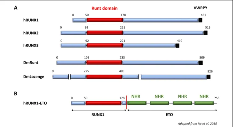
The RUNX family is composed of Core Binding Factor (CBF) transcription factors strongly conserved across evolution of bilaterian metazoans (Rennert et al. 2003). Its founding member, the product of the runt gene, was described for the first time in Drosophila melanogaster in 1980 and was shown later on to be essential for embryonic segmentation (Gergen et Wieschaus 1986). In Drosophila, there are four RUNX genes: runt, RUNXA, RUNXB and lozenge (lz), and in mammals only three: RUNX1, RUNX2, RUNX3 (Figure 3). These
Figure 3: RUNX transcription factors
A. Schematic representation of the three human RUNX proteins, along with the Drosophila Runt and Lozenge RUNX factors. Runt is the founding member of the RUNX family; Lozenge is a RUNX gene ortholog, and the only identified RUNX protein involved Drosophila hematopoiesis. All RUNXs possess a highly conserved Runt domain responsible for DNA binding and a VWRPY motif involved in interactions with co-repressors. B. Schematic representation of the leucemogenic fusion protein RUNX1-ETO: the full-length Runt domain of RUNX1 is fused to the ETO protein, which contains four Nervy Homology Region (NRH) domains that are mostly known to allow interaction with repressors.
(Adapted from Ito et al, 2015)
hRUNX1
hRUNX2
Runt domain VWRPY
hRUNX3 DmLozenge 0 50 178 451 0 92 221 513 410 221 92 0 0 275 403 826 DmRunt 509 233 105 0 hRUNX1-ETO 0 50 178 NHR NHR NHR NHR 753 RUNX1 ETO A B
12
transcription factors are involved in many processes like hematopoiesis, neurogenesis and bone development (Ito 2008), as well as in various diseases (Chuang, Ito, et Ito 2013; C.-C. Sun et al. 2019). They all contain a highly conserved runt domain, which allows both DNA binding on a specific TGYGGTY recognition motif and heterodimerization with their partner CBFb (Adya, Castilla, et Liu 2000; Ito, Bae, et Chuang 2015). Furthermore, they possess a C-terminal VWRPY motif responsible for the interaction with co-repressors, such as the Groucho/TLE family (Aronson et al. 1997; Levanon et al. 1998).
In human, RUNX1 was first identified as the target of chromosomal translocations associated to the development of acute myeloid leukemia (AML; Miyoshi et Ohki 1991; Erickson et Gao 1992). It is one of the most frequently mutated loci, as RUNX1 is altered in 26% of AML cases, either by a single point mutation or by chromosomal rearrangements (Haferlach et al. 2014). A RUNX1 mutation in AML is almost always linked with poor prognosis. This association of RUNX1 mutations with the development of severe pathologies is indicative of a prominent role of RUNX1 during normal hematopoiesis (see below). The second mammalian RUNX transcription factor, RUNX2, was identified on the basis of its sequence homology with RUNX1 (Kamachi et al. 1990) and is notably involved in the ossification process (Takarada et al. 2016). Finally, RUNX3 was identified in 1994 (Levanon et al. 1994) and has been shown to be a tumor suppressor gene involved in many cancers (notably colorectal cancer; Bae et Choi 2004). Furthermore, it has been shown that in human gastric carcinoma cells, RUNX3 is responsible for the expression of miR-182, which in turn inhibits cancer growth (Yu et al. 2017). Together, RUNX1 and RUNX3 are involved in breast cancer, where they have a protective effect against epithelial to mesenchyme transition (Kulkarni et al. 2018). RUNX3 not only has roles in pathologies, but is also involved in normal neural development (Appel et al. 2016) and in hematopoiesis, where it cooperates with RUNX1 for the differentiation of several subtypes of T lymphocytes (Woolf et al. 2003, 3; Li et al. 2012, 3). Furthermore, RUNX3 has a role of its own; it modifies the chromatin landscape during Cytotoxic T lymphocyte formation (D. Wang et al. 2018).
It thus appears that RUNX genes are key regulators of normal and pathological development, and that their regulation is essential to maintain homeostasis of several tissues, including the hematopoietic tissue, where RUNX1 plays a crucial role in the development of AML.
13
a) RUNX1 in Acute Myeloid Leukemia
• RUNX1-ETO at the onset of AML
RUNX1 is the target of several chromosomal rearrangements that lead to the induction of AML, among which the most common is the t(8;21) translocation that fuses RUNX1 with the Eight Twenty-One gene (ETO) (Erickson et Gao 1992; S. C. Bae et al. 1993). Little is known about ETO function, but it is mostly involved in gene repression, since it can recruit several co-repressors (Salat et al. 2008; Issay Kitabayashi et al. 1998) through its nervy homology (NRH) domains (Y. Liu et al. 2006). This translocation is the most common in de novo AML (Grimwade et al. 2010) and affects mostly elder people. It provokes an inhibition of wild-type RUNX1 and leads to a block in myeloid differentiation, associated with a high and abnormal self-renewal rate of progenitors (Okuda et al. 1998), which gives the AML phenotype. It was shown recently that the RUNX1-ETO (also called RUNX1-RUNX1T1) fusion protein interacts with several factors such as Lmo2 and the ETS factor PU.1, to inhibit the expression of differentiation genes and activate the expression of stem cell genes (Ptasinska et al. 2014; X.-J. Sun et al. 2013). Furthermore, RUNX1-ETO drives changes in the chromatin landscape that modify the epigenome and facilitate leukemic functions (Loke et al. 2017). However, it has been proposed that the t(8;21) translocation is not able to trigger leukemogenesis on its own, and that it has to be combined with a secondary mutation (Grisolano et al. 2003; Kelly et Gilliland 2002). In about 20-40 % of cases, a mutation in the c-KIT gene (a crucial factor in HSC development) was concomitantly identified (W. Xie et al. 2019; Y.-Y. Wang et al. 2005).
• Other leukemogenic mutations of RUNX1
Another translocation affecting RUNX1 is the t(3;21) leading to the RUNX1-EVI1 fusion gene (Nucifora et al. 1994; Mitani et al. 1994). This translocation is mostly found as a secondary hit in already established blood pathologies, like chronic myeloid leukemia (Nukina et al. 2014). It leads to the same phenotype as RUNX1-ETO, even if the molecular targets are not the same (Loke et al. 2017). In addition to chromosomal rearrangements, several point mutations in the RUNX1 gene were identified, all involved in the initiation of AML (Gaidzik et al. 2011).
14
Therefore, RUNX1 appears to be a major target in pathologies affecting hematopoiesis, and its function during the malignant process is extensively studied. But the fact that its deregulation leads to severe disorders, is an indication pointing towards its important role during normal hematopoiesis.
b) RUNX1 during normal hematopoiesis
RUNX1 is expressed in the progenitors of the two definitive embryonic waves (second and third waves), as well as in some primitive macrophages (T. North et al. 1999) and in adult HSCs (Ng et al. 2010; Nottingham et al. 2007). It is a major regulator of both embryonic and adult hematopoiesis; indeed, RUNX1 is crucial for EMP and HSC emergence during EHT (Chen et al. 2009, 1; T. E. North et al. 2002; Yzaguirre et al. 2018; Liakhovitskaia et al. 2014, 41). RUNX1 mutant mouse embryos are very pale and lack any fetal liver hematopoiesis (Okuda et al. 1996, 1), phenotypes that lead to early lethality due to severe hemorrhages. This evidence underlines its importance during the establishment of hematopoietic cells, and in particular for HSC development and fetal liver colonization. Consistently, primitive hematopoiesis of the first embryonic wave does not seem to be affected in the RUNX1 mutant, showing that this wave is RUNX1 independent. It is not clear in the literature if RUNX1 is involved in HSC maintenance, but several studies show that it is expressed in the bone marrow compartment (Ng et al. 2010; Nottingham et al. 2007). Finally, it is known that during adult hematopoiesis, RUNX1 is involved in myeloid differentiation and in particular for megakaryocytes formation (Draper et al. 2016). RUNX1 is also responsible for B and T lymphoid development (Chi et al. 2018; Woolf et al. 2003, 3; Li et al. 2012, 3; Taniuchi et al. 2002).
RUNX1 expression and function are regulated and mediated by several factors that play a role upstream, downstream, or in collaboration with RUNX1, in order to generate every blood cell not only in the embryo but also in the adult. Here I describe RUNX1’s main partners.
15
c) RUNX1 partners and regulators during normal hematopoiesis
• Notch is a major regulator of RUNX1 expression
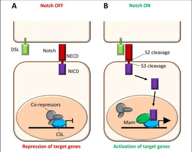
The Notch pathway is crucial during development and can be involved in several processes, like proliferation, asymmetrical cell division (and thus differentiation), or even cell death. Notch is a membrane receptor composed of two parts: an intracellular domain (NICD) responsible for the transcriptional activity of the pathway and an extracellular domain (NECD) responsible for the interaction with its ligands Delta/Serrate/Jagged (DSL). Once the ligand/receptor interaction occurs, the Notch receptor is cleaved multiple times, a first time (S2 cleavage) by the ADAM metalloproteinase and a second time (S3 cleavage) by the g-secretase complex. This separates NECD from NICD, and the released intracellular part will then relocalize to the nucleus. There, it interacts with a complex composed of the Notch pathway transcription effector called CSL (CBF1/RBPJ, Supressor of Hairless(Su(H)), Lag-1), Mastermind (Mam) and other coactivators to regulate their target genes (Figure 4; reviewed in Bray 2006; Kovall et al. 2017). In mammals, there are four Notch receptors: Notch1-4.
As described above, HSCs arise from the hemogenic endothelium of the major embryonic arteries, and in particular in the dorsal aorta. Its hemogenic potential is controlled and maintained by Notch signaling from the interaction between the ligand Jag1 and the receptor Notch1 (Kumano et al. 2003, 2; À. Robert-Moreno et al. 2008, 1; Clarke et al. 2013).
Notch1 directly regulates the expression of GATA2, a marker of hematopoietic cells within the hemogenic endothelium required for HSC development (A. Robert-Moreno 2005; de Pater et al. 2013, 2), and is at the origin of the onset of RUNX1 expression in hematopoietic cells (Nakagawa et al. 2006). Then, Notch expression is downregulated in the cells undergoing EHT (Richard et al. 2013). Although Notch1 is required during the process of HSC formation, it has been shown that RUNX1 allows the emergence of EMPs from the yolk sac hemogenic endothelium independently of Notch1 (Hadland 2004; Bertrand et al. 2010).
• GATA transcription factors are known RUNX interactors
The GATA family is composed of transcription factors conserved across evolution. They are characterized by the presence of one or two zinc finger domains that allow DNA binding
Figure 4: Overview of the Notch signaling pathway.
A. When the pathway is inactive, the CSL transcription factor is bound by co-repressors and Notch target genes are repressed. B. Notch pathway is activated by the interaction between one Delta Serrate Ligand (DSL) and the Notch ExtraCellular Domain (NECD) of the receptor. Upon interaction, the receptor undergoes a series of cleavages (S2 and S3) that lead to the translocation of the Notch IntraCellular Domain (NICD) into the nucleus. There, it interacts with its CBF1/RBPJ, Supressor of Hairless (Su(H)), Lag-1 (CSL, blue) partner and with Mastermind (Mam, green) to replace co-repressors and activate transcription of target genes. (Adapted from Bray 2006)
Notch OFF Notch ON
DSL Notch
NECD
NICD
Repression of target genes Activation of target genes
CSL Co-repressors
S2 cleavage S3 cleavage
Mam
Adapted from Bray 2006
16
or interaction with their cofactors Friend Of GATA (FOG). In mammals there are six GATA factors (GATA1-6), but only three of them (GATA1-3) are required during the hematopoietic process. GATA1 is a major actor of the hematopoietic development, as it is required for erythrocyte formation (described above, Fujiwara et al. 1996, 1) and megakaryocyte differentiation (Chang et al. 2002; Iwasaki et al. 2003, 1). GATA2 is absolutely required for HSC emergence and survival in the embryo (de Pater et al. 2013, 2; Ling et al. 2004, 2; Tsai et al. 1994). Initially, GATA3 was only shown to be required for the differentiation of T-helper-type-2 cells (G. R. Lee, Fields, et Flavell T-helper-type-2001; Ho, Tai, et Pai T-helper-type-2009, 3). However, it has been shown that GATA3 is expressed in lymphoid progenitors, where it is crucial for the formation of the T cell lineage (Rothenberg 2013), and some recent findings suggested that it also plays a role during HSC emergence in the embryo (Zaidan et Ottersbach 2018).
RUNX1 is required for megakaryocyte differentiation during definitive myelopoiesis (Draper et al. 2016), and it has been shown that GATA1 cooperates with RUNX1 in this process (Elagib 2003; Goldfarb 2009; Pencovich et al. 2011; Tijssen et al. 2011). It is interesting to note that Elagib et al show that the fusion protein RUNX1-ETO inhibits GATA1 during megakaryocytic differentiation in cell culture, which they propose to be a mechanism involved in the blocking of myeloid differentiation during AML development.
Furthermore, RUNX1 and GATA2 are both present in HSCs in the bone marrow but have never been shown to interact with each other. However, they cooperate during HSC emergence in the embryo (Wilson et al. 2010), and it has recently been shown that they are involved in a complex regulating the expression of the SET gene (which is a potent Inhibitor of Protein Phosphatase 2A) in two AML cell lines (HL-60 and HEL cells; Pippa et al. 2017).
• CBFb, RUNX heterodimerization partner
CBFb, a RUNX1 heterodimerization partner seems to be involved during the hematopoietic process, because a loss of function of this gene in the mouse embryo leads to the same phenotypes as a RUNX1 mutant (Niki et al. 1997). It is likely that all the roles played by RUNX factors during hematopoiesis are dependent on CBFb. Indeed, its loss of function specifically in the adult, has broader phenotypes than the ones of RUNX1 alone, indicating that CBFb interacts with other partners, which may be RUNX factors during this process.
17
• Monocytic Leukemia Zinc-Finger protein (MOZ)
MOZ is a Lysine Acetyltransferase (KAT) involved during hematopoiesis at several levels, that I will develop later in more detail. It was shown that in cultured myeloid mouse cells, MOZ was part of the RUNX1 complex and required for its transcriptional activity, therefore participating in myeloid differentiation (I. Kitabayashi 2001, 1; Yoshida et Kitabayashi 2008, 1). The authors propose that MOZ acts independently of its catalytic activity in this process, but this hypothesis has never been developed further. They also demonstrate that MOZ is able to acetylate RUNX1 in vitro, but this modification was never identified in vivo. Another study showed that MOZ and RUNX1 cooperate for the transcription of the Macrophage Inflammatory Protein 1a (MIP-1a) gene in human Jurkat T-cells (Bristow 2003), providing a new example of MOZ/RUNX1 interaction during hematopoiesis.
The epigenetic regulator MOZ and its paralog MOZ-Related Factor (MORF) are themselves common targets of chromosomal rearrangements leading to the induction of AML, and I will describe their role in normal and pathological hematopoiesis in the next part.
3. MOZ/MORF and the MYST family of Lysine Acetyl-Transferases
The MYST family is named after its founding members: MOZ, Yfb2, Sas2 and Tip60, and is a very conserved family from yeast to human (Figure 5). In mammals there are five members, MOZ/MORF, Tip60, Male absent of the First (MOF) and Human acetylase Binding to ORC1 (HBO1, reviewed in (Yang 2004; X.-J. Sun et al. 2015). KATs of the MYST family were first shown to acetylate histone tails, and by doing so, to participate in the remodeling of the chromatin landscape (Grant 2001; Goll 2002). However, studies over these last years yielded increasing evidence that they have a much broader range of substrates than just histones (reviewed in Sapountzi et Côté 2011). KATs of the MYST family are involved in a variety of pathologies, but I will focus here on the role of MOZ and its paralog MORF in hematopoietic disorders.
Figure 5: MYST family of acetyl transferases
A. Schematic representation of human and Drosophila MYST proteins. All of them possess a conserved MYST domain with catalytic activity. B. Schematic representation of MOZ and the main fusion proteins involved in leukemia. In the MOZ-CBP fusion, the N-terminal part of MOZ comprises a functional MYST domain fused to the C-terminal part of CBP. In the MOZ-TIF2 fusion, the same breakpoint in MOZ is fused to the CBP Interacting Domain (CID) domain of TIF2, which is responsible for CBP binding.
(Adapted from Yang 2004 ; Yoshida and Kitabayashi 2008)
MOZ KIX Bromo HAT MOZ-CBP MOZ-TIF2 MOZ MOZ TIF2 CBP
Adapted from Yoshida and Kitabayashi 2008
B
CID AD
t(8;16)(p11.2;p13.3)
inv(8)(p11;q13)
Adapted from Yang 2004
18
a) MOZ and MORF in AML
The name MOZ comes from Monocytic leukemia Zinc-finger protein, because it was first identified as a target of chromosomal translocations responsible for the development of AML. These rearrangements involve in particular the CREB-binding Protein (CBP) (Borrow et al. 1996; Chaffanet et al. 2000; Crowley et al. 2005) and TIF-2 (inv(8)(p11q13); Figure 5; Carapeti et al. 1999). MOZ protein contains several important domains that have been characterized and linked to specific functions. The MYST domain bears the acetylation catalytic activity and contains a C2HC zinc-finger involved in DNA binding (Holbert et al. 2007). This conserved MYST domain is also required for the interaction with MOZ partners Bromodomain-PHD finger proteins (BRPF) 1 2 and 3 (Ullah et al. 2008). The C-terminal domain of MOZ is called SM for Serine/Methionine-rich domain and is involved in transcription activation. This domain is also involved in the interaction with p53 (Susumu Rokudai et al. 2009; S. Rokudai et al. 2013; Tham et al. 2015), which is a transcription factor involved in cell cycle regulation, autophagy and apoptosis (Sabapathy et Lane 2019). Finally the N-terminal part of MOZ, is composed of the NEMM domain (for N-terminal of Enok MOZ/MORF; these proteins are the only members of the MYST family that contain this N-terminal part) and a double PHD (Plant Homeodomain) finger, which contains a recognition domain for histones and is involved in transcription inhibition (Dreveny et al. 2014; Xiong et al. 2016).
MOZ and CBP are both important factors for HSC maintenance and quiescence (Katsumoto 2006; Thomas 2006; Rebel et al. 2002; Bilal N. Sheikh et al. 2017), and TIF2 (Transcriptional Intermediary Factor 2) is a direct partner of CBP (Demarest et al. 2002).
The leukemogenic MOZ-CBP fusion protein contains the N-terminal part of MOZ with its intact MYST domain, and almost the entire CBP protein (Chan et al. 2007). As a result, the recognition repertoire of CBP is aberrantly acetylated by MOZ and leads to the proliferation of leukemic progenitors and to the AML phenotype. Similarly, MOZ-TIF2 has been shown to recruit CBP via the CID interaction domain of TIF-2, which results in CBP inhibition and leads to leukemic development (Deguchi et al. 2003; Kindle et al. 2005). It also has been shown that MOZ-TIF2 represses senescence in AML stem cells and thus leads to their expansion (Largeot et al. 2016). It is noteworthy that MORF, a paralog of MOZ, is also involved in a chromosomal
19
translocation t(10;16) with CBP, associated to the development of AML (Panagopoulos 2001), suggesting that MORF could play a role during normal blood cell formation.
In addition to its involvement in leukemia, MOZ has also been linked to the development of other pathologies, such as intellectual disability (Tham et al. 2015) and esophageal adenocarcinoma (Dulak et al. 2013), and its transcriptional deregulation leads to metastasis of medulloblastoma and colorectal cancer (Wu et al. 2012; Mohammadi et al. 2018). It is interesting to note that in a recent study, Baell et al identified a new inhibitor of MOZ that is sufficient to stop cancer growth in a model of mouse lymphoma, by inducing senescence in cancer cells (Baell et al. 2018).
As for RUNX1, MOZ is an important target of leukemogenic mutations and rearrangements, which prompted many research groups to study its role during normal hematopoietic development.
b) MOZ during normal hematopoiesis
MOZ was first shown to display a KAT activity on histone tails (N. Champagne, Pelletier, et Yang 2001) and has since been studied mainly considering this property. Indeed, in a murine model carrying a point mutation in the catalytic site of MOZ, Perez-campo et al showed that MOZ-mediated acetylation is required for proliferation and expansion of hematopoietic progenitors (F. M. Perez-Campo et al. 2009). Later, the same group showed that MOZ catalytic activity is required to prevent HSCs from entering into replicative senescence, thus promoting their self-renewal capacity (Flor M. Perez-Campo et al. 2014). MOZ is also required in for their maintenance, since its loss of function in the embryo leads to the absence of hematopoietic progenitors and adult-repopulating cells in the fetal liver (Katsumoto 2006; Thomas 2006). Furthermore, specific MOZ loss of function in the adult bone marrow compartment leads to a rapid loss of HSCs (B. N. Sheikh et al. 2016), as measured by their loss of ability to reconstitute the hematopoietic system in an irradiated mouse recipient. Finally, it was shown that MOZ acts in collaboration with the polycomb group protein BMI1 to maintain HSCs in a quiescent state (Bilal N. Sheikh et al. 2017), ensuring their maintenance over time.
20
In addition to its function in HSCs, MOZ is required for myeloid lineage and lymphoid B-cells formation: it is a coactivator of RUNX1, in particular for MIP-1a expression, and is required for normal macrophage and B-cell development (Jiang et al. 2019; Katsumoto 2006; Good-Jacobson et al. 2014 ).
As I previously mentioned, MOZ acts as a transcriptional coactivator of RUNX1, in a KAT-independent manner (I. Kitabayashi 2001). There are consistent differences between the phenotypes of mice completely deprived of any MOZ activity (Katsumoto 2006) (embryonic lethality and strong depletion of the hematopoietic system) and phenotypes of mice specifically deprived of its catalytic function (F. M. Perez-Campo et al. 2009) (viable with reduced progenitor proliferation, but no effect on differentiation). These observations strongly support the hypothesis that MOZ has other non-catalytic functions during both development and hematopoiesis.
It has been shown that MOZ has broad functions during development, in addition to its roles in hematopoiesis. Indeed, it is an important player during embryonic development, as it regulates histone acetylation of HOX genes during mouse body segmentation (Voss et al. 2009; Bilal N. Sheikh et al. 2015).
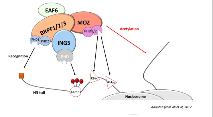
c) MOZ and the ING5 acetylation complex
KATs of the MYST family generally act as members of acetylation complexes (reviewed in Lee et Workman 2007). It has been shown that MOZ is a part of such an acetylation complex, working within the ING5 complex, along with Esa1-Associated-Factor 6 (EAF6), Inhibitor of Growth 5 (ING5) and BRPF1 2 or 3 (Figure 6; Ullah et al. 2008) to specifically modify histone H3. In this complex, MOZ interacts directly with BRPF factors via its MYST domain, and BRPF itself interacts directly with EAF6 and ING5 (Ullah et al. 2008). BRPF proteins are epigenetic readers that recognize specific histone modifications in order to bind chromatin and allow proper modification of the chromatin landscape. BRPF1 has been shown to recognize in particular H3K14ac, therefore directing MOZ to its substrates (Poplawski et al. 2014). In addition, BRPF1 shares common functions with MOZ; indeed, BRPF1 participates in the expression of HOX genes during development in zebrafish (Hibiya et al. 2009), and it is also
Figure 6: The ING5 acetylation complexes
MOZ, which is the catalytic subunit of the complex, directly interacts with the BRPF factors. The other members, EAF6 and ING5, also specifically interact with BRPF but not directly with MOZ. Together, the complex is able to recognize precise chromatin motifs that allow acetylation of a specific repertoire. The BRPF factor recognizes the histone H3 tail via its PHD fingers, ING5 recognizes trimethylation of H3K4, and MOZ recognizes acetylated H3K9 and H3K14. In turn, MOZ will acetylate another H3 tail on lysine 9 or 14. (Adapted from Ali et al 2012)
EAF6
MOZ
BRP
F1/
2/3
ING5
PHD K4me3 PHD1 PHD2 PHD1/2 K9ac K14ac H3 tail Recognition Acetylation Nucleosome21
essential for the normal establishment of fetal liver HSC (You et al. 2016). ING5 is a Tip60 cofactor (N. Liu et al. 2013) and is important for cell proliferation in human cultured cells (Linzen et al. 2015). Furthermore, it has been shown to recognize histone H3 modification H3K4me3 via its PHD domains (K. S. Champagne et al. 2008, 3; Ormaza et al. 2019, 4). The function of EAF6 has not been described so far.
In summary, the ING5 complex provides an environment that enables a specific recruitment of MOZ on its target repertoire, allowing MOZ to specifically acetylate its substrates (Ali et al. 2012).
Mammalian hematopoiesis is a very complex system. Indeed, the need to produce numerous cell types in physiological proportions requires a high level of regulation and therefore a lot of molecular interplay. Moreover, the classical model for hematopoiesis in mammals is the mouse model, which is complicated to manipulate. Mice possess 20 chromosomes and achieve puberty at 2 months of age, which makes it a poor genetic model. However, the hematopoietic process is strongly conserved in vertebrates, both at the developmental and genetic levels. All the molecular factors described above (Notch, RUNX, GATA, MOZ…) are highly conserved across evolution, and in addition to vertebrate models,
Drosophila melanogaster has proven these last fifteen years to be a very powerful model to
study the molecular mechanisms controlling the development of innate immunity.
C.
Drosophila as a model to study hematopoiesis
1. Generalities on the model
Drosophila, which means “who likes dew”, also called the fruit fly, is an animal model used
in many research fields, and in particular in genetic and developmental studies. In labs, the subfamily melanogaster (“black belly”) is widely used.
Thomas Hunt Morgan, a zoologist, was the first to use this model in his lab (later called the Fly Room) in 1910 and received the Nobel Prize for his discovery concerning the role of
22
chromosomes during the process of heredity. He and his group identified many genes, which are still used today as genetic markers for experiments, like the white gene that is responsible for the red color of the eye.
This animal model is particularly easy to use in genetic studies, because it only has four pairs of chromosomes, three pairs of autosomes and one pair of sexual chromosomes. In addition, meiotic recombination does not occur in males, which is of particular interest when doing genetic experiments. Moreover, Drosophila generation time is quite short, around 10 days at 25°C, which makes it easy to design in vivo experiments.
Finally, it is a highly relevant model concerning medical research, because it has been shown that 75 % of disease-related genes have a sequence similarity in the fly, and that 50 % of fly proteins have mammalian homologs (Reiter 2001).
The fruit fly has a life cycle with four stages: embryo, larva, pupa and adult (Figure 8A). The embryonic stage lasts for twenty-four hours, ending with the emergence of the larva. This larval stage is itself composed of three stages called L1, L2 and L3 and lasts for five days. Then, the larva produces an ecdysone (a steroid hormone) signal that leads to a “wandering” behavior and finally to pupariation. The pupal stage also lasts for five days, and during this stage the individual undergoes the metamorphosis process, in which tissues are remodeled and give rise to the adult morphology. Finally, the adult fly emerges from the pupa and begins to mate eight to twelve hours thereafter.
Many developmental processes are conserved throughout evolution like neurogenesis, organ formation, stem cell biology, and of course hematopoiesis. Drosophila is an ideal model to study these processes and many discoveries in Drosophila participated in our understanding of mammalian (and particularly human) development. Here, I will describe the process of hematopoiesis in flies and how it can be used as a model for this process in normal and pathological situations.
23
2. Drosophila hematopoiesis
Drosophila is an organism that possesses an open circulatory system in which circulates a
liquid called the hemolymph. In larvae, the dorsal vessel (or aorta), which is positioned dorsally in the antero-posterior axis, ends with the “heart” in the posterior part. This heart beats to generate a flow of hemolymph that circulates in all the tissues and enables blood cells to migrate. In Drosophila, blood cells share functional similarities with the mammalian myeloid lineage. In 1957, Rizki characterized three cell types: the plasmatocytes, the crystal cells and the lamellocytes (Figure 7; M. T. M. Rizki 1957) which are called hemocytes.
a) Drosophila blood cell types
• Plasmatocytes
Plasmatocytes are the most abundant cells, as they represent about 95 % of all blood cells in Drosophila. They are phagocytes with functional homology to macrophages. They are crucial players of innate immunity in Drosophila, since they are responsible for the elimination of bacteria (Charroux et Royet 2009; Defaye et al. 2009; Nehme et al. 2011) or virally-infected cells (Nainu et al. 2015). Plasmatocytes are also partly responsible for the production of antimicrobial peptides after bacterial infection (Irving et al. 2005).
Moreover, they play an essential role during development, as they phagocytose apoptotic bodies both in the embryo and in the pupa, particularly after tissue remodeling during metamorphosis (Defaye et al. 2009; Wood et Martin 2017; Lanot et al. 2001; Regan et al. 2013). Furthermore, plasmatocytes are responsible for the secretion of extra-cellular matrix during development, in particular around the ventral nervous system and Malpighian tubules (Olofsson et Page 2005; Bunt et al. 2010).
• Crystal cells
Crystal cells represent around 5 % of the total circulating cells. They were first identified because of the crystalline inclusions they have in their cytoplasm (M. T. M. Rizki 1957); these
Figure 7: Drosophila blood cells
Schematic representation of blood cell types in Drosophila. Progenitors, or prohemocytes, give rise to plasmatocytes, which are macrophages, and to crystal cells, which have crystalline inclusions and are functional homologs of the mammalian megakaryocytes. Upon an immune challenge like wasp infestation, prohemocytes in the larva are able to differentiate into lamellocytes; these are big cells able to encapsulate the wasp egg. It also has been shown that plasmatocytes in the larval stages are able to transdifferentiate into crystal cells or upon infestation into lamellocytes. (Adapted from Rizki
1957)
Prohemocyte
Crystal cell
Plasmatocyte
Lamellocyte
24
inclusions are in fact accumulation of prophenol-oxydases (PPOs) involved in the arthropod-specific melanization reaction (Dudzic et al. 2015). Melanization reactions are responsible for wound healing by promoting coagulation; this makes crystal cells the functional homologs of mammalian megakaryocytes. These PPOs are processed into active phenoloxydases by a proteolytic cascade (Nam et al. 2012), when they are released in circulation after crystal cell rupture (Bidla, Dushay, et Theopold 2007). This activation has also been shown to contribute to survival after bacterial infection (Binggeli et al. 2014). I will describe later, in more detail, how crystal cells are formed, in particular during the larval stage.
• Lamellocytes
Lamellocytes are the third and last blood cell type in Drosophila. They are large cells produced specifically when parasitoid wasps lay eggs inside the larval cuticle (M. T. M. Rizki 1957; T. M. Rizki et Rizki 1992; Crozatier et al. 2004). Lamellocytes can be formed in circulation by the transdifferentiation of plasmatocytes (Avet-Rochex et al. 2010; Stofanko, Kwon, et Badenhorst 2010; Anderl et al. 2016), and also directly from progenitors in a specialized organ called the lymph gland (Oyallon et al. 2016). Their function is to encapsulate objects too big to be phagocytized. It has been shown that in a wasp infestation condition, lamellocytes arise from the pool of intermediate progenitors in the lymph gland, and that this process occurs at the expense of crystal cell formation, suggesting that the choice between the two cell types is mutually exclusive (Krzemien et al. 2010). Although lamellocytes are produced in response to an immune stress, they can also be generated both in the lymph gland and in circulation, in some genetic conditions (Avet-Rochex et al. 2010).
b) Drosophila blood cell formation
As for mammals, blood cells in Drosophila are formed during successive waves of hematopoiesis: an embryonic wave that gives rise to all circulating hemocytes in the larva, and a larval wave that generates the pool of hemocytes required during metamorphosis and later on, during adult life.
25
c) Embryonic wave of hematopoiesis
Blood cells in the embryo first originate from the procephalic mesoderm as bipotent progenitors (they can differentiate into both plasmatocytes and crystal cells; Tepass et al. 1994; Holz 2003). These progenitors called prohemocytes, emerge, and differentiate into the two embryonic blood cell types. Plasmatocytes then migrate throughout the embryo (Tepass et al. 1994; Wood, Faria, et Jacinto 2006), while crystal cell lineages stay in a cluster next to the proventriculus (Lebestky 2000; Bataillé et al. 2005). At the end of embryogenesis, about 700 plasmatocytes and 30 crystal cells are generated. It is important to note that since lamellocytes are produced after an immune stress like parasitism, they only appear at the larval stage and are completely absent from the embryo (Figure 8A).
Hemocytes formed after this first hematopoietic wave during embryogenesis will persist in all stages of life. Indeed, during the larval stages, plasmatocytes will aggregate in stereotyped clusters along the cuticle that will become new hematopoietic sites called hematopoietic pockets (HPs; Figure 8B). These HPs provide a microenvironment in which plasmatocytes proliferate (K. Makhijani et al. 2011; Leitão et Sucena 2015) and give rise to the pool of larval circulating hemocytes. Interestingly, these clusters of self-renewing plasmatocytes have been shown to share functional similarities with resident macrophages in mammals (reviewed in Gold et Brückner 2015). Plasmatocytes are attracted to HPs, because of chemotaxis signals produced by peripheral neurons (Kalpana Makhijani et al. 2017), and when stimulated by appropriate signals, they can differentiate into either crystal cells (Leitão et Sucena 2015) or lamellocytes (Honti et al. 2009). It also has been shown recently that plasmatocytes located near pericardial cells of the dorsal vessel, are also able to transdifferentiate into crystal cells, seemingly after Notch induction (Cevik et al. 2019). All the cells produced during this wave subsequently persist until adulthood.
d) Larval wave of hematopoiesis
The second wave of hematopoiesis, which takes place during the larval stage in a specialized organ called the lymph gland (Figure 8C; reviewed in Banerjee et al. 2019), has its
Figure 8: Drosophila life cycle and hematopoiesis
A. During embryonic development, cells of the procephalic mesoderm and cardiogenic mesoderm (in green) develop to give rise to both circulating hemocytes (blue and purple) and to the lymh gland (green), apposed on the cardiac tube (orange). The embryo develops into a larva, then a pupa and finally an adult, where hematopoiesis occurs within Hematopoietic Hubs (HB). B. Representation of larval hematopoietic pockets during the third larval stage. In this microenvironment, plasmatocytes (brown) are in contact with neurons (green) that emit signals to promote plasmatocyte homing and proliferation. Within this microenvironment, plasmatocytes can give rise to larval crystal cells by transdifferentiating. C. Schematic representation of the lymph gland during the third larval stage. In the medullary zone of the anterior pair of lobes (MZ, blue), progenitors are mostly quiescent with a low rate of proliferation. They give rise to differentiated cells (plasmatocytes, red, and crystal cells, purple) located in the cortical zone (CZ). During wasp infestation, progenitors in the MZ are instructed by the PosteriorSignaling Center (PSC) to become lamellocytes. The posterior lobes (also blue) along the cardiac tube will in the adult stage become parts of the HB. (Adapted from Letourneau et al 2016;
Gold and Brückner 2014).
Plasmatocyte Neurons
Adapted from Letourneau et al 2016
Adapted from Gold and Brückner 2014