HAL Id: tel-01558535
https://tel.archives-ouvertes.fr/tel-01558535
Submitted on 8 Jul 2017
HAL is a multi-disciplinary open access
archive for the deposit and dissemination of sci-entific research documents, whether they are pub-lished or not. The documents may come from teaching and research institutions in France or abroad, or from public or private research centers.
L’archive ouverte pluridisciplinaire HAL, est destinée au dépôt et à la diffusion de documents scientifiques de niveau recherche, publiés ou non, émanant des établissements d’enseignement et de recherche français ou étrangers, des laboratoires publics ou privés.
Morgane Rosendale
To cite this version:
Morgane Rosendale. Visualisation and Perturbation of the Spatio-Temporal Dynamics of Endocytosis. Human health and pathology. Université de Bordeaux, 2015. English. �NNT : 2015BORD0071�. �tel-01558535�
Pour l'obtention du
D
OCTORAT DE
L'U
NIVERSIT
É
DE
B
ORDEAUX
Ecole doctorale Sciences de la Vie et de la Santé
Mention : Sciences, Technologies et Santé
Option : Neurosciences
Présentée le
18/06/2015
Par
Morgane ROSENDALE
Visualisation et Perturbation de la
Dynamique Spatio-Temporelle de l’Endocytose
-
Visualisation and Perturbation of the
Spatio-Temporal Dynamics of Endocytosis
Membres du Jury
Dr. Nathalie SANS
Présidente du jury
Pr. Aurélien ROUX
Rapporteur
Pr. Volker HAUCKE
Rapporteur
Pr. Britta QUALMANN
Examinateur
The true delight is in the finding out, not in the knowing
3
A
CKNOWLEDGEMENTS
I would like to start by thanking the members of my jury for accepting to read and judge this work: Dr. Nathalie Sans, Pr. Aurélien Roux, Pr. Volker Haucke, and Pr. Britta Qualmann.
Merci également à Daniel de m’avoir laissée travailler dans ton équipe. Un ENORME merci, bien évidemment, à David, “le patron”, pour ta disponibilité, ton encadrement, tes idées, tes conseils et bien bien plus (je ne citerai pas les divers barbecues, bières pariées et autres sorties en catamaran qui n’auraient pas leur place dans ce document officiel ;)). Merci à Matthieu pour ton soutien, tes commentaires et tes conseils au delà de la simple collaboration.
Merci à tou(te)s celles et ceux qui font que l’IINS est un chouette lieu de travail. Merci à toute l’équipe Choquet bien sûr mais aussi en dehors. Merci aux grands (Dam le ‘ti chef, Lolo, Mimi, Nanel, Juju…) et aux petits (Charlotte, Benjamin, William, Abinaya, Ivy, Anna, Alvaro…). Thanks to Andy, Jennifer, Philipp, Ciarán, Martin, Juan, Mirelle, Ezekiel, Thomas, Sara etc. for making the lab feel like a Spanish apartment! Merci à tous les ITA qui nous rendent la vie facile et sont toujours dispos (Camillette, Fabinou, Zeynep, maman Delphine et bien d’autres). Merci à Eric pour être l’une de ces rares personnes qui demandent encore “ça va” en le pensant. Special thanks should go the “the girls”, starting with Dolors, my first collaborator. It was a true pleasure to work with you, both professionally and personally. I wish you all the best with your new start in life and hope we’ll have the chance to work together again. A Isa, pour ton amitié, pour toutes les pauses thé, les ramassages à la p’tite cuillère, les “mfpfiou!” intempestifs (t’as vu, j’ai immortalisé l'orthographe ;)), les bons ptits plats et j’en passe. A Julia pour ton sourire et ta sérénité et à Ngoc pour ton sourire et ta folie contagieuse!
And of course, thank you so much to all the people, in and out of the lab, who have shaped me into who I am today. Thanks to my various teachers and supervisors who made a crazy little scientist out of me: Mr. Dodeman, Mme. Le Bras, Daniela, Claudio, Seeong Long… Et surtout, merci à celles et ceux qui m’ont vu et fait grandir bien avant celà. By having the chance to meet you, I was able to change, in one way or the other, pour le meilleur ou pour le pire, à vous de juger :). Vous m’avez inspirée, guidée et influencée, peut-être sans vous en rendre compte, et méritez votre nom ici, dans un ordre chronologique, car un quelconque ordre d’importance serait bien impossible: bien sûr à mon nounours préféré, Damien, le frérot, pour m’avoir formée aux aléas de la vie à force de chatouilles et câlins, boutades et conseils ainsi qu'à son p'ti bout de moitié qui le rend heureux; à Laurence, mon idole, pour aller au bout de tes rêves; à Mai-Lan, ma pt’ite pétasse, pour m’avoir accueillie à Rennes; à mon pti Chou, pour tout; à Morgan, pour m’avoir appris à penser; à Victorien, mon inlassable ami; à Zhen Yu, binôme un jour, binôme toujours ainsi qu’à JeN, Clem, Martin(e) et tout le CSOne pour avoir fait
4
parti de la plus belle année de ma vie; à ma prof de clown, qui ne lira jamais ces lignes, mais que j’ai tant admirée; to Chris, even though I may never get to see you again; à Marco, à ma poule, à Thibaud et surtout, surtout, à ma smurchouille, la seule, l’unique, pour votre amitié; a Christian e Mirella per un’estate granidissimo; a Chloé y Mau para enseñarme español y mucho mas; to Arvind for being as crazy as I am; to Mo, for his grand ideas (and his cool name ;)) together with Tim, Yovi, Chrishan, my lil critter, Brian, Wei Shan and Mark, for making me feel on holidays even at work; to the lovely iranian gang in Singapore, for being so genuine; to Maria, for introducing me to svensk kultur and to Gema, Luis and Bobby, for never ending discussio… no parti… no scienc…. no politics… wait, what was it again we did together?; A tous les SwingTimers pour leur énergie et leur joie de vivre; to Tush, S², Kevin and Cary for making me feel alive again… to all of you: MERCI!
Most importantly, I give my deepest thanks to the one without whom I wouldn't be here, literally and metaphorically speaking. The one who has watched her little baby girl fly to the other side of the world over and over again with only words of support, who has encouraged me every time I changed my mind, who has panicked and celebrated with me each time I missed and got an opportunity, celle qui a suivi chaque étape du periple de mes années d’études, celle qui m’aime et me soutient infailliblement,... Oui ma petite maman, tu t'es bien reconnue, à toi, merci!
And to use a famous phrase: last but not least (though I feel like saying first and utmost), I would like to thank, from the bottom of my heart, Fabien, my one and only, mon ange, mon amour, mon chéri. Thank you for being with me, supporting me, bearing with me and loving me. Je pourrais t’écrire un pavé aussi long que ce manuscript juste pour te remercier de tout ce que tu fais et as fait pour moi. Alors je vais me contenter de cette dernière année de rédaction puisqu’il s’agit de remerciements de thèse… Thanks for having listened to me, or at least pretended to ;), while I philosophised about the wonders of photoactivation and photocontrol, while I theorised about how "my little peptides" may affect the grand machinery of endocytosis (endo-whaaat?!), while I dreamt out loud about the beauty of neuroscience and the miracle contained in each synapse. Thanks for never complaining about my coming home late, but rather having a delicious meal ready for me to enjoy (well..., sometimes ;)). Thanks for all the chocolates, liquors and pictures you brought home to let me travel the world with you while rooted in my office. Et par dessus tout, merci d'être toi. Jtm.
5
L’endocytose dépendante de la clathrine (EDC) est un processus fondamental des cellules eucaryotes. Elle se caractérise par la formation d’invaginations à la membrane plasmique aboutissant à la création de petites vésicules par l’action de la dynamine. Dans le cerveau, elle est impliquée dans la dépression synaptique à long terme, un corrélat cellulaire de la mémoire. La morphologie complexe des neurones et le contrôle précis du code neuronal suggèrent qu’elle puisse être régulée spatialement et temporellement dans ces cellules. Le but de mon travail a été de développer de nouveaux outils pour visualiser et perturber l’EDC afin d’étudier ce type de régulation. Le premier de ces outils est pHuji, un senseur de pH rouge génétiquement encodable. Je l’ai utilisé avec un senseur de pH vert existant pour montrer que dans les cellules NIH-3T3, le récepteur β2-adrénergique est internalisé dans une sous-population de vésicules contenant le récepteur à la transferrine constitutivement endocyté. Le deuxième est une nouvelle méthode d’imagerie permettant de visualiser l’activité d’endocytose de structures recouvertes de clathrine optiquement stables dans des neurones d’hippocampe. J’ai ainsi pu suivre pour la première fois la cinétique d’internalisation de récepteurs au glutamate de type AMPA dans des conditions de plasticité. Enfin, j'ai élaboré un test combinant imagerie et patch-clamp afin de développer un bloqueur peptidique spécifique de l'EDC. En utilisant des peptides dimériques, j’ai montré que la dynamine se lie à ses partenaires via des interactions multimériques. En conclusion, ce travail propose une boite à outils permettant d’élucider les mécanismes de l’EDC avec une grande résolution spatiale et temporelle.
Mots-clés : Endocytose dépendante de la clathrine, dynamine, récepteur à la
transferrine, récepteur Beta2 adrénergique, protéines fluorescentes, récepteur AMPA, dépression synaptique à long terme, photoactivation, interactions protéine-protéine, peptides multivalents.
6
Clathrin mediated endocytosis (CME) is a fundamental process of all eukaryotic cells. At the level of the plasma membrane, it is characterised by the formation of deep invaginations resulting in the creation of small vesicles after membrane scission by dynamin. In the central nervous system, it is involved in the expression of synaptic long term depression, a proposed cellular correlate of learning and memory. The complex morphology of neurons and the precise timing of neuronal firing suggest that endocytosis may be spatially and temporally regulated in those cells. The aim of the work presented here was to develop new tools to visualise and perturb CME in order to study such regulation. The first tool to be characterised was pHuji, a genetically encoded red sensor. I used it in combination with an existing green pH-sensor to demonstrate that in NIH-3T3 cells, the β2-adrenergic receptor was internalised in a subset of vesicles containing the constitutively endocytosed transferrin receptor. The second tool is a new imaging method that allowed me to monitor the endocytic activity of optically stable clathrin coated structures in hippocampal neurons. I was thus able to visualise for the first time the kinetics of internalisation of AMPA-type glutamate receptors under plasticity inducing conditions. Finally, I set up an assay combining imaging and cell dialysis in order to develop a specific peptide-based inhibitor of CME. Using dimeric peptides, I found that the interplay between dynamin and its binding partners relies on multimeric interactions. Altogether, this work provides a toolbox to decipher the mechanisms of vesicle formation with high spatial and temporal resolution.
Keywords : Clathrin mediated endocytosis, dynamin, Transferrin Receptor, Beta2
adrenergic receptor, fluorescents proteins, AMPA receptor, long-term synaptic depression, photoactivation, protein-protein interactions, multivalent peptides.
7
P
UBLICATIONS
A
ND
C
OMMUNICATIONS
Publications
Published:
Shen Y.1, Rosendale M.1, Campbell R. E., Perrais D.
pHuji, a pH sensitive red fluorescent protein for imaging of exo- and endocytosis
Journal of Cell Biology, Nov. 2014; 207(3):419-32 doi: 10.1083/jcb.201404107.
1 shared first authorship
In preparation:
Rosendale M., Jullié D., Choquet D., Perrais D.
Visualising the dynamics of endocytic zones in neuronal dendrites
In preparation
Rosendale M., Grillo-Bosh D., Gauthereau I., Choquet D., Sainlos M., Perrais D.
Caged peptides for the acute blockade of endocytosis in living cells
In preparation
Cauvin C., Rosendale M., Larraufie P., Perrais D., Echard A.
Precise spatiotemporal recruitment of the Lowe syndrome phosphatase OCRL in the early steps of endocytosis by the Rab35 GTPase
In preparation
Poster Communications
Jacques Monod Conference " Molecular basis for membrane remodelling and organization" - Roscoff, France - 2011
Rosendale M. & Perrais D.
Visualisation of single endocytic vesicle formation and control of cytosol composition by simultaneous imaging and patch clamp recordings in live cells
15th Annual meeting of the "Exocytosis-Endocytosis club" - L'isle sur la Sorgue, France -
2012
Rosendale M. & Perrais D.
Simultaneous imaging and patch clamp recordings for the visualisation and blocking of endocytic vesicle formation
Best poster presentation award
8th FENS Forum - Barcelona, Spain - 2012
Rosendale M., Grillo-Bosh D., Gauthereau I., Choquet D., Sainlos M., Perrais D.
8
16th Annual meeting of the "Exocytosis-Endocytosis club" - La Grande Motte, France - 2013
Rosendale M., Jullié D., Abdou W., Perrais D.
Detecting the formation of endocytic vesicles in the soma and dendrites of live cultured neurons
European Neuroscience Conference by Doctoral Students - Bordeaux, France -2013
Rosendale M., Jullié D., Abdou W., Perrais D.
Detecting the formation of endocytic vesicles in the soma and dendrites of live cultured neurons
EMBO Conference "Systems Dynamics in Endocytosis" - Villars, Switzerland - 2013
Rosendale M., Grillo-Bosh D., Gauthereau I., Choquet D., Sainlos M., Perrais D.
Optimisation of endocytic blockers and validation by simultaneous imaging and patch clamp recordings
Best poster presentation award
9th FENS Forum - Milan, Italy - 2014
Rosendale M., Grillo-Bosh D., Gauthereau I., Choquet D., Sainlos M., Perrais D.
Simultaneous imaging and patch clamp recordings in live cells to visualise and interfere with endocytic vesicle formation
Oral Communications
17th Annual meeting of the "Exocytosis-Endocytosis club" - Pornichet, France - 2014
Rosendale M., Jullié D., Perrais D.
Visualising the spatio-temporal dynamics of endocytosis in neuronal dendrites
1st European Meeting in Neuroscience by PhD Students - Grenoble, France - 2014
Rosendale M., Grillo-Bosh D., Gauthereau I., Choquet D., Sainlos M., Perrais D.
Simultaneous imaging and patch clamp recordings in live cells to visualise and interfere with endocytic vesicle formation
Training
INSERM Workshop
Photocontrol and optogenetic of biological systems and functions
Symposium: Bordeaux, France - Technical workshop: Paris, France - September 2012
7th Frontiers in Neurophotonics Summer School
9
T
ABLE OF CONTENTS
A
CKNOWLEDGEMENTS3
R
ÉSUMÉ5
A
BSTRACT6
P
UBLICATIONSA
NDC
OMMUNICATIONS7
L
ISTO
FT
ABLESA
NDF
IGURES14
A
BBREVIATIONSA
NDD
EFINITIONS16
F
OREWORD22
Introduction
25
E
NDOCYTOSIS–
O
RH
OWC
ELLSG
ETT
HEIRO
UTSIDEI
N25
A. One step of a complex machinery 25
A.1. Membrane trafficking: Overview and definitions 26
A.2. Membrane trafficking markers and regulators 27
A.3. Endocytosis in membrane trafficking 29
B. The various modes of endocytosis 30
B.1. Phagocytosis 31
B.2. Macropinocytosis 31
B.3. Clathrin and caveolin independent pathways 32
B.4. Caveolin dependant endocytosis 32
B.5. Clathrin mediated endocytosis 33
C. Functional roles of endocytosis 34
C.1. Endocytosis and nutrient uptake 34
C.2. Endocytosis and degradation 34
C.3. Endocytosis and recycling 35
C.4. Endocytosis and signalling 35
C.5. Endocytosis and cell migration 36
C.6. Endocytosis and adhesion 36
C.7. Endocytosis and cytokinesis 36
C.8. Endocytosis and cell polarity 37
C.9. Endocytosis and neurotransmission 37
C.10. Endocytosis and pathologies 37
D. Focus on Clathrin Mediated Endocytosis 38
D.1. The major molecular players 39
D.2. Nucleation: getting started 42
D.3. Maturation: lattice expansion and cargo selection 42
10
D.5. Scission: when vesicles are born 47
D.6. Uncoating and onwards: the start of a new story 50
E. Visualising endocytosis: assumptions and controversies 50
E.1. Cargo uptake 50
E.2. Observing clathrin dynamics 51
E.3. Morphological determinants 54
E.4. Observing membrane scission 54
N
EUROTRANSMISSION–
O
RH
OWB
RAINC
ELLSC
OMMUNICATE57
F. Components of the brain 57
F.1. Glial cells 58
F.2. Neurons 58
F.3. Synapses 60
G. Principles of neurotransmission 63
G.1. The action potential and synaptic currents 63
G.2. Axons and the presynapse 64
G.3. The synaptic cleft 65
G.4. Dendrites and the postsynapse 65
H. Synaptic plasticity 69
H.1. Short term plasticity 69
H.2. Long term plasticity 70
M
EMBRANET
RAFFICKINGI
NN
EURONS–
A
M
ULTIPURPOSEB
IOLOGICALP
HENOMENON73
A. Trafficking at the presynapse 73
A.1. Neurotransmitter release via exocytosis 74
A.2. Synaptic vesicle recycling via endocytosis 75
A.3. Traffic regulated short term plasticity 77
B. Trafficking at the postsynapse 78
B.1. Lateral diffusion of receptors 79
B.2. Long term potentiation via exocytosis 80
B.3. Long term depression via endocytosis 81
M
EMBRANET
RAFFICKINGI
NN
EURONS–
A
D
EMANDINGT
ECHNOLOGICALC
HALLENGE87
A. The need for imaging techniques 87
A.1. Seeing is believing 87
A.2. Imaging endocytosis in the presynapse 90
A.3. Imaging endocytosis at the postsynapse 93
B. From visualisation to perturbation 94
B.1. Inhibiting protein function 94
B.2. Blocking endocytosis in cell lines 97
B.3. Blocking endocytosis in neurons: overall perturbation 100
11
B.5. Blocking endocytosis in neurons: the postsynapse 103
C. All optical monitoring and control 106
C.1. Biosensors 106 C.2. Photo(in)activation 107
Objectives
111
Methodological developments
112
C
ELLC
ULTURE113
A. Cell lines 113A.1. Choosing NIH-3T3 cells among others 113
A.2. Maintenance 113
A.3. Transfection 114
A.4. Seeding 114
B. Neurons 115
B.1. The model 115
B.2. The Banker protocol 115
B.3. Transfection 115
T
HES
ETUP116
A. Overview 117
A.1. Components 117
A.2. The use of triggers 118
A.3. The second setup 118
B. Imaging 119
B.1. Total Internal Reflection Fluorescence Microscopy 119
B.2. The optical path 120
B.3. Fast solution exchange 120
C. Cell dialysis and patch-clamp recordings 121
C.1. Paradigm 121 C.2. Electrical wiring 122 C.3. Intracellular solution 122 C.4. Parameter monitoring 125 D. Uncaging 126 D.1. Motivation 126 D.2. Preliminary experiments 126
T
HE PPH
PROTOCOL126
A. The 2005 original protocol 126
A.1. Description 127
A.2. New application for a collaborative work 127
B. Newly developed variants: trials and successes 128
B.1. Following single receptor internalisation: quenching Q-dots 128
12
B.3. Induced internalisation: the ppH protocol using β2AR-SEP 129
B.4. Dual colour imaging: the ppH protocol with a red pH-sensor 129
C. The ppH on neurons 130
C.1. pH induced currents 130
C.2. The pulsed-Trypan variation 130
C.3. Induction of chemical LTD 131
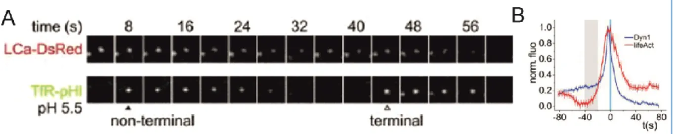
C.4. Visualising AMPAR endocytosis 132
P
OST-
ACQUISITION ANALYSIS133
A. Multi-dimentional Image Analysis: MIA 133
B. Newly developed Matlab routines 134
B.1. Partial quenching of surface receptors 134
B.2. Distance measurements 134
B.3. Quantifying the frequency of endocytic events 136
C
HEMICALB
IOLOGY137
A. Peptide synthesis 137
A.1. Design 137
A.2. Synthesis 139
B. Biochemical and biophysical characterisation 140
Results
141
P
H
UJI:
A
PH
S
ENSITIVER
ED-F
LUORESCENTP
ROTEINF
ORI
MAGINGO
FE
XO-
A
NDE
NDOCYTOSIS142
V
ISUALISINGT
HED
YNAMICS OFE
NDOCYTICZ
ONESI
NN
EURONALD
ENDRITES162
T
OWARDST
HED
EVELOPMENT OFA
P
HOTOACTIVATABLEI
NHIBITOR OFE
NDOCYTOSIS188
P
RECISES
PATIOTEMPORALR
ECRUITMENT OF THEL
OWES
YNDROMEP
HOSPHATASEOCRL
IN THEE
ARLYS
TEPS OFE
NDOCYTOSIS BY THER
AB35
GTP
ASE227
Discussion and Perspectives
231
A
DVANTAGESO
FT
HE PPH
P
ROTOCOL232
A. Plaques, pits, buds and vesicles 232
B. Pinpointing cargo internalisation 233
I
MAGINGE
NDOGENOUSE
NDOCYTOSIS234
A. Limitations of cargo overexpression 234
B. Perspectives for the visualisation of endogenous cargo internalisation 235
P
HOTOACTIVATIONI
NN
EURONS236
13
B. Postsynaptic spatio-temporal regulation 237
Conclusion
239
References
240
Appendix
269
R
ÉSUMÉL
ONGE
NF
RANÇAIS269
A. INTRODUCTION 269
B. OBJECTIFS 275
C. MATERIELS ET METHODES 276
D. RESULTATS 278
14
L
IST
O
F
T
ABLES
A
ND
F
IGURES
Figure 1: Major vesicular membrane trafficking pathways 26
Figure 2: Lipid composition of membrane compartments and lipid recognition domains 28
Figure 3: Subcellular localisation of Rab GTPases 29
Figure 4: Multiple internalisation routes at the plasma membrane 30
Figure 5: Modules of the clathrin mediated endocytosis interactome 39
Figure 6: The clathrin cage 39
Figure 7: Crystal structure of AP-2 40
Figure 9: Canonical view of the various stages of clatrhin mediated endocytosis 41
Figure 8: Domain organisation of dynamin 41
Figure 10: Adaptors are required for cargo concentration and selection at CCPs 43 Figure 11: Various shapes of clathrin structures – from flat to highly curved lattices 44
Figure 12: Example crystal structures of dimeric BAR domains 45
Figure 13: Membrane bending by helix insertion 46
Figure 14: Five proposed models for dynamin's function in vesicle scission 47 Figure 15: GTP-dependent conformational changes induce a helical twist in the dynamin ring 49
Figure 16: Example transferrin uptake and antibody feeding assays 51
Figure 17: Protein recruitment profiles aligned to the time of clathrin disappearence 52 Figure 18: Monitoring CCS lifetimes as a readout of endocytic activity and inhibition 53 Figure 19: Protein signatures and morphological changes undergone by forming CCVs 54
Figure 20: The pulsed-pH assay reveals new dynamics of endocytosis 55
Figure 21: Brain cells and their interactions 57
Figure 22: Complexity of neuronal morphologies 59
Figure 23: Neuronal communication 60
Figure 24: Anatomical features of synapses 62
Figure 25: Induction of an action potential 64
Figure 26: Molecular organisation of the postsynaptic density 66
Figure 27: Crystal structure of the AMPA receptor 67
Figure 28: Special features of the NMDA receptor 68
Figure 29: Shared characteristics of LTP and memory 70
Figure 30: Long lasting bidirectional modulation of synaptic strength 71
Figure 31: Trafficking of synaptic vesicles in a presynaptic terminal 74
Figure 32: Molecular components regulating SV exocytosis 75
Figure 33: AMPA receptor trafficking at the postsynapse 78
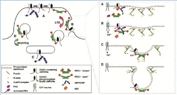
Figure 34: Mobile AMPARs regulate the recovery from paired-pulse depression 79 Figure 35: Possible regulation of AMPAR endocytosis by GluA2 interacting proteins 82
Figure 36: Visualising endocytosis in postsynaptic compartments 84
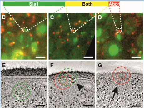
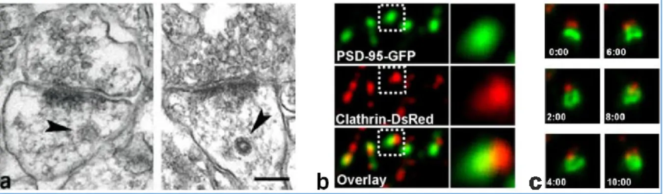
Figure 37: Clathrin coated structures are found in close proximity to the postsynaptic density 85 Figure 38: Imaging the dynamics of clathrin structures reveals several endocytic mechanisms 88 Figure 39: Optical advantages of TIRF illumination for the study of endocytosis 89
15 Figure 41: Monitoring exocytosis and endocytosis using pHluorin tagged proteins 92
Figure 42: Monitoring AMPAR endocytosis 93
Figure 43: Small molecule inhibitors of endocytosis 96
Figure 44: Principles of biomimetism to inhibit protein-protein interactions 97 Figure 45: Interfering with clathrin or dynamin perturbs the pathway at different stages 99 Figure 46: SH3-domain interactions are necessary for vesicle scission 102
Figure 47: Effect of D15 on synaptic transmission and plasticity 104
Figure 48: Major photoactivation strategies 108
Figure 49: NIH-3T3 cells in culture 113
Figure 50: The setup used for simultaneous imaging, patch-clamp recording and uncaging 116
Figure 51: The triggering paradigm 118
Figure 52: Definition of the critical angle 119
Figure 53: Principle of TIRF illumination applied to biology 119
Figure 54: Simultaneous imaging and cell dialysis 122
Figure 55: Comparison of the intracellular solutions used in this project 124
Figure 56: Induction of chemical LTD 132
Figure 57: Detection of endocytic events originating from partially quenched surface clusters 134 Figure 58: Measuring the distance between endocytic events and synapses 135 Figure 59: Normalisation procedure and quantification of the efficacy of endocytosis blockers 136
Figure 60: Sequence alignment of D15 and AL14 137
Figure 61: Truncation peptides synthesised for this project 138
Figure 62: Sequences of the dimeric peptides synthesised for this project 139 Figure 1000: Me writting this manuscript... and onwards? A concluding metaphore 284
Table 1: Peptides interfering with protein-protein interactions involved in presynaptic endocytosis 103 Table 2: Peptides interfering with protein-protein interactions involved in postsynaptic endocytosis
16
A
BBREVIATIONS
A
ND
D
EFINITIONS
General
A
a.a : amino-acid
AAK1 : adaptor-associated kinase 1
AMPA : L-α-amino-3-hydroxy-5-methyl-4-isoxazolepropionic acid
AMPAR : AMPA-type glutamate Receptor
amph-SH3 : SH3 domain of amphiphysin
ANTH : AP-180 N-Terminal Homology
AP-2 : Adaptor Protein 2 - A complex composed of four subunits (α, β2, µ2 and σ2) binding the plasma membrane and clathrin, and mediating a number of interaction with receptor cargo and associated proteins.
AP : Action potential
APPL1 : A4 precursor protein like 1
Arc : Activity Regulated Cytoskeleton-associated protein
Arf6 : ADP-ribosylation Factor 6
ARH : Autosomal Recessive Hypercholesterolemia
Arp2/3 : Actin-Related Proteins 2 and 3 complex - An actin filament nucleator
AMP : Adenosine MonoPhosphate
ASIC : Acid Sensing Ion Channel (can be blocked by amiloride)
ATP : Adenosine TriPhosphate
Axon : Nerve fibre carrying electrical impulses coding for neuronal information.
B
β2AR : β2-Adrenergic Receptor
BAR : BIN/Amphiphysin/Rvs
BCG : BromoChresol Green
BPB : BromoPhenol Blue C
°C : Celsius degrees
CAMKII : Calcium/calmodulin-dependent protein kinase II
cAMP : cyclic Adenosine MonoPhosphate
Clathrin : Trimeric protein polymerising into a spherical scaffold around invaginating membranes. Its terminal domains are major protein- protein interaction hubs.
CCP : Clathrin Coated Pit
CCS : Clathrin Coated Structure
CCV : Clathrin Coated Vesicle
CLASP : Clathrin-coat Associated Sorting Protein
17
Chem-LTD : Chemically induced Long Term Depression
CME : Clathrin Mediated Endocytosis
COP : COat Protein complex D
D15 : 15 amino-acid long peptide mimicking part of the PRD of dynamin and interacting with the SH3-domain of amphiphysin
Dab2 : Disabled homolog 2
Dendrite : Neuronal processes specialised in integrating multiple electrical input signals.
DIV : Days in vitro
Dyn : Dynamin - Large GTPase involved in the fission reaction of endocytic vesicles from the plasma membrane. Dynamin-1 and 3 are
predominantly neuronal. Dynamin-2 is ubiquitous. E
ECM : ExtraCellular Matrix
EGFP : Enhanced Green Fluorescent Protein
EGFR : Epidermal Growth Factor
EH : Eps15 homology - modular domain providing proteins with membrane binding properties
EM : Electron Microscopy
EMCCD : Electron Multiplying Charge Coupling Device
Endosome : (or endosomal compartment) - Membrane bound intermediate in the endocytic machinery to which newly formed vesicles fuse and from which recycling vesicles bud off. Endosomes eventually fuse with lysosomes for content degradation
endo-SH3 : SH3 domain of endophilin
Eps15 : Epidermal growth factor pathway substrate 15
EPSP : Excitatory PostSynaptic Potential
ER : Endoplasic Reticulum F
FALI : Fluorophore Assisted Light Inactivation
FCHo : Fer/Cip4 homology domain-only proteins 1 and 2
FEME : Fast Endophilin Mediated Endocytosis
FITC : Fluorescein IsoThioCyanate
FlAsH : Fluorescein Arsenical Helix binder
(or 4′,5′-bis(1,3,2-dithioarsolan-2-yl)fluorescein)
FYVE : Fab1, YOTB/ZK632.12, Vac1 and Early endosome antigen 1 - Modular domain providing proteins with membrane binding properties
G
GABA : Gamma-AminoButyric Acid
GAK : cyclin G-Associated Kinase (aka auxilin)
18
GED : GTPase effector domain
GFP : Green Fluorescent Protein
GPCR : G-Protein Coupled Receptor - A family of proteins largely involved in cellular signalling pathways.
GPI : GlycoPhosphatidylInositol - GPI anchors allow proteins to be targeted to the plama membrane and remain membrane bound.
GPI-AP : GPI Anchored Protein
GRIP : Glutamate Receptor-Interacting Protein
GTP : Guanosine TriPhosphate
GTPase : GTP-binding protein with GTP hydrolysis activity H
HEPES : 4-(2-hydroxyethyl)-1-piperazineethanesulfonic acid – pH 7.4 buffering agent)
Hsc70 : Heat shock cognate protein 70 kDa I
IPSP : Inhibitory PostSynaptic Potential
iGluR : Ionotropic Glutamate Receptor L
LDLR : Low Density Lipoprotein Receptor
LTD : Long Term Depression
LTP : Long Term Potentiation M
MALDI-TOF : Matrix-assisted laser desorption ionisation – Time Of Flight MES : 2-(N-morpholino)ethanesulfonic acid – pH5.5 buffering agent
MIA : Multi-dimentional Image Analysis
mGluR : Metabotropic Glutamate Receptor N
NMDA : N-Methyl-D-Aspartic acid
NMDAR : NMDA-type glutamate Receptor
NSF : N-Ethylmaleimide-Sensitive Fusion protein O
OCRL : OculoCerebroRenal syndrome of Lowe P
19
PH : Pleckstrin Homology - modular domain providing proteins with membrane binding properties
pH : Acidity of an aqueous solution
PICK1 : Protein Interacting with C Kinase 1
PI : PhosphoInositides - Negatively charged lipid constituents of membranes. Distiguished by their phosphoylation state.
PiPES : 1,4-PiPerazinediEthaneSulfonic acid
pKa : Logarithmic acidity constant PKA : Protein Kinase A
PKC : Protein Kinase C
PLC : PhosphoLipase C
PP1 : Protein Phosphatase 1
PP19 : 19 amino-acid long peptide mimicking part of the PRD of synaptojanin and interacting with the SH3-domain of endophilin
ppH : pulsed pH protocol
PRD : Proline and aRginine-rich Domain - modular domain involved in protein-protein interactions with SH3 domains
PSD : Post Synaptic Density
PSP : PostSynaptic Potential
PX : PhoX homology - modular domain providing proteins with membrane binding properties
R
RP-HPLC : Reverse-phase high performance liquid chromatography
RRP : Readily Releasable Pool - Functionally defined pool of SVs releasable by moderate stimulation of synaptic terminals
RP : Recycling Pool - Functionally defined pool of SVs releasable by sustained stimulation of synaptic terminals
R2D2 : Reel2-Dialog2
S
SEP : Super-Ecliptic pHluorin - a pH sensitive mutant of EGFP
SH3 : Src Homology 3 - modular domain involved in protein-protein
interactions with proline rich motifs.
SHIP2 : SH2-domain-containing Inositol 5'-Phosphatase
SNAP : Synaptosomal-Associated Protein
SNARE : Soluble N-ethylmaleimide-sensitive-factor Attachment protein REceptor
Soma : Cell body of neurons
Src : Sarcoma
STDP : Spike timing Dependent Plasticity
STED : STimulated Emission Depletion
STORM : STochastic Optical Reconstitution Microscopy
SV : Synaptic Vesicle - Refers to presynaptic neurotransmitter filled vesicles
Synapse : Specialised contact site enabling neuronal communication T
20
TARP : Transmembrane AMPAR Regulatory Proteins
Tfn : Transferrin
TfR : Transferrin Receptor - a model receptor to study CME as it is constitutively being internalised.
TGN : TransGolgi Network
TIRF : Total Internal Reflection Fluorescence microscopy - Imaging method used to study processes occurring in close proximity to the glass interface on which a sample is present. (aka evanescent wave microscopy)
TTL : Transistor-Transistor Logic
TTX : Tetrodotoxin - A blocker of sodium channels inhibiting AP firing U
UV : Ultraviolet (light characterised by a wavelength shorter than 400 nm) V
VAMP : Vesicle-Associated Mebrane Protein (aka synaptobrevin)
VGCC : Voltage-Gated Calcium Channel W
21
Amino-acid single letter code
A (Ala) : AlanineC (Cys) : Cysteine
D (Asp) : Aspartic acid (Aspartate) E (Glu) : Glutamic acid (Glutamate) F (Phe) : Phenylalanine G (Gly) : Glycine H (His) : Histidine I (Ile) : Isoleucine K (Lys) : Lysine L (Leu) : Leucine M (Met) : Methionine N (Asn) : Asparagine P (Pro) : Proline Q (Gln) : Glutamine R (Arg) : Arginine S (Ser) : Serine T (Thr) : Threonine W (Trp) : Tryptophan V (Val) : Valine Y (Tyr) : Tyrosine
X : Any amino-acid in a consensus sequence
ɸ : Any hydrophobic amino-acid in a consensus sequence
Chemical products
Ca : Calcium Cl : Chloride CO2 : Carbon diOxyde DMNPE : 1-(4,5-DiMethoxy-2-NitroPhenyl)Ethyl H20 : WaterHBS : HEPES Buffered Saline
HEPES : 4-(2-HydroxyEthyl)Piperazine-1-EthaneSulfonic acid - pKa(25°C) = 7.5
K : Potassium
MBS : MES Buffered Saline
MES : 4-MorpholineEthaneSulfonic acid - pKa(25°C) = 6.1
Mg : Magnesium
N : Nitrogen
Na : Sodium
OH : Hydoxyl
PiBS : PiPES Buffered Saline
22
F
OREWORD
New technologies and ground-breaking discoveries often go hand in hand. Investigators can answer increasingly detailed and complex questions as new methodologies are made accessible to them, just as much as they drive the development of ever more advanced techniques by perpetually taking up more demanding challenges. Neuroscience is one of those fields which illustrates this stimulating feed-forward phenomenon best. The brain, the cells that constitute it and the contacts made between them are so small, yet so highly organised, that we continuously need to seek new ways to look at them, monitor them, perturb and even control them if we one day hope to understand their function. Biologists, chemists, physiologists and many others all contribute to this goal, and the present thesis is but one of the many efforts made towards it.
The work presented in the following pages was performed in the Interdisciplinary Institute for Neuroscience, in the team of Dr. Daniel Choquet "Dynamic Organization and Function of Synapses", under the supervision of Dr. David Perrais. It will deal with understanding the spatial and temporal molecular dynamics of endocytosis while focusing on the development of new tools to help with this understanding. This work will be defended in a public oral communication in front of Dr. Nathalie Sans (Neurocentre Magendie, INSERM U862, Bordeaux University, France), Pr. Aurélien Roux (NCCR Chemical-Biology and Biochemistry Department, University of Geneva, Switzerland), Pr. Volker Haucke (Leibniz Institute for Molecular Pharmacology and Freie University Berlin, Germany) and Pr. Britta Qualmann (Institute for Biochemistry I at the University Hospital, Friedrich Schiller University Jena, Germany).
The introduction will aim at presenting key notions and concepts about endocytosis in general and in neurons: what is known about it from a mechanistic point of view, what roles does it play in maintaining and shaping synaptic transmission and finally what tools have been used so far to study it.
I will then provide a detailed description of the methodologies I have used and developed in order to visualise and perturb endocytosis with high spatial and temporal resolution.
The results of my work will be presented in the shape of three scientific communication articles and one article abstract. The first article has been published in the Journal of Cell Biology in 2014. It has been written in collaboration with the group of R.E. Campbell (University of Alberta, Canada) and characterises a new red pH-sensor
23
that enables dual-colour imaging of receptor endocytosis. The second article is in preparation. It describes a novel imaging method that detects the dynamics of endocytic vesicle formation in neurons. The third article, also in preparation, was written in collaboration with chemists of our institute. It reports on the ongoing development of a photoactivatable inhibitor of endocytosis. The fourth article will be shortly presented as a summary, illustrated by one figure. It introduces a collaborative work, undertaken with the group of A. Echard (Institut Pasteur, Paris), that uses imaging to characterise the role of Rab35 in recruiting OCRL on nascent endocytic vesicles.
Finally, I will provide an extended discussion on the advantages and limitations of the tools and protocols I developed as well as propose some perspectives for the continuation of the project based on the obtained results.
25
Introduction
Visualising endocytosis unambiguously is a challenging task, and perturbing it is a necesary one for a deeper understanding of its regulation. When combined with the intrinsic complexity of neurons, it becomes an exciting field of study. In this introduction, I shall first provide a description of the molecular mechanisms regulating clathrin mediated endocytosis and an overview of the general principles of neurotransmission. I will then combine these two bodies of knowledge to describe the functional roles of endocytosis in shaping neuronal communication as well as provide technical insights on the existing methods to study it, insisting on their advantages and limitations.
E
NDOCYTOSIS
–
O
R
H
OW
C
ELLS
G
ET
T
HEIR
O
UTSIDE
I
N
A. One step of a complex machinery
Endocytosis is a universal feature of all eukaryotic cells. Literally meaning getting inside (“endo”) the cell (“cyto”), the term was coined by Christian deDuve during a symposium entitled “The Lysosome” in 1963. Nowadays, it refers to the process by which a substance, proteins, lipids as well as components in suspension or solubilised in the extracellular fluid, gains entry inside the cell by being engulfed in a membrane surrounded vesicle. This mechanism is to be opposed to processes allowing substances to pass directly through the membrane by diffusion or with the help of transporters or channels. The formation and packaging of these vesicles, their transport throughout the cell interior, their fusion with other cellular compartments, their recycling to the cell exterior and their degradation are all part of a complex phenomenon known as
26
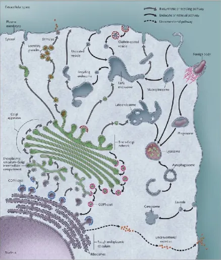
Figure 1: Major vesicular membrane trafficking pathways Vesicular trafficking occurs at various cellular compartments: between the Endoplamic Reticulum (ER) and the Golgi for protein synthesis and maturation as well as lipid biogenesis; from the Golgi to endosomes as well as directly to the plasma membrane for protein sorting and localisation; from recycling endosomes to the plama membrane for recycling and exocytosis; and from the plasma membrane to early endosomes and lysosomes for sorting, recycling and degradation. Some vesicles require coat proteins such as COPI, COPII, caveolin or clathrin for their formation whereas others seem to be able to protrude or invaginate without the need for such a scaffold. Pathogens such as bacteria or viruses are capable of hijacking some of these entry routes.
Source: (De Matteis and Luini, 2011)
Endocytosis is a major step in this grand machinery that ranges from the budding of vesicles from the plasma membrane to the processing if these vesicle inside the cell as they are being transported to their final destination. It controls the biochemical composition of a cell’s outer plasma membrane and serves a multipurpose
role to regulate complex, yet vital, cellular processes such as nutrient uptake, cell
adhesion, cell migration, cell polarity, cytokinesis (splitting of a mother cell into two daughter cells), biochemical signalling and neurotransmission.
A.1. Membrane trafficking: Overview and definitions
The plasma membrane (PM) is a lipidic bilayer that forms the boundary that defines the intracellular and extracellular compartments of a cell. It is composed of phospholipids, a subcategory of which, known as phosphoinositides (PIs), is particularly important in regulating membrane trafficking events. Many proteins are embedded in the PM and ensure proper communication between the cell and its exterior. Within the cell, many other membrane bound compartments, or organelles, co-exist: the nucleus is the site of gene transcription and controls the cell cycle by which cells divide. The nuclear membrane is continuous with the membrane of the
27
transcription, they fold in the ER. Folded proteins can either be secreted as such or proceed to the Golgi apparatus via COPII (coat protein complex II) coated vesicles where they will be further modified by post-translational modifications including glycosylation and phosphorylation. Modified proteins as well as lipids are then transported via COPI coated vesicles to the transgolgi network (TGN) where they will be packaged into different vesicles according to their destination: some will go to
lysosomes (or late endosomes) to ensure the acidic environment and the presence of
hydrolytic enzymes in this compartment specialised in protein degradation; others will directly reach the PM to be secreted on demand upon stimulation via exocytosis; some are constitutively trafficked to the cell surface where they will be exocytosed and let the proteins and lipids they contained diffuse in the bulk of the PM; and finally, some will be trafficked to early endosomes which constitute sorting platforms that can decide the fate of proteins. These early endosomes are also an intermediate step of the inverse route, when proteins, lipids and other cargo molecules go from the outside to the inside of the cell via endocytosis. Proteins and lipids contained in early endosomes will then be packaged again, either to reach lysosomes for degradation or to be sent to recycling
endosomes that will bring them back to the PM for a new cycle.
A.2. Membrane trafficking markers and regulators
The various membrane delineated subcellular organelles display identity markers that are believed to both help with their function as well as coordinate the communication and routing of vesicles trafficking from one organelle to another. These markers consist of lipids and proteins known as phosphoinositides (PIs) and Rab GTPases.
PIs result from the phosphorylation of phosphatidylinositol, a type of
amphiphatic lipid which polar head is composed of an inositol that can be phosphorylated at positions 3, 4 and/or 5. The family of PIs thus includes the following lipids: PI(3)P, PI(4)P, PI(5)P, PI(3,4)P2, PI(3,5)P2, PI(4,5)P2 and PI(3,4,5)P3. The different
subcellular membrane compartments are differentially enriched in the various PIs. This is believed to play an important role in targeting regulatory proteins to the right compartment using lipid recognition domains (Figure 2). PI(4,5)P2 is particularly
important in regulating endocytosis and signalling cascades. It is thereby often simply referred to as PIP2. It can be cleaved into the signalling molecules IP3 and diacyglycerol
by the activity of the phospholipase PLCγ. It is also the substrate of kinases and phosphatases that regulate its phosphorylation state. Its biosynthesis from PI(4)P is thus ensured by the 5’-kinases PIP5Kα, β and γ. Conversly, its turnover into PI(3,4,5)P3
or PI(4)P is achieved by the 3’-kinase PI3K and the 5’-phophatases SHIP2, synaptojanin or OCRL respectively (Antonescu et al., 2011). As illustrated in Figure 2, PIP2 is
particularly enriched at the plasma membrane but early and recycling endosomes are predominantly composed of PI(3)P. The turnover of PIP2 into PI(3)P is therefore
28
proposed that the formation of PI(3)P from PIP2 relied on the formation PI(3,4)P2 as an
intermediate species via the action PI(3)K C2α (Posor et al., 2013). The 4'-phosphatase(s) that would then lead to PI(3)P is(are) however currently unknown. Interestingly, the 4'-phosphatase Sac2/INPP5F, has recently been identified on endosomes but in vitro assays suggest that it specifically dephosphorylates PI(4)P over other PIs, including PI(3,4)P2 (Nakatsu et al., 2015).
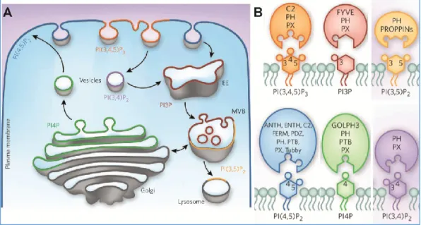
Figure 2: Lipid composition of membrane compartments and lipid recognition domains
(A) Subcellular localisation of various PIs. The plasma membrane is mostly enriched in PIP2 while early
endosomes (EE) are mostly enriched in PI(3)P. (B) Many proteins involved in membrane trafficking (coloured ellipses) exhibit lipid recognition domains. Some of these domains recognise all PIs such as PH and PX domains. Others are specific of a given PI such as the ANTH and ENTH domains for PIP2 or the FYVE domain for PI(3)P. PIs
are represented with hexagonal headgroups phosphorylated at positions 3, 4 and/or 5 (circles).
Source: adapted from (Kutateladze, 2010)
Rab GTPases form a large family of 60 to 70 molecular switches cycling from
a GTP bound state to a GDP bound state. Each Rab is specific for a subcellular compartment and can even define membrane microdoamains within a given compartment (Figure 3). They therefore play an essential role in spatially and temporally controlling the recruitment of functionally active molecules to their due site of action. The switch from GDP- to GTP-bound state is catalysed by a guanine exchange factor (GEF) while the switch from GTP- to GDP-bound state occurs upon hydrolysis of the GTP nucleotide and is regulated by a GTPase-activating protein (GAP). Rab proteins undergo major conformational rearrangements upon binding to GTP which confers them the ability to recruit a variety of effectors. Rab effectors have been implicated in all aspects of membrane trafficking such as vesicle budding, vesicle uncoating, vesicle motility and vesicle fusion (Stenmark, 2009). Interestingly, Rab GTPases could also be implicated in defining the lipidic identity of the membranes they are bound to by recruiting PI kinases or phosphatases as effectors. Recently, Rab GTPases have also been proposed to directly interact with endocytic cargo molecules, thereby controlling their
29
fate in being routed towards recycling or degradation pathways for example (Aloisi and Bucci, 2013).
Figure 3: Subcellular localisation of Rab GTPases
The family of Rab GTPases specifies the identity of intracellular organelles. They are involved in routing vesicles from one compartment to another by recruiting effectors to their correct subcellular localisation. For example Rab 1 and 2 regulate ER to Golgi trafficking. Rab 8 is localised at the TGN and regulates direct exocytosis. Rab 5 is found in newly formed endocytic vesicles and in early endosomes together with Rab 4 which in turn is responsible for rapid recycling to the PM. Rab 11 and 35 on the other hand are found on recycling endosomes and regulate the slow recycling. Rab 15 regulates the trafficking from early to recycling endosomes whereas Rab 7 and 9 are found on late endosomes and may regulate the maturation from late endosome to lysosome.
Source: (Stenmark, 2009)
A.3. Endocytosis in membrane trafficking
Strictly speaking, endocytosis refers to the whole process through which solute, proteins and lipids, together referred to as cargo, are internalised and then fuse with an endosome that will either mature into a lysosome or will reach a recycling endosome. Many endocytic pathways co-exist within a cell. Their internalisation mechanisms vary a great deal and require the involvement of various molecular players. However, many features of these pathways overlap and they all lead to the eventual fusion of the newly formed vesicle with an early endosome. Why cells would use a great variety of entry mechanisms to finally merge all the vesicular contents in the same structure is currently poorly understood. Whether the endosomal population within a given cell is actually heterogeneous or whether endosomes display a nanoscale organisation such that, as vesicles coming from different routes fuse with them, they retain this information into subdomains for future sorting remains largely unknown.
30
It should also be noted, and it will often be the case in this manuscript, that the word "endocytosis" is quite commonly used to solely refer to the internalisation step, when vesicles are being formed: how cargo is selected at the level of the PM, which mechanisms enable membrane invagination and finally how membrane fission is achieved are complex enough questions to deserve a whole field of study of their own, however fascinating the fate of these vesicles is.
The best studied form of endocytosis is known as clathrin mediated endocytosis (CME). It is characterised by the packaging of membrane and proteinacious components into small vesicles coated with clathrin proteins. The mechanisms regulating the internalisation step of this endocytic pathway involve many accessory proteins and will be the main focus of the present study. However, clathrin independent forms of endocytosis have increasingly drawn the attention of the cell biologist community and will be briefly described in the following section.
B. The various modes of endocytosis
Figure 4: Multiple internalisation routes at the plasma membrane
Phagocytosis specialises in the uptake of large particles such as bacteria or debris whereas pinocytosis originally refers to fluid phase uptake. However, most of the mechanisms not only internalise purely extracellular components such as nutrients and enzymes but also enable the internalisation of membrane bound proteins and membrane lipids. The various forms of endocytosis are defined by their molecular machinery and characterised by the nature of their cargo and the size of the vesicles formed.
31 B.1. Phagocytosis
Phagocytosis refers to the process of engulfing large solid particles into the cell interior. It is a major mechanism used by cells of the immune system (macrophages in the body and microglia in the nervous system) to clear cell debris and pathogens such as post-apoptotic cells, bacteria or viruses. The internalised particle ends up in a phagolysosome, a transient compartment containing lytic enzymes and reactive oxygen species that will degrade the particle before disappearing (Nüsse, 2011). The first step in phagocytosis is target recognition, a process mediated by antigen receptors present at the cell surface. Then internalisation occurs as the membrane protrudes into a phagocytic cup by the action of the actin cytoskeleton with the help of actin modulators such as WASP and the Arp2/3 complex (Rougerie et al., 2013). The activity of dynamin, a large GTPase that will be extensively discussed later in this manuscript, is believed to mediate the fission reaction leading to the creation of the phagosome (Gold et al., 1999).
B.2. Macropinocytosis
Macropinocytosis is one of those confounding processes that can be readily observed but lack features to specifically describe them. It consists in the protrusion of plasma membrane into the extracellular space that leads to fluid phase uptake into a macropinosome upon closure. However, at the molecular level, it is rather characterised by what doesn't regulate it than by what does. It indeed does not exhibit any cargo specificity, the macropinosome is devoid of coat proteins and the mechanism for membrane closure is currently unknown. Nonetheless, macropinocytosis is known to be greatly enhanced by the presence of growth factors in the extracellular medium and shares many common features with phagocytosis such as the requirement for actin and actin regulators as well as the importance of the lipid PI(3)P (Jones, 2007). The non specificity of macropinocytosis makes it a favourite entry route for several pathogens including viruses (Mercer and Helenius, 2009).
Together, endocytic pathways that are neither phagocytosis nor macropinocytosis form a functional category known as micropinocytosis. They are characterised by smaller, stereotypical vesicles (~100 nm compared to the one or more micron big phagosomes and macropinosomes) that form by membrane invagination rather than membrane protrusion. After their formation, these vesicles converge to fuse with sorting endosomal compartments. Micropinocytosis can be categorised by the molecular players involved in the internalisation step, especially according to whether or not vesicle formation requires the presence of a coat protein.
32
B.3. Clathrin and caveolin independent pathways
Some poorly understood pathways are known to occur in cells because they allow internalisation of certain cargos without depending on canonical entry routes. The best studied of those pathways is the CLIC-GEEC pathway, initially identified as the entry route for the cholera toxin (Kirkham et al., 2005). Vesicles forming through this pathway converge to intracellular compartments concentrated in GPI-anchored proteins (GPI-APs). For lack of other candidate molecular players, this endocytic pathway is thus best described as forming CLathrin-Independent Compartments (CLIC) that fuse to GPI-AP-enriched Early Endosomal Compartments (GEECs). Because of the lack of a rigid spherical coat around the invaginating membrane, tubular structures are observed when this pathway is being studied. They do not appear to require coat proteins and the involvement of dynamin in membrane fission is either unclear or unknown (Doherty and McMahon, 2009). Other clathrin and caveolin independent pathways include the
Arf6 dependent pathway and the flotilin dependent pathway which enable the
internalisation of MHC class I proteins (involved in the immune system) and of proteoglycans (components of the extracellular matrix) respectively (Mayor and Pagano, 2007; Doherty and McMahon, 2009). Intrestingly, these forms of endocytosis could be initiated by invaginations induced by extracellular factors, such as pathogens like Shiga toxin B (Römer et al., 2007) or SV40 virus (Ewers et al., 2010), and endogenous lectins (Lakshminarayan et al., 2014), which further increases the complexity of defining these pathways. Moreover, a novel endocytic pathway, reported under the name FEME, standing for fast endophilin mediated endocytosis, has been recently described as forming endophilin coated tubular invaginations. It appears to be responsible for the internalisation of classical clathrin independent cargos such as activated β1-adrenergic receptors, interleukin-2 receptors and cholera toxin, on a timescale of a few seconds (Boucrot et al, 2014).
B.4. Caveolin dependant endocytosis
Caveolae are small 60-80 nm wide flask-shaped invaginations found at the PM of all cell types but red and white blood cells and neurons, and are particularly numerous in muscle cells. They are coated by oligomerised caveolins and cavin complexes and are enriched in cholesterol and sphingolipids. Caveolae are preassembled at the level of the Golgi, where they are already coated with caveolin. As they are trafficked to the PM, they are stabilised by cavins and probably adopt their final shape by interacting with PACSIN, aka syndapin, a curvature sensing molecule. Caveolae are quite stable structures but can be disrupted by cholesterol depletion due to disruption of the caveolin/cavin interaction (Parton and del Pozo, 2013). For many years, caveolae have been described as a constitutive endocytic route for various cargos such as palmitoylated G-Protein Coupled Receptors (GPCRs) (Patel et al., 2008), ubiquitylated epidermal growth factor receptor (EGFR) and GPI-APs, highlighting a strong overlap between this pathway and the CLIC/GEEC pathway (Mayor and Pagano,
33
2007). However, inconsistent results and recent advances in the understanding of caveolae have brittled this belief. Caveolae are now sometimes viewed as
mechanosensors, capable of flattening out when mechanical stress is inflicted to the
PM, thereby regulating membrane tension. They do undergo dynamin dependant endocytosis but either retain their identity and recycle back to the PM by exocytosis without dissociating or are targeted to late endosomes for degradation. This feature is different from clathrin mediated endocytosis, described below, where coat components as well as vesicular identity upon fusion with early endosomes are rapidly lost. This intriguing stability of caveolae has therefore been proposed to play a key role in intracellular signalling by spatial segregation. By regulating the localisation of signalling receptors into cholesterol and sphingolipid rich microdomains, called lipid rafts at the PM, endocytosis of caveolae may play a role in modulating their activity (Parton and del Pozo, 2013).
B.5. Clathrin mediated endocytosis
Clathrin mediated endocytosis (CME) is by far the most studied and best understood endocytic pathway. A recent, and rather audacious, study even claims that it represents the only endocytic pathway used by the cells for cargo internalisation purposes under normal conditions (Bitsikas et al., 2014). It is a fundamental endocytic pathway which regulatory mechanisms are greatly conserved throughout eukaryotes, from yeast to mammals. The first observation of such internalisation from the PM dates back to the 1960's, with a study of ferritin uptake (Rosenbluth and Wissig, 1964). It was originally called receptor mediated endocytosis but this term was abandoned as more and more receptors were found to be internalised via clathrin independent routes. Clathrin coated pits (CCPs) assemble at the plasma membrane which then invaginates to form clathrin coated vesicles (CCVs) of sizes ranging from 80 nm in the brain to 150 nm in other mammalian cells. This pathway is known to be responsible for the internalisation of a variety of signalling receptors, such as GPCRs and growth factor receptors, ion channels, such as aquaporins and AMPA receptors, and other transmembrane proteins such as integrins and tyrosin receptor kinases. It is also specifically implicated in the internalisation of the transferrin receptor (TfR), making the latter a good marker for studying CME. Being involved in so many cellular functions, CME is unsurprisingly highly regulated by a myriad of molecular interactors. These include adaptor proteins (e.g. AP-2, AP-180, β-arrestin) which in combination with clathrin form the coat around the vesicle, and clathrin-coat associated sorting proteins (CLASPs) which help with specific cargo selection and pathway directionality (Traub, 2009). Molecular mechanisms inducing the subsequent invagination of the CCP are still poorly understood but are known to involve the recruitment of BIN-Amphiphysin-Rvs167 (BAR) domain containing proteins. Dimers of such proteins form banana shaped structures believed to sense and/or induce curvature onto the membrane they bind to (Qualmann et al., 2011). As for the final scission of the vesicle from the membrane, there is no controversy about the necessary requirement of dynamin (Robinson, 1994; Roux
34
et al., 2006). The involvement of actin in late stages of yeast CME is widely accepted but although it has consistently been observed right after membrane scission, its necessity in mammalian cells is still being debated (Boulant et al., 2011). PIP2 is then turned into
PI(3)P and vesicles are being uncoated before fusing with early endosomes.
Being the main interest of the present study, the mechanisms regulating CME will be described in much further detail in section D. of this chapter. However, in order to emphasise the vital importance of this pathway, focus will first be given to the many cellular functions in which it is involved.
C. Functional roles of endocytosis
The descriptions made in the following paragraphs are meant as an overview, and the, probably non exhaustive, listing of functional roles briefly presented here was inspired from two reviews (McMahon and Boucrot, 2011) and (Gould and Lippincott-Schwartz, 2009). Each of them are described in great detail in dedicated reference works, with their own regulatory mechanisms and their specific molecular modulators. Most examples cited here are taken from mammalian endocytosis, even though vast amounts of work performed in yeast or other model organisms could illustrate the same concepts.
C.1. Endocytosis and nutrient uptake
Endocytosis primarily refers to fluid phase uptake. The extra cellular fluid contains many nutrients, e.g. amino-acids, ATP, growth factors and oxygen, as well as metabolites, such as cholesterol and iron, important for cell function. Nutrient depletion is known to induce phagocytosis mediated autophagy, i.e. regulated cell death (Russell et al., 2014). CME of receptors involved in sensing those factors can occur in two different ways: constitutively or upon stimulation. TfR for example is constitutively endocytosed, meaning that it does not have to be bound by its ligand transferrin (Tfn, an iron binding protein) to be internalised. On the other hand, EGFR dimerises upon ligand binding and this step is necessary for its binding to AP-2 and subsequent internalisation.
C.2. Endocytosis and degradation
Proteins have a defined lifetime. Either because downregulation is needed in response to an environmental change, or simply to ensure that cellular function is properly fulfilled, cells regularly degrade proteins and provide newly synthesised copies. Proteins targeted for degradation are tagged with ubiquitin, a 76 amino-acid protein that can oligomerise to form polyubiquitin chains. After internalisation by CME,