1 23
Acta Parasitologica ISSN 1230-2821 Volume 64 Number 1 Acta Parasit. (2019) 64:112-128 DOI 10.2478/s11686-018-00013-9Evaluation of Fasciola hepatica Infections
in Cattle in Northeastern Algeria and
the Effects on Both Enzyme and Hepatic
Damage, Confirmed by Scanning Electron
Microscopy
Amina Taibi, Miriem Aissi, Khaled
Harhoura, Safia Zenia, Houria Zait &
Boussad Hamrioui
1 23
Your article is protected by copyright and
all rights are held exclusively by Witold
Stefa#ski Institute of Parasitology, Polish
Academy of Sciences. This e-offprint is for
personal use only and shall not be
self-archived in electronic repositories. If you wish
to self-archive your article, please use the
accepted manuscript version for posting on
your own website. You may further deposit
the accepted manuscript version in any
repository, provided it is only made publicly
available 12 months after official publication
or later and provided acknowledgement is
given to the original source of publication
and a link is inserted to the published article
on Springer's website. The link must be
accompanied by the following text: "The final
publication is available at link.springer.com”.
Acta Parasitologica (2019) 64:112–128 https://doi.org/10.2478/s11686-018-00013-9
ORIGINAL PAPER
Evaluation of Fasciola hepatica Infections in Cattle in Northeastern
Algeria and the Effects on Both Enzyme and Hepatic Damage,
Confirmed by Scanning Electron Microscopy
Amina Taibi1 · Miriem Aissi1 · Khaled Harhoura1 · Safia Zenia1 · Houria Zait2 · Boussad Hamrioui2
Received: 11 March 2018 / Accepted: 29 November 2018 / Published online: 28 January 2019 © Witold Stefański Institute of Parasitology, Polish Academy of Sciences 2019
Abstract
Purpose The aim of our study is to establish the presence of Fasciola hepatica on farms in northeastern Algeria.
Methods 143 blood and coprological samples of 15 males and 128 females of different breeds and ages were analysed.
Results Our study indicates a heterogeneous level of the anti-f2 antibodies to Fasciola hepatica in response to the infection.
The overall seroprevalence was about 22.37%, and 9 out of 13 investigated farms were infected, with rates varying from 5.88% to 70%. To explain the intrinsic variability of the infection, we identified age, sex and breed as potential risk factors. Based on this, we collected information about their relevance. There was a significant difference for age (p = 0.018) and coprol-ogy (p < 0.0001). Independently, sex and breed had no impact on the infection, although males were more infected (27%) than females (22%). Of the five investigated breeds, Holstein cattle (31.11%) were most affected, followed by Montbeliard (20.27%) and crossed breed (13.64%). Multivariate comparisons showed that the presence of faecal eggs reflects the active infectious status of cattle (p < 0.0001), while age (p = 0.011) and sex (p = 0.040) significantly impact the chance of acquir-ing the infection. To evaluate liver parenchyma integrity and its functionality, hepatic enzymes were examined and showed relatively low levels of aminotransferases, excluding cytolisis. Although sensitive to distomatosis, γGT and PAL values were inconsistent with the infection rate. The relatively high levels of proteins and albumin eliminate hepatic insufficiency.
Conclusion Our results suggest a chronic fasciolosis, confirmed by histology and SEM.
Article Highlights
• The northeast of Algeria is classified as an area with fas-ciolosis.
• Confirmation of enzymatic data by chronology of histo-logical events.
• Invalidation of the relationship between anti-f2 antibod-ies to Fasciola hepatica and the Gamma GT levels, iden-tified as one of the markers of the canalicular enzymes. • Superposition of macroscopic observations to SEM /
EDX images showing calcified structures referring to histological data.
Keywords Cattle · Elisa · Fasciola hepatica · Liver enzyme · Liver injuries
Abbreviations
γGT Gamma glutamyl transferase PAL Alkaline phosphotase
SEM Scanning electron microscopy
Introduction
Of the many parasitic infections that occur in northern Africa, fasciolosis appears to be one of the most common zoonotic diseases in domestic ruminants (cattle, sheep, and goats) [32]. The prevalence of Fasciola hepatica in Tuni-sia affects approximately 14.3% of cattle [17] and between 17.1 and 23.8% of goats in Morocco [20]. In Algeria, epide-miological surveys carried out at slaughterhouses revealed * Amina Taibi
aminataibi.ensv@yahoo.com
Extended author information available on the last page of the article
113 Acta Parasitologica (2019) 64:112–128
percentages of 52.4% in cattle at El Taref [9], 27% at Jijel and 9.1% at Constantine [23]. Serological surveys carried out on a few farms showed a frequency of 6.3% in Constan-tine and 43.7% in Bouinan [3]. The cosmopolitan distribu-tion of this parasitosis affects both northwest and northeast Algeria.
The ecological conditions (climatic and ecopedological conditions) of the fluvial zones favour the proliferation of mollusks, and the presence of the intermediate host (Galba
truncatula) in trampling areas is a link in the overall
infesta-tion pattern.
Given its elective location, liver is the seat of many syn-theses that expose it to many major metabolic disorders, causing economic losses such as weight loss and decreased milk production.
Frequently, the chronic and silent aspect of fasciolosis is only discovered in the slaughterhouse [16].
In this context, the objective of our study was to evalu-ate the parasitic status of livestock in north eastern Alge-ria. Although the coprological diagnosis is rapid and easy, it is often conditioned by the presence and/or absence of
Fasciola hepatica eggs. Their intermittent excretion
unde-niably suggests a confirmation by an immuno-enzymatic test. For this, we combined liver markers to evaluate the liver damage. Thus, a histopathological study of the liver was performed to visualise the chronological pattern of the infection.
Materials and Methods
Study Area
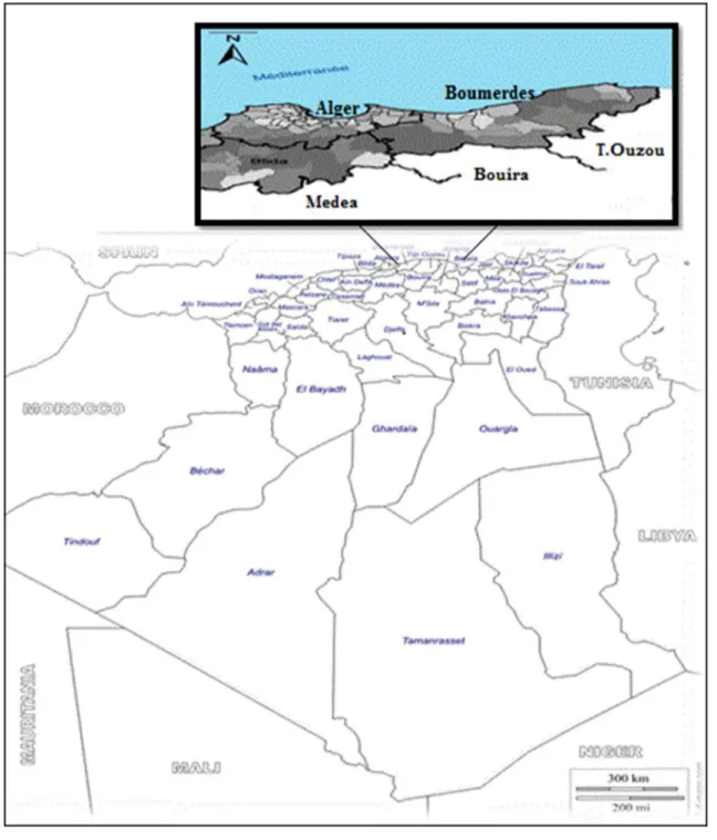
The study was performed from July to November 2015 at privately owned farms located in northeastern Algeria (Fig. 1). The prevailing climate at the time of the study as subhumid, with 70% of humidity, influenced by a high average rainfall in winter (168.40 mm) and a high tempera-ture in summer (26 °C) https ://www.onm.org.dz (National Meteorological Office-Avenue Khemisti, Dar El Beida, BP 153, Alger, Algeria). The experimental farms are located on uneven ground that may contain Galba because of their proximity to water points (lakes).
Experimental Groups
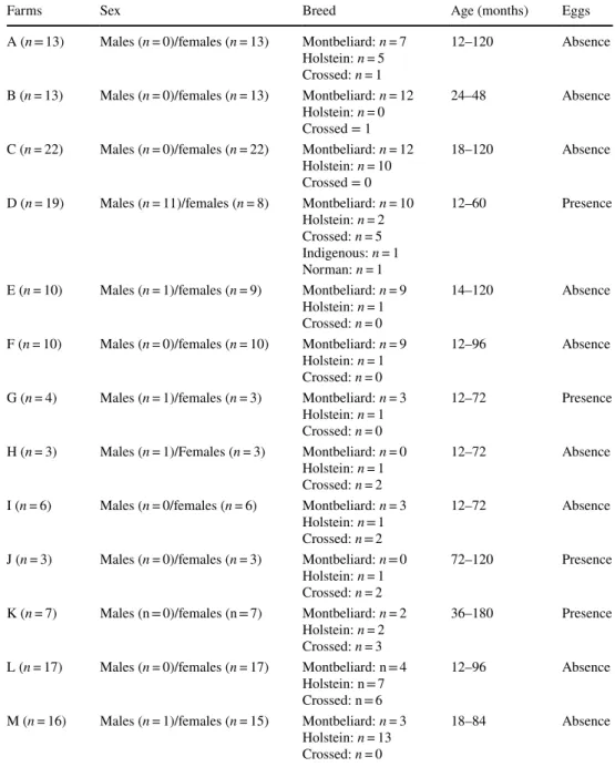
The survey was conducted randomly on a total of 143 cattle, 15 males, and 128 females, from different breeds: Holstein (n = 45), Montbeliard (n = 74), crossed (n = 22), indigenous (n = 1), and Norman (n = 1). The animals included in the experiment were 1–10 years.
Study Design and Sampling
We simultaneously collected 143 faeces and blood; the fae-ces were taken from the rectum of each animal, placed in plastic bags and labelled. The presence of F. hepatica eggs in faecal samples was evaluated by a modified sedimentation method [18]; briefly, 250 g of faeces were mixed in 1 L of tap water and filtered through a tea sieve. The glass content was sedimented for 30 min, stained with methylene blue and then microscopically observed at a magnification of ×40.
Blood samples were collected from the jugular vein, cen-trifuged at 3000 rpm for 10 min, and sera was recovered and stored at − 20 °C until used for biochemical analysis and antibody detection.
Determination of the transaminases (Alat: alanine ami-notransferase, Asat: aspartate Aminotransferase), alkaline phosphatase (Pal), gamma glutamyltransferase (γGT), albu-min, and total protein was performed on commercially avail-able kits (BIOLABO SAS, Matzy, France). The working solutions were prepared in accordance with the supplier’s recommendations.
Immunological Diagnosis of Fasciolosis in the Serum via the ELISA Methods
Serum IgG antibodies were assayed using specific, com-mercially available F. hepatica antigens (IDEXX Fasciolosis Verification, Montpellier, France). Microplates were coated in alternance with control antigen (−Ag) and f2 antigen (+Ag) in both odd and even columns, respectively; 10 µl of sera in duplicate were added to each well without dilution and incu-bated for 1 h at 37 °C. After washing, 100 µl of peroxidase-conjugated anti-bovine IgG antibody were incorporated in each well and incubated 30 min at 37 °C to bind to the immune-complex antigen–antibody. After another washing, 100 µl of the enzyme–substrate were added to the conjugate (18–26 min protected from light), resulting in the formation of a blue compound which becomes yellow after blocking (100 µl). Optical density (OD) was read at 450 nm, using a plate reader (Bio-Rad, USA). The diagnosis was established by comparing the net optical density of the sample with the mean net optical density of the positive control, according to the following equation:
Calculation of Positive Control
CPX = (CP1+ AgA(450) + CP2+ AgA(450))∕2
NEXCP=[(CP1+AgA(450) − CP1−AgA(450)) +(CP2+AgA(450) − CP2−AgA(450))]∕2
114 Acta Parasitologica (2019) 64:112–128
where CPX is the average optical density value of the posi-tive control, NEXCP is the average net optical value of the positive control, and NECN = average net optical value of the negative control.
NECN = CN+AgA(450) − CN−AgA(450), Calculation of the Sample
where NE is the net optical value.
NE = E+AgA(450) − E−AgA(450) ⋯ ⋯ E∕P % = 100 × (NE∕NEXCP),
Fig. 1 Representative map of the study area
115 Acta Parasitologica (2019) 64:112–128
Histopathology Assessment
One bovine liver with fasciolosis, obtained from the slaugh-terhouse of Thenia, was used. One 18-month-old Montbeli-ard cattle (male) was obtained from one of the farms studied and sacrificed in the presence of the veterinary inspector of the department. The presence of F. hepatica was confirmed macroscopically. A fragment of hepatic lobe was seized, fixed in 10% of neutral buffered formalin, and embedded in paraffin. Sections (5 µm) were stained with Goldner’s modified Masson Trichrome to determine the damaged liver areas. To obtain more details, we analysed the histologi-cal hepatic fragment via FEI Quanta 250 scanning electron microscopy (SEM/EDX). The samples were not pre-treated,
and the images were taken at 10.00 kV in a low-vacuum (10–130 Pa) secondary electron (LFD) detector at a magni-fication of ×41 to 600.
Statistical Analysis
The data were initially organised on an excel spreadsheet and then imported into the XLSTAT statistical SOFTWARE (Version 2017). The prevalence to F. hepatica was calculated as a percentage value, and analysis of variance (ANOVA) was used to determine the statistical relationship between infection rates and three epidemiological risk factors (age, sex, and breed).
Table 1 Summary table
showing the distribution of age, sex and breed in the 13 farms studied
Farms Sex Breed Age (months) Eggs
A (n = 13) Males (n = 0)/females (n = 13) Montbeliard: n = 7 Holstein: n = 5 Crossed: n = 1
12–120 Absence B (n = 13) Males (n = 0)/females (n = 13) Montbeliard: n = 12
Holstein: n = 0 Crossed = 1
24–48 Absence C (n = 22) Males (n = 0)/females (n = 22) Montbeliard: n = 12
Holstein: n = 10 Crossed = 0
18–120 Absence D (n = 19) Males (n = 11)/females (n = 8) Montbeliard: n = 10
Holstein: n = 2 Crossed: n = 5 Indigenous: n = 1 Norman: n = 1
12–60 Presence
E (n = 10) Males (n = 1)/females (n = 9) Montbeliard: n = 9 Holstein: n = 1 Crossed: n = 0
14–120 Absence F (n = 10) Males (n = 0)/females (n = 10) Montbeliard: n = 9
Holstein: n = 1 Crossed: n = 0
12–96 Absence G (n = 4) Males (n = 1)/females (n = 3) Montbeliard: n = 3
Holstein: n = 1 Crossed: n = 0
12–72 Presence H (n = 3) Males (n = 1)/Females (n = 3) Montbeliard: n = 0
Holstein: n = 1 Crossed: n = 2
12–72 Absence I (n = 6) Males (n = 0/females (n = 6) Montbeliard: n = 3
Holstein: n = 1 Crossed: n = 2
12–72 Absence J (n = 3) Males (n = 0)/females (n = 3) Montbeliard: n = 0
Holstein: n = 1 Crossed: n = 2
72–120 Presence K (n = 7) Males (n = 0)/females (n = 7) Montbeliard: n = 2
Holstein: n = 2 Crossed: n = 3
36–180 Presence L (n = 17) Males (n = 0)/females (n = 17) Montbeliard: n = 4
Holstein: n = 7 Crossed: n = 6
12–96 Absence M (n = 16) Males (n = 1)/females (n = 15) Montbeliard: n = 3
Holstein: n = 13 Crossed: n = 0
18–84 Absence
116 Acta Parasitologica (2019) 64:112–128 Multiple comparison tests were performed to compare the immunological response to infection (serological presence of antibody anti-f2 of F. hepatica) and hepatic enzymes, including both protein and albumin rates, between farms (A to M).
The relationship between variables was considered statis-tically significant at a p value of less than 0.05, with a 95% confidence level.
Results
Relationship Between Hepatic Fasciola hepatica Infection and Changes in Serum Parameters
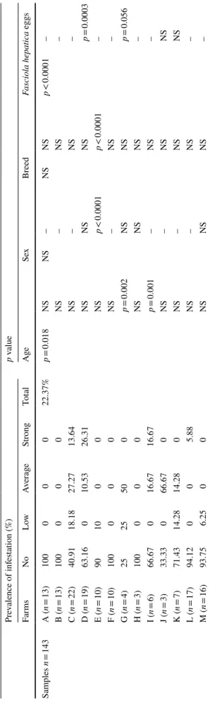
To investigate the serological prevalence to F. hepatica infection, 143 cattle from 13 farms were tested. The details of the distribution of the population in terms of age, sex, and breed are summarised in Table 1. The ELISA test results showed a heterogeneous distribution of the anti-f2 antibod-ies in response to the infection. As depicted in Table 2, the overall infestation rate was about 22.37%, which corresponds to a ratio of 0.69; i.e., 9 farms out to 13 were affected. We observed three levels of infestation, as described in Table 2. The most affected farms reached levels exceeding 50%, as recorded for farms C (59.09%), G (70%), and J (66.67%). Farms A, B, F, and H were free of this zoonosis (100%).
Serological confirmation of F. hepatica infection has broadened the study by integrating multiple combina-tions referring to age, sex, and breed. Based on the sta-tistical analysis, there was a significant difference related to age (p = 0.018) and the presence of F. hepatica eggs (p < 0.0001). Regarding sex and breed, no significant dif-ference was recorded for the 143 samples studied.
The factors mentioned above suggest their involvement in the appearance of the infection observed in farms.
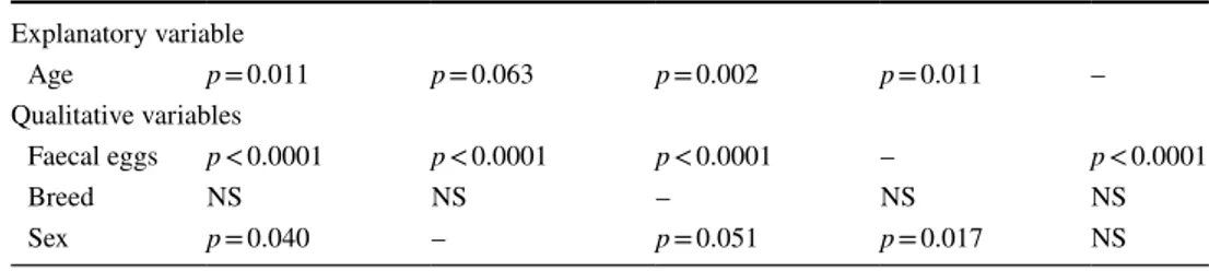
Table 3 shows the results of the multiple comparison test, which was performed to obtain the in vivo state by setting the dependent (anti-f2 antibodies) and explanatory (age) variables by integrating faecal eggs, breed and sex into the equation.
In each column selected, significant effects were noticed at different degrees because of the importance of integrated variables. The first column indicates that the presence of faecal eggs reflects the active infectious status of cattle (p < 0.0001), while age (p = 0.011) and sex (p = 0.040) will increase chance of acquiring the infection.
This was identical for all the categories studied except for column 5, where no explanatory variable (age) was selected.
The clinical value of the determination of serum param-eters relates to the exploration of liver function. In fact, the most commonly observed variations affect total proteins including anti-f2 antibodies and albumin. Because some
Table 2 Pr ev alence of f asciolosis in nor th eas ter n f ar ms (A–M) of Alg er ia accor ding t o ag e, se x, br eed and F. hepatica eggs based on t he ser ological r
esults of anti-f2 antibodies
NS non significant, p v alue pr obability Pr ev alence of inf es tation (%) p v alue Far ms No Low Av er ag e Str ong To ta l Age Sex Br eed Fasciola hepatica eggs Sam ples n = 143 A ( n = 13) 100 0 0 0 22.37% p = 0.018 NS NS – NS NS p < 0.0001 – B ( n = 13) 100 0 0 0 NS – NS – C ( n = 22) 40.91 18.18 27.27 13.64 NS – NS – D ( n = 19) 63.16 0 10.53 26.31 NS NS NS p = 0.0003 E ( n = 10) 90 10 0 0 NS p < 0.0001 p < 0.0001 – F ( n = 10) 100 0 0 0 NS – NS – G ( n = 4) 25 25 50 0 p = 0.002 NS NS p = 0.056 H ( n = 3) 100 0 0 0 NS NS NS – I ( n = 6) 66.67 0 16.67 16.67 p = 0.001 – NS – J ( n = 3) 33.33 0 66.67 0 NS – NS NS K ( n = 7) 71.43 14.28 14.28 0 NS – NS NS L ( n = 17) 94.12 0 0 5.88 NS – NS – M ( n = 16) 93.75 6.25 0 0 NS NS NS –
117 Acta Parasitologica (2019) 64:112–128
enzymes are interesting from a lesional point of view, we confined the investigations to analysis of aspartate ami-notransferase (AST), alanine amiami-notransferase (ALT), gamma glutamyltransferase (γGT), and alkaline phosphatase (PAL).
Table 4 shows the expression of the serological parame-ters divided into two categories: proteins (anti-f2 antibodies, total proteins, and albumin) and hepatic enzymes accord-ing to age. With the exception of IgG, which was statisti-cally significant (p = 0.018) for all cattle studied, proteins and albumin were not significant. Table 4 indicates that the mean serum value of the anti-f2 antibodies increases equivalently in the farms C, D, G, and J, affected by fascio-losis. The maximum percentages were observed on farms G (79.75 ± 37.64%, p = 0.002) and J (79.76 ± 40.58%), while minimum values were recorded on farms A (3.18 ± 1.49%), B (3.86 ± 1.41%), F (1.14 ± 0.25%), and M (3.06 ± 2.03%). Table 4 also shows the slight fluctuations in the distribu-tion of total proteins between different farms. The impact of fasciolosis on the profile of total proteins is similar to that on albumin, albeit with some exceptions. The average ranges were between 107.56 ± 5.54 and 69.45 ± 5.50 g/l for farms A and G. Albumin levels were highest and lowest for farms F and G, respectively, with 806.75 ± 75.04 and 363.26 ± 42.93 µmol/l. Unlike albumin, which showed no significant difference within farms and for the entire herd, proteins were significantly different within farms B (106.71 ± 3.79 g/l, p = 0.049), H (76.00 ± 7.31 g/l, p = 0.005), and I (97.77 ± 4.97 g/l, p = 0.004).
The responses of liver enzymes to the parasitic infection are summarised in Table 4; average values of 143 cattle were below the standard. However, for all liver enzymes, only PAL was highly significant (p < 0.0001) for age. Sub-jected to this parameter, a maximum (123.89 ± 56.41 UI/l) and minimum levels (42.82 ± 3.76 UI/l) were observed, respectively, for farms H and K. Fasciolosis also causes the elevation of AST, as seen in farms G (73.77 ± 28.62 UI/l) and K (67.84 ± 8.76 UI/l), suggesting liver injury in terms of individual values. The results also showed that farm D was statistically significant for PAL (101.85 ± 12.80 UI/l,
p = 0.015), γGT (13.25 ± 1.58 UI/l, p = 0.041) and ALT
(6.63 ± 0.62 UI/l, p = 0.009).
Another aspect explored in our study was the effect of parasitosis on the liver enzymes released. This approach allowed us to note that γGT (r = 0.401, p < 0.0001) and AST (r = 0.186, p = 0.027) responded significantly to the infection. The different farms showed differences in terms of parameters. For example, for farm A, we observed a highly significant liver production of albumin (p = 0.004) in response to circulating IgG levels during the infection–secre-tion cycle. This tendency was expressed differently in farms B (p = 0.009) and D (p = 0.001), where γGT did not reach extremely high, but still significant levels. Unlike farm I (total proteins: p = 0.013, AST: p = 0.038, ALT: p = 0.046), which perfectly expressed the infectious power of distoma-tosis, the remaining farms (C, E, F, G, H, J, K, L, and M) seemed to be unscathed (p > 0.05).
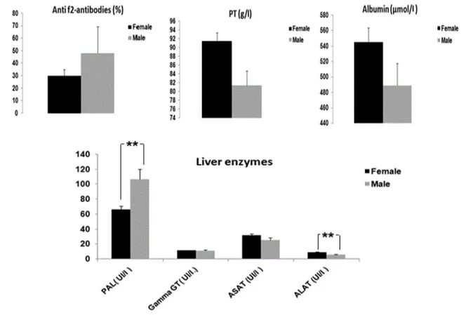
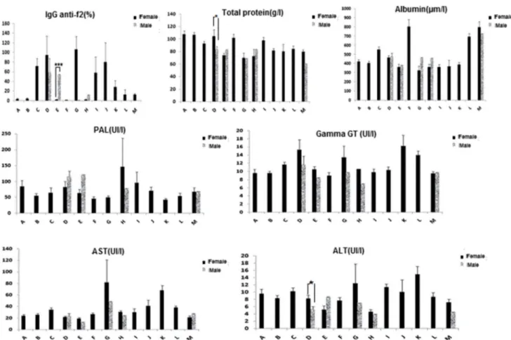
Regarding sex, no significant differences were recorded for anti-f2 antibodies, proteins, albumin, γGT, and AST, except for both PAL (p = 0.005) and ALT (p = 0.005). In our study, males were more infected (27%) than females (22%), as shown in Fig. 2. Although the numerical superiority of females is established (128 females vs. 15 males), only 10% of the males of the farm E showed a significant difference (p < 0.0001) in terms of anti-f2 antibodies to Fasciola
hepat-ica (Fig. 3). A relatively close infestation rate between males (15.79%) and females (26.31%) was observed for farm D. In addition, farms C, D, G, I, J, K, L, and M showed infestation levels of females ranging from 6 to 75%.
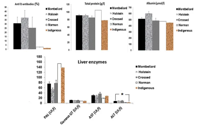
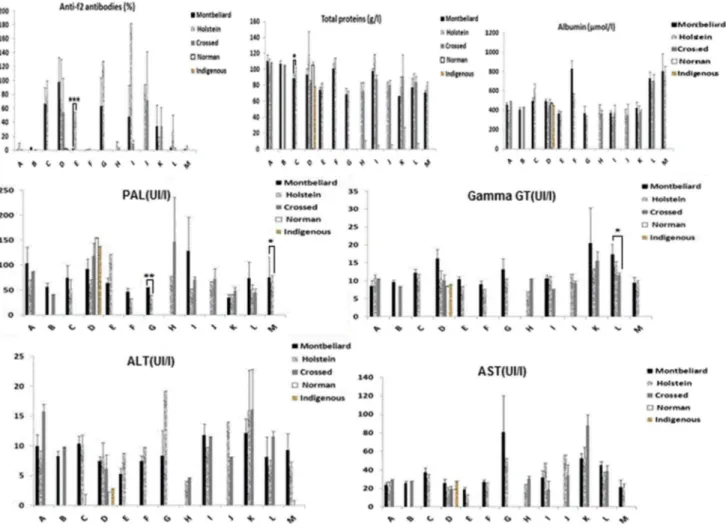
The post-analysis results of the 143 samples showed that five distinct breeds [Montbeliard (M), Holstein (H), Crossed (C), Norman (N), and Indigenous (I)] forming herds, with some marked racial disparity among the 13 farms. In view of the results shown in Fig. 4, the Holstein breed appears to be the most affected (31.11%), followed by the Montbe-liard (20.27%) and the crossed breed (13.64%). With the exception of ALT (p = 0.027), no significant difference was observed for breed. As illustrated above (Fig. 5), farm E showed an infestation rate of 10%, with a highly signifi-cant difference (p < 0.0001) for the Holstein breed. In addi-tion, we observed the following details: C (M 27.27% vs. H 31.82%); D (M 26.31% vs. H 5.26% vs. C 5.26%); G (M 50% vs. H 25%); I (M 16.67% vs. H 16.67%); J (H 33.33% vs. C 33.33%); K (M 14.28% vs. C 14.28%); L (H 5.88%);
Table 3 Analysis of differences between modalities (faecal eggs, breeds and sex) by fixing the dependent and explanatory variables: anti-f2 antibodies and age, respectively
Fixed dependent variable (anti-f2 antibodies) Explanatory variable Age p = 0.011 p = 0.063 p = 0.002 p = 0.011 – Qualitative variables Faecal eggs p < 0.0001 p < 0.0001 p < 0.0001 – p < 0.0001 Breed NS NS – NS NS Sex p = 0.040 – p = 0.051 p = 0.017 NS
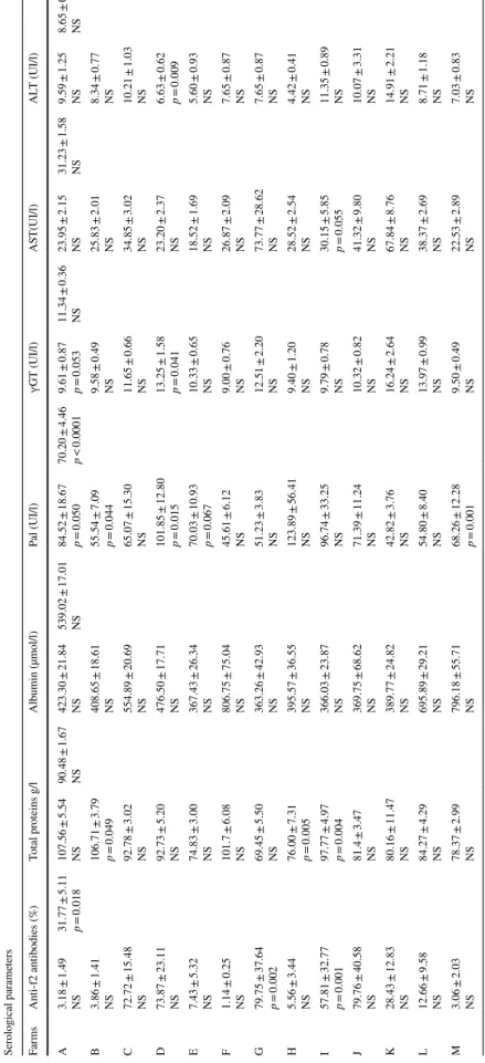
118 Acta Parasitologica (2019) 64:112–128 Table 4 Ser ological par ame ters (X ± SEM) af ter F. hepatica inf ection accor ding t o t he ag e in nor theas ter n f ar ms of Alg er ia Ser ological par ame ters Far ms Anti-f2 antibodies (%) To tal pr oteins g/l Albumin (µmol/l) Pal (UI/l) ɣG T (UI/l) AS T(UI/l) AL T (UI/l) A 3.18 ± 1.49 NS 31.77 ± 5.11 p = 0.018 107.56 ± 5.54 NS 90.48 ± 1.67 NS 423.30 ± 21.84 NS 539.02 ± 17.01 NS 84.52 ± 18.67 p = 0.050 70.20 ± 4.46 p < 0.0001 9.61 ± 0.87 p = 0.053 11.34 ± 0.36 NS 23.95 ± 2.15 NS 31.23 ± 1.58 NS 9.59 ± 1.25 NS 8.65 ± 0.37 NS B 3.86 ± 1.41 NS 106.71 ± 3.79 p = 0.049 408.65 ± 18.61 NS 55.54 ± 7.09 p = 0.044 9.58 ± 0.49 NS 25.83 ± 2.01 NS 8.34 ± 0.77 NS C 72.72 ± 15.48 NS 92.78 ± 3.02 NS 554.89 ± 20.69 NS 65.07 ± 15.30 NS 11.65 ± 0.66 NS 34.85 ± 3.02 NS 10.21 ± 1.03 NS D 73.87 ± 23.11 NS 92.73 ± 5.20 NS 476.50 ± 17.71 NS 101.85 ± 12.80 p = 0.015 13.25 ± 1.58 p = 0.041 23.20 ± 2.37 NS 6.63 ± 0.62 p = 0.009 E 7.43 ± 5.32 NS 74.83 ± 3.00 NS 367.43 ± 26.34 NS 70.03 ± 10.93 p = 0.067 10.33 ± 0.65 NS 18.52 ± 1.69 NS 5.60 ± 0.93 NS F 1.14 ± 0.25 NS 101.7 ± 6.08 NS 806.75 ± 75.04 NS 45.61 ± 6.12 NS 9.00 ± 0.76 NS 26.87 ± 2.09 NS 7.65 ± 0.87 NS G 79.75 ± 37.64 p = 0.002 69.45 ± 5.50 NS 363.26 ± 42.93 NS 51.23 ± 3.83 NS 12.51 ± 2.20 NS 73.77 ± 28.62 NS 7.65 ± 0.87 NS H 5.56 ± 3.44 NS 76.00 ± 7.31 p = 0.005 395.57 ± 36.55 NS 123.89 ± 56.41 NS 9.40 ± 1.20 NS 28.52 ± 2.54 NS 4.42 ± 0.41 NS I 57.81 ± 32.77 p = 0.001 97.77 ± 4.97 p = 0.004 366.03 ± 23.87 NS 96.74 ± 33.25 NS 9.79 ± 0.78 NS 30.15 ± 5.85 p = 0.055 11.35 ± 0.89 NS J 79.76 ± 40.58 NS 81.4 ± 3.47 NS 369.75 ± 68.62 NS 71.39 ± 11.24 NS 10.32 ± 0.82 NS 41.32 ± 9.80 NS 10.07 ± 3.31 NS K 28.43 ± 12.83 NS 80.16 ± 11.47 NS 389.77 ± 24.82 NS 42.82 ± 3.76 NS 16.24 ± 2.64 NS 67.84 ± 8.76 NS 14.91 ± 2.21 NS L 12.66 ± 9.58 NS 84.27 ± 4.29 NS 695.89 ± 29.21 NS 54.80 ± 8.40 NS 13.97 ± 0.99 NS 38.37 ± 2.69 NS 8.71 ± 1.18 NS M 3.06 ± 2.03 NS 78.37 ± 2.99 NS 796.18 ± 55.71 NS 68.26 ± 12.28 p = 0.001 9.50 ± 0.49 NS 22.53 ± 2.89 NS 7.03 ± 0.83 NS
119 Acta Parasitologica (2019) 64:112–128
and M (H 6.25%). The other farms (A, B, F, and H) had healthy animals.
Histopathological Alterations and Changes in Liver Tissue
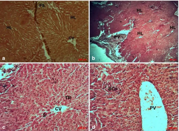
Microscopic observation (Fig. 6) showed a section of healthy liver (a, b, c, and d) with hepatocyte lobules arranged radi-ally around the central vein (a), forming Remak bays sepa-rated by sinusoidal channels and delimited by the Glisson’s capsule (b). At the contact angles of the hepatic lobules are the portal spaces (c and d): the triad (portal vein, bile duct, and hepatic artery).
Figure 7 shows the architectural changes of a parasitised liver, caused by the presence of Fasciola hepatica. The path-ogenic process shows the formation of fibrous trabeculae between the hepatic lobules, defining annular and biliary fibrosis evolving towards a sclerosis (a). The absence of the centrilobular vein would explain the presence of regenera-tion lobules following a partial destrucregenera-tion of the hepatic parenchyma (b). The periportal fibrosis (c) progresses to the formation of porto-portal bridges separating the hepatic parenchyma in nodules (dissection nodules). Hepatocytes trapped in the connective tissue septa form cholangioles (d
and e). The histological aspect of Fig. 7f refers to a diffuse portal leukocyte infiltrate indicative of a response to parasit-ism. Trapped in the bile ducts (Fig. 7g, h and i), liver fluke appears as debris. The scar tissue (fibrosis) observed around the liver lobules suggests chronic hepatitis.
Figure 8 shows bile duct glandular epithelium prolifera-tion, forming a new bile ductule surrounded by thick con-nective tissue (a). Indeed, portal fibrosis showed liver fluke in the lumen of the intra-hepatic bile duct, destroying the bile duct (b) with heavy infiltration of inflammatory cells (c, d, and e) in the fibrous connective tissue between newly formed bile ductules.
As depicted in Fig. 8f, g, h, and i, a yellowish longitu-dinal acellular form was observed at different magnifica-tions, infiltrating the hepatic parenchyma and causing its disorganisation. In the centre, cellular debris surrounding an amorphous material was observed.
Macroscopic lesions observed in Fig. 9a, b, c, and d show a circumferential thickening of the wall of the bile ducts with narrowing of light, suggesting an inflammatory cholangiopa-thy. This ductular reaction is thought to be secondary to the presence of the liver fluke. The scanning electron micros-copy (SEM) image (a1) of the biliary duct showed a dense structure containing a limestone formation, confirmed by the
Fig. 2 Evaluation of levels of Fasciola hepatica anti-f2 antibodies, total proteins, albumins and liver enzymes in response to F. hepatica infec-tion according to sex. Results are expressed as X ± SEM
120 Acta Parasitologica (2019) 64:112–128
pictures a2, a3, a4, and a5. In fact, the SEM mode allowed collecting fine structural and topological data, together with insights into the interior of the sample. In addition to the above-mentioned pathological calcifications (b1), porous formations (b2, b3, b4) appeared at the level of the bile ducts, suggesting a remodelling of the latter.
Discussion
The evaluation of fasciolosis in cattle and its geographical distribution was the subject of our study. The northeastern areas of Algeria have a subhumid climate and border water reservoirs with the presence of snails. We focused on the upstream detection of zoonotic infections, which are often discovered in the slaughterhouse.
There is a great variability in the prevalence of natural F.
hepatica infections in domestic ruminants between
slaugh-terhouses and farms. Unlike the slaughterhouse model, farms show a heterogeneous distribution of infection, as reported in southwest Tunisia with 14.3% [17]. In the same way, the seroprevalence examined via a number of blood samples in
cattle revealed averages ranging from 6.3 to 35.5% in Algeria [23]. Similarly, the plain of Mitidja, with its different locali-ties, showed an infestation rate close to 18.8% [3]. Morocco is also affected by this parasitosis, which is reflected by relatively high levels (49.1%). However, this prevalence has been determined according to the seasons [24]. Epidemiol-ogy of bovine fasciolosis in the Nile Delta region of Egypt does not exceed 9.77% [14]. All these data place fasciolosis around the Mediterranean basin, although it is not specific to underdeveloped countries, since it occurs in Romania (63%) [27], France (75%) [29], Equatorial Guinea (44.1%) [7], Tur-key (65.2%) [35], and Black Africa (21.39%) [16].
For some countries cited above, this cosmopolitan zoono-sis is closer to the values obtained in our study, because the serological investigations of F. hepatica infection were around 22.37%, according to their incidences on the dif-ferent farms: C (59.09%), D (36.84%), E (10%), G (70%), I (33.34%), J (66.67%), K (28.56%), L (5.88%), and M (6.25%). The higher prevalence rate of fasciolosis in the pre-sent study might be due to the variation in agro-ecological conditions favouring snail occurrence.
Fig. 3 Evaluation of levels of Fasciola hepatica anti-f2 antibodies, total proteins, albumins and liver enzymes in response to F. hepatica infection according to the sex of all animals of the 13 selected farms
(A–M). Results are expressed as X ± SEM. PAL alkaline phosphatase, Gamma GT gamma glutamyl transpeptidase, AST aspartate ami-notransferase, ALT alanine aminotransferase
121 Acta Parasitologica (2019) 64:112–128
Because hepatobiliary distomatosis is highly unobtru-sive (a chronic form in cattle), many cases of fasciolosis have been reported in several slaughterhouses during vet-erinary inspections. Slaughterhouse discoveries provide information on the frequency of this lesion and its extent. In Haiti, for example, 10.67% of cattle killed confirmed this parasitosis [8]. In northeastern Nigeria, the overall percentage of Fasciola gigantica antibodies detected was around 72%, distributed seasonally [11]. On the basis of post-mortem examinations, in the industrial abattoir located in Gondar (northwestern Ethiopia), 79.83% of the infection were due to F. hepatica, with a near-normal dis-tribution [43.37% (2005), 70.68% (2006), 53.44% (2009), and 52.35% (2010)] between 2005 and 2010 [25]. The picture that we describe here is far from being exhaustive, since Algeria has extended this list with natural infections to Fasciola in cattle and sheep from slaughterhouses of Constantine (9.1%) and Jijel (27.0%) [3]. Countries such as USA are not unscathed to this parasitosis, and fasciolosis is quite present, with nine cases reported in humans [4].
The abattoir and areas with a prevalence of fascio-losis fluctuate considerably between strangely high and relatively low values. This heterogeneity can be associ-ated with numerous factors, such as animal breeding sites, favourable ecological biotopes, climate, age, soil ecopedology, or the lack of livestock management systems
[9]. Beyond classic and known data on infestations, other approaches have been considered, such as the study of snail populations and snail age to explain this variability [12].
Unlike previous studies, we can confirm that our herd has not benefited from any anthelmintic treatment, which is an excellent indicator of the infestation level.
Although the coprological examination shows its own limitations, it remains consistent with the serological data on the infection (presence of anti-f2 antibodies against F.
hepatica) in our study. However, the opposite is not
neces-sarily right.
To explain the intrinsic variability to the infection (anti-f2 antibodies), we independently integrated age, sex, and breed as explanatory variables. In a previous study, age was an important factor for the prevalence of fasciolosis in ruminants [15]. We observed that age greatly influenced the prevalence of the infection, which was expected. Sim-ilar results have been described by [35], who report that older animals ≥ 6 years were more infected than young ani-mals ≤ 2 years, which was the case at least for sheep because of the relaxation of their immunity [2]. Our findings are in agreement with those of [24], who consider older sheep as the main reservoir of this parasitosis. In view of our results, we find that the degree of infestation with F. hepatica on the basis of breed showed no statistically significant difference.
Fig. 4 Evaluation of levels of Fasciola hepatica anti-f2 antibodies, total proteins, albumins and liver enzymes in response to F. hepatica infec-tion according to breed
122 Acta Parasitologica (2019) 64:112–128
The infection rate in the Holstein breed (31.11%) was higher than that in the Montbeliard breed (20.27%) and the crossed breed (13.64%), which could be due to the genetic potential of the Holstein breed because of its significant milk produc-tion, making it more sensitive. Our results disagree with a study performed in Ethiopia, which favours the local breed in terms of infections [34]. In regard to the effect of sex and despite the fact that females were numerically superior (15 male for 128 females), the infestation rates of males were higher than those of females (27% vs. 22%). Most likely, this can be explained by the fact that most female cattle are kept for lactation. However, there was no statistically significant difference in infection rates between males and female, as described by [34]. We conclude the independent of the risk factors (age, sex, and breed) in the expression of
F. hepatica parasitism.
The identification of potential risk factors for liver fluke infection has been complicated by setting independent vari-ables one by one. However, based on multivariate model-ling calculations, we were able to demonstrate that age,
sex, breed, and coprology provided a wealth of information about their relevance. The model in column no. 1 (Table 3) represents the best state encountered in vivo, most closely reflecting the field conditions.
The macroscopic observation, the histopathology, and the serological detection of anti-f2 antibodies as well as liver enzymes are concordant, which allows us to attribute the majority of observed liver lesions to distomic infestations that mark their chronicity.
The macroscopic liver lesions caused by fasciolosis in infected cattle showed an increase in liver volume, with a lighter colour. White-haired longitudinal trabeculae were present on the surface of the parenchyma, and there was a screeching during the post-mortem examination of the organ, possibly caused by calcareous structures. In addition, many canalicular lesions referring to the bile ducts showed thickened walls containing liver flukes of different sizes. Our visual observations were similar to the pathological investi-gations of liver fluke infections of slaughtered black Bengal goats in Bangladesh [31].
Fig. 5 Evaluation of levels of Fasciola hepatica anti-f2 antibodies, total proteins, albumins and liver enzymes in response to F. hepatica
infec-tion according to the breed of the animals of the 13 selected farms (A–M)
123 Acta Parasitologica (2019) 64:112–128
Microscopically, the histological lesions are characterised by diseased fibrosis around hepatic lobules, forming junc-tions between the portal spaces and leading to the destructur-ation of the hepatic parenchyma. There is also compensatory hyperplasia of the bile ducts secondary to the infection with
Fasciola hepatica, as described by [28]. The most prevalent alteration in chronic fibrosis was the proliferation of the bile duct glandular epithelium, forming new bile ductules sur-rounded by thick connective tissue. Similar results have been reported by [13] at the portal space and, more specifically, around the bile duct, an inflammatory reaction with eosino-phils and neutroeosino-phils was observed, suggesting an immune response of the host during the migration of the young fluke, as also reported by [21] and [1]. Variable degrees of prolif-eration and calcification were also observed in the lumen of biliary ducts, revealing chronic infection. Our results are in agreement with two previous studies [5, 33].
Hepatic enzyme activities and biochemical variables in examined cattle serum show parasitic infestation related to
histopathology examination. Although AST and ALT are not specific for liver disease, they can be used in combination with other enzymes (PAL and γGT) to monitor the course of various liver disorders.
The serological results reported in our study enabled us to evaluate the integrity of the liver parenchyma. Indeed, the relatively high levels of total proteins (90.68 ± 1.67 g/l) and albumin (539.02 ± 17.01 µmol/l), except for some farms (F, L and M), can explain the inflammation [6], even though some means (albumin) recorded remain low with the exam-ple of the farms E, G, H, I, J, and K (see Table 4), indicating a discrete, reversible hepatic insufficiency.
In the chronic form, adult flukes cause cholangitis by attracting inflammatory cells, which is consistent with histopathology.
As previously described, hepatobiliary distomatosis did not completely destroy the hepatic parenchyma with regard to the few disseminated hepatic lobules, testifying to a brief regeneration of the latter, which was confirmed by the
Fig. 6 Liver biopsy showing the juxtaposition of hepatic lobules (a
magnification 10× and c magnification 20×) and portal triad (b mag-nification 4× and d magmag-nification 20×) in a healthy bovine liver
recovered at the slaughterhouse and used as a negative control, col-oured with Masson’s Trichrome
124 Acta Parasitologica (2019) 64:112–128
absence of the centrilobular vein. Our results contradict the work of [6, 13], which situates proteins below the reference levels, indicating hypoproteinemia secondary to liver disease with protein synthesis deficiency.
The activity of liver enzymes is determined according to the pathogenesis of the disease. Thus, to validate the chronic and/or the acute form, it seemed interesting to observe the variations in γGT and PAL, which are canalicular enzymes. The hypothetical hepatic disturbances expected by the bio-chemical analysis place γGT within the physiological limits (9.6–39 IU/l) in the same way as PAL (27–107 IU/l) [22], with certain individual exceptions in C3 (256.15 IU/l), D16 (179.85 IU/l) and E7 (121.53 IU/l), where we encoun-tered very high values, suggesting biliary malfunction (cholestasis).
This difference is due to the great intra-species variability as described previously [26], and such isolated cases are
scattered throughout the flock. These enzymes, which are highly sensitive to F. hepatica infection because of their intra-membranous localisation (bile duct membrane), did not reveal canalicular involvement as described previously by [30] in Zebu West Africa, although hyperplased at some places in the hepatic parenchyma. On the other hand, the presence of artifacts assimilated to cellular debris show-ing liver fluke trapped and destroyed in situ consolidates our results, even if we were inspired by a single clinical case that proved positive (strongly infested). To clarify this, we analysed the individual values of all animals and found that the percentage of the anti-f2 antibodies to F.
hepatica of the majority, i.e., the heavily infected animals,
was not compatible with the γGT levels. As examples, we report the values for farms: C6 (202.14% –11.88 IU/l), D3 (282.26% –29.69 IU/l), E7 (54.79% –8.48 IU/l), G2
Fig. 7 Liver biopsy showing a disruption of liver integrity (a 4×, b: 10×, c 4×, d 10×, e 10×, f 40×, g 4×, h 10×, i 10×), with a large periportal
and lobular leukocyte
125 Acta Parasitologica (2019) 64:112–128
(127.77% –10.6 IU/l), I1 (182.1% –11.24 IU/l), J1 (141.37% –9.97 IU/l), K7 (88.01% –16.69 IU/l), L15 (165.2% –12.73 IU/l), and M2 (33.28% –7 IU/l), each time one or more cases confirming our hypothesis, namely, the non-specificity of γGT as an indicator of liver damage.
Although the measurement of the enzymatic activity of transaminases in ruminants is no specific to the liver, transaminases show a fairly high catalytic concentration in this organ. As a result, the insignificant mean levels of AST and ALT found in our study indicate chronic fasciolosis, although this implication contradicted by the work of [6,
10], who report cell destruction and increased membrane permeability to enzymes (cytolysis) during larval migration into the hepatic parenchyma (see also [19]).
Conclusions
Based on immuno-enzymatic (anti-f2 antibodies of F.
hepatica), coprological and biochemical tests, we were
able to determine the infectivity of the study zone and cir-cumscribe it as the focus of fasciolosis. In addition, our study evaluated the hepatocellular integrity and the meta-bolic function of the liver. Evaluation of the serological importance of the liver enzymes confirmed the installa-tion of the chronicity during pathology and discarded a hepatic insufficiency relative to both hyperproteinemia and albuminemia. The monitoring of PAL, γGT, AST, and ALT data also provided some insights, resulting in the
Fig. 8 (a 40×) Bile duct hyperplasia surrounded by a thick layer of fibrous connective tissue, (b 10×), (c, d, e 40×). Severe fibrosis of the bile duct, with encapsulated and degenerating immature fluke suspi-cious of an acellular structure, suggesting calcified Fasciola hepatica (f 10×, g 20×, h 40×, i 100×). HL hepatic lobules, CV centrolobu-lar vein, PS periportal space, PV portal vein, TR trays of remak, S
sinusoids, BC biliary canal, FT fibrous tissue, GC glisson capsule, IC inflammatory cells, EPG eosinophilic polynuclear granulocytes, IG inflammatory granuloma, NPG neutrophil polynuclear granulocytes, PT portal triad, H hepatocyte, HA hepatic artery, AS acellular struc-ture, D debris, DLF degenerated liver fluke, RC red cells
126 Acta Parasitologica (2019) 64:112–128
non-establishment of cholestasis and cytolysis. The few variables (age, sex, and breed) introduced into our experi-mental model, treated independently and in combination, greatly influenced the response of animals to F. hepatica infection, albeit to varying degrees, which justifies their importance in the expression of the pathology.
Compliance with ethical standards
Conflict of interest There is no conflict of interest between co-authors and another institution
References
1. Abraham J.T., Jude I.B. 2014. Fascioliasis in cattle and goat slaughtered at Calabar abattoirs. Journal of Biology, Agriculture and Healthcare, 4(18), 34–41.
2. Afshan K., Qayyum M., Rizvi S.S.R., Mukhtar M., Mushtaq M., Miller J.E. 2013. Serological and coprological comparison for rapid diagnosis of Fasciola hepatica infection in small ruminants from sub-tropical area of Pakistan. Small Ruminant Research,
113(1), 267–272. DOI: https ://doi.org/10.1016/j.small rumre s.2013.01.020.
3. Aissi M., HarhouraK.,Gaid S., Hamrioui B. 2009. Etude prélim-inaire sur la prévalence de la fasciolose due à Fasciola hepat-ica dans quelques élevages bovins du nord centre algérien (la Mitidja). Bulletin de la Société de PathologieExotique, 102(3), 177–178. https ://doi.org/10.3185/pathe xo325 1.
4. Alatoom A., Cavuoti D., Southern P., Gander R. 2008. Fasciola hepatica Infection in the United States. Laboratory Medicine, 39(7), 425–428. DOI: https ://doi.org/10.1309/mlffx a77ub bfh4g p. 5. Andriamanantena D., Rey P., Perret J.L., Klotz F. 2005.
Distomatoses. EMC -Maladies Infectieuses, 2,105–118. DOI:
https ://doi.org/10.1016/j.emcmi .2005.03.001.
6. Baldissera M.D., Bottari N.B., Mendes R.E., Schwertz C.I., Lucca N.J., Dalenogare D et al. 2015. Activity of cholinest-erases, pyruvate kinase and adenosine deaminase in ratsex-perimentally infected by Fasciola hepatica: Influences of these enzymes on inflammatory response and pathological findings. Pathology Research and Practice, 211(11), 871–876. DOI: https ://doi.org/10.1016/j.prp.2015.09.006.
7. Barry A.M., Keita S. 2010. Sondage sérologique de la fasciolose bovine enGuinée.Revue d’élevage et de médecinevétérinaire des pays tropi-caux, 63, 13–16. DOI:https ://doi.org/10.19182 /remvt .10103 . 8. Blaise J. 2001. Prévalence et fréquence des lésionsparasitaires
du foie et du poumon des ruminants enHaïti. Revue de Médeci-neVétérinaire,152 (3), 269–274.
Fig. 9 Left, macroscopic picture of F. hepatica debris in the biliary canal (a, b, c and d) showing liver injuries. Right, details of patho-logical calcifications (Mag 45×, 180×, 150×, 41×, 80×), observed
via scanning electron microscopy (Fig. 9a). Bar lengths represent: a1:
1 mm, a2: 500 µm, a3: 500 µm, a4: 2 mm, a5: 1 mm, b1: 500 µm, b2: 100 µm, b3: 1 mm, b4: 400 µm
127 Acta Parasitologica (2019) 64:112–128
9. Boucheikhchoukh M., Righi S., Sedraoui S., Mekroud A., Benakhla A. 2012. Principaleshelminthoses des bovins: enquêteépidemiologique au niveau de deux abattoirs de larégiond’ElTarf (Algérie). Tropicultura, 30(3), 167-172. 10. Braun J.P., Bézille P., Rico A.G.1986. Sémiologiebiochimique du
foie chez les ruminants. Reproduction Nutrition et Developpement, 26 (1B), 227-243. DOI: https ://doi.org/10.1051/rnd:19860 206. 11. Damwesh S.D., Ardo M.B.2013. Detection of Fasciola
giganti-caantibodies using Pourquier ELISA kit. Sokoto Journal of Vet-erinary Sciences, 11(2),43–48. DOI:https ://doi.org/10.4314/sokjv s.v11i2 .7.
12. Dreyfuss G., Vignoles P., Rondelaud D. 2008. Paramphistomum daubneyi: The number of sporocysts developing in experimen-tally and naturally infected Galba truncatula.ParasitologyRe search, 103(2), 345–349. DOI: https ://doi.org/10.1007/s0043 6-008-0978-4.
13. El-Hallawany H.A., Abdel-Aziz M.Z. 2012. Clinico-histopatho-logical studies on the correlation between some parasitic infesta-tion on liver and ovarian efficiency in small ruminants. Journal of Reproduction and Infertility, 3(3), 67–76. DOI: http://doi. org/10.5829/idosi .jri.2012.3.3.7110.
14. El-Tahawy A.S., Bazh E.K., Khalafalla R.E. 2017. Epidemiology of bovine fascioliasis in the Nile Delta region of Egypt: Its preva-lence, evaluation of risk factors, and its economic significance.Ve terinaryWorld,10(10), 12411249. DOI: https ://doi.org/10.14202 / vetwo rld.2017.1241-1249.
15. Espinoza J.R., Maco V., Marcos L., Saez S., Neyra V., Terashima A et al.2007.Evaluation of FAS2-ELISA for the serological detec-tion of Fasciola hepatica infecdetec-tion in humans. The American Journal of Tropical Medecine and Hygiene, 76(5), 977–982. DOI:
https ://doi.org/10.4269/ajtmh .2007.76.977.
16. Girmay T., Teshome Z., Hailemikael A. 2015. Prevalence and economic losses of bovinefasciolosis at Hawzien Abattoir, Tigrey Region, Northern Ethiopia. Journal of veterinary advances, 5(5), 945-951. DOI: https ://doi.org/10.5455/jva.20150 40902 1806. 17. Hammami H., Hamed N., Ayadi A. 2007. Epidemiological studies
on Fasciola hepaticain Gafsa oases (South West of Tunisia). Para-site, 14(3), 261–264. DOI: https ://doi.org/10.1051/paras ite/20071 43261 .
18. Happich F.A., BorayJ.C. 1969. Qantitative diagnosis of chronic fasciolosis: 1.Comparative studies on quantitative faecal exami-nations for chronic Fasciola hepatica infection in Sheep. Aus-tralian Veterinary Journal, 45 (7), 326–328. DOI: https ://doi. org/10.1111/j.1751-0813.1969.tb050 09.x.
19. Hodžić A., Zuko A., Avdić R., Alić A., Omeragić J., Jažić A. 2013.Influence of Fasciola hepatica on serum biochemical param-eters and vascular and biliary system of sheep liver. Iran Journal of Parasitology, 8(1), 92–8.
20. Khallaayoune K.,El Hari M.1991.Variations saisonnières de l’infestation par Fasciola hepatica chez la chèvre dans la région du Haouz (Maroc).Annales de Recherches Vetérinaires, 22, 219–226.
21. Kishida K., Ohkusu-Tsukada K., Hori M., Konnai M., Abiko C., Suzuki Y et al. 2013. Investigation of the roles of fascioliasis and food allergy in intrahepatic eosinophilic proliferative pylephlebitis in Japanese Black cattle. The Veterinary Journal, 196(2), 160– 66. DOI: https ://doi.org/10.1016/j.tvjl.2012.10.006.
22. Lallemand M. 2014. L’hématologie et la biochimie des bovins.In: (Eds David Francoz and Yvon Couture) Manuel de medecine des bovins.MED’COM, pp.5–16.
23. Mekroud A., Benakhla A., Vignoles P., Rondelaud D., Dreyfuss G. 2004.Preliminary studies on the prevalence of natural fasciolosis in cattle, sheep, and the host snail (Galba truncatula) in north-eastern Algeria. Parasitology Research, 92(6), 502- 505. DOI:
https ://doi.org/10.1007/s0043 6-004-1072-1.
24. Mrifag R., Belghyti D., EL Kharrim K., Laamri M., Boukbal M. 2012. Enquête sur la fasciolose bovine dans la commune de Khni-chet (Maroc). World Journal of Biological Research, 5(1), 13–8. 25. Nega M., Bogale B., Chanie M., Melaku A., Fentahun T. 2012.
Comparison of coprological and postmoretem examinations techniques for the determination of prevalence and economic sig-nificance of bovine fasciolosis. Journal of Advanced Veterinary Research, 2(1), 18–23.
26. Ouedraogo G.A. 1986. Contribution a la connaissance des valeurs seriques des enzymes du Zebu gobra (PAL, TGP, TGO, GGT ET LDH). PhD Thèse, Ecole inter-etats des sciences et médecine vétérinaires, Dakar.
27. Predescu G., Gaina M., Cozma V. 2009. Serological screening profile with ELISA assay in chronic bovine fasciolosis in Bistrita-Nasaud area. Scientia Parasitologica, 1–2, 56–58.
28. Sajadi S.M., Taki B. O.A. 1997. Quantitative histopathology of liver and portal lymphnode in chronic bovine Fascioliasis. Indian Journal of Animl Sciences, 67(4), 270–274.
29. Salem A., Chauvin A., Braun J.P., Jacquiet P.H., Dorchies P.H. 2011. Comparaison de six méthodes de dépistage de Fasciola hepatica chez les bovins naturellement infestés.Revue de Mede-cine Vétérinaire, 162(1), 18–24.
30. Sawadogo G.J., Ouedraogo G.A., Ilboudo A.J., Benard P., Braun J.P. 1993. Influence de la Fasciolose naturelle sur les marqueurs hépatiques sériques chez le Zebu en Afrique de l’Ouest. Revue de Medecine Vétérinaire, 10, 787–789.
31. Talukder S., Bhuiyan M., Hossain M., Uddin M., Paul S., How-lader M. 2011. Pathological investigation of liver fluke infec-tion of slaughtered black Bengal goat in a selected area of Bangladesh.Bangladesh Journal of Veterinary Medicine, 8(1), 35–40. DOI: https ://doi.org/10.3329/bjvm.v8i1.7717.
32. Tasawar Z., Minir U., HayatC.S., Lashari M.H. 2007. The preva-lence of Fasciola hepatica in goats around Multan. Pakistan Vet-erinary Journal, 27(1), 5–7.
33. Trivilin O.L., de Sousa D.R., Nunes L de C., Rocha N de S., Mar-tins IVF. 2014. Histopathology aspects and fibrosis evaluation of bovine naturally infected livers by Fasciolahepatica. Archives of Veterinary Science, 19(1), 61–69. DOI: https ://doi.org/10.5380/ avs.v19i1 .32506 .
34. Tsegaye B., Abebaw H., Girma S. 2012. Study on coprological prevalence of bovine fasciolosis in and around Woreta, Northwest-ern Ethiopia. Journal of Veterinary Medicine and Animal Health, 4(7), 89–92. DOI: https ://doi.org/10.5897/JVMAH 12.018. 35. Yildirim A., Ica A., Duzlu O., Inci A. 2007. Prevalence and risk
fac-tors associated withFasciola hepatica in cattle from Kayseri prov-ince, Turkey. Revue de Medecine Vétérinaire, 158(12), 613–617.
Publisher’s Note Springer Nature remains neutral with regard to jurisdictional claims in published maps and institutional affiliations.
128 Acta Parasitologica (2019) 64:112–128
Affiliations
Amina Taibi1 · Miriem Aissi1 · Khaled Harhoura1 · Safia Zenia1 · Houria Zait2 · Boussad Hamrioui2
Miriem Aissi aissimiriem@yahoo.fr Khaled Harhoura harhourakhaled@yahoo.fr Safia Zenia Safia_Zenia@yahoo.fr Houria Zait houria.zait@yahoo.fr Boussad Hamrioui spmma@gmail.com
1 Laboratoire de Parasitologie et Mycologie, Laboratoire de
Recherche Santé et Production Animale, École Nationale Supérieure Vétérinaire, B.P. 228, Rue Issad Abbes, El Alia, Oued Smar, Alger, Algeria
2 Laboratoire de Parasitologie et Mycologie, Centre
Hospitalo-Universitaire Mustapha Pacha, Place du 1er Mai, Alger, Algeria