L'INFLUENCE DU RECEPTEUR A L'OESTROGENE a SUR LA
DYNAMIQUE CHROMATINIENNE DANS LES CANCERS DU SEIN
HORMONO-DEPENDENTS
PAR
AMY SVOTELIS
These soumis au Departement de biologie
pour I'obtention du diplome de philosophiae doctor (Ph.D.)
FACULTE DES SCIENCES
UNIVERSITE DE SHERBROOKE
Library and Archives Canada Published Heritage Branch Bibliotheque et Archives Canada Direction du Patrimoine de I'edition 395 Wellington Street Ottawa ON K1A 0N4 Canada 395, rue Wellington Ottawa ON K1A 0N4 Canada
Your file Votre reference ISBN: 978-0-494-83329-2 Our file Notre reference ISBN: 978-0-494-83329-2
NOTICE:
The author has granted a
non-exclusive license allowing Library and Archives Canada to reproduce, publish, archive, preserve, conserve, communicate to the public by
telecommunication or on the Internet, loan, distrbute and sell theses
worldwide, for commercial or non-commercial purposes, in microform, paper, electronic and/or any other formats.
AVIS:
L'auteur a accorde une licence non exclusive permettant a la Bibliotheque et Archives Canada de reproduire, publier, archiver, sauvegarder, conserver, transmettre au public par telecommunication ou par Plnternet, preter, distribuer et vendre des theses partout dans le monde, a des fins commerciales ou autres, sur support microforme, papier, electronique et/ou autres formats.
The author retains copyright ownership and moral rights in this thesis. Neither the thesis nor substantial extracts from it may be printed or otherwise reproduced without the author's permission.
L'auteur conserve la propriete du droit d'auteur et des droits moraux qui protege cette these. Ni la these ni des extraits substantiels de celle-ci ne doivent etre imprimes ou autrement reproduits sans son autorisation.
In compliance with the Canadian Privacy Act some supporting forms may have been removed from this thesis.
While these forms may be included in the document page count, their removal does not represent any loss of content from the thesis.
Conformement a la loi canadienne sur la protection de la vie privee, quelques formulaires secondares ont ete enleves de cette these.
Bien que ces formulaires aient inclus dans la pagination, il n'y aura aucun contenu manquant.
Le27mai2011
lejury a accepte la these de Madame Amy Svotelis dans sa version finale.
Membres du jury
Professeur Nicolas Gevry Directeur de recherche Departement de biologie
Professeur Luc R. Gaudreau Codirecteur de recherche
Departement de biologie
Professeur Daniel Lafontaine Membre
Departement de biologie
Professeur Andre Tremblay Membre externe
Universite de Montreal, CHU Ste-Justine
Professeur Viktor Steimle President rapporteur Departement de biologie
TABLE OF CONTENTS
ABSTRACT iv RESUME VI ACKNOWLEDGEMENTS VIII
LIST OF ABBREVIATIONS X
LIST OF TABLES XII LIST OF FIGURES XIII
CHAPTER 1 1 I. INTRODUCTION 1
1.1. T H E EUKARYOTIC GENOME 1
/. /. /. Structure of the eukaryotic genome 2
1.1.2. Chromatin remodelling via A TP-dependent complexes 5 1.1.3. Incorporation ofhistone variants into the nucleosome 7
1.1.3.1. The his tone variant H2A.Z 10
1.1.4. Post-translational h is ton e modifications 14 1.2. CHROMATIN ARCHITECTURE AND REGULATABLE GENE EXPRESSION 19
1.2.1. RNA Polymerase II transcription of genes 20 1.2.2. Transcription factor mediated gene regulation 22 1.2.3. Chromatin architecture at regulatory elements 24 1.2.4. Mechanisms of transcriptional activation and roles ofH2A.Z in gene expression 25
1.2.5. Gene silencing and the resolution of poised chromatin states 29
1.3. NUCLEAR RECEPTOR MEDIATED TRANSCRIPTION 34
1.3.1. ERa-regulated gene expression 37
1.3.1.1. The ERa genomic pathway 37 1.3.1.2. Non-classical ERa pathways 40
1.3.2. ERa-dependent breast cancer and anti-estrogen resistance 41
CHAPTER II 47 II. REGULATION OF GENE EXPRESSION AND CELLULAR PROLIFERATION BY
HISTONEH2A.Z 47 II.l. PREAMBLE 47 H.2. MANUSCRIPT 49
CHAPTER HI 78 III. H2A.Z OVEREXPRESSION PROMOTES CELLULAR PROLIFERATION OF BREAST
CANCER CELLS 78 111.1. PREAMBLE 78 111.2. MANUSCRIPT 80
CHAPTER IV 101 IV. H3K27 DEMETHYLATION BY JMJD3 AT A POISED ENHANCER OF
ANTI-APOPTOTIC GENE BCL2 DETERMINES ERa LIGAND-DEPENDENCY 101
IV. 1. PREAMBLE 101 IV.2. MANUSCRIPT 103
CHAPTER V 148 V. DISCUSSION 148
V.l. ERa MODULATION OF CHROMATIN SIGNATURES AFFECTS THE CELL CYCLE 149 V.2. T H E ROLE OF EPIGENETIC SIGNATURES IN ANTI-ESTROGEN RESISTANCE 153
V.3. GENERAL CONCLUSIONS AND PERSPECTIVES 156
ABSTRACT
The human genome is organised into a DNA-protein complex called chromatin, of which the main repeating unit is the nucleosome. Chromatin is generally repressive to gene expression, rendering RNA Polymerase II regulatable gene expression dependent on chromatin remodelling complexes. These complexes will displace nucleosomes or change the nucleosomal structure by incorporating histone variants or by the post-translational modification of histone. Chromatin remodelling works in concert with transcription activators on regulatory elements of regulatable genes. The histone variant H2A.Z has a major role in creating a permissive structure at gene regulatory elements. Conversely, the di- and tri-methylation of histone H3 lysine 27 (H3K27) has a repressive effect on gene regulation, but can be reversed by the recently identified demethylase, JMJD3. The steroid hormone estrogen (E2) and its intracellular receptor estrogen receptor a (ERa) stimulate transcription of target genes by promoting local changes in hormone-responsive promoters embedded in chromatin. ERa-dependent cancers demonstrate deregulated proliferation, and the treatment of these cancers with anti-estrogens (AE) occasionally leads to resistant cancer subtypes. H2A.Z overexpression has been associated with ERa target gene expression and breast cancer. In addition, an interesting link exists between ERa and H3K27me3 related chromatin remodelling complexes.
We thus hypothesized that ERa-mediated transcription in normal and antiestrogen-resistant breast cancer implicates the modification of chromatin signatures on target genes and results in proliferation. We observed that H2A.Z overexpression is related to ERa levels and leads to increased proliferation in low E2 concentrations and in the presence of the AE tamoxifen. We also show that the perturbation of a repressive epigenetic mark that normally controls the expression of the proto-oncogene BCL2 in response to E2 leads to its constitutive transcriptional activation and deregulation of the apoptosis program in AE-resistant breast cancer cells.
Therefore my doctoral studies present the following conclusions: (1) epigenetic modifications are useful prognostic markers for breast cancer severity; (2) the deregulation of these modifications leads to carcinogenesis; and (3) continued deregulation of these pathways can lead to more severe breast cancer and AE resistance.
RESUME
L'organisation du genome dans un complexe d'ADN et proteines, appele la chromatine presente une barriere a l'expression des genes regule par l'ARN Polymerase II. L'activite de complexes de remodelage de la chromatine sert a modifier la structure de la chromatine pour la rendre permissive a la transcription via 1'incorporation des variants d'histone ou la modification post-traductionnelle des histones dans le nucleosome. De plus, ces activites sont en partie recrutees aux sequences regulatrices par des activateurs de la transcription. La dynamique de recrutement des complexes de remodelage de la chromatine sur les genes dependant de la recepteur a l'cestrogene a (ERa) est controlee par un processus epigenetique antinomique qui est initie par et qui se termine par un remodelage de la chromatine. Les cancers du sein ERa-dependent cancers demontrent une proliferation anormale, et le traitement de ces cancers avec des anti-oestrogenes (AE) peut mener a des cancers resistants a ces traitements. L'incorporation cyclique de H2A.Z aux genes dependants d'ERa est necessaire a l'initiation de la transcription de ces genes. Au contraire, la di- et tri-methylation de l'histone H3 sur la lysine 27 (H3K27) jouent un role important dans la formation d'une structure de la chromatine ayant un effet negatif sur la transcription, et cette marque peut etre enlevee par la demethylase JMJD3.
L'objectif general de ce projet de recherche est d'ameliorer les connaissances sur les mecanismes moleculaires qui gouvernent l'expression des genes dependants de ERa et ce, dans le contexte de la structure de la chromatine et l'etablissement du cancer du sein et la resistance aux traitements AE. On a demontre que la surexpression de H2A.Z existe dans les cancers du sein, et celle-ci est liee avec l'expression d'ERa. La surexpression de H2A.Z dans les cellules de cancer du sein hormono-dependent augmente la proliferation des cellules dans des conditions depourvue d'oestrogene et en presence d'un AE tamoxifene. Aussi, on a demontre la regulation du gene anti-apoptotique BCL2 par ERa et JMJD3 via la demethylation de H3K27me3, une regulation qui est perdue dans les cancers resistant aux AE.
En resume, mes etudes doctorales m'ont amene a proposer les conclusions suivantes: (1) les modifications epigenetique sont des outils interessant dans la detection de cancer du sein avec un mauvais pronostique; (2) la deregulation de ces modifications peut mener a la carcinogenese; et (3) une deregulation prolongee des voies qui affectent ces modifications peut mener aux cancers plus severe, dont des cancers resistants aux traitements AE.
Mots cles: Receptor a l'cestrogene, transcription, chromatine, H2A.Z, modifications d'histones
ACKNOWLEDGEMENTS
The process of acknowledging all of those who have contributed to my doctoral studies brings about a great amount of nostalgia. As a result, it is almost impossible to mention everyone that has contributed to my learning process. Notoriously known as an emotional and sensitive person (in both the positive and negative realms of these terms), please bear with me, as this will be the only intensely personal section of an otherwise scientific thesis.
First and foremost, I must thank Nicolas Gevry. His unrelenting support of me over the years is second only to my family. Starting out as an annoying returning student with an immeasurable number of questions, I can only hope to live up to the expectations he now has of me. I count myself lucky to have worked so closely with him all these years, and have revelled in the ever-changing relentless idea machine with his off-key singing, whistling, strange expressions and sense of humour, as well as the many moments we have worked, discussed, and laughed together. Thank you Nic for being such an incredible boss, mentor, and friend.
I cannot forget my co-director Luc Gaudreau, who originally believed in me and pushed me to believe in myself when I didn't, and tried his best to get his "Calimero" to toughen up! Your success in research is an inspiration, and thanks you for all that you have taught me.
Special thanks are reserved for the jury members of my thesis, who will be taking time to review my work. The job of a researcher is a noble one, although rarely recognised and appreciated, so much of their time dedicated to volunteer work. This is why I first thank Andre Tremblay, my external evaluator. Viktor Steimle has been a non-stop source of knowledge and interesting tidbits through the years, and I would never understand FACS (or many other things) without him. Just for your own personal enjoyment Viktor, I have included a few well-placed "proper"s in my thesis. I also appreciate the more recent yet significant contribution of Daniel Lafontaine to my studies, always there for a good pep talk. Finally, Benoit Leblanc rivals Viktor in the knowledge department, and his contribution to project-related and non-scientific silly discussions will always be remembered.
My Gevry lab-mates need to be specially mentioned. Gabrielle lived through my pregnancy with me, keeping the others in line and suffered through my slave-driver ways in the preparation of the most recent article. Mylene was always there for scientific and non-scientific discussion, and I
apologise for the incessant chatter that may have led to human error in your experiments... I hope to have helped Anne-Marie, Lydia, Guylaine, and Ma'fka in their learning processes, since I have sincerely enjoyed their company during the past few years and they have been the subject of my constant teasing and silliness. I only hope the newcomers, Stephanie and Alexei, will become as used to this as you all have! My colleagues in Luc's lab, Alain, Maud, Liette, Sylvain, Marc, Annie, Sebas, Joelle and yes, even my nemesis Ben, have all contributed in their own unique way to my progression through the years. I especially thank the overworked and underappreciated Manon, who is the best Mommy to my cells I could ask for. Impossible to forget, although not lab-mates, my department colleagues Gabi, Jean-Phillipe, Simon, Jean-Francois, Jerome and Benoft played an important role in my psychological wellness over the years. Thanks as well to Andree-Anne, who renewed my faith in the youngun's...
Last, but most definitely not least, I thank my family with all my heart. My husband, Francis Jacques, was the essential in my re-entry into research in 2005 and has suffered through countless moments of tardiness, existential and professional questioning, and the long hours spent at the lab. If it was not for his help during my studies, my pregnancy and the birth of our child, Noah, I would have quit many times. As I sit writing this on a wintery Saturday afternoon alone in the lab, he is at home, taking care of our little Noah. Saying this, I also have to thank Noah, who was such a good fetus and baby once he came into this world, letting Mommy finish her Ph.D.! The support of my mother will stay with me my whole life, my number one fan. She is always backed up by the cheerleaders, my sisters. Finally, I hope I have made my inspiration, my father, proud.
"No! Try not. Do or do not. There is no try." - Yoda, a long time ago in a galaxy far, far away
LIST OF ABBREVIATIONS 3C 3D ACF AD AE AF-1 AF-2 ATP ATPase bp BRD CAF CBP CENP CHD ChlA-PET ChIP ChlP-PET ChlP-seq CRC C-terminal DBD DNA DSB E2 EGFR ER ERE EZH FISH GNAT H2A H2B H3 H4 HAT HDAC HER2 HMT HP1 HRE HZAD
chromosome conformation capture three-dimensional
ATP-utilizing chromatin-assembly and -modifying factor activation domain anti-estrogen activation function 1 activation function 2 adenosine-5'-phosphate ATP hydrolysis base pairs bromodomain
chromatin assembly factor CREB-binding protein centromere protein chromodomain
chromatin interaction analysis with paired-end tag sequencing chromatin immunoprecipitation
ChIP analysis with paired-end tag sequencing ChIP analysis with deep sequencing
chromatin remodelling complexes carboxy terminal
DNA-binding domain deoxyribonucleic acid double strand break estrogen
epidermal growth factor receptor estrogen receptor
estrogen response element enhancer of zeste
fluorescence in situ hybridization Gcn5-related acetyltransferase histone 2 A histone 2 B histone 3 histone 4 histone acetyltransferase histone deacetylase
human epidermal growth factor receptor 2 histone methy transferase
heterchromatic protein 1 hormone response element ^/-activated domain
ISWI Jmj kb KDM LBD LSD MAPK MLL MYST NAP N-terminal NR PcG PCR pEZH2 PI3K PIC PRC PRMT pTEF-b RCAF RE RNA RNAi RNAPII SET SWI/SNF TFII trxG TSS Ub imitation switch jumonji kilobases lysine demethylase ligand-binding domain lysine-specific demethylase Mitogen activated protein kinase Mixed Lineage Leukaemia
MOZ, Ybf2/Sas3, Sas2 and TIP60 nucleosome assembly protein amino terminal
nuclear receptor polycomb group
polymerase chain reaction phosphorylated EZH2
phosphotidylinositol-3 kinase pre-initiation complex
polycomb respressive complex peptidylarginine methyltransferases positive transcription elongation factor b replication coupling assembly factor response elements
ribonucleic acid RNA interference RNA polymerase II
su(var)3-9, enhancer of zeste, trithorax SWItch/Sucrose NonFermentable transcription factor II
trithorax group transcription start site ubiquitin
LIST OF TABLES
CHAPTER I : INTRODUCTION
TABLE 1-1: CHROMATIN CONFORMATIONS AT DIFFERENT PROMOTER TYPES 25
CHAPTER III : H2A.Z OVEREXPRESSION PROMOTES CELLULAR PROLIFERATION OF
BREAST CANCER CELLS
TABLE 3-1: HIGH GRADE BREAST CANCER CORRELATES WITH HIGH LEVELS OF H2A.Z 89
TABLE 3-2: HIGH LEVELS OF H2A.Z CORRELATE WITH HIGH LEVELS OF IMPORTANT CELL CYCLE
REGULATORS 89
CHAPTER IV : T H E CONTROL OF H3K27 METHYLATION STATUS AFFECTS THE APOPTOTIC
PROGRAM IN HORMONE-DEPENDENT BREAST CANCER AND ANTI-ESTROGEN RESISTANCE
TABLE 4-S1: E2-REGULATED GENES NEGATIVELY AFFECTED BY THE KNOCKDOWN OF JMJD3 139
TABLE 4-S2 : POTENTIAL ANTI-APOPTOTIC GENES NEGATIVELY REGULATED BY THE KNOCKDOWN OF JMJD3
140
LIST OF FIGURES
CHAPTER I : INTRODUCTION
Figure 1-1 : The 3 main mechanisms of site exposure on DNA through chromatin remodelling....5
Figure 1-2 : Phytogeny of H2A variants 9
Figure 1-3 : Epigenetic tools 15 Figure 1-4 : Gene promoter structure in inducible gene expression 22
Figure 1-5 : Chromatin conformations at different promoter types 25 Figure 1-6 : Polycomb-mediated repression and model of cell fate decision on poised promoters
33
Figure 1-7 : The different pathways of gene regulation by ERa 36
CHAPTER II : REGULATION OF GENE EXPRESSION AND CELLULAR PROLIFERATION BY
HISTONE H2A.Z
Figure 2-1 : Proposed model for the control of cellular proliferation by H2A.Z in mammalian
cells 69
CHAPTER III: H2A.Z OVEREXPRESSION PROMOTES CELLULAR PROLIFERATION OF BREAST CANCER CELLS
Figure 3-1 : The overexpression of H2A.Z promotes E2-independent cell growth 85 Figure 3-2 : H2A.Z is overexpressed in several different cancerous cell lines and tissues and
correlates with ERa levels 86 Figure 3-3 : High grade breast cancer correlates with high levels of H2A.Z 88
Figure 3- 4 : ERa regulates the expression of H2A.Z in breast cancer cells 91
CHAPTER IV : T H E CONTROL OF H3K27 METHYLATION STATUS AFFECTS THE APOPTOTIC PROGRAM IN HORMONE-DEPENDENT BREAST CANCER AND ANTI-ESTROGEN RESISTANCE
Figure 4-1: JMJD3 causes apoptosis in ERa-positive breast cancer cells 109 Figure 4-2: JMJD3 regulates BCL2 gene transcription in ERa-positive cells 112
Figure 4-3 : JMJD3 colocalizes with ERa at the BCL2 promoter 114 Figure 4-4 : Presence of ERa and JMJD3 at BCL2 are interdependent 116 Figure 4-5 : AE-resistant cells have a modified H3K27me3 chromatin signature that alters BCL2
transcriptional activity 121 Figure 4-6 : Inactivation of the EZH2 methyltransferase activity by the HER2/AKT pathway bypasses the
need for JMJD3 at the BCL2 enhancer region in AE-resistant cells 125 Figure 4-7: Summary model of the role of JMJD3 and H3K27 methylation in the control of BCL2 gene
expression in tamoxifen sensitive and resistant breast cancer cells 130 Supplementary figures
Figure 4-S1 : The shRNA-mediated knockdown of JMJD3 affects apoptosis in MCF7 cells 134
Figure 4-S2 : The effect of JMJD3 knockdown is specific to ERa-positive cells 135 Figure 4-S3 : The H3K27me3 demethylase UTX does not affect BCL2 expression 135 Figure 4-S4 : H3K27me3 status does not affect chromatin remodelling at the BCL2 enhancer region 136
Figure 4-S5: Anti-estrogen resistant cells exhibit an altered epigenetic pattern and regulation of BCL2.. 136
Figure 4-S6 : Depletion of PRC complex member SUZ12 induces expression of BCL2 137 Figure 4-S7 : Inhibition of pEZH2 in AE resistant cells decreases BCL2 expression 137 Figure 4-S8 : A decrease in H3K27me3 due to EZH2 phosphorylation leads to resistance to
CHAPTER I
I. INTRODUCTION
1.1. The eukaryotic genome
The world we live in is teeming with diverse forms of life, all based on the same simple structure: a series of nucleotides (A, T, C, G) which are arranged into base pairs (bp) in a double helix called DNA. The specific arrangement, or sequence, of these nucleotides into genes will determine the different characteristics of an organism, from a simple-cell prokaryotic or eukaryotic organism such as a bacteria or yeast, to complex multicellular eukaryotic organisms that are composed of multiple organs or tissues, such as plants and animals. The entirety of genes in an organism is called its genome, and generally the more complex the organism, the larger the genome. However, the size of each cell does not necessarily vary accordingly to accommodate the increasing amount of DNA. For example, the yeast Saccharomyces cerevisiae genome is 12.1 million bp, approximately 6 300 genes, would measure if placed end to end almost 1 centimetre (cm) and fits in a cell of 5-10 micrometers (um) in diameter (Alberts et al., 2002). The human genome is composed of 3.2 billion bp that code for approximately 30 000 genes, and would measure if placed end to end 2 metres long, but fits into cells that measure on average 10 urn (almost 200 times smaller than the size of a pinhead) (Alberts et al., 2002).
To package a genome this size into such a small space is one of the greatest organising feats ever achieved, using the sorting of sequences into different sections, or
chromosomes, and the folding of these DNA sections by proteins into a structure called chromatin. In addition, multicellular organisms are composed of a number of different cell types, but the expression of every single gene simultaneously would create chaos. At the same time, the expression of the wrong genes at the wrong time leads to a number of pathophysiologies, such as cancer. Therefore, the establishment of cell identity depends largely on the timely differential expression of genes that are present in each cell. The effect of chromatin structure on gene expression decisions in normal and cancerous cells has been the subject of my doctoral studies.
1.1.1. Structure of the eukaryotic genome
Chromatin is a complex of DNA and proteins that is separated into several levels, achieved via the use of several different proteins and complexes. The first level of chromatin organisation is defined by the nucleosome, 146 base pairs (bp) of DNA wrapped around an octamer of canonical histones (2 of each H2A, H2B, H3 and H4), with generally 50-80 bp of linker DNA between nucleosomes (Kornberg and Thomas, 1974). Histones are proteins with a high proportion of positively charged amino acids, and the central domain consists of three a-helixes separated by loop domains, known as the "histone fold" domain (Arents et al., 1991). It is believed that these two characteristics permit the histones to tightly bind the DNA via the sugar phosphate backbone, mostly in the minor groove of DNA (Luger et al., 1997). The main variations between the histones occur in the N-terminal tails, which are known to be subject to specific post-translational modifications (Strahl and Allis, 2000). The protruding N-terminal tails interact with DNA, neighbouring nucleosomes, and non-chromatin proteins (Schwarz et al., 1996).
Chromatin is basically separated into two forms in the cells: heterochromatin and euchromatin. Heterochromatin is known as the most highly condensed form and generally remains always condensed (Craig, 2005). For example, regions of heterochromatin present at the end of chromosomes, the telomeres, and at the interaction point between chromosomes, the centromere, rarely contain genes and are known as constitutive heterochromatin (Craig, 2005). However, some regions of chromatin present in different areas of the chromosome have the capacity to condense in the response of cellular signals, known as facultative heterochromatin (Craig, 2005). The histone protein HI, larger than the core histones, can bind to the DNA-nucleosomal axis and change the path of the linker DNA to help in further compaction of chromatin (Bustin et al., 2005). Additional levels of DNA packaging exist in order to organise the genetic information into a more efficiently compacted structure and to preserve the DNA during cell division, where the condensation of chromatin into the chromosomes occurs (Felsenfeld and McGhee, 1986).
Euchromatin makes up the majority of chromatin in eukaryotic cells, of which 10% is actively being transcribed at any moment, giving it the designation of "active chromatin" (Cairns, 2009). Euchromatin is defined by its less condensed structure than heterochromatin (Cairns, 2009). However, the simple presence of nucleosomes provides an important barrier to gene expression and DNA replication mechanisms that must be overcome (Knezetic and Luse, 1986; Lorch et al., 1987). Thus, gene expression is dependent on complexes and mechanisms that modify nucleosomal positions and structure. Therefore, there exist several factors that determine nucleosomal assembly on a DNA template, and genome-wide studies in different organisms have revealed the requirements for nucleosome positioning in vivo (Albert et al., 2007; Field et al., 2009; Field et al., 2008; Kaplan et al., 2009; Lee et al., 2007b; Mavrich et al., 2008a; Mavrich et al., 2008b; Ozsolak et al., 2007; Peckham et al., 2007; Schones et al., 2008; Shivaswamy et al., 2008; Valouev et al., 2008; Yuan et al., 2005). First, the DNA sequence in itself encodes preferential locations for nucleosome assembly. Considering that a mechanical
stress is created when DNA must be wrapped around the nucleosome, sequences with the least resistance to the sharp bending, such as periodic stretches of AA/TT dinucleotides, are preferred nucleosome locations, whereas long tracts of rigid sequences are often devoid of nucleosomes (Segal and Widom, 2009). The original positioning of nucleosomes on DNA can then be forcibly modified by complexes that affect the histone-DNA interaction in different ways.
Chromatin remodelling complexes (CRC) are large complexes composed of a variable number of proteins that direct nucleosome dynamics in the cell by regulating DNA accessibility. There are three main ways that the eukaryotic cell remodels chromatin: ATP-dependent site exposure, the incorporation of histone variants into the nucleosome, and post-translational modification of the histones (Figure 1-1; (Saha et al., 2006)). A common feature of CRC is the presence of protein domains that are required for nucleosome interaction: bromodomains (BRD) bind to acetylated residues; chromodomains (CHD) recognise methylated residues; and plant homeodomain (PHD) fingers and Tudor domains are known to bind methylated histones (Saha et al., 2006; Zhang, 2006). These complexes also contain subunits with catalytic activity, such as ATPase, acetylase and/or methylase activities, which are essential in executing the chromatin remodelling mechanisms. Each of these mechanisms is crucial for cell viability and the equilibrium between these mechanisms is central in proper gene regulation.
Positioning ATP-de pendent CRC Composition ATP-dependent CRC Canonical nucleosome Modification CRC with histone modifying enzymes • Variant nucleosome
Figure 1-1 : The 3 main mechanisms of site exposure on DNA through chromatin remodelling
The action of chromatin remodelling complexes can modify nucleosomal positions on DNA or the composition of nucleosomes by incorporating histone variants. Nucleosomes can also be modified by the actions of chromatin remodelling complexes that contain proteins with histone modifying enzymatic capacity. Adapted from (Saha et al., 2006).
1.1.2. Chromatin remodelling via ATP-dependent complexes
In general, ATP-dependent CRC will affect chromatin structure via intrinsic ATPase activity contained in the main subunit of the complex. The ATPase activity will permit the disruption of histone-DNA contacts while the remodelling factor subunits will either reposition the nucleosome along DNA, eject the nucleosome, or unwrap the DNA from
the nucleosome surface (Figure 1-1) (Clapier and Cairns, 2009; Saha et al., 2006). To date, there exist four known main families of ATP-dependent CRC with different ATPase activities in eukaryotic organisms, whose composition and function varies in each organism: SWI2/SNF2 (Switching/Sucrose Non-Fermenting), ISWI (Imitation Switch), CHD (chromodomain), and INO80 families (Clapier and Cairns, 2009; Eberharter and Becker, 2004).
Assembly of the nucleosome during replication occurs through the concerted action of the CAF-1 (Chromatin Assembly Factor 1), RCAF (Replication-Coupling Assembly Factor), ACF (ATP-utilizing Chromatin assembly and remodelling Factor), and NAP-1 or NAP-2 (Nuclesome Assembly Protein 1 or 2) complexes (Ito et al., 1999; Loyola et al., 2001; Rodriguez et al., 1997; Tyler et al., 1999). CAF-1 and RCAF aid in the formation of an H3-H4 tetramer and its deposition onto DNA (Smith and Stillman, 1989, 1991) while Nap-1 or Nap-2 can bind H2A-H2B dimers and act as histone chaperone proteins to deposit the dimer onto the H3-H4 tetramer, aided by the ACF complex, which contains the ISWI ATPase (Ito et al., 1999; Rodriguez et al., 1997). Studies in human cells have demonstrated that assembly is dependent on the ATP-dependent RSF (human Remodelling and Spacing Factor) complex, also an ISWI-containing complex, and can take place without the use of a histone chaperone (Loyola et al., 2001).
The ATPase activity of CRCs increases the accessibility to DNA and aids in the regular spacing of nucleosomes along DNA (Ito et al., 1999). Both the ISWI and human INO80 complexes can displace nucleosomes in cis by "sliding" the nucleosomes along DNA (Jin et al., 2005b; Langst and Becker, 2001). Members of the SWI2/SNF2 families of chromatin remodelling complexes can displace nucleosomes in cis and trans, completely disrupting chromatin structure in order to move a nucleosome to a different position on DNA (Fan et al., 2003a; Imbalzano et al., 1994; Mizuguchi et al., 2004). The members of the CHD family are characterised by the presence of a chromodomain-containing subunit, used to bind methylated lysine residues on proteins, and often have a histone deacetylase
associated with the complex (Delmas et al., 1993; Feng and Zhang, 2003; Kelley et al., 1999).
The actions of each of these complexes are also implicated directly in the regulation of gene expression, often by the intrinsic activity of the complexes or by the association with other histone modifiers. For example, the actions of ISWI in the modification of translational positions of nucleosomes along DNA can uncover regulatory regions, thereby modifying gene expression and DNA replication (Fazzio and Tsukiyama, 2003; Langst et al., 1999; Xella et al., 2006). ISWI complexes have also been associated with histone modifying activity (Kal et al., 2000; Wysocka et al., 2006). Of the INO80 members, the ATPase chromatin remodelling activity of the yeast SWRl, and human SRCAP and p400 complexes have a special ability to specifically target H2A/H2B dimers to exchange them with a dimer containing H2A.Z, an H2A variant strongly associated with gene regulation (Gevry et al., 2009; Jin et al., 2005b; Kobor et al., 2004; Krogan et al., 2004; Krogan et al., 2003; Mizuguchi et al., 2004; Ruhl et al., 2006; Wu et al., 2005). CHD family CRC have been associated with active histone modifications and demonstrates a role in relieving torsional stress on DNA during transcriptional and replication processes (Hall and Georgel, 2007; Srinivasan et al., 2008; Zhang et al., 2005b). Taken together, the increasing knowledge of the functions and subunits of CRC have revealed that the three chromatin remodelling mechanisms are strongly inter-related, and have crucial effects on DNA-related activity.
1.1.3. Incorporation of histone variants into the nucleosome
Histone variants are paralogues of the canonical histones, differing from the latter by slight to major changes in sequence (Malik and Henikoff, 2003). Recent studies on these variants have shown that they tend to be conserved across species, delineating their
importance (Talbert and Henikoff, 2010). Histone variants are generally expressed throughout the cell cycle but are specifically deposited into chromatin at specialized instances via a replication-independent mechanism, contributing to epigenetic control in the cell (Henikoff et al., 2004; Jin et al., 2005a). Both histones H3 and H2A are known to have several variants in mammalian cells: CENP-A, H3.lt, H3.1, H3.2 and H3.3 for H3; and macroH2A, H2A.Bbd, H2A.X, H2A.Z1 (H2A.Z) and H2A.Z2 (H2A.F/Z) for H2A (Hake and Allis, 2006; Pusarla and Bhargava, 2005; Talbert and Henikoff, 2010). Each of the histone variants conveys a special characteristic to the chromatin fiber once they are incorporated. CENP-A (or CenH3) is associated with the formation of the highly compacted centromeric chromatin (Bernad et al., 2009). In contrast, H3.3 is located at gene regulatory regions and areas that are enriched with covalent modifications associated with active chromatin, such as acetylation (Bulger et al., 2003; Elsaesser et al., 2010; McKittrick et al., 2004).
H2A variants are numerous and provide a great variability in the nucleosomes they inhabit by modifying nucleosome structure, stability and targeting by CRC, conferring the nucleosome an important role in transcription, DNA repair and chromosome compaction (Figure 1-2; (Talbert and Henikoff, 2010)). The existence of variants of H2A was first observed in mammalian cells, in a study that demonstrated two as-yet-unidentified forms of H2A via gel and peptide analysis (West and Bonner, 1980). H2A is the major form of histone H2A proteins in eukaryotic cells, with the variants H2A.X and H2A.Z constituting a minor fraction of total H2A in S phase (Wu and Bonner, 1981). H2A.X, in its phosphorylated form of y-H2A.X, plays a pivotal role in the marking of DNA damage in the form of double-strand breaks (DSB) and the recruitment of DNA repair proteins to these sites (Dickey et al., 2009). H2A.Z has been shown to have an increasingly complex role in several organisms, notably in mammalian cells, with varying roles in transcriptional regulation and nucleosome stability (Svotelis et al., 2009; Zlatanova and Thakar, 2008).
" — " * "*~ Trypanosoma bmca M2A Z ~ Texrahym&na ihermophia H2A I
"Cryptosporidium parvum H2A £ Toxoplasma gondii H2A Z
Ambtdopms thattana HTM
™ Arobktepsli fifw&Jrta H2A Z ™™— 0>!»5Btt« H2A 2 rffcmo sapient H2AZ1 uDontor«r«>M2A.Z
Homo K#wns H2A Z2 ^O0flusgfl&«H2A.Z2
Dr)rMOWK>H2A.v j Trichofjtax odhopntra H2A 7
-DresppMo nwlonogoster H2Av 5oc^*anpmyic«5 cenrvrstoe H2AZ - SehteosoxfiOfDmyGK pcmbe H2A.Z
- — ™ -Dtetyosttitum dfceoMewn H2A.Z ~7r>p<jri050ox» hwm H2A
-<J&^iam6taH2A.X -DfCtyoSfcSurr* <AscokfcwiH2A.X
Homo sapiens HMBbd
Bos taumt HM Sbd ' MUJ muscv&tf H2A.BW J Mus rrmstiAjs H2AU
• • A*rt museums H2AL1 C&n* femtferfe H2AI
* AJwandnum romonwis* H2A X , f ™ Homo saptem tv&<2A\.2
*" Go&ftgofc«mH2A12 j Homo septem mH2AU
"•• 0mto/*rfomH2AU
~ Homo *opfen mH2A2 GaSfus go«us mH2A2
Danio rerio mH2A2
" Nematoste&a wcwnss mH2A
" Ixodes scapufarts mH2A - Hyvfre maigrwpap*lfota mHZA
- Strongyhcentrotls purpuratui mH2A Trfchep&wt odrwewns mH2A
™" OypfosporwAtirri parvum H2A X * rarqs&tsmo^o^f HiA X
tf
*— Temsfrymem thermoptvla HM X ArobkbfHK thobano HMJftCK - / * « * * * » & rMSJnaHM — • O y m SoBvo H2A AVafcitfwsisrtefeiMoHMX 1 r Li c O0tfastftfvaH2AJ( —Si*cdl»aror»vc«iDewWsfe» H2A X 5cfir/OSOCt/)ctrOfr<yc»i port?t* H2A.X'OstdagO moydH K2A.X
Hpmo sapwrrw H2AX Monodefcfta domesriOJ H2A.X Omtifcrfyrtc/tus onotinus H2A X Oewtor*noH2AX
Danto/*rfoH2A *tomo wp«m H2A
Calks ga8usH2A
TWdwpta* «#x»rwu H2AJC r Mus musctAs H2A
f*A#smrtefer3H2A ~Aps/rwJtffaraH2A.X
"AdSoew voga H2Abd2 AdrwttJ w^ga H2Afadl ——j^&aeta vaga H2Abd 0 01 differences per site
Kingdom Excavate Amoebo/oan Alveolate Plant Fungt Animal Historic variant H2AZ — HTA4 »rtd H2AV — H2AX •—- Canc*itcal H2A — &detto<i rotifer H2A
Plant taiter bmdmg HM — Mammattian H2AJJbd
and sperm H2A — AntmaimH2A
Figure 1-2 : Phylogeny of H2A variants
A phylogenetic tree of H2A variants, showing that H2A.Z and H2A.X diverged from other H2A variants before the diversification of eukaryotes, and may have preceded canonical H2A (Talbert and Henikoff, 2010).
1.1.3.1. The histone variant H2A.Z
The study of H2A.Z and H2A.Z-containing nucleosomes has been an exciting adventure over the past few years, providing irrefutable evidence that H2A.Z is an essential variant for normal eukaryotic cell function. Originally identified in several organisms, it was found that H2A.Z is significantly more conserved across species (90% sequence identity) than H2A (Iouzalen et al., 1996; Jiang et al., 1998), and it has been suggested that either H2A.Z or H2A.X evolved before the canonical H2A (Figure 1-2; (Talbert and Henikoff, 2010)). It has recently been shown that there are two distinct genes in vertebrates that produce H2A.Z proteins, H2A.Z1 and H2A.Z2 (Eirin-Lopez et al., 2009). H2A.Z2 differs from H2A.Z1 by only three amino acids, but does not seem to be completely functionally redundant with H2A.Z1, shown by the lethality of the murine simple knockout of H2A.Z1 (Eirin-Lopez et al., 2009; Faast et al., 2001). For simplicity, all future references to H2A.Z in this thesis apply to H2A.Z1, unless specifically stated. The crucial differences between H2A and H2A.Z seem to lie within the C-terminal region of the protein.
Although its deletion conferred roles in transcription and chromosome stability in Saccharomyces cerevisiae and Schizosaccharomyces pombe, H2A.Z (htzl in yeast) is not essential for survival (Adam et al., 2001; Carr et al., 1994; Jackson and Gorovsky, 2000; Krogan et al., 2004; Larochelle and Gaudreau, 2003; Santisteban et al., 2000). However, the overexpression of H2A.Z in H2A yeast gene knockout strains (Ahtal/Ahta2) could not rescue the lethal phenotype associated with this strain, and the overexpression of either of the H2A genes could not rescue the bhtzl phenotypes (Jackson and Gorovsky, 2000; Santisteban et al., 2000). Also, replacement of the C-terminus of H2A.Z with the equivalent region in H2A could not rescue the Ahtzl phenotype, while a fusion of the H2A.Z C-terminus with H2A lacking its C-terminus restored wild-type function (Adam etal.,2001).
The importance of H2A.Z is more evident in metazoan models, such as Tetrahemena thermophila, Xenopus laevis, Drosophila melanogaster, and Mus musculus, where deletion of the gene is lethal (Faast et al., 2001; Iouzalen et al., 1996; Liu et al., 1996a; van Daal et al., 1988). Complementation assays in Drosophila embryos via the injection of H2A.Z in early larval stages rescues this lethality (van Daal et al., 1988). More recently, RNA interference (RNAi) has now permitted us to explore the cellular pathways affected by a decrease in the expression of H2A.Z while escaping the knockout lethal phenotype in metazoan cells. Taken together, these results suggest that H2A.Z has a role in the development of multi-cellular organisms, notably in the regulation of gene expression, which will be discussed in the section 1.2.4.
The differences between H2A and H2A.Z function may stem from the differences in the H2A- or H2A.Z-containing nucleosome structures. Despite a homology of only 60% between the primary protein structures of H2A.Z and H2A, crystal structure analysis of reconstituted nucleosomes containing mouse H2A.Z demonstrated only a few major differences from H2A (Suto et al., 2000). A chimeric construct of H2A.Z with the C-terminal region of H2A introduced into Drosophila embryos lacking the wild-type histone variant leads to lethality or major developmental defects, while other areas appear to be interchangeable (Clarkson et al., 1999). Although subtle, these changes lead to a possible steric hindrance between H2A and H2A.Z, which was suggested to lead to homotypic nucleosomes, containing either H2A.Z or H2A in metazoan cells (Suto et al., 2000). However, the existence of heterotypic "ZA" nucleosomes (one H2A/H2B dimer coupled to a H2A.Z/H2B dimer) has been observed in reconstituted vertebrate nucleosomes in vitro and confirmed in vivo by immunoprecipitation in S. cerevisiae and Drosophila melanogaster (Ishibashi et al., 2009; Luk et al., 2010; Weber et al., 2010). The heterotypic nucleosome tends to be less stable than homotypic H2A.Z nucleosomes (Ishibashi et al., 2009). Many unresolved issues remain concerning the particularities of the stability of the H2A.Z-containing nucleosome particle, which seems to depend on
nucleosome composition and H2A.Z acetylation (Jin et al., 2009; Le et al., 2010; Zlatanova and Thakar, 2008).
The specificity of H2A.Z-related responses is due in part by it targeting to chromatin in response to cellular signals independent of nucleosome formation during replication. There has been speculation on the specific sequence characteristics that may determine H2A.Z nucleosome positioning. Large-scale analysis of the DNA sequences that immunoprecipitate with H2A.Z by ChlP-chip, or by a massive DNA sequencing analysis of ChIP DNA fragments (ChlP-seq) (Venters and Pugh, 2009) have provided a multitude of information on H2A.Z in chromatin. Using previously obtained data from ChlP-chip and Chip-seq experiments, bioinformatics analyses have shown that H2A.Z-containing nucleosomes protect a smaller region of DNA in both yeast an humans (120bp as compared to 147 in H2A-containing nucleosomes), and that the underlying sequence is more GC-rich and more rigid (Fu et al., 2008; Gervais and Gaudreau, 2009; Tolstorukov et al., 2009). However, a recent genome-wide analyses of sequences from purified nuclesomes in human HeLa cells has further determined that nucleosomes containing only H2A.Z are rich in more flexible AT motifs, while "double-variant" nucleosomes (H2A.Z and H3.3), the nucleosomes that are most present at gene regulatory areas, are more GC-rich (Jin et al., 2009; Le et al., 2010). Also, it appears that DNA methylation will also affect the deposition of H2A.Z into chromatin, as they are anti-correlated in plant, mouse and human genomes (Conerly et al., 2010; Edwards et al., 2010; Kobor and Lorincz, 2009; Zemach et al., 2010; Zilberman et al., 2008).
The main determining factor of H2A.Z presence in chromatin remains is its deposition as an H2A.Z/H2B dimer by the ATP-dependent remodelling CRC Swrl.com in yeast, and SRCAP or p400/Tip60 complexes in metazoans (Cai et al., 2005; Gevry et al., 2007; Kobor et al., 2004; Krogan et al., 2003; Mizuguchi et al., 2004; Ruhl et al., 2006). In yeast, in addition to the copurification of H2A.Z, Swrl, and Swrl.com components, ChIP assays in strains lacking swrl or other components of the complex showed a decrease in
the presence of H2A.Z in euchromatin that borders silenced areas of chromatin (Kobor et al., 2004; Krogan et al., 2003). The role of Swrl.com in the catalysis of the exchange of H2A/H2B dimers for H2A.Z/H2B dimers was confirmed in vitro, using an exchange assay with reconstituted nucleosomes in the presence of ATP (Mizuguchi et al., 2004). This activity appears to be genome-wide, as shown by the Swrl-dependent localization of H2A.Z in chromatin (Li et al., 2005; Zhang et al., 2005a). The existence of heterotypic "ZA" nucleosomes in S. cerevisiae and Drosophila melanogaster suggests a mechanism by which the exchange occurs in a stepwise manner, replacing one dimer at a time (Ishibashi et al., 2009; Luk et al., 2010; Weber et al., 2010). Orthologs of the catalytic subunit Swrl in metazoan cells were suggested to be SRCAP and p400 (Cai et al., 2005; Wu et al., 2005). The H2A.Z protein was present in an SRCAP immunoprecipitated complex, and the ATP-dependent exchange of H2A/H2B dimers for H2A.Z/H2B by the SRCAP complex was demonstrated in vitro (Cai et al., 2005; Ruhl et al., 2006). Although not present in the p400/Tip60 immunoprecipitated complex (Fuchs et al., 2001), H2A/H2B dimers can be exchanged for H2A.Z/H2B dimers by p400/Tip60 in vitro (Gevry et al., 2007).
Once deposited into chromatin, the H2A.Z-containing nucleosome confers distinct properties to the chromatin environment. Genome expression and ChIP analysis of an htzl knockout strain suggested that H2A.Z was important in close proximity to telomeres in yeast (Guillemette et al., 2005; Kobor et al., 2004; Krogan et al., 2004; Krogan et al., 2003; Meneghini et al., 2003). These clusters are referred to as Htzl-activated domains, serving to protect the spread of the Sir-dependent repressive effect (Meneghini et al., 2003). Immunofluorescence and ChIP studies in mouse and human cells showed an association between H2A.Z and HP la in constitutive and facultative heterochromatin, as well as in important staining at the silent X chromosome (Fan et al., 2004; Rangasamy et al., 2003; Sarcinella et al., 2007). However, H2A.Z is also present in a punctuate manner throughout the chromosomes (Sarcinella et al., 2007). Accordingly, H2A.Z was found to have a role in the prevention of heterochromatic spreading at insulator regions of the
chicken P-globin locus, regions of highly compacted chromatin that define chromatin domains within the euchromatic regions of DNA (Bruce et al., 2005). The role of H2A.Z in insulator definition is supported by the correlation between H2A.Z and the insulator protein CTCF in human cells (Barski et al., 2007; Fu et al., 2008; van de Nobelen et al., 2010). Along the same lines, the genome-wide ChlP-seq profiling of Plasmodium falciparum showed that H2A.Z was found to demarcate intergenic regions and correlated with H3K4me3 and H3K9ac (Bartfai et al., 2010). However, this localisation could not be associated with a specific activity since it was stably present throughout the developmental stages and no deletion analyses were performed (Bartfai et al., 2010). So, early studies on H2A.Z positioning in the metazoan system corroborated yeast studies that claimed H2A.Z was most important in heterochromatic areas of chromatin. However, the majority of studies on H2A.Z confer a crucial role of this histone variant in the control of gene expression in all model organisms, which will be discussed in more detail in the section 1.2. Also, nucleosomes containing H2A.Z can be modified by the post-translational modificaiton of this histone, as is the case for the canonical histone, discussed in the following section.
1.1.4. Post-translational histone modifications
The post-translational modification of histones in the nucleosome has shown to be one of the most important epigenetic marks, affecting chromatin compaction, gene regulation, and lineage determination in multi-cellular organisms (Kouzarides, 2007). The association of nucleosomes with chromatin remodeling complexes and complexes involved in gene expression is often affected by markers created by the post-translational modification of histone proteins (Tarakhovsky, 2010). This results in major effects on protein function and structure of the nucleosome by affecting the ability to fold or by creating a new surface for protein interaction (Figure 1-3) (Tarakhovsky, 2010). The
main types of modifications that histones are subjected to are: acetylation, methylation, phosphorylation and ubiquitinylation (Kouzarides, 2007). The abbreviated nomenclature used for identifying histone marks begins with the histone that is modified, followed by the residue, the modification and the number of marks placed on the residue (Kouzarides, 2007). For example, the tri-methylation of histone H3 on lysine 27 is identified as H3K27me3. The integration of acetyl, methyl and phosphorylation marks on the canonical histone as well as their variants creates what can be known as an "epigenetic signature" of different transcriptional states and cell lines (Lee et al., 2010). The cross-talk between these modifications can affect the establishment of these different signatures in the process of differentiation or the development of different cancers, an area of great interest in recent research.
Writing Erasing Reading
Acetylases, Deacetylases, Bromodomain, methylases demethylases. chromodomain
Figure 1-3 : Epigenetic tools
The post-translational modification of histones in the nucleosome starts with the "writing" of these marks by enzymes, such as acetylases and methylases. Following these modifications, they can be removed by "erasers" (deacetylases or demethylases) or serve as signals for the binding of proteins to the chromatin template through the presence of domains such as bromo- and chromodomains. Adapted from (Tarakhovsky, 2010)
The acetylation of histones was one of the first studied histone modifications, due to the observation that the acetylation of chromatin led to a more relaxed, therefore accessible, structure (Mellor, 2005). The acetylation of lysine residues on histone tails neutralises the positive charge and releases possible interactions with DNA and neighbouring nucleosomes, "opening" the chromatin structure in the area of histone acetylation (Mellor, 2005). However, the change in charge is not the only purpose of histone acetylation, as this modification can also lead to the recruitment of regulatory proteins (or complexes containing regulatory proteins) that possess a bromodomain (Mellor, 2005; Sanchez and Zhou, 2009) (Figure 1-3). Histone acetylation is performed by complexes that contain HAT (Histone Acetyl-Transferase) activity of which there are three main families in mammalian cells, each with various substrates: GNAT (Gcn5-related acteyltransferase); MYST (MOZ, Ybf2p/Sas3p, Sas2p, Tip60) and p300/CBP (CREB-binding protein) (Kouzarides, 2007; Marmorstein, 2001; Sterner and Berger, 2000). All four canonical histones can be multiply acetylated in their N-terminal tails (Kouzarides, 2007). The most common acetyl marks that have been studied to date are H3K9ac, H3K14ac, H3K27ac, and H4K16ac (Lee et al., 2010). In addition to the canonical histones, histone variants can also be modified, adding another dimension to the variant-containing nucleosome. For example, H2A.Z was shown to be acetylated in yeast and chicken in areas of transcriptional activation and in regions required for the maintenance of genome integrity and heterochromatin boundaries (Babiarz et al., 2006; Bruce et al., 2005; Fan et al., 2002; Keogh et al., 2006; Millar et al., 2006). Histone acetylation is not a permanent modification, and the removal of histone acetylation is associated with decreased gene expression. The action of class I and II HDAC (Histone Deacetylase) and class III Sir-family NAD-dependent enzyme complexes will remove the acetyl marks on histone tails (Boffa et al., 1978; Kouzarides, 2007; Workman and Kingston, 1998). The deacetylation of regions of chromatin can also lead to the susceptibility to the methylation of lysine residues or to the methylation of DNA (Dobosy and Selker, 2001; Rice and Allis, 2001). Both the actions of HATs and HDACs are considered to have multiple substrate specificity, leading to a global effect on chromatin structure.
Histone methylation has varied effects on the chromatin state without altering the charge of the histone tail. The effect of the methylation of histone tails varies depending on which residue is modified, how many methyl groups are added, cellular context, other modifications present in the nucleosome and the protein complexes that are recruited to the modified nucleosome. The action of HMT (Histone Methyltransferase) complexes adds methyl groups to lysine and arginine residues, with the mono-, di- or tri-methylation of lysines on H3 and H4 becoming the subject of recent interest: H3K4, H3K9, H3K27, H3K36, H3K79, and H4K20 (Sawan and Herceg, 2010). The three main classes of HMTs are: the SET (Su(var)3-9, Enhancer-of-zeste, Trithorax)-domain family; the non-SET domain enzyme family, and the PRMT family (protein arginine methyltransferase) (Wu and Zhang, 2009). The SET family contains members, of the MLL (Mixed Lineage Leukaemia) group, which are important in the mono-, di-, and tri-methylation of H3K4, an important mark in gene regulation (Ansari and Mandal, 2010). Other members of the SET family, Polycomb (PcG) Repressive complex (PRC) and Trithorax group (trxG), are important in heterochromatin formation by the trimethylation of H3K9me3 and H3K27me3 (Ng et al., 2009; Wu and Zhang, 2009). The heterochromatic protein HPla binds to both H3K9me3 and H3K27me3 via its chromodomain, leading to further DNA compaction (Zeng et al., 2010). Of interest, the PRC and trxG complexes were also identified as important regulators of homeotic (hox) gene expression in Drosophila, and have been shown to be very important in mammalian development and cell identity (Richly et al., 2010; Soshnikova and Duboule, 2009). The interplay between the two complexes on related loci occurs due to the capacity of PRC to bind H3K27me3 and trxG to bind H3K4me3 in the balance between repression and activation of developmental genes, discussed in section 1.2.5 (Cao and Zhang, 2004; Czermin et al., 2002; Egli et al., 2008; Kuzmichev et al., 2002; Muller et al., 2002; Niessen et al., 2009; Richly et al., 2010; Soshnikova and Duboule, 2009).
Once considered a non-reversible chromatin mark associated with heterochromatic or repressed regions, the discovery of lysine demethylases (KDM) has shown that this mark is dynamically regulated. To date, there are two main families of KDMs: the KDM1/LSD1 (Lysine specific demethylase) and JmjC (Jumonji C) domain families (Pedersen and Helin, 2010). The JmjC domain family is composed of five different subfamilies: KDM2, KDM3, KDM4, KDM5, KDM6, PHF and JMJD6 (Pedersen and Helin, 2010). The actions of KDMs have been shown to be pivotal in the regulation of differentiation, by altering the balance of histone modification in key gene regulatory areas, resulting in important cell fate decisions. For example, the depletion of members KDM6A (UTX) and KDM6B (JMJD3) of the KDM6 subfamily led to developmental defects in several systems in human and porcine cells, due to the loss of H3K27me3 demethylation on specific genes during the developmental process (Agger et al., 2007; Dai et al., 2010; Gao et al., 2010; Lan et al., 2007; Seenundun et al., 2010). Taken together, studies have shown that although a very important modification, histone methylation has a more specific rather than generalised effect on chromatin architecture within euchromatin.
The phosphorylation and ubiquitinylation of histone tails has been noted to some extent, yet remains less characterised when compared to acetylation or methylation. The addition of a phosphate group to serine or threonine residues on histone tails is aided by the action of kinases and can render the histone more sensitive to acetylation (Barratt et al., 1994). All of the core histones are found to be phosphorylated, yet the phosphorylation of H3 at residues S10, S28, and Tl 1 have been the most studied seeing as they are important in cell cycle progression (Clayton and Mahadevan, 2003; Goto et al., 1999; Gurley et al., 1978; Preuss et al., 2003). In addition, the histone variant H2A.X is subject to phosphorylation in response to DNA damage in several model organisms and serves to mark the sites of DNA double-strand breaks (DSB) (Dickey et al., 2009). Ubiquitinylation is a post-translational modification that adds one to several ubiquitin (Ub) proteins to the target protein, which may lead to the degradation of the target protein
via the proteasome (Pickart, 2004). Histones H2A and H2B are known to be modified by the addition of Ub in their C-terminal tails (Margueron et al., 2005). There have been some indications in the mammalian system that H2A.Z can be Ub (Chen et al., 2006; Sarcinella et al., 2007). Furthermore, immunoflourescence analysis of H2A.Z and mutant forms localized mono-Ub H2A.Z to the inactive X chromosome in female cells, suggesting that this modification could direct H2A.Z to specific chromatin environments (Sarcinella et al., 2007). In summary, histone phosphorylation and ubiquitinylation serve important roles in affecting histone function and may define different chromatin domains.
1.2. Chromatin architecture and regulatabie gene expression
The architecture of chromatin is such that it is generally not conducive to gene expression, or the transcription of genes. It is obvious that not all genes in the eukaryotic genome are expressed at all times, and a large majority of genes are regulatabie in response to the presence (or absence) of extra- and intracellular stimuli (Weake and Workman, 2010). Transcription requires the fine-tuned orchestration of several different players that ultimately all modify the binding or activity of the basal transcription apparatus, known as regulatabie gene expression (Weake and Workman, 2010). The study of chromatin structure genome-wide by ChlP, ChlP-chip, ChlP-seq, and ChlP-PET (ChlP-paired end diTag) has clarified the functioning of regulated gene expression in vivo (Venters and Pugh, 2009). Most importantly, these techniques have permitted us to elucidate the role of regulatory sequences that either activate or repress transcription via their interaction with regulatory factors. In addition to the regulation of the initiation of transcription, the underlying architecture of the structure of the eukaryotic genome, chromatin, plays one of the most important roles in the regulation of expression.
1.2.1. RNA Polymerase II transcription of genes
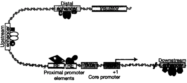
If one were to imagine DNA as the simple double helix structure devoid of nucleosomes, the transcription process would be fairly simple. Transcription begins in the crucial 5' region of a gene, the core promoter which consists of the transcription start site, known as + 1 or TSS; the initiator region (Inr), the area that will open to permit transcription of the gene; and occasionally a TATA box, at approximately -25 bp in metazoans, serving as a recognition site for the basal transcription apparatus (Figure 1-4) (Juven-Gershon et al., 2008; Levine and Tjian, 2003; Sandelin et al., 2007). However, for transcription to occuer, genes require the use of up/downstream elements that are controlled by the binding of different regulators, including chromatin remodelling complexes, transcription factors and coactivators and repressors (Figure 1-4) (Juven-Gershon et al., 2008; Levine and Tjian, 2003; Sandelin et al., 2007).
Basal transcription in eukaryotes of protein-encoding genes is performed by the pre-initiation complex (PIC), composed of the RNA polymerase II holoenzyme complex (RNAPII), general transcription factors and coactivator complexes (Buratowski, 2009; Conaway et al., 2005; Malik and Roeder, 2010). RNAPII is composed of: catalytic subunits that catalyze the addition of ribonucleotides (Rpbl and Rpb2) which also form a channel through which the DNA-RNA hybrid moves during the transcription process (Cramer, 2004a, b; Zhang et al., 1999); proteins with endonuclease activity that are important for "proofreading" activity; and proteins required for the interaction with transcription initiation and elongation coregulators (Malik and Roeder, 2000). The largest subunit of RNAPII, Rpbl, is generally responsible for the initiation of elongation via the phosphorylation of its C-terminal tail (Buratowski, 2009; Reese, 2003).
The general transcription factors are major components of the holoenzyme and have roles in recognition of the promoter, initiation and elongation of transcription (Figure 1-4;
(Koleske and Young, 1994)). They are designated TFII, for transcription factor for RNAPII, and have multiple roles in promoter recognition (Reese, 2003). The most characterised to date are the TFIID and TFIIH complexes. The general transcription factor complex TFIID is composed of several subunits: TBP (TATA-Binding Protein) which binds to the TATA region distorting the DNA, and the TAFs (TBP-Associated Proteins) which have multiple roles in promoter selectivity in TATA and TATA-less promoters (Chen and Hampsey, 2002). The multiple subunits of TFIIH (Cdk7/cyclin H in humans) contain ATP-dependent helicase activity needed to unwind the DNA template and the kinase activity to phosphorylate RNAPII at serine 2 in the C-terminal domain during transcriptional elongation. Transcription continues following the phosphorylation of RNAPII at serine 5 by the factor positive transcription elongation factor b (p-TEFb) (Dvir et al., 2001; Garriga and Grana, 2004). Although transcription generally does involve these simple steps, the specific regulation of gene expression is a very important process in order to control cellular proliferation, differentiation and response to stimuli in multicellular organisms. So, to this process we add the concerted action of several complexes that bind to multiple regulatory regions of genes and to the core promoter, which serve as modifiers of chromatin and activators or repressors of transcription (Levine and Tjian, 2003). Although basal transcription may occur through the core promoter, the key elements of gene regulation lie in areas surrounding the TSS (proximal regulatory elements), and sometimes several kilobases (kb) from the core promoter (distal regulatory elements) (Levine and Tjian, 2003).
Figure 1-4 : Gene promoter structure in regulatable gene expression
The representation of a gene promoter and the multiple possible regulatory elements, including proximal (response elements (RE), TATA, INR, +1) and distal elements (enhancers, insulators). Adapted from (Levine and Tjian, 2003)
1.2.2. Transcription factor mediated gene regulation
There are numerous fundamental mechanisms in the control of regulatable gene expression. First, within the DNA sequence itself, there are c/s-acting regulatory sequences in areas surrounding the gene: response elements (RE) that permit the binding of specific proteins (transcription factors, or activators); and enhancers that are bound by positive transcriptional regulators and/or negative transcriptional regulators (Figure 1-4; (Villard, 2004; Weake and Workman, 2010)). Both of these regulatory elements can be found proximal or distal to the TSS.
The binding of activators to their RE is an essential step in transcription activation (Weake and Workman, 2010). These factors generally have two main domains: a DNA-binding domain (DBD) that includes usually a dimerisation domain that permits the
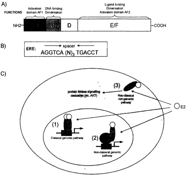
formation of homo- or heterodimers; and an activating domain (AD) that binds to the proteins of the transcriptional machinery (Kumar and Thompson, 1999; Weake and Workman, 2010) Some activators have ligand-binding domains (LBD) that respond to cellular signals such as hormone presence (Kumar and Thompson, 1999). The ensemble of regions bound by one transcription factor in the genome has been recently come to be known as its "cistrome" (Lupien et al., 2008). Activator binding to a RE generally activates transcription by interacting directly or indirectly with the transcription machine (Ptashne, 2005; Weake and Workman, 2010). For example, presence of the hormone estrogen (E2) in mammalian cells will bind the estrogen receptor (ER) transcription factor, permitting their dimerisation, nuclear transport and subsequent interaction with estrogen response elements (ERE) on DNA, resulting in the transcriptional activation of target genes (further detailed in section 1.3) (Beekman et al., 1993; Welboren et al., 2009). Also, regulatory factors often do not act alone, utilizing different types of co-regulatory proteins to respond to specific cell signals. Repressors can bind co-regulatory sequences and prevent the binding of the transcriptional machinery or can also act indirectly by interfering with the action of activators (Beekman et al., 1993; Kumar and Thompson, 1999; Weake and Workman, 2010; Welboren et al., 2009). The activators ERa and p53 notably recruit the help of histone modifiers CBP/p300, PRMT and CARM1 during induction of their target genes (An et al., 2004; Gu et al., 1997; Lill et al.,
1997; Metivier et al., 2004; Shang et al., 2000). The action of these regulatory proteins is not restricted to promoter proximal elements, but can also be observed at numerous distal elements.
Distal response elements that influence transcription once bound by a trans-acting factor and coregulatory proteins are known as enhancers. These areas are often large in size, from 100 bp up to several kb, and can be located up- or downstream of the TSS in variable positions and orientations with regard to the core promoter (Mitchell and Tjian, 1989; Weake and Workman, 2010). Some coregulator complexes prepare the regulatory areas for the binding of different factors, and help form the bridge between activators and
RNAPII, aiding in the formation of the PIC (Malik and Roeder, 2010). These bridges can result in the creation of specific 3-dimensional (3-D) structures, chromatin loops, that affect the accessibility of the transcriptional machinery to the TSS and other promoter elements (Bondarenko et al., 2003). This phenomenon is an area of much interest in recent studies, and in part explains how distal elements will influence transcription of a gene (Bondarenko et al., 2003; Hou et al., 2008; Ling et al., 2006; Splinter et al., 2006). Similar structures are created at insulator regions, which define gene regions and limit the interactions of enhancers with other promoters (Hou et al., 2008). The protein CTCF has been shown by ChIP and chromosome conformation capture (3C) assays to be a master regulator in the creation of insulator regions, of which H2A.Z is an important component (Barski et al., 2007; Fu et al., 2008; Hou et al., 2008; Ling et al., 2006; Splinter et al., 2006). Enhancer function and chromatin looping demonstrates the importance of chromatin structure in the gene regulation process.
1.2.3. Chromatin architecture at regulatory elements
It has become widely accepted that main criteria in controlling regulatable gene expression is the modification of the chromatin structure that surrounds the promoter region of a gene. The architecture of a promoter is known as the composition of the DNA-protein complex that surrounds the TSS and regulatory elements (Cairns, 2009). The application of high-resolution genome-wide analyses performed targeting histone modifications and histone variants has provided a "chromatin signature" of enhancers and promoters in human cells (Barski et al., 2007; Fu et al., 2008; Guenther et al., 2007; Jin et al., 2009; Lupien et al., 2008; Mikkelsen et al., 2007; Schones et al., 2008; Sharov and Ko, 2007; Zhou et al., 2010). Generally, there exist three different transcriptional states for a promoter: active, in which the chromatin is organised and modified to be permissive to transcription; repressed, where repressive chromatin marks dominate; and poised, a
state in which the promoter can be activated or repressed (Table 1-1; (Sakabe and Nobrega, 2010; Zhou et al., 2010)). Numerous different chromatin modifications will define these states, but this section will explore in more detail the effects on the fate of transcription of one of the most influential histone variants, H2A.Z (1.2.4), and the modification of H3K4 and H3K27 methylation (1.2.5).
Table 1-1: Chromatin conformations at different promoter types
Different histone modifications define inactive, poised or active promoters, and inactive or active enhancers. Adapted from (Sakabe and Nobrega, 2010).
Genomic Element Enrichment of Methylated Histories Enrichment of Acetylated Histories Active genes (around TSS) H3K4me1 H3K4me2 H3K4me3 (me1, me2, me3
increase from 3' to 5')
Active genes (transcribed regions) Enhancers
Repressed genes
Bivalent domains'1
H3K9me1 H2A.Z
H2BK5me1 (5' end) H3K27me1 (5'end) H4K20Mel (5' end) H3K36me3 (3" end) H3K4me1 (H3K4me3)* H2A.Z
H3K27me2, H3K27me3 H3K79me3 H3K9me2, H3K9me3 (weak correlation)
H3K4me3 'activating' H3K27me3 'repressive'
H2AK9ac, H2BK5ac. H3K9ac, H3K18ac, H3K27ac, H3K36ac H4K91ac
H2BK12ac. H2BK20ac, H2BK120ac, H3K4ac. H4K5ac, H4K8ac, H4K12ac and H4K16ac H2BK12ac, H2BK203C, H2BK120ac, H3K4ac,
H4K5ac, H4K8ac, H4K12ac and H4K16ac H3K27ac
1.2.4. Mechanisms of transcriptional activation and roles of H2A.Z in gene expression
Active promoters are generally defined as having an open promoter conformation, defined by "available" regulatory sites (TSS, RE, or enhancers). This availability is achieved by the epigenetic signature that is present at these promoters: highly acetylated histone tails, and the presence of H3K4me3, H3.3 and H2A.Z in nucleosomes surrounding the TSS (Azuara et al., 2006; Barski et al., 2007; Dreijerink et al., 2006; Fu
et al., 2008; Guenther et al., 2007; Jin et al., 2009; Lupien et al., 2008; Mikkelsen et al., 2007; Schones et al., 2008; Sharov and Ko, 2007; Zhang et al., 2005b; Zhou et al., 2010).
First, transcriptionally active areas of chromatin are generally known to be hyperacetylated and enriched in H3K4 methylation (Mellor, 2005; Zhou et al., 2010). Interestingly, several coactivators of transcription are actually HAT-containing complexes that effect transcription, and transcriptional activators have been shown to interact with and recruit chromatin remodelling complexes to promoters, inducing either the displacement or modification of nucleosomes (Szutorisz et al., 2005). For example, the p300/CBP complex is a coactivator of transcription that interacts with the basal transcriptional machinery and several known transcription factors (Kalkhoven, 2004), yet also contains HAT activity that targets K12 and K15 of H2B, K14 and K18 of H3 and K5 and K8 of H4 (Schiltz et al., 1999). Notably, the p300/CBP complex colocalises with H3K4 methylation, and specific methylation of H3K4 has rapidly become one of the most recognised determinants of gene activity (Azuara et al., 2006; Barski et al., 2007; Dreijerink et al., 2006; Fu et al., 2008; Guenther et al., 2007; Jin et al., 2009; Lupien et al., 2008; Mikkelsen et al., 2007; Schones et al., 2008; Sharov and Ko, 2007; Zhang et al., 2005b; Zhou et al., 2010). More specifically, active promoters are defined by H3K4me3 in mammalian cells, where enhancers exhibit H3K4mel/2, H3K27ac and p300/CBP (Azuara et al., 2006; Barski et al., 2007; Dreijerink et al., 2006; Fu et al., 2008; Guenther et al., 2007; Jin et al., 2009; Lupien et al., 2008; Mikkelsen et al., 2007; Schones et al., 2008; Sharov and Ko, 2007; Zhang et al., 2005b; Zhou et al., 2010). In addition to these histone modifications, the histone variant H2A.Z has generally been shown to be very important in transcriptional regulation, and several lines of evidence suggest its incorporation in chromatin promotes an active state.
Genome-wide analyses in yeast shed new light on the positioning of H2A.Z and its role in gene activation in this model system. H2A.Z was mainly present at the 5' ends of genes genome-wide, in the promoter regions in the nucleosomes that border the