High-Light Stress Is Controlled by OXI1 Kinase
1
Leonard Shumbe, Anne Chevalier, Bertrand Legeret, Ludivine Taconnat, Fabien Monnet, and Michel Havaux*
CEA, Direction des Sciences du Vivant, Institut de Biologie Environnementale et de Biotechnologie, F-13108 Saint-Paul-lez-Durance, France (L.S., A.C., B.L., F.M., M.H.); CNRS, UMR 7265 Biologie Végétale et
Microbiologie Environnementales, F-13108 Saint-Paul-lez-Durance, France (L.S., A.C., B.L., F.M., M.H.); Aix-Marseille Université, F-13284 Aix-Marseille, France (L.S., A.C., B.L., F.M., M.H.); POPS Transcriptomic Platform, Institute of Plant Sciences Paris-Saclay IPS2, Rue de Noetzlin, 91405 Orsay, France (L.T.); and Université Avignon et des Pays de Vaucluse, 84000 Avignon, France (F.M.)
ORCID ID: 0000-0001-7912-7220 (F.M.).
Studies of the singlet oxygen (1O2)-overproducing flu and chlorina1 (ch1) mutants of Arabidopsis (Arabidopsis thaliana) have
shown that 1O2-induced changes in gene expression can lead to either programmed cell death (PCD) or acclimation. A
transcriptomic analysis of the ch1 mutant has allowed the identification of genes whose expression is specifically affected by each phenomenon. One such gene is OXIDATIVE SIGNAL INDUCIBLE1 (OXI1) encoding an AGC kinase that was noticeably induced by excess light energy and1O2stress conditions leading to cell death. Photo-induced oxidative damage and cell death
were drastically reduced in the OXI1 null mutant (oxi1) and in the double mutant ch1*oxi1 compared with the wild type and the ch1 single mutant, respectively. This occurred without any changes in the production rate of1O2but was cancelled by
exogenous applications of the phytohormone jasmonate. OXI1-mediated1O2signaling appeared to operate through a different
pathway from the previously characterized OXI1-dependent response to pathogens and H2O2and was found to be independent
of the EXECUTER proteins. In high-light-stressed plants, the oxi1 mutation was associated with reduced jasmonate levels and with the up-regulation of genes encoding negative regulators of jasmonate signaling and PCD. Our results show that OXI1 is a new regulator of1O
2-induced PCD, likely acting upstream of jasmonate.
Reactive oxygen species (ROS) are unavoidable by-products of photosynthesis in plants. Indeed, the pho-tosynthetic processes involve electron-transfer mecha-nisms as well as generation of excited molecules, which can interact with molecular oxygen leading to the for-mation of reduced forms of oxygen such as superoxide and of singlet-excited oxygen (1O2; Apel and Hirt, 2004;
Asada, 2006; Li et al., 2009). Superoxide can spontane-ously or enzymatically disproportionate to hydrogen peroxide that can ultimately lead to the production of the hydroxyl radical in the presence of metals. These phenomena can be strongly amplified in plants exposed
to environmental stress conditions that inhibit the photosynthetic activity and can lead to a situation where light energy is absorbed in excess to what can be used by the photosynthetic processes. The resulting over-reduction of the electron transport chain and in-creased lifetime of singlet-excited chlorophylls favor leakage of electrons and/or excitation energy to O2,
leading to enhanced ROS production. As a result, oxidative damage to the photosynthetic apparatus can occur, lowering photosynthetic efficiency and ultimately leading to cell death.
1O
2is produced within the photosystems (PS) from
chlorophyll molecules in the triplet excited state (Krieger-Liszkay, 2005; Triantaphylidès and Havaux, 2009).1O2is believed to be the main ROS produced in the chloroplasts under stress and excess light (González-Pérez et al., 2011) and to play a major destructive role during the execution of ROS-induced cell death in leaf tissues (Triantaphylidès et al., 2008).1O2is a very reactive species that can readily oxidize macromolecules, partic-ularly those containing double bonds (Triantaphylidès and Havaux, 2009). Because of its high reactivity,1O2has a short lifetime in biological tissues, likely, 0.5 to 1 ms in plant cells (Bisby et al., 1999; Redmond and Kochevar, 2006; Li et al., 2012), making difficult the study of this ROS in planta. A major breakthrough in the study of1O2 effects on plants was the isolation and identification of
1This work was supported by the French National Research
Agency through the SLOSAM project (ANR-14-CE02-0010). L.S. was supported by a CEA IRTELIS studentship.
* Address correspondence to michel.havaux@cea.fr.
The author responsible for distribution of materials integral to the findings presented in this article in accordance with the policy de-scribed in the Instructions for Authors (www.plantphysiol.org) is: Michel Havaux (michel.havaux@cea.fr).
L.S., F.M., and M.H. designed the experiments; L.S. performed most experiments; A.C. performed some qRT-PCR analyses of gene expressions; L.T. performed the microarray-based transcriptomic analyses; B.L. performed the UPLC-MS analyses of jasmonates; L.S., F.M., and M.H. interpreted the data; L.S. and M.H. wrote the article.
www.plantphysiol.org/cgi/doi/10.1104/pp.15.01546
Arabidopsis (Arabidopsis thaliana) mutants that specifi-cally produce1O2under particular conditions. One such mutant is flu, a conditional mutant that accumulates a chlorophyll precursor in the dark (Meskauskiene et al., 2001). Upon transfer of flu seedlings from darkness to light, the accumulated chlorophyll precursor molecules act as photosensitizers in the light and bring about the production of high amounts of1O2 (op den Camp et al.,
2003). Theflu mutant appeared to be a very good ex-perimental tool to generate1O2in leaf tissues in a specific
and relatively controlled manner. Thanks to this mutant, it was demonstrated that1O2is not only a toxic molecule, but it can also function as a signal molecule inducing changes in gene expression that can lead to programmed cell death (PCD; op den Camp et al., 2003). This 1O2
signaling pathway was found to be dependent on two chloroplastic proteins, EXECUTER1 (EX1) and EX2 (Wagner et al., 2004; Lee et al., 2007). Accordingly,1O2 -induced cell death was suppressed in theflu*ex1 double mutant deficient in the EX1 protein, and this took place without affecting the rate of1O2formation.
Another 1O2-overproducing Arabidopsis mutant,
chlorina1 (ch1), has emerged more recently as a useful tool to study noninvasively the in vivo signaling role of
1O
2(Ramel et al., 2013a). Contrary to theflu mutant, this
mutant produces1O2in the PSII reaction centers, i.e. in
the natural site of 1O2 production in plants (Krieger-Liszkay, 2005; Ramel et al., 2012a). Because of the in-creased release of 1O2 from the PSII centers, the ch1
mutant is very photosensitive, exhibiting extensive leaf damage under light conditions that have little effect on the wild type (Havaux et al., 2007; Dall’Osto et al., 2010; Ramel et al., 2013a). The ch1 mutant confirmed that1
O2
can trigger changes in the expression of nuclear genes (Ramel et al., 2013a). Depending on the levels of1O2
production induced by light, the1O2-triggered signal-ing pathway in ch1 was found to lead either to cell death or to an acclimation process (Ramel et al., 2013a). Transcriptomic analyses of ch1 plants exposed to both conditions revealed marked difference in the gene ex-pression profiles. Strikingly, most genes involved in the jasmonate biosynthesis pathway were strongly induced in ch1 leaves under photooxidative stress conditions, while acclimatory conditions repressed those genes (Ramel et al., 2013a). Those contrasted changes in gene expression correlated with changes in the jasmonate concentrations in the leaves. Jasmonate, and possibly other phytohormones, appeared to be major players in the orientation of the1O2 signaling pathway toward a
particular response (cell death or acclimation; Danon et al., 2005; Ramel et al., 2013b; Laloi and Havaux, 2015). A double mutant, ch1*dde2, that cannot synthesize jasmonate was found to be constitutively tolerant to photooxidative stress (Ramel et al., 2013b), affirming the function of jasmonate as a cell-death-promoting regula-tor under1O2stress conditions (Laloi and Havaux, 2015).
Other remarkable differences in the transcriptome of ch1 plants exposed to conditions leading to cell death or to acclimation concern genes known to play a role in the response to ROS and in cell death regulation. For
instance, the gene encoding OXIDATIVE SIGNAL INDUCIBLE1 (OXI1) was observed to be specifically induced by stress conditions that led to cell death in ch1 leaves (Ramel et al., 2013a). OXI1 is a Ser/Thr kinase that was shown to be regulated by H2O2(Rentel et al., 2004). The OXI1 gene is expressed in all plant organs and was principally localized at the cell periphery and in the nucleus (Anthony et al., 2004). This kinase was previ-ously found to be necessary for ROS-mediated processes such as root hair growth (Rentel et al., 2004; Anthony et al., 2004) and plant-pathogen interactions (Rentel et al., 2004; Petersen et al., 2009; Camehl et al., 2011). OXI1 is believed to link oxidative burst signals and downstream responses through activation of mitogen-activated protein kinases (MAPKs; Rentel et al., 2004). This work was undertaken to investigate the possible implication of OXI1 in the responses of Arabidopsis to high-light stress and1O2. The results of this study reveal a crucial role for OXI1 in the control of1O2-induced PCD
in Arabidopsis plants exposed to excess light energy, which operates by a mechanism different from the OXI1-dependent pathway triggered by H2O2and pathogens.
RESULTS
Transcript Profile of the OXI1 Gene in Arabidopsis Leaves under High-Light Stress
A previous microarray-based transcriptomic analysis of the1O2-overproducing Arabidopsis ch1 mutant ex-posed to high-light stress (Ramel et al., 2013a) identified OXI1 as a potentially interesting gene: it was markedly induced during light stress conditions that led to leaf damage but not differentially expressed during accli-mation to 1O2, suggesting that it might be a specific component of the1O2signaling pathway leading to cell
death. We have confirmed the microarray data for this gene using quantitative real-time PCR (qRT-PCR;
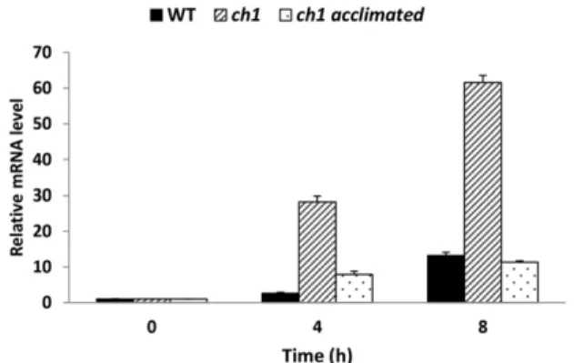
Figure 1. Transcript profile of the OXI1 gene analyzed by qRT-PCR in wild-type Arabidopsis (WT) and in thech1 mutant (control or photoacclimated) exposed for 4 or 8 h to high-light stress (1500mmol m22s21at 7˚C/14˚C, day/night). Photoacclimation was induced in thech1 mutant by a 2-d treatment at a moderately elevated PFD of 450mmol m22s21at 20˚C/18˚C (day/night), as by Ramel et al. (2013a). Data are normalized to the values at time 0. Results are mean values of three biological replicates +SD.
Fig. 1). OXI1 was induced in wild-type leaves during high-light stress: the transcript level was increased by a factor of 2.6 after 4 h in high light and a factor of 13 after 8 h. This induction was strongly enhanced in the ch1 mutant (330 and 360 after 4 and 8 h, respectively) compared with the wild type, indicating high respon-siveness of this gene to 1O2stress. The ch1 mutant was also acclimated to high-light stress by pre-exposing the plants for 2 d to a moderately elevated photonflux density (PFD; 450mmol m22s21at 20°C), as previously described (Ramel et al., 2013a). The acclimated ch1 plants were then exposed to high-light stress for 4 or 8 h. Interestingly, photoacclimation of the ch1 mutant blocked the induction of the OXI1 gene after transfer to high-light stress (compared with ch1 plants exposed to stress without acclimation) to similar values as in the wild type. Photoacclimation of ch1 was previously shown not to modify the1O2levels in leaves exposed to high PFDs (Ramel et al., 2013a). So, the low transcript levels in photoacclimated leaves are due to a down-regulation of the OXI1 gene. The data in Figure 1 sug-gest a specific role of OXI1 in photooxidative stress, with tolerance to1O2stress being associated with inhi-bition of the up-regulation of this gene.
Photosensitivity of the Arabidopsisoxi1 Mutant
An Arabidopsis mutant (oxi1) deficient in OXI1 was previously studied in the context of pathogen attacks (Camehl et al., 2011). Although OXI1 is required for
normal root development (Rentel et al., 2004; Anthony et al., 2004), oxi1 mutant shoots did not exhibit any growth phenotype under normal growth conditions compared with its wild type (not shown, but see Figure 2B, panel i, for rosette size). We crossed this mutant with the ch1 mutant to generate a1O2-overproducing,
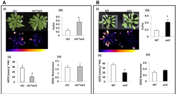
OXI1-deficient double mutant (ch1*oxi1). Compared with ch1, the ch1*oxi1 double mutant did not show any alter-ation of its shoot-growth capacities under normal con-ditions. Ch1 and ch1*oxi1 mutants were transferred from normal growth light (PFD 120mmol photons m22s21) to high-light stress (1100 mmol m22s21). Air temperature was simultaneously decreased to 7°C to prevent over-heating of the leaves during high-light stress and to maintain leaf temperature in the range of the control leaf temperature (;18°C). As shown in Figure 2A (panel i, top), ch1*oxi1 plants appeared to be more resistant to high-light stress than ch1 plants as depicted by decreased leaf bleaching. In leaves, lipids are highly unsaturated (Douce and Joyard, 1980) and therefore very sensitive to oxidation (Mène-Saffrané et al., 2009; Farmer and Mueller, 2013). Consequently, photooxidative damage to plant leaves can be estimated by measuring the extent of lipid peroxidation. Accumulation of lipid peroxides can be visualized via the light emission associated with their slow spontaneous decomposition (Havaux et al., 2006; Birtic et al., 2011). Figure 2A (panel i, bottom) shows that the autoluminescence of high-light-treated ch1*oxi1 plants was substantially lower than the lumines-cence signal emitted by ch1 plants, indicating a lower
Figure 2. Effect of the oxi1 mutation on the tolerance to high-light stress of1O
2-overproducingch1 mutant plants (A) and wild-type Arabidopsis plants (WT; B). The high-light treatments were 2 d at 1100 and 1500mmol m22s21at 7˚C/14˚C (day/night) for A and B, respectively. i, Picture of the plants after the high-light stress treatment (top) and autoluminescence imaging of lipid peroxidation (bottom). Color scale indicates signal intensity from 0 (blue) to saturation (white). ii, Lipid peroxidation measured by HPLC quantification of hydroxy fatty acids (HOTE) after high-light stress. iii, PSII photochemical efficiency as measured by the Fv/Fmchlorophyll fluorescence ratio after high-light stress. iv,
1
O2production measured in leaves by fluorescence of the SOSG probe after exposure of SOSG-infiltrated plants to high light for 30 min. Background fluorescence produced when SOSG-infiltrated plants were placed in the dark for 30 min was subtracted from fluorescence produced in light. Asterisk indicates significant difference betweenoxi1/ch1*oxi1 plants and wild-type/ch1 plants, respectively, at P , 0.05 (Student’s t test). Results are mean values of five measurements (ii) and 10 measurements (iii and iv) +SD.
accumulation of lipid peroxides in the former plants after high-light stress. Differential lipid peroxidation was confirmed by HPLC analyses of hydroxyoctatrienoic acid (HOTE; the oxidation product of linolenic acid C18:3, the major fatty acid in Arabidopsis leaves), which showed drastic decrease in the ch1*oxi1 double mutant relative to the ch1 single mutant (Fig. 2A, panel ii). The decrease in lipid peroxidation in ch1*oxi1 relative to ch1 correlated with a lesser diminution of the PSII photochemical effi-ciency measured by the Fv/Fmchlorophyllfluorescence
ratio (Fig. 2A, panel iii).
We also compared the responses of the wild type and the oxi1 single mutant to high-light stress (Fig. 2B). Be-cause wild-type plants are much more tolerant to high-light stress compared with ch1 plants (Ramel et al., 2013a) , it is necessary to use a higher PFD (1500mmol m22s21) to achieve comparable stress intensities. Similar to what we observed in the ch1 genetic background, the oxi1 mutation showed decreased leaf photodamage, with reduction of leaf bleaching (panel i), decrease in lipid peroxidation as measured by autoluminescence im-aging (panel i) and HOTE levels (panel ii), and more preserved PSII activity (panel iii) after high-light stress. To ascertain the fact that the observed photoresistant phenotype of oxi1 and ch1*oxi1 mutants under high-light stress compared with the wild type and ch1, re-spectively, was not imputable to different levels of1O2 production,1O2produced in leaves after 30 min of high-light treatment was measured by fluorescence of the
1O
2-specific Singlet Oxygen Sensor Green (SOSG)
flu-orescent probe (Flors et al., 2006). No difference in1O2
production was observed between ch1 and ch1*oxi1 (Fig. 2A, panel iv) or between the wild type and oxi1 (Fig. 2B, panel iv). However, as expected (Dall’Osto et al., 2010; Ramel et al., 2013a),1O2accumulation in the
ch1 background (Fig. 2A, panel iv) was noticeably higher than the production in the wild-type back-ground (Fig. 2B, panel iv). A drawback of SOSG is that it can act as a photosensitizer producing1O2upon ex-posure to UV radiation and, to a lesser extent, visible light (Ragàs et al., 2009). We exposed a SOSG solution to the light conditions used in this study (1500 mmol photons m22 s21 for 30 min), and we observed that SOSG fluorescence at 525 nm was slightly increased (Supplemental Fig. S1), confirming the idea that some
1O
2 photosensitization by SOSG can occur upon
illu-mination. However, this light-induced increase in SOSGfluorescence was low compared with the effect of
1O
2 produced by Rose Bengal. Moreover, this
phe-nomenon is likely to be minor in our analyses of1O2 production by Arabidopsis leaves. Indeed, SOSG fluo-rescence was much higher in ch1 leaves compared with wild-type leaves (Fig. 2, A, panel iv, and B, panel iv), although the light treatment was less intense, in-dicating that this difference cannot be merely due to a direct effect of light on SOSG. In addition, the com-parison between the wild type and oxi1 or between ch1 and ch1*oxi1 in Figure 2 was done at the same PFD (1500 or 1100 mmol m22 s21, respectively), and, therefore, the interference of 1O2 photosensitization
by SOSG, if any, can be supposed to be the same for both genotypes in each comparison. Nevertheless, the SOSG data must be considered as semiquantitative only. Based on the results presented in Figure 2, OXI1 thus appeared to play a crucial role in the develop-ment of cellular damage during high-light stress in Arabidopsis.
OXI1-Mediated Cell Death in Arabidopsis Is Independent of the EX Proteins
PCD is used by plants as a means of defense against many ROS-mediated stresses (Van Breusegem and Dat, 2006; Gadjev et al., 2008). One peculiar feature of ROS-mediated PCD is that the ROS triggers reprogramming of expression of defense genes. Influ, EX1 and EX2 are principal mediators of1O2-triggered cell death (Wagner
et al., 2004; Lee et al., 2007). Cell death and up-regulation of almost all1O2-specific genes induced by
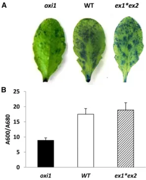
a dark-to-light transfer in theflu mutant were abolished in theflu*ex1*ex2 triple mutant (Lee et al., 2007). How-ever, our study showed that the EX proteins do not mediate PCD in the wild-type Columbia (Col)-0 genetic background under conditions of excess light energy induced by high PFD and low temperature: ex1*ex2 double mutant plants showed similar susceptibility to high-light stress as wild-type plants, depicted by simi-lar levels of leaf bleaching and lipid peroxidation measured by autoluminescence imaging (Fig. 3A) and HOTE levels (Fig. 3B).
To analyze the implication of OXI1 and EX1 and EX2 in the process of1O2-mediated cell death, we measured cell death in mature leaves of oxi1 and ex1*ex2 mutants by Evans blue staining relative to the wild type (Fig. 4). As shown in Figure 4A, wild-type leaves retained more dye than oxi1 mutant leaves, indicating an increased extent of cell death in the former leaves. This phenom-enon was quantified in Figure 4B, revealing an almost doubling of the amount of dye retained in wild-type leaves relative to oxi1 leaves. This confirms the role of OXI1 in high-light-induced PCD in Arabidopsis. On the contrary, ex1*ex2 double mutant leaves retained similar amounts of the dye as wild-type leaves, thus strength-ening the irrelevance of the EX proteins in cell death mediation in the Arabidopsis Col-0 background during high-light stress.
The1O2Signaling Pathway Is Different from the
OXI1-Dependent H2O2Pathway Involving MAPKs OXI1 is known to be essential for the full activation of MAPK3 and MAPK6 in plants submitted to pathogen attacks (Rentel et al., 2004). Activation of OXI1 and downstream components of the signaling pathway are known to be achieved by sequential phosphorylation. The presumed pathway for OXI1 signaling during pathogen attack has previously been established (Hirt et al., 2011), as illustrated in Supplemental Figure S2. This pathway indicates PDK1-dependent
and PDK1-independent routes for activation of OXI1 by pathogens. The major ROS triggering the signaling during pathogen attack is H2O2. We were interested to
determine if the OXI1 pathway for PCD triggered by
1O
2 is similar to that portrayed during H2O2 stress.
Whole wild-type and oxi1 plants were sprayed with 5% H2O2 and allowed for 2 h under normal growth conditions (140mmol photons m22s21light and 20°C/18°C day/night). On the other hand,1O2-specific stress was induced by exposing ch1 and ch1*oxi1 plants to high-light stress (1100mmol photons m22s21). The general transcript profiles of the OXI1-dependent signaling pathway showed great differences between H2O2 treatment and1O2stimulation as illustrated in Figure 5. Both H2O2 and 1O2 induced OXI1 expression and
repressed the upstream activator of OXI1, PDK1 (At5g04510). As expected (Rentel et al., 2004), activation of OXI1 by H2O2led to an increase in transcript levels for the downstream components PTI1-4 (At2g47060), PTI1-1 (At1g06700), MAPK3 (At3g45640), and MAPK6 (At2g43790) in the wild type. In the oxi1 mutant, the H2O2-induced changes in these downstream
compo-nents were perturbed as previously shown (Rentel et al., 2004): PTI1-4, PTI1-1, and MAPK6 were slightly repressed (instead of being induced), while MAPK3 was not fully activated. In striking contrast with the H2O2-induced changes in gene expressions, neither PTI-1 and PTI1-4 nor MAPK3 and MAPK6 were induced by
1O
2in the ch1 mutant exposed to high light, and the oxi1
mutation in the ch1*oxi1 double mutant did not affect these responses (Fig. 5). The differential response of the PTI1 and MAPK genes between H2O2 and 1O2 treat-ments and the similar responses portrayed by these genes in the ch1 and ch1*oxi1 mutants clearly indicate that 1O2-induced signaling for cell death regulated
by OXI1 occurs by a pathway different from that of the OXI1-mediated signaling pathway induced by
pathogen attack and H2O2 stress, which involves
MAPK3 and MAPK6 activation.
Microarray-Based Transcriptomic Analysis of theoxi1 Mutant under Photooxidative Stress Conditions
To gain more insight into the effect of oxi1 mutation during high-light stress, a transcriptomic analysis was performed on oxi1 mutant plants compared with wild-type plants, using the CATMAv6 microarray system (Hilson et al., 2004). Three comparisons were per-formed: oxi1-control versus wild type-control to iden-tify transcripts affected as a result of mutation in the OXI1 gene; oxi1-stress versus wild type-stress to dissect transcripts affected in the oxi1 mutant under stress, which may explain the phenotype observed when compared with the wild type; and oxi1-stress versus oxi1-control to identify transcripts affected in the oxi1 mutant specifically due to high-light stress. Among the 38,356 genes identified in the CATMAv6 array, 1221 genes were differentially expressed (with P value , 0.05) in the oxi1-control versus wild type-control com-parison, corresponding to 3.6% of the total genes. This comprised 1.5% genes up-regulated and 2.1% genes down-regulated (Fig. 6A). Functional classification of these differentially expressed genes using the DAVID gene functional classification tool revealed that the mutation affects a number of important biological and metabolic processes, such as response to endoge-nous stimuli, ethylene-mediated signaling pathway, and toxin metabolic processes that were repressed. Glucosinolate and anthocyanin biosynthetic processes, carbohydrate biosynthetic processes, responses to en-dogenous stimuli (ethylene, jasmonic acid, etc.), and lipid transferase activity were induced (Supplemental Table S1).
Figure 3. Lipid peroxidation in wild-type Arabidopsis (WT) and in the ex1*ex2 double mutant after high-light stress (2 d at 1500mmol m22s21and 7˚C/14˚C, day/night). A, Autoluminescence imaging of lipid peroxides. B, HPLC quantification of hydroxy fatty acid (HOTE). Data are mean values of five measurements +SD.
Comparing oxi1-stress versus oxi1-control, 3262 genes were differentially expressed (with P value , 0.05), corresponding to 4% up-regulated and 5.7% down-regulated genes (Fig. 6A). Functional classifi-cation of genes differentially expressed using the DAVID tool revealed that the down-regulated genes coded for proteins involved in processes such as re-sponse to endogenous/hormone stimuli, glucosino-late biosynthesis/metabolism, response to abiotic stress, photosynthesis, and oxylipin/jasmonate biosynthesis/ metabolism (Supplemental Table S2). Interestingly, most of the genes of the jasmonate biosynthetic pathway, including LOX3 (At1g17420), LOX2 (At3g45140), AOS (At5g42650), OPR3 (At2g06050), AOC1 (At3g25760), JMT (At1g19640), and AOC4 (At1g13280), were down-regulated. Thisfinding shows a behavior very different from the responses to1O2 in theflu and ch1 mutants,
which have been shown to be associated with a strong induction of the jasmonate pathway (Przybyla et al., 2008; Ramel et al., 2013a). Considering the fact that jasmonate is associated with1O2-induced cell death in the flu and ch1 mutants (Laloi and Havaux, 2015), this find-ing suggests that the OXI1 action durfind-ing high-light stress could involve a regulation of jasmonate synthesis and/or activity. The genes up-regulated in oxi1 by high-light stress were mainly involved in the processes of ribonu-cleoprotein and cellular protein biosynthesis,flavonoid biosynthesis, helicase activity, ATPase activity, toxin
metabolism, and response to abiotic stimuli, represented by overexpression of genes implicated in detoxification processes, such as glutaredoxin-C14 (At3g62960), Peroxiredoxin-C2 (At1g65970), glutathione S-transferases (ATGSTU1 [At2g29490], ATGSTU11 [At1g69930], and ATGSTU16 [At1g59700]), CAT2 (At4g35090), HSP70 (At3g12580), and the transcription factors HY5 (At5g11260) and MYB32 (At4g34990; Supplemental Table S2).
Of great importance to us was to assess the transcript profile of the oxi1 mutant relative to the wild type under stress conditions, which was achieved by the compar-ison oxi1-stress versus wild type-stress in the micro-array analysis. A total of 1568 genes were differentially expressed after stress (P value , 0.05), which account for 4.7% of the total genes analyzed (Fig. 6A), with up-regulation and down-up-regulation representing half of the 1568 genes each (Fig. 6B). The majority of the genes up-regulated and down-regulated in this comparison were found to have log2intensity ratios between 1 and
2, which corresponds to 2- to 4-fold up-regulation/ down-regulation of the genes in the stressed oxi1 mu-tant compared with stressed wild-type plants (Fig. 6C). As observed in the oxi1-stress versus oxi1-control comparison, functional classification of down-regulated genes using the DAVID functional classification tool also revealed down-regulation of genes implicated in the re-sponse to hormonal stimuli, mainly auxin, as represented by genes such as PIN4 (At2g01420), AUX1 (At2g38120), IAA15 (At4g14560), and IAA14 (At4g14550); ethylene, as depicted by genes such as DREBA1A (At4g25480), DREBA2B (At3g11020), ERF8 (At1g53170), and ERF015 (At4g31060); and gibberellic acid, as represented by genes such as RGA1 (At2g01570), GA2 (At1g79460), GA3 (At5g25900), and GASA4 (At5g15230; Supplemental Table S3). Other processes in this category included re-sponse to abiotic stress, photosynthesis, oxidoreductases, and organic acid biosynthetic processes (Supplemental Table S3). As far as the jasmonate biosynthesis pathway is concerned, repression in high-light-stressed oxi1 leaves compared with stressed wild-type leaves concerned mainly the AOC4 gene (At1g13280). Also, similar to up-regulation in the oxi1-stress versus oxi1-control compar-ison, we observed an up-regulation of genes coding for specific processes, such as ribosome biogenesis, response to abiotic stimuli (genes such as CAT2 [At4g35090], HSP70 [At3g12580], and HY5 transcription factor [At5g11260] involved in ROS-dependent stress regula-tion), flavonoid, anthocyanin, and other pigment biosynthesis. In addition, there was up-regulation of genes related to salicylic acid, such as GRP23 (At1g10270), PBS3 (At5g13320), MYB77 (At3g50060), and CCA1 (At2g46830), as well as genes involved in response to jasmonate, predominantly the TIFY genes that code for the JAZ proteins (JAZ5 [At1g17380], JAZ6 [At1g72450], JAZ7 [At2g34600], and JAZ10 [At5g13220]), which are negative regulators of jasmonate (Supplemental Table S3). To sum up, relative to the wild type, high-light stress in oxi1 leaves causes significant down-regulation of photosynthesis, growth processes, and phytohormones, and up-regulation of structural component biogenesis Figure 4. High-light-induced cell death measured in leaves by Evans
blue staining. Wild-type Arabidopsis (WT) is compared with the single mutantoxi1 and the double mutant ex1*ex2. The high-light treatment was 2 d at 1500mmol photons m22s21and 7˚C/14˚C (day/night). A, Visualization of dead cells in leaves which retain the Evans blue stain. B, Quantification of cell death in leaves by measuring the absorbance ratio (A600/A680) of the Evans blue stain solution extracted from the leaves. Data are mean values of five measurements +SD.
and pigment biosynthesis, all of which are regulated by an interplay of the hormones auxin, ethylene, jasmonic acid, salicylic acid, and gibberellins.
Interestingly, in the transcriptomic comparison of oxi1 and the wild type, we noticed the up-regulation in oxi1 leaves of a number of genes proposed to antagonize cell death processes (Cominelli et al., 2000; Danon et al., 2004; Alonso-Peral et al., 2010). This was confirmed by qRT-PCR analyses (Fig. 6D): MYB90 (At1g66390), DAD1 (At1g32210), and MYB114 (At1g66380) were up-regulated in stressed oxi1 plants compared with wild-type plants. These gene expression changes are consistent with the phenotype of the mutant that is more resistant to high light and less prone to cell death than the wild type (Figs. 1 and 4). This observation raises the possi-bility that OXI1 is a master regulatory gene that controls PCD induced by1O2stress.
Interactions between Jasmonate and OXI1-Dependent Signaling
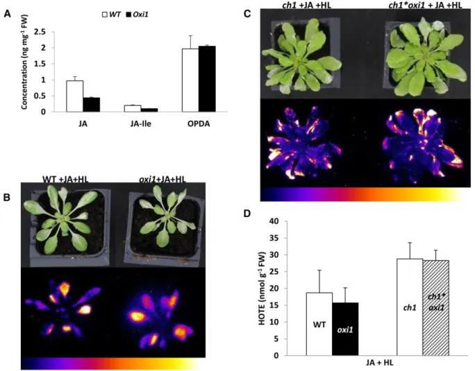
The significant down-regulation of jasmonate-related genes in high-light-stressed oxi1 mutant plants ob-served in the transcriptome studies suggests a possible involvement of decreased levels of jasmonic acid in cell death attenuation in this mutant. In line with this idea, oxi1 plants after exposure to high-light stress for; 26 h portrayed ; 2-fold lower levels of jasmonic acid and jasmonyl-Ile (JA-Ile) compared with wild-type plants (Fig. 7A). In contrast, the levels of the jasmonic acid precursor oxophytodienoic acid (OPDA) were similar in both wild-type and oxi1 plants.
We also examined the effect of exogenous jasmonate applications on the response of the oxi1 mutant to high-light stress (Fig. 7). Oxi1 plants recovered the susceptibility Figure 5. Transcript profiling of genes involved in the OXI1-dependent signaling pathway (PDK1, OXI1, PTI1-1, PTI1-4, MAPK3,
andMAPK6), during H2O2stress compared with1O2stress. H2O2stress was generated by spraying wild-type (WT) andoxi1 mutant plants with 5% H2O2and incubating for 2 h in low light (150mmol photons m22s21).1O2stress was generated by ex-posure ofch1 and ch1*oxi1 plants to high-light stress (HL, 1100 mmol photons m22s21at 7˚C). Asterisk indicates significant difference in gene expression between wild type andoxi1 and between ch1 and ch1*oxi1 at P , 0.05 (Student’s t test). Data are mean values of three biological replicates +SD.
phenotype of wild-type plants under high-light stress when sprayed with methyl-jasmonate, as illustrated by the autoluminescence images of lipid peroxidation shown in Figure 7B. This also corresponded to a similar accumula-tion of HOTE in the oxi1 mutant and the wild type (Fig. 7D). Thesefindings indicate enhanced stress in the oxi1 mutant after high-light exposure in the presence of exog-enously applied methyl-jasmonate compared with the substantial reduction of lipid peroxidation in oxi1 relative to the wild type observed when the high-light treatment was imposed without applying exogenous jasmonate (Fig. 2B, panel ii). The effects of exogenous jasmonate were also observed in the ch1 genetic background: the photo-tolerance of the ch1*oxi1 double mutant was abolished by the jasmonate treatment (compare Figs. 7C and 2A; see also Fig. 7D). Spraying wild-type, oxi1, ch1, or ch1*oxi1 plants with methyl-jasmonate in low light did not induce
leaf damage or lipid peroxidation, excluding a direct tox-icity of jasmonate on the plants (data not shown). DISCUSSION
OXI1 Is a New Regulator of High-Light-Induced Cell Death in Arabidopsis
Singlet oxygen is generated from triplet excited chlorophyll molecules and is therefore intimately as-sociated with light absorption and photosynthesis in plants. 1O2 has been shown to be the main ROS pro-duced in plant cells in high light (González-Pérez et al., 2011) and the major ROS involved in photooxidative damage to plant leaves (Triantaphylidès et al., 2008). This study has identified OXI1 as a link between 1O
2
production in the chloroplasts under excess light Figure 6. Microarray analysis of the transcriptome of oxi1 relative to the wild type (WT). A, Venn diagram showing the number of
genes differentially expressed in three comparisons, as well as genes commonly expressed in the different comparisons. B, Plot of mean log2signal intensities of genes differentially induced (red), repressed (green), and nondifferentially expressed (gray) atP , 0.05, under high-light-stress conditions inoxi1 mutant relative to the wild type. Signal intensities , 7 correspond to genes that are very weakly expressed, between 7 and 10 are weakly to averagely expressed,. 10 are highly expressed, and . 13 are the most abundant transcripts. Points above and below the blue lines represent genes inoxi1 with respectively . and , 2-fold intensities compared with the wild type after stress. C, Distribution of genes induced (red) and repressed (green) in the wild type-stress versus oxi1-stress comparison by the log2ratio of folds expression. Negative sign represents repression. D, qRT-PCR profile of genes of apoptosis regulators (MYB90 [At1g66390], DAD1 [At1g32210], and MYB114 [At1g66380]) identified in the transcriptome as differentially expressed inoxi1 and the wild type. Data are mean values of three biological replicates +SD.
energy and the resulting development of leaf cell death. The OXI1 gene was strongly induced by1O2(Figs. 1 and
5), and mutational suppression of OXI1 in the oxi1 null mutant drastically reduced the extent of photooxidative damage and cell death in high-light-stressed leaves (Fig. 2). This occurred without any change in the 1O2 accumulation levels, indicating that OXI1 is required for the 1O2 signaling process leading to cell death. Moreover, acclimatory conditions that increase toler-ance to1O2stress and prevent photooxidative damage blocked the1O2-induced up-regulation of OXI1. Thus, this study confirms the involvement of OXI1 kinase in ROS signaling and extends the area of intervention of this protein, which was previously limited to immune responses to pathogens, into high-light and1O2stress. Incidentally, because the extent of photoinduced cell death was noticeably reduced by the oxi1 mutation, this work also confirms in a mutant different from flu that
the damaging effects of1O2involve a genetic program and result only partially from the direct toxicity of this ROS.
Regulation of 1O2-induced cell death by OXI1 re-sembles the function previously attributed to the EX proteins in theflu mutant (Wagner et al., 2004; Lee et al., 2007). PCD induced in this mutant by a dark-to-light transition was drastically reduced when EX1 and/or EX2 was inactivated. However, the signaling pathway that leads to cell death in wild-type plants under high-light stress was found here to be independent of the EX proteins (Fig. 3). Previously, the gene expression profile induced by 1O2in the ch1 mutant was also observed to be unaffected by the ex1 mutation (Ramel et al., 2013a). Similarly, a recent study of Arabidopsis cell suspen-sions treated with the 1O2 generator Rose Bengal has
shown that the induced changes in gene expression led to PCD by a mechanism that appears to be distinct from Figure 7. Implication of jasmonate in OXI1-mediated response to photooxidative stress. A, UPLC-MS quantification of jasmonic
acid (JA), its active form JA-Ile, and its precursor OPDA, from wild-type (WT) andoxi1 plants exposed to 26 h of high-light and low-temperature stress (1500mmol m22s21and 7˚C). Data are mean values of five measurements +SD. B, Autoluminescence image of lipid peroxidation of wild-type andoxi1 plants sprayed with methyl-jasmonate under high-light and low-temperature stress (JA+HL). C, Autoluminescence imaging ofch1 and ch1*oxi1 mutant plants sprayed with methyl-jasmonate (JA + HL) under high-light and low-temperature stress conditions (1100mmol m22s21and 7˚C). Color scales indicate signal intensity from 0 (blue) to saturation (white). D, HPLC measurements of hydroxy fatty acids (HOTE). Data are mean values of at least four measurements +SD.
the EX-dependent pathway described influ (Gutiérrez et al., 2014). Thus, the role of the EX proteins as cell death modulators does not apply to all conditions that promote1O2production.
A major difference between ch1 (and the wild type) andflu is the site of1O2production: in theflu mutant, 1O
2 is formed in thylakoid membranes from the
chlo-rophyll precursor protochlochlo-rophyllide that does not accumulate normally in wild-type membranes while it is formed from chlorophyll in the photosynthetic com-plexes in wild-type and ch1 leaves. One can then spec-ulate that the signaling pathway of1O2depends on how and where1O2is formed in leaf cells. It is also important
to mention that1O2 can oxidize the b-carotene mole-cules located in the PSII reaction centers, producing various oxidized derivatives (Ramel et al., 2012a). Some of thoseb-carotene metabolites were shown to act as upstream messengers in the gene responses leading to acclimation to1O2(Ramel et al., 2012b; Shumbe et al.,
2014). Similarly,1O2oxidation of protochlorophyllide in
flu mutant leaves could possibly generate oxidized molecules that are not normally present in the wild type, and we cannot exclude an effect of these metab-olites in gene responses. Consistently, it is known that porphyrins can operate as cell death regulators, even in the unexcited state (Hirashima et al., 2009). Thus, dif-ferent signal molecules may be generated in the ch1 and flu mutants, possibly leading to different signaling pathways. In agreement with our results, Kim and Apel (2013) reported an EX-dependent1O2signaling in wild-type Arabidopsis under mild conditions only; more severe conditions that lead to photooxidative damage and cell death in leaves corresponded to a different mechanism. Another factor that could possibly modu-late the responses to1O2influ versus the wild type and
ch1 is the time scale: in theflu mutant,1O2concentration rises immediately after the dark-to-light shift (within minutes or less), and this probably occurs transiently since this phenomenon depends on preaccumulation of the photosensitizer protochlorophyllide in darkness. In contrast, the1O2level is likely to rise more gradually in ch1 leaves with a production lasting as long as the light stress is maintained (and the PSII reaction centers are not severely damaged), leading to a more progressive
1O
2stress. Independently of the factors underlying the
involvement of EX in plant responses to1O2, the results of this study demonstrate that high-light-induced cell death is predominantly mediated by OXI1, not EX, in wild-type Arabidopsis leaves (Figs. 2–4).
The OXI1-Dependent1O
2Signaling Pathway Operates
through a Different Route from the H2O2 Signaling Pathway
OXI1 belongs to the AGC kinase proteins, which constitute a large family of kinase proteins both in an-imals and plants (Pearce et al., 2010). The AGC kinase family (named after cAMP-dependent protein kinase A [PKA], cGMP-dependent protein kinase G [PKG], and phospholipid-dependent protein kinase C [PKC]) is a
group of Ser/Thr protein kinases that share sequence similarities in their catalytic kinase domains with PKA, PKG, and PKC (Pearce et al., 2010) and are conserved among eukaryotic genomes. In mammalian systems, AGC kinases are important regulators of cell growth, metabolism, and cell death (Pearce et al., 2010). Although the catalytic kinase domains of plant AGC kinases share sequence similarities with those of mammalian AGC ki-nases, their molecular functions and targets are largely unknown (Garcia et al., 2012). In the Arabidopsis ge-nome, there are 39 AGC kinase members (Bögre et al., 2003). To date, in different model plant species, several AGC kinases have been implicated in various develop-mental and physiological processes, including growth, morphogenesis, defense, and symbiosis (Rentel et al., 2004; Abuqamar et al., 2008; Fu et al., 2009; Garcia et al., 2012; Rademacher and Offringa, 2012; Zhu et al., 2015). In Arabidopsis, OXI1 is required for immunity against the pathogenic oomycete Hyaloperonospora arabidopsidis and the bacterial biotrophic pathogen Pseudomonas syringae pv tomato DC3000 (Rentel et al., 2004; Petersen et al., 2009). The rice (Oryza sativa) AGC kinase OsOXI1, an ortholog of the Arabidopsis OXI1, phosphorylates the residue Thr-233 of OsPTI-1a and then releases OsPTI-1a-mediated inhibition of disease resistance, resulting in positive contribution to basal resistance to the blast fun-gus Magnaporthe oryzae pv oryzae (Matsui et al., 2010). How the ROS signal is perceived in those responses is currently unknown. However, a signal transduction cascade, including the activation of protein kinases and downstream transcription factors, has been proposed in Arabidopsis as a mechanism of ROS sensing (Mittler et al., 2004; Van Breusegem et al., 2008; Meng and Zhang, 2013). Accordingly, the Arabidopsis AGC kinase, OXI1, is required for the full activation of MAPK3 and MAPK6 under H2O2stress (Rentel et al., 2004). Clearly, this is not the case under 1O2 stress: exposing the 1O2 -overproducing ch1 mutant to high-light stress did not induce the expression of MAPK3 and MAPK6, and this insensitivity to1O2was confirmed in the double mutant
ch1*oxi1 (Fig. 5). Another remarkable difference was found in the regulation of PTI1-1 and PTI1-4. In Arabi-dopsis and rice, Ser/Thr kinases of the PTI1 family were identified as interacting partners and kinase targets of OXI1, making them downstream components of the OXI1-dependent signaling pathway (Anthony et al., 2006; Matsui et al., 2010; Forzani et al., 2011). Arabidopsis PTI1-4 was recently shown to form protein complexes with MAPK3 and MAPK6 and could therefore mediate OXI1 regulation of the MAPKs (Forzani et al., 2011). However, the PTI1-1 and PTI1-4 genes were up-regulated by H2O2, but not by 1O2 (Fig. 5). Based on those observations, we can conclude that the OXI1-dependent signaling pathway of 1O2 is distinct from
the OXI1-dependent immune responses to pathogens. Interestingly, many AGC kinases contain phospholipid-binding domains (Pearce et al., 2010). In Arabidopsis plants exposed to pathogens, OXI1 is regulated by PDK1, and PDK1 is able to bind various lipids in vitro, resulting in a stimulation of its activity (Anthony et al.,
2004). As mentioned above,1O2oxidation of the
carot-enoid b-carotene in PSII generates signal molecules involved in gene regulations mediated by1O2. It is also
known that, because of their high unsaturation levels, lipids in plant leaves are sensitive to oxidation, with lipid peroxidation being usually a primary event asso-ciated with photooxidative stress (Mène-Saffrané et al., 2009). Oxidation of lipids can produce a variety of re-active derivatives, such asa,b-unsaturated aldehydes, with strong electrophilic properties (Farmer and Mueller, 2013). Those lipophilic reactive electrophilic species (RES) can function as secondary messengers in signal transduction and activation of redox sensitive transcription factors (Farmer and Davoine, 2007; Mueller and Berger, 2009; Farmer and Mueller, 2013). In the green microalga Chlamydomonas, the promoter of many1O2-responsive genes contains an electrophile
re-sponse element that is required for their induction by lipid RES (Fischer et al., 2012). Whether reactive lipid derivatives can bind to plant AGC kinases is unknown. Nevertheless, considering that lipid RES can induce PCD (Farmer and Mueller, 2013) and the light stress conditions that induced PCD in wild-type and ch1 Arabidopsis plants promoted lipid peroxidation (Figs. 2 and 3), this would be an attractive possibility for the regulation of the OXI-dependent pathway by high light and would de-serve to be investigated in the future. However, OXI1 was not found to be localized in plastids (Anthony et al., 2004). Moreover, it has no N-terminal transit peptide, although this is not an unambiguous parameter since there are chloroplast proteins without cleavable transit peptides (Armbruster et al., 2009). The cytosolic localization of OXI1 could limit its interaction with chloroplast-generated lipid products.
Links between OXI1 and Oxylipins
In bothflu and ch1 mutants, several genes that encode proteins involved in the biosynthesis or signaling of oxylipins are up-regulated after the release of 1O2
(Przybyla et al., 2008; Ramel et al., 2013a; Laloi and Havaux, 2015). Moreover, those gene expression changes associated with 1O2-induced cell death are correlated with changes in oxylipin concentrations, suggesting that this type of compound could be in-volved in triggering cell death. In this work, high-light-stress-induced cell death in wild-type Arabidopsis was also associated with an accumulation of jasmonate and its precursor: the concentrations of jasmonate, JA-Ile, and OPDA in light-stressed leaves (Fig. 7A) were no-ticeably higher than the control concentrations (0.12, 0.07, and 0.23 ng mg21 fresh weight, respectively). The role of oxylipins in1O2-mediated PCD was previ-ously investigated by crossing theflu and ch1 mutants with jasmonate-depleted mutants (Danon et al., 2005; Ramel et al., 2013a, 2013b). The analysis of cell death in the double mutant lines, in combination with or with-out the addition of exogenous jasmonate, revealed that jasmonate promotes the1O2-mediated cell death reac-tion, whereas OPDA seems to antagonize this effect.
Our transcriptomic analysis of the effects of high-light stress on the oxi1 Arabidopsis mutant indicated a down-regulation of genes of the jasmonate pathway. This effect was substantiated by the quantification of jasmonates in light-stressed leaves, which showed a substantial reduction of jasmonate and its active form, JA-Ile, in oxi1 relative to the wild type (Fig. 7A). In addition, the microarray-based transcriptomic analysis revealed a down-regulation of ethylene biosynthesis and response genes, auxin-responsive genes, and gibberellin-mediated signaling genes in the oxi1 mutant relative to the wild type, whereas genes that respond to salicylic acid were predominantly up-regulated (Supplemental Table S3). Salicylic acid is known to enhance tolerance in plants exposed to abiotic stresses (Mateo et al., 2006; Khan et al., 2015), while ethylene has been shown to function in synergy with jasmonic acid in regulation of stress tolerance (Xu et al., 1994; Lorenzo et al., 2003). Gibberellins have also been shown to play a role in regulation of tolerance to abiotic stresses, with increased gibberellins levels leading to increased sus-ceptibility to these stresses (Colebrook et al., 2014). In addition, jasmonic acid activates the expression of auxin biosynthetic/responsive genes and vice versa (Dombrecht et al., 2007; Tiryaki and Staswick, 2002), implying a synergistic role of these hormones. Thus, the down-regulation of jasmonic acid-, gibberellin-, and ethylene-related genes coupled with the up-regulation of salicylic acid responsive genes may account for the enhanced tolerance of oxi1 to high-light stress relative to the wild type.
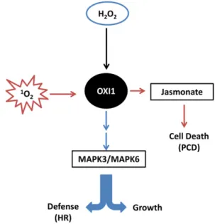
Figure 8. Proposed OXI1-dependent signaling pathways in plants after stimulation with1O2stress produced predominantly during high-light stress (red arrows) and H2O2stress (blue arrows). PCD triggered by
1 O2 is possibly regulated downstream of OXI1 by jasmonic acid, whereas during the hypersensitivity reaction (HR) triggered by H2O2,cell death is regulated by MAPK3 and MAPK6 downstream of OXI1.
Because jasmonate is a cell-death-promoting hor-mone in the presence of1O2 (Ramel et al., 2013b), the decreased levels of jasmonate and JA-Ile in oxi1 mutant plants could participate in the phototolerance of this mutant. This idea is also supported by thefinding that exogenous methyl-jasmonate cancelled the tolerance to cell death in the oxi1 and ch1*oxi1 mutants relative to the wild type and ch1, respectively, suggesting that jasm-onate acts downstream of OXI1. Moreover, the tran-scriptome comparison between oxi1 and the wild type indicated an up-regulation of genes coding for JAZ proteins in the oxi1 mutant, which are negative regu-lator of jasmonate-induced transcriptomic modifica-tions (Chini et al., 2007). This suggests that the oxi1 mutation could also modify jasmonate signaling pro-cesses. Interestingly, high-light induction of genes en-coding negative regulators of PCD was also noticeably stimulated in the oxi1 mutants compared with the wild type (Fig. 6D). Thus, effects on jasmonate levels, on the abundance of negative regulators of jasmonate signal-ing, and on the expression levels of defense genes against PCD could contribute to the regulation of cell death by OXI1. Figure 8 summarizes the role of OXI1 as a central hub for ROS signaling where the H2O2and1O2
signaling pathways intersect.
CONCLUSION
Data on1O2-induced PCD in wild-type Arabidopsis or in other plant species are scarce; most information on the1O2intracellular signaling in vascular plants is derived from studies on the 1O2-overproducing
Arabidopsis mutantsflu and ch1. Compared with those mutants, the responses to high-light stress are possibly different in the wild type since several ROS can be produced simultaneously in the chloroplasts and sev-eral superimposing signaling pathways can be ex-pected. Nevertheless, using a biochemical approach based on1O2-specific lipid oxidation markers,
Tri-antaphylidès et al. (2008) found that photodamage to Arabidopsis leaves is always associated with 1O2
-induced lipid peroxidation, indicating a central role for this high-energy form of oxygen in the execution of high-light-induced cell death in plants. Moreover, a good correlation was found between transcriptomes of numerous stresses, whether from light or dark treat-ments, and the transcriptome of theflu mutant (Mor et al., 2014), indicating that the role of 1O2 in plant
stress regulation and response could be ubiquitous and not necessary restricted to high-light stress. Here, based on a previous transcriptomic analysis of the ch1 mutant exposed to different light conditions, we have identified a kinase that controls high-light-induced cell death in wild-type Arabidopsis. This regulator is part of a new signaling pathway, distinct from the H2O2-induced
pathway and the EX-dependent1O2signaling pathway, with a possible link with jasmonate signaling. Further studies will have to clarify this link and to characterize the molecular targets of the OXI1 kinase during1O2stress.
MATERIALS AND METHODS
Plant Types, Growth Conditions, and Stress Treatments
Arabidopsis (Arabidopsis thaliana) lines used in this work were the wild-type ecotype Col-0, the1O
2-overproducing ch1 mutant (Ramel et al., 2013a), the oxi1 null mutant (Gabi_355H08) deficient in the OXI1 kinase (Camehl et al., 2011), the ch1*oxi1 double mutant, and the ex1*ex2 double mutant deficient in the EX1 and EX2 proteins (Lee et al., 2007). Plants were grown for 5 to 8 weeks in short-day conditions (8 h/16 h, short-day/night) under a moderate PFD of 120 to 160mmol photons m22s21, controlled temperature (20°C/18°C, day/night), and relative humidity (65%). Photooxidative stress was obtained by subjecting plants of the Col-0 background (wild type, oxi1, and ex1*ex2) to 1500mmol m22s21PFD, 7°C/14°C day/night temperature, and 380 ppm CO2in a growth chamber. Because of their high photosensitivity (Ramel et al., 2013a), plants of the ch1 background (ch1 and ch1*oxi1) were subjected to less severe stress conditions: 1100mmol photons m22s21PFD, 7°C/14°C temperature day/night, respec-tively, and 380 ppm CO2. Photoacclimation in the ch1 background was achieved by exposing plants to a moderate PFD of 450mmol photons m22s21and day/night temperatures of 20°C/18°C for 2 d, as previously described (Ramel et al., 2013a).
Lipid Peroxidation Quantification and Imaging
Lipids were extracted from; 0.5 g of leaves frozen in liquid nitrogen. The leaves were ground in an equivolume methanol/chloroform solution contain-ing 5 mMtriphenyl phosphine, 1 mM2,6-tert-butyl-p-cresol (5 mL g21fresh weight), and citric acid (2.5 mL g21fresh weight), using an Ultra-Turrax blender. Internal standard 15-HEDE was added to a final concentration 100 nmol g21fresh weight and mixed properly. After centrifugation at 700 rpm and 4°C for 5 min, the lower organic phase was carefully taken out with the help of a glass syringe into a 15-mL glass tube. The syringe was rinsed with; 2.5 mL of chloroform and transferred back into the tube. The process was repeated, and the lower layer was again collected and pooled to thefirst collection. The sol-vent was evaporated under N2gas at 40°C. The residues were recovered in 1.25 mL of absolute ethanol and 1.25 mL of 3.5NNaOH and hydrolyzed at 80°C for 30 min. The ethanol was evaporated under N2gas at 40°C for; 10 min. After cooling to room temperature, pH was adjusted to 4 to 5 with 2.1 mL of citric acid. Hydroxy fatty acids were extracted with hexane/ether 50/50 (v/v). The organic phase was analyzed by straight phase HPLC-UV, as previously de-scribed (Montillet et al., 2004). HOTE isomers (9-, 12-, 13-, and 16-HOTE derived from the oxidation of the main fatty acid in Arabidopsis leaves, linolenic acid) were quantified based on the 15-HEDE internal standard.
Lipid peroxidation was also visualized in whole plants by autoluminescence imaging. Stressed plants were dark adapted for 2 h, and the luminescence emitted from the spontaneous decomposition of lipid peroxides was captured by a highly sensitive liquid N2-cooled CCD camera, as previously described (Birtic et al., 2011). The images were treated using Image J software (National Institutes of Health).
PSII Photochemical Activity
The maximum quantum yield of PSII was determined by the chlorophyll fluorescence Fv/Fmratio, measured in dark-adapted attached leaves using a PAM-2000fluorometer (Walz), as previously described (Ramel et al., 2012a). The variablefluorescence Fvwas calculated as the difference between maxi-mumfluorescence Fm(obtained with an 800-ms pulse of intense white light) and the initial levelfluorescence Fo(obtained with a 2-s pulse of far-red light).
Detection of1O2Production
Production of1O
2was measured in attached leaves byfluorescence of the SOSGfluorescent probe (Invitrogen), as described previously (Ramel et al., 2012a). With the help of a 1-mL syringe (without the needle), 100mMSOSG was pressure infiltrated into the leaves through the lower surface. Plants with SOSG-infiltrated leaves were exposed to a PFD of 1400 mmol m22s21at 7°C for 30 min. As a control treatment, plants with SOSG-infiltrated leaves were placed in the dark at room temperature for 30 min. SOSGfluorescence was then measured from leaf discs punched from the SOSG-infiltrated leaves using a fiberoptics-equipped Perkin-Elmer spectrofluorometer (LS 50B) with a 475-nm excitation light. SOSGfluorescence at 524 nm was normalized to fluorescence of chloro-phyll a (at 647 nm) for each leaf disc.
Cell Death Determination
Cell death was measured in detached leaves of stressed plants by Evans blue staining as described by Shi et al. (2007) with slight modifications. Fourth to sixth rosette leaves were vacuum infiltrated with 0.1% (w/v) Evans blue for 40 min and then maintained under vacuum for 14 to 15 h. Leaves were thor-oughly rinsed with water to remove unbound dye. Dye bound to dead cells was solubilized in 50% (v/v) methanol/H2O containing 1% SDS for 14 to 15 h. Cell death was then estimated spectroscopically by measuring the absorbance ratio A600/A680of the resulting dye solution.
RNA Isolation and qRT-PCR
Total RNA was isolated from 100 mg of leaves using the Nucleospin RNA plant kit (Macherey-Nagel). The concentration was measured on a Nano-Drop2000 (Thermo Scientific). First-strand cDNA was synthesized from 1 mg of total RNA using the PrimeScript reverse transcriptase kit (Takara). qRT-PCR was performed on a Lightcycler 480 real-time PCR system (Roche). Three mi-croliters of a reaction mixture comprising SYBR Green I Master (Roche), 10mM forward and reverse primers, and water was added to 2mL of 10-fold diluted cDNA sample in a 384-well plate. The PCR program used was as follows: 95°C for 10 min, then 45 cycles of 95°C for 15 s, 58°C for 15 s, and 72°C for 15 s. At least three biological replicates were performed for each gene tested. Primers for genes examined (Supplemental Table S4) were designed using Primer3Plus software (http://www.bioinformatics.nl/cgi-bin/primer3plus/primer3plus.cgi).
Microarray Transcriptomic Analysis
Transcript analysis was performed on a CATMAv6.2 (Complete Arabidopsis Transcriptome Microarray) system at the POPS Transcriptomic Platform of the Institute of Plant Sciences Paris-Saclay, France. A single high-density CAT-MAv6.2 microarray slide contains 12 chambers, each containing 219,684 primers representing all the Arabidopsis genes: 30,834 probes corresponding to CDS TAIRv8 annotation (including 476 probes of mitochondrial and chloroplast genes) + 1289 probes corresponding to EUGENE software predictions. More-over, it included 5352 probes corresponding to repeat elements, 658 probes for miRNA/MIR, 342 probes for other RNAs (rRNA, tRNA, snRNA, soRNA), and, finally, 36 controls. Each long primer is triplicate in each chamber for robust analysis and in both strands. The transcriptome compared RNA from wild-type and oxi1 plants under control growth conditions or after high-light and low-temperature stress (1500mmol photon m22s21and 7°C/14°C day/night). RNA was extracted using the Nucleospin RNA plant kit (Macherey-Nagel). Synthesis of cDNA, amplification, labeling, hybridization, and scanning of slides were performed as described elsewhere (Lurin et al., 2004). Three different com-parisons were performed: oxi1-control versus wild type-control, oxi1 stress versus wild-type stress, and oxi1 stress versus oxi1 control. For each compari-son, three biological replicates (RNA from three individual plants for each treatment) were performed. Two technical replicates were performed for each comparison, with a reversal offluorochrome for each pool of RNA (one dye was swapped per comparison). The hybridization and washing were performed according to NimbleGen Arrays User’s Guide v5.1 instructions. Two-micron scanning was performed with an InnoScan900 scanner (Innopsys), and raw data were extracted using Mapix software (Innopsys).
Experiments were designed with the statistics group of the IPS2. For each array, the raw data comprised the logarithm of median feature pixel intensity at wavelengths 635 nm (red) and 532 nm (green). For each array, a global intensity-dependent normalization using the loess procedure (Yang et al., 2002) was performed to correct the dye bias. The differential analysis is based on the log ratios averaging over the duplicate probes and over the technical replicates. Hence, the number of available data for each gene equals the number of bio-logical replicates and is used to calculate the moderated t test (Smyth, 2004). Under the null hypothesis, no evidence that the specific variances vary between probes is highlighted by Limma, and, consequently, the moderated t statistic is assumed to follow a standard normal distribution. To control the false dis-covery rate, adjusted P values found using the optimized FDR approach of Storey and Tibshirani (2003) were calculated. We considered as being differ-entially expressed the probes with an adjusted P value# 0.05. Analysis was done with the R software. The function SqueezeVar of the library limma has been used to smooth the specific variances by computing empirical Bayes posterior means. The library kerfdr has been used to calculate the adjusted P values.
Functional classification and analysis of the differentially expressed genes were performed using the DAVID functional classification tool (Huang et al.,
2009a, 2009b). Enrichment clusters/terms with EASE score (P value), 0.05 were considered as significantly different.
Jasmonate Treatment
Wild-type and oxi1 mutant plants were subjected to high-light stress (1500 mmol m-2s21PFD and 7°C/14°C day/night temperature) and simultaneously sprayed twice a day with a solution of 1 mMmethyl-jasmonate in 25 mM phosphate buffer and 0.1% Tween 20 (; 400 mL per plant), as previously de-scribed (Ramel et al., 2013a). Similar treatment was performed on ch1 and ch1*oxi1 plants at PFD of 1100mmol m22s21. After; 48 h of stress, the effect of jasmonate on oxi1 and ch1*oxi1 plants was estimated relative to wild-type and ch1 plants, respectively, by autoluminescence imaging and HPLC quantifica-tion of HOTE.
Jasmonate Quantification
Jasmonic acid, its active form JA-Ile, and its precursor OPDA were quantified by UPLC-MS as described by Stingl et al. (2013), with some modifications. Frozen leaves from high-light-stressed wild-type and oxi1 plants were ground under liquid N2, and 100 mg of powder was used for extraction in a 1.5-mL screw cap tube. Extraction solution, ethylacetate/formic acid (99/1), internal standard jasmonyl nor-Val, and a bead were added to the ground leaf tissue and agitated for 3 min at 20 Hz. After centrifugation, the supernatant was evaporated under N2gas. The residue was reconstituted in 100mL of acetoni-trile/ water (1/1). Aliquots were then analyzed by UPLC-MS on a C8 Kinetex column (Phenomenex) of dimension 150 mm3 2.1 mm 3 1.7 mm, maintained at 45°C. Elution was achieved by gradually increasing gradient of solvent B (90% methanol/10% isopropanol) from 37% to 97% relative to solvent A (90% water/ 10% methanol) at a rate of 0.3 mL min21.
Accession Numbers
Sequence data from this article can be found in The Arabidopsis Information Resource or GenBank/EMBL database under the following accession numbers (in parentheses): OXI1 (At3g25250), EX1 (At4g33630), EX2 (At1g27510), and CH1 (At1g44446).
Microarray data from this article were deposited at Gene Expression Om-nibus (http://www.ncbi.nlm.nih.gov/geo/, accession no. GSE61478) and at CATdb (http://urgv.evry.inra.fr/CATdb/; Project CEA13-01_oxi1) according to the“Minimum Information About a Microarray Experiment” standards.
Supplemental Data
The following supplemental materials are available.
Supplemental Figure S1.Effect of visible light on thefluorescence of a SOSG solution.
Supplemental Figure S2.OXI1-dependent pathway in plants during path-ogen attack.
Supplemental Table S1.Functional classification list of induced and re-pressed genes in the comparison oxi1-control versus wild type-control. Supplemental Table S2.Functional classification list of induced and
re-pressed genes in the comparison oxi1-stress versus oxi1-control. Supplemental Table S3.Functional classification list of induced and
re-pressed genes in the comparison oxi1-stress versus wild type-stress. Supplemental Table S4.Primer sequences used in qRT-PCR transcript
profiling.
ACKNOWLEDGMENTS
We thank Christophe Laloi (Aix Marseille University) for providing ex1*ex2 double mutant seeds and for critical reading of the article. The oxi1 single mu-tant was a kind gift from Ana Victoria Garcia and Heribert Hirt (URGV, INRA Evry). We would like to thank Martin Mueller (Wuerzburg University) for
providing standards for jasmonate quantification, and Philippe Ortet (CEA) for his help in analyzing the transcriptomic data. We also acknowledge the support of the Phytotec platform and the HelioBiotec platform funded by the European Union (FEDER), the Région Provence Alpes Côte d’Azur, the French Ministry of Research, and CEA.
Received October 5, 2015; accepted January 7, 2016; published January 8, 2016.
LITERATURE CITED
Abuqamar S, Chai MF, Luo H, Song F, Mengiste T(2008) Tomato protein kinase 1b mediates signaling of plant responses to necrotrophic fungi and insect herbivory. Plant Cell 20: 1964–1983
Alonso-Peral MM, Li J, Li Y, Allen RS, Schnippenkoetter W, Ohms S, White RG, Millar AA(2010) The microRNA159-regulated GAMYB-like genes inhibit growth and promote programmed cell death in Arabi-dopsis. Plant Physiol 154: 757–771
Anthony RG, Henriques R, Helfer A, Mészáros T, Rios G, Testerink C, Munnik T, Deák M, Koncz C, Bögre L(2004) A protein kinase target of a PDK1 signalling pathway is involved in root hair growth in Arabi-dopsis. EMBO J 23: 572–581
Anthony RG, Khan S, Costa J, Pais MS, Bögre L(2006) The Arabidopsis protein kinase PTI1-2 is activated by convergent phosphatidic acid and oxidative stress signaling pathways downstream of PDK1 and OXI1. J Biol Chem 281: 37536–37546
Apel K, Hirt H(2004) Reactive oxygen species: metabolism, oxidative stress, and signal transduction. Annu Rev Plant Biol 55: 373–399 Armbruster U, Hertle A, Makarenko E, Zühlke J, Pribil M, Dietzmann A,
Schliebner I, Aseeva E, Fenino E, Scharfenberg M, et al(2009) Chlo-roplast proteins without cleavable transit peptides: rare exceptions or a major constituent of the chloroplast proteome? Mol Plant 2: 1325–1335 Asada K(2006) Production and scavenging of reactive oxygen species in
chloroplasts and their functions. Plant Physiol 141: 391–396
Birtic S, Ksas B, Genty B, Mueller MJ, Triantaphylidès C, Havaux M (2011) Using spontaneous photon emission to image lipid oxidation patterns in plant tissues. Plant J 67: 1103–1115
Bisby RH, Morgan CG, Hamblett I, Gorman AA(1999) Quenching of singlet oxygen by Trolox C, ascorbate, and amino acids: effects of pH and temperature. J Phys Chem A 103: 7454–7459
Bögre L, Okrész L, Henriques R, Anthony RG(2003) Growth signalling pathways in Arabidopsis and the AGC protein kinases. Trends Plant Sci 8:424–431
Camehl I, Drzewiecki C, Vadassery J, Shahollari B, Sherameti I, Forzani C, Munnik T, Hirt H, Oelmüller R(2011) The OXI1 kinase pathway mediates Piriformospora indica-induced growth promotion in Arabi-dopsis. PLoS Pathog 7: e1002051
Colebrook EH, Thomas SG, Phillips AL, Hedden P(2014) The role of gibberellin signalling in plant responses to abiotic stress. J Exp Biol 217: 67–75 Cominelli E, Gusmaroli G, Conti L, Allegra D, Petroni K, Tonelli C(2000)
Role of Arabidopsis MYB transcription factors in osmotic stress. In JH Cherry, RD Locy, A Rychter, eds, Plant Tolerance to Abiotic Stresses: Role of Genetic Engineering. Kluwer, Dordrecht, The Netherlands, pp 181–194
Chini A, Fonseca S, Fernández G, Adie B, Chico JM, Lorenzo O, García-Casado G, López-Vidriero I, Lozano FM, Ponce MR, et al(2007) The JAZ family of repressors is the missing link in jasmonate signalling. Nature 448: 666–671
Dall’Osto L, Cazzaniga S, Havaux M, Bassi R (2010) Enhanced photo-protection by protein-bound vs free xanthophyll pools: a comparative analysis of chlorophyll b and xanthophyll biosynthesis mutants. Mol Plant 3: 576–593
Danon A, Miersch O, Felix G, Camp RG, Apel K(2005) Concurrent acti-vation of cell death-regulating signaling pathways by singlet oxygen in Arabidopsis thaliana. Plant J 41: 68–80
Danon A, Rotari VI, Gordon A, Mailhac N, Gallois P(2004) Ultraviolet-C overexposure induces programmed cell death in Arabidopsis, which is mediated by caspase-like activities and which can be suppressed by caspase inhibitors, p35 and Defender against Apoptotic Death. J Biol Chem 279: 779–787
Dombrecht B, Xue GP, Sprague SJ, Kirkegaard JA, Ross JJ, Reid JB, Fitt GP, Sewelam N, Schenk PM, Manners JM, et al(2007) MYC2 differ-entially modulates diverse jasmonate-dependent functions in Arabi-dopsis. Plant Cell 19: 2225–2245
Douce R, Joyard J(1980) Plant galactolipids. In PK Stumpf, ed, The Bio-chemistry of Plants, Vol 4. Academic Press, New York, pp 321–362 Farmer EE, Davoine C(2007) Reactive electrophile species. Curr Opin Plant
Biol 10: 380–386
Farmer EE, Mueller MJ(2013) ROS-mediated lipid peroxidation and RES-activated signaling. Annu Rev Plant Biol 64: 429–450
Fischer BB, Ledford HK, Wakao S, Huang SG, Casero D, Pellegrini M, Merchant SS, Koller A, Eggen RIL, Niyogi KK (2012) SINGLET OXYGEN RESISTANT 1 links reactive electrophile signaling to singlet oxygen acclimation in Chlamydomonas reinhardtii. Proc Natl Acad Sci USA 109: E1302–E1311
Flors C, Fryer MJ, Waring J, Reeder B, Bechtold U, Mullineaux PM, Nonell S, Wilson MT, Baker NR(2006) Imaging the production of singlet oxygen in vivo using a newfluorescent sensor, Singlet Oxygen Sensor Green. J Exp Bot 57: 1725–1734
Forzani C, Carreri A, de la Fuente van Bentem S, Lecourieux D, Lecourieux F, Hirt H(2011) The Arabidopsis protein kinase Pto-interacting 1-4 is a common target of the oxidative signal-inducible 1 and mitogen-activated protein kinases. FEBS J 278: 1126–1136
Fu D, Uauy C, Distelfeld A, Blechl A, Epstein L, Chen X, Sela H, Fahima T, Dubcovsky J (2009) A kinase-START gene confers temperature-dependent resistance to wheat stripe rust. Science 323: 1357–1360 Gadjev I, Stone JM, Gechev TS(2008) Programmed cell death in plants:
new insights into redox regulation and the role of hydrogen peroxide. Int Rev Cell Mol Biol 270: 87–144
Garcia AV, Al-Yousif M, Hirt H (2012) Role of AGC kinases in plant growth and stress responses. Cell Mol Life Sci 69: 3259–3267
González-Pérez S, Gutiérrez J, García-García F, Osuna D, Dopazo J, Lorenzo Ó, Revuelta JL, Arellano JB(2011) Early transcriptional de-fense responses in Arabidopsis cell suspension culture under high-light conditions. Plant Physiol 156: 1439–1456
Gutiérrez J, González-Pérez S, García-García F, Daly CT, Lorenzo O, Revuelta JL, McCabe PF, Arellano JB(2014) Programmed cell death activated by Rose Bengal in Arabidopsis thaliana cell suspension cul-tures requires functional chloroplasts. J Exp Bot 65: 3081–3095 Havaux M, Dall’Osto L, Bassi R (2007) Zeaxanthin has enhanced
antioxi-dant capacity with respect to all other xanthophylls in Arabidopsis leaves and functions independent of binding to PSII antennae. Plant Physiol 145: 1506–1520
Havaux M, Triantaphylidès C, Genty B(2006) Autoluminescence imaging: a non-invasive tool for mapping oxidative stress. Trends Plant Sci 11: 480–484
Hilson P, Allemeersch J, Altmann T, Aubourg S, Avon A, Beynon J, Bhalerao RP, Bitton F, Caboche M, Cannoot B, et al(2004) Versatile gene-specific sequence tags for Arabidopsis functional genomics: transcript profiling and reverse genetics applications. Genome Res 14: 2176–2189 Hirashima M, Tanaka R, Tanaka A(2009) Light-independent cell death
induced by accumulation of pheophorbide a in Arabidopsis thaliana. Plant Cell Physiol 50: 719–729
Hirt H, Garcia AV, Oelmüller R(2011) AGC kinases in plant development and defense. Plant Signal Behav 6: 1030–1033
Huang W, Sherman BT, Lempicki RA(2009a) Systematic and integrative analysis of large gene lists using DAVID bioinformatics resources. Nat Protoc 4: 44–57
Huang W, Sherman BT, Lempicki RA(2009b) Bioinformatics enrichment tools: paths toward the comprehensive functional analysis of large gene lists. Nucleic Acids Res 37: 1–13
Khan MIR, Fatma M, Per TS, Anjum NA, Khan NA(2015) Salicylic acid-induced abiotic stress tolerance and underlying mechanisms in plants. Front Plant Sci 6: 462
Kim C, Apel K(2013) Singlet oxygen-mediated signaling in plants: moving fromflu to wild type reveals an increasing complexity. Photosynth Res 116:455–464
Krieger-Liszkay A(2005) Singlet oxygen production in photosynthesis. J Exp Bot 56: 337–346
Laloi C, Havaux M(2015) Key players of singlet oxygen-induced cell death in plants. Front Plant Sci 6: 39
Lee KP, Kim C, Landgraf F, Apel K(2007) EXECUTER1- and EXECUTER2-dependent transfer of stress-related signals from the plastid to the nu-cleus of Arabidopsis thaliana. Proc Natl Acad Sci USA 104: 10270–10275 Li H, Melø TB, Arellano JB, Razi Naqvi K(2012) Temporal profile of the singlet oxygen emission endogenously produced by photosystem II re-action centre in an aqueous buffer. Photosynth Res 112: 75–79