HAL Id: hal-03011557
https://hal.sorbonne-universite.fr/hal-03011557
Submitted on 18 Nov 2020
HAL is a multi-disciplinary open access
archive for the deposit and dissemination of
sci-entific research documents, whether they are
pub-lished or not. The documents may come from
teaching and research institutions in France or
abroad, or from public or private research centers.
L’archive ouverte pluridisciplinaire HAL, est
destinée au dépôt et à la diffusion de documents
scientifiques de niveau recherche, publiés ou non,
émanant des établissements d’enseignement et de
recherche français ou étrangers, des laboratoires
publics ou privés.
Extracellular Control of Radial Glia Proliferation and
Scaffolding During Cortical Development and Pathology
Julien Ferent, Donia Zaidi, Fiona Francis
To cite this version:
Julien Ferent, Donia Zaidi, Fiona Francis. Extracellular Control of Radial Glia Proliferation and
Scaffolding During Cortical Development and Pathology. Frontiers in Cell and Developmental Biology,
Frontiers media, 2020, 8, pp.578341. �10.3389/fcell.2020.578341�. �hal-03011557�
fcell-08-578341 October 12, 2020 Time: 15:46 # 1 REVIEW published: 16 October 2020 doi: 10.3389/fcell.2020.578341 Edited by: Victor Borrell, Instituto de Neurociencias, CSIC − UMH, Spain Reviewed by: Simone Diestel, University of Bonn, Germany Samuel Pleasure, University of California, San Francisco, United States Hidenori Tabata, Aichi Human Service Center, Japan *Correspondence: Fiona Francis Fiona.Francis@inserm.fr
Specialty section: This article was submitted to Cell Adhesion and Migration, a section of the journal Frontiers in Cell and Developmental Biology Received: 30 June 2020 Accepted: 08 September 2020 Published: 16 October 2020 Citation: Ferent J, Zaidi D and Francis F (2020) Extracellular Control of Radial Glia Proliferation and Scaffolding During Cortical Development and Pathology. Front. Cell Dev. Biol. 8:578341. doi: 10.3389/fcell.2020.578341
Extracellular Control of Radial Glia
Proliferation and Scaffolding During
Cortical Development and Pathology
Julien Ferent
1,2,3, Donia Zaidi
1,2,3and Fiona Francis
1,2,3*
1Inserm, U 1270, Paris, France,2Sorbonne University, UMR-S 1270, IFM, Paris, France,3Institut du Fer á Moulin, Paris,
France
During the development of the cortex, newly generated neurons migrate long-distances
in the expanding tissue to reach their final positions. Pyramidal neurons are produced
from dorsal progenitors, e.g., radial glia (RGs) in the ventricular zone, and then migrate
along RG processes basally toward the cortex. These neurons are hence dependent
upon RG extensions to support their migration from apical to basal regions. Several
studies have investigated how intracellular determinants are required for RG polarity
and subsequent formation and maintenance of their processes. Fewer studies have
identified the influence of the extracellular environment on this architecture. This review
will focus on extracellular factors which influence RG morphology and pyramidal
neuronal migration during normal development and their perturbations in pathology.
During cortical development, RGs are present in different strategic positions: apical RGs
(aRGs) have their cell bodies located in the ventricular zone with an apical process
contacting the ventricle, while they also have a basal process extending radially to reach
the pial surface of the cortex. This particular conformation allows aRGs to be exposed
to long range and short range signaling cues, whereas basal RGs (bRGs, also known
as outer RGs, oRGs) have their cell bodies located throughout the cortical wall, limiting
their access to ventricular factors. Long range signals impacting aRGs include secreted
molecules present in the embryonic cerebrospinal fluid (e.g., Neuregulin, EGF, FGF, Wnt,
BMP). Secreted molecules also contribute to the extracellular matrix (fibronectin, laminin,
reelin). Classical short range factors include cell to cell signaling, adhesion molecules
and mechano-transduction mechanisms (e.g., TAG1, Notch, cadherins, mechanical
tension). Changes in one or several of these components influencing the RG extracellular
environment can disrupt the development or maintenance of RG architecture on which
neuronal migration relies, leading to a range of cortical malformations. First, we will detail
the known long range signaling cues impacting RG. Then, we will review how short
range cell contacts are also important to instruct the RG framework. Understanding
how RG processes are structured by their environment to maintain and support radial
migration is a critical part of the investigation of neurodevelopmental disorders.
Keywords: apical radial glia, cortical development, neuronal migration, scaffold, cell-cell interaction, cell signaling, extracellular matrix
fcell-08-578341 October 12, 2020 Time: 15:46 # 2
Ferent et al. Extracellular Cues and RG Scaffolding
INTRODUCTION
The cerebral cortex is an intricate brain structure responsible
for many precise functions such as thinking, decision making
and long term memory, and is required for the final processing
of sensory inputs and motor control. These functions rely
on the way the neuronal network is precisely organized. The
structure of the cortex is composed of different layers of
neuronal subtypes (
Taverna et al., 2014
;
De Juan Romero
and Borrell, 2015
). In the mouse, these layers are established
during embryonic development in an inside-out manner via the
successive migration of young neurons generated directly or
indirectly from apical radial glial cells (aRGs) in the ventricular
zone (VZ) to their final location in distinct superficial regions
(
Rakic, 1972
;
Kriegstein and Gotz, 2003
). aRGs have a particular
morphology as they grow processes that extend from the apical
to the basal side of the cortex. In both rodent and primate,
aRGs generate further basal intermediate neurogenic progenitors
(IPs) residing in the subventricular zone (SVZ). In gyrencephalic
species such as humans and other primates, neurons can also be
generated from basal radial glia (bRGs), also called outer radial
glia (oRG), which are distributed in an outer SVZ (
Penisson et al.,
2019
;
Matsumoto et al., 2020
). bRGs can extend processes to the
apical, the basal or both surfaces of the cortex (
Betizeau et al.,
2013
). In all situations their structure provides a linear support
for neuronal migration. Therefore, RGs are not only the source
of neurons during embryonic cortical development but also the
scaffold necessary for their proper distribution throughout the
expanding cortex. The formation and maintenance of the RG
scaffold is essential for the correct positioning of neurons and
thus, the organization of the neuronal network.
Several cellular processes are important to consider for
proper RG morphology. As they are dividing and self-renewing
progenitors, RGs have been widely studied in the context of
the mechanisms underlying their proliferative features (
Taverna
et al., 2014
;
Uzquiano et al., 2018
). This will have an impact on
the density of fibers available for supporting migration. RGs (e.g.,
expressing factors such as Pax6, Sox2, Hes5) can self-renew via
symmetric divisions but can also carry out asymmetric divisions
giving rise to different progeny including Tbr2 + IPs (Figure 1;
Taverna et al., 2014
;
De Juan Romero and Borrell, 2015
). RGs
are also able to directly produce neurons. In particular, cell
intrinsic mechanisms acting on mitotic spindle orientation or
nucleokinesis via cytoskeletal or polarity proteins are tightly
linked to daughter cell production and fate. At the structural
level, how the radial processes critical to RG function are
created, modulated or maintained relies on additional molecular
mechanisms, which is the topic of this review.
Since aRGs are structured in a very defined way, with their
soma and apical processes at the border of the ventricle, they
are exposed to many secreted factors from the embryonic
cerebrospinal fluid (eCSF). In particular, the primary cilium
extends inside the ventricle and this is a crucial signaling center
for the activation of numerous molecular cascades (
Sarkisian
and Guadiana, 2015
). At the level of their cell bodies, aRGs
and bRGs are both exposed to cell−cell and cell−environment
interactions. They interact with each other as well as with
additional cell types such as IPs or neurons. Importantly they
interact with the surrounding extracellular matrix (ECM). For
example, human bRGs have been shown to produce specific
proteins which interact with the ECM in their basal position
(
Pollen et al., 2015
). Finally, their basal processes are also
exposed to external signals throughout the intermediate zone
(IZ), cortical plate (CP), marginal zone (MZ), and at the pial
surface (Figure 2). First, we will review the different secreted
molecules involved in the establishment or maintenance of RG
morphology from the eCSF or in the ECM. Then, we will
describe how short range interactions between cells are essential
for these processes. Finally, we will detail the impact of relevant
molecular players on the origin and evolution of several human
neurodevelopmental diseases.
ROLE OF SECRETED PROTEINS
DERIVED FROM THE CSF IN THE
FORMATION AND MAINTENANCE OF
THE RG SCAFFOLD
Secreted Factors From the Embryonic
Cerebrospinal Fluid (eCSF)
The cortex develops primarily from the neuroepithelium during
embryonic development. Between E8.5 and E9.5 in mice, the
neural tube closes, forming the ventricular cavity in which the
amniotic fluid is sequestered and forms the basis for the eCSF
(
Lowery and Sive, 2009
). Later during mouse brain development,
the choroid plexus arises and secretes many factors, modifying
the composition of the ventricular fluid (
Chau et al., 2015
).
The deepest apical region of the developing brain is composed
of neuroepithelial-derived aRG progenitors from E10.5. These
aRGs are exposed to a variety of secreted factors from the
ventricle during development. Proteomics analyses of the CSF
indicates that the precise composition of secreted molecules
varies during development. For instance, the concentration of
Bone Morphogenic Proteins (BMPs) is higher in the amniotic
fluid than in the eCSF, Sonic Hedgehog (Shh) concentration
is higher in the eCSF at the beginning of aRG development
(E10.5) and decreases thereafter, whereas the concentration of
retinoic acid (RA) is higher at later stages (E14.5) (
Chau et al.,
2015
). These variations in composition are required to induce the
production of RGs (Sox2+) at the right time during the formation
of the cortex (
Chau et al., 2015
). These data also suggest that
certain secreted proteins or combinations of proteins in the eCSF
during murine corticogenesis are required for evolving aspects of
RG production and maintenance.
The composition of secreted factors in the eCSF not only
changes with developmental stages but is also specific to different
ventricles. Indeed, the different choroid plexus tissues present
in each ventricle develop in a sequential manner (
Lehtinen
et al., 2011
). Firstly, the choroid plexus from the fourth ventricle
appears (E11 in the mouse), then the choroid plexus develops
in the lateral ventricles (E12) and lastly, it develops in the third
ventricle by E14. Each type of choroid plexus will express a
different panel of secreted factors. For example, Shh is mainly
fcell-08-578341 October 12, 2020 Time: 15:46 # 3
Ferent et al. Extracellular Cues and RG Scaffolding
FIGURE 1 | Radial glias function as both the source and the support of newborn neurons in the developing cortex. Apical radial glia (aRG) extend an apical process reaching the ventricular surface, where they expose their primary cilia, as well as a basal process reaching the cortical surface. Basal radial glia (bRG) have their cell bodies located in more basal areas of the cortical wall. Apical and basal processes from these cells (blue) establish the scaffold across the whole cortical wall. RGs undergo cell division, giving birth to a daughter cell which can be either another RG (apical or basal – symmetric division) or a basal progenitor (asymmetric division, intermediate progenitors are represented in orange). These cells give rise to migrating neuroblasts (green) which move along RG basal processes to reach their final position within the cortical layers. First deep layer neurons are generated, then upper layer neurons are born.
produced in the fourth ventricle by the choroid plexus close
to the hindbrain (
Huang et al., 2010
), whereas many other
proteins are found only in the lateral ventricles (
Zappaterra
et al., 2007
). More recently, proteomics data were integrated
with RNA sequencing datasets, comparing telencephalic and
hindbrain choroid plexuses (
Lun et al., 2015
). This spatial
heterogeneity of their secretomes argue in favor of a precise and
specific regulation of different brain areas. Overall, the eCSF plays
multiple important roles in the formation of the nervous system
(for review,
Fame and Lehtinen, 2020
). In this part of our review,
we will describe the functional role of the main secreted factors
present in the eCSF for the maintenance of RGs and therefore the
formation of the RG scaffold.
Growth Factors
As mentioned in the previous section, several cytokines are found
in the eCSF. Different types of molecules can be found within this
family such as growth factors like Transforming Growth Factors
(TGF, developed later on in this review). But not all of these
cytokines have a direct effect on radial scaffolding. For example,
chemokines are best known for their action on neurons (
Zhu and
Murakami, 2012
). On the other hand growth factors are diffusible
cytokines widely known to activate RG proliferation and/or
sustain cell survival. Therefore, we first provide a non-exhaustive
list of eCSF-derived growth factors (Figures 3, 4 and Table 1)
necessary for cortical development and in particular for the
integrity of the RG scaffold.
Multiple Fibroblast growth factor (FGF) ligands are
expressed in the developing telencephalon. At early stages (E10–
E12), FGFs 8, 17, and 18 are expressed in the frontal midline
area where they act as morphogens (see section “Morphogens”
below). In the ventral telencephalon, FGF15 is expressed (
Rash
and Grove, 2006
;
Cholfin and Rubenstein, 2007
;
Hebert and
Fishell, 2008
), whereas in dorsal regions, FGFs 2, 9, and 10 are
expressed (
Vaccarino et al., 1999
;
Raballo et al., 2000
;
Sahara
and O’Leary, 2009
). Here, we focus on FGF2, which increases
the total number of neurons in the mouse cerebral cortex and
promotes self-renewal of cortical progenitor cells (
Vaccarino
et al., 1999
;
Raballo et al., 2000
).
Fibroblast growth factor 2 is one of the most important
growth factors for the production and maintenance of RGs
during cortical development. Initially, FGF2 proteins were
fcell-08-578341 October 12, 2020 Time: 15:46 # 4
Ferent et al. Extracellular Cues and RG Scaffolding
FIGURE 2 | Extracellular factors controling the scaffolding of RGs. RGs are exposed to a variety of extracellular cues. These signals can be secreted molecules (blue boxes) or received directly from other cells (green boxes). In apical regions aRGs receive signals from the eCSF as their cell bodies and primary cilia are in contact with the ventricles. They also establish contacts between themselves and with the extracellular matrix (ECM). In basal regions, RG basal processes are exposed to secreted cues from the meninges and from already differentiated neurons. These interactions can occur while neurons are migrating along them. Basal processes also exhibit interactions between themselves.
fcell-08-578341 October 12, 2020 Time: 15:46 # 5
Ferent et al. Extracellular Cues and RG Scaffolding
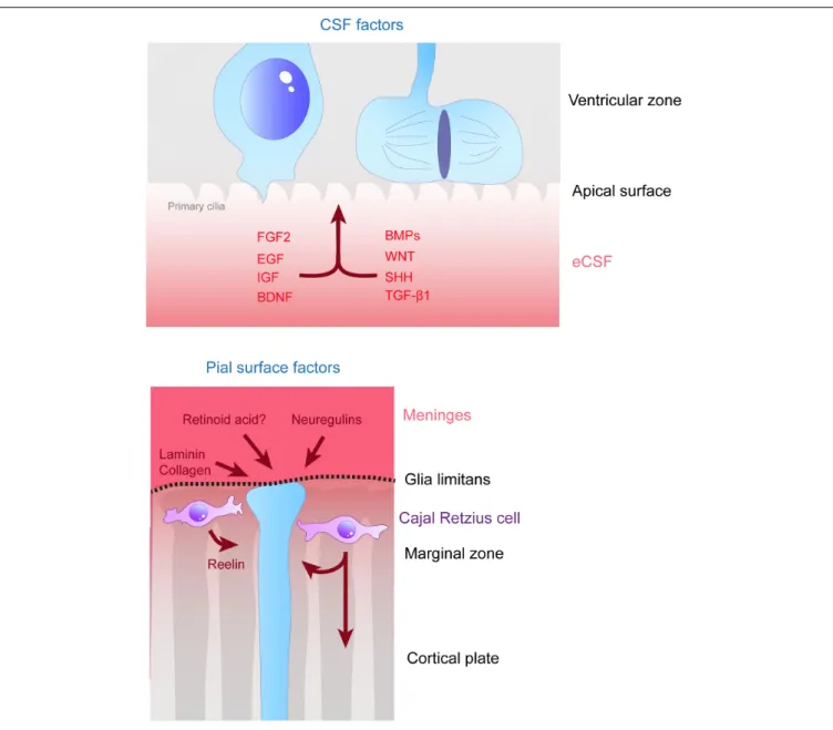
FIGURE 3 | Remote extracellular factors controling the scaffolding of RGs. Some of the extracellular cues controlling RG development are produced and secreted from relatively remote locations. Here are represented the factors present in the CSF (upper schema) which are detailed in this review, namely FGF2, EGF, IGF, BDNF, BMPs, WNT, SHH, and TGF-β1. On the bottom schema, extracellular cues derived from the meninges and acting on the extremities of basal processes are depicted, namely, laminin, collagen, neuregulins and retinoid acid. Cajal Retzius cells (in purple) are migrating cells which in early stages of development tangentially move in the MZ of the developing cortex. These cells are a source of Reelin amongst other molecules which influence RG scaffolding.
found present in the VZ of the murine developing cortex
by immunohistochemistry (
Dono et al., 1998
). The source of
the protein was not clearly defined but later the protein was
detected in chicken eCSF (HH25) by western blot experiments
(
Martin and Groves, 2006
), suggesting that it might be produced
remotely and captured at the ventricular surface. In this study,
authors show that the actual origin for FGF2 production in the
chick embryo is the notochord, the mesonephros, the hepatic
primordia and the brain neuroectoderm. The receptor for FGF2
(FGF2R) is expressed in the mouse VZ (E14.5) as shown by
in situ hybridization (
Dono et al., 1998
). More recently single
cell RNA-seq data shows that the
Fgf2r gene is expressed in
mouse RGs (
Telley et al., 2019
), suggesting that these cells can
receive the FGF2 signal from the eCSF. The cortex of
Fgf 2 mutant
mice is thinner and there is abnormal distribution of neurons
in the cortical wall. Indeed, pulse chase analyses indicate an
increase of neurons generated at E14.5 in the deep layers of the
cortex (
Dono et al., 1998
). This suggests a defect in the ability of
these cells to colonize their final target place in more superficial
layers. Defects in proliferation were also identified in
Fgf2 KO
embryonic cortices in a separate study explaining the decrease in
the size of the cortex (
Raballo et al., 2000
). This is in agreement
with the fact that FGF2 is one of the major factors necessary
for the renewal of RGs
in vitro (
Sun et al., 2011
). The knockout
fcell-08-578341 October 12, 2020 Time: 15:46 # 6
Ferent et al. Extracellular Cues and RG Scaffolding
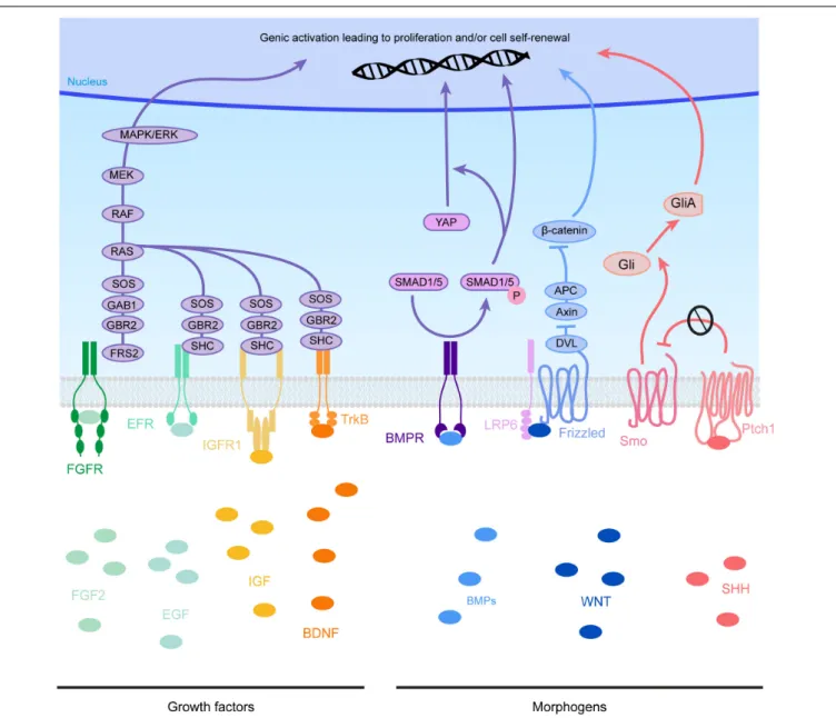
FIGURE 4 | Molecular pathways triggered by eCSF-derived factors. The growth factors found in the eCSF are mainly known to trigger the mitogen-activated protein kinases (MAPK) pathway (also known as the RAS-RAF-MEK-ERK pathway). This molecular signaling pathway is involved in the regulation of several essential cellular processes such as proliferation, differentiation, survival and death. BMP receptors (BMPR) activate the phosphorylation of SMAD1/5, which can activate directly transcription of target genes or act via the translocation of YAP into the nucleus. WNT molecules activate the Frizzled receptors and LRP6 co-receptors which will allow Disheveled (DVL) to inhibit the Axin-APC complex. This complex is a major inhibitor ofβ-catenin. Therefore, upon WNT activation, β-catenin is free to be directed into the nucleus to activate its target genes. Finally SHH binds to its receptor Patched1 (Ptch1), which then releases the 7 transmembrane protein Smoothened (Smo) from its inhibition. Smo activation triggers the cleavage of Gli transcription factors into their active form (GliA). GliA is then enriched in the nucleus to allow transcription of target genes (such as Cyclin D1 or Gli itself).
(KO) of
Fgfr genes in the anterior neural plate using
Foxg1-Cre, inhibits the formation of the telencephalon, leaving just the
midline (
Paek et al., 2009
). When Fgfrs are removed only from
RG, their development is impaired resulting in lower numbers
of
Pax6 and Hes5 + cells (
Kang et al., 2009
). These combined
data show how crucial FGF2 is for the maintenance of RGs and
therefore, the formation of the cortex. Moreover, gain of function
experiments performed by
in utero injection of FGF2 first in
the telencephalic ventricles of rat embryos at E15 (
Vaccarino
et al., 1999
), then in mouse embryos at E11.5 (
Rash et al.,
2013
), induces an increase in proliferation. When FGF2 signaling
is overactivated locally by these manipulations, this induces
the formation of gyri in the mouse cortex (
Rash et al., 2013
).
Although gyrification can be associated with the appearance
of bRGs during evolution (
Penisson et al., 2019
), in this case
FGF2 injections did not appear to increase the proportion of
bRGs in the cortical wall. This suggests that FGF2 modified the
development of the architecture of the cortex via other unknown
mechanisms. At the molecular level, FGF2 triggers the
mitogen-activated protein kinase (MAPK) pathway to induce cell cycle
fcell-08-578341 October 12, 2020 Time: 15:46 # 7
Ferent et al. Extracellular Cues and RG Scaffolding
and proliferation (for review,
Iwata and Hevner, 2009
, Figure 4).
FGF2, as well as Notch signaling, can also induce calcium (Ca2+)
bursting which can support communication along the RG fiber
(
Rash et al., 2016
). Indeed, both along the RG fiber and the
communication with neurons can be mediated by calcium waves,
in a bidirectional manner.
Fibroblast growth factor 2 can be used in culture in
combination with Epidermal growth factor (EGF). Indeed, the
action of EGF on cortical progenitors has been studied for
many years (
Burrows et al., 1997
;
Lillien and Raphael, 2000
).
Recently, FGF2 and EGF were shown to regulate self-renewal
of rat cortical progenitors in organotypic cultures
in vitro
(
Lamus et al., 2020
). These two growth factors can activate
the same molecular pathways to initiate proliferation. The
action of FGF2 and EGF is not a simple synergy since FGF2
can modulate the responsiveness of RGs to EGF (
Lillien and
Raphael, 2000
). RGs are first responsive to FGF2 alone and
later during cortical development, start to be also responsive
to EGF (
Ciccolini and Svendsen, 2001
). Moreover, the effect
of EGF on the proliferation of cortical progenitors is
dose-dependent (
Nelson et al., 2008
). The combined action of EGF
and FGF2 is therefore essential for the development of RGs
and their maintenance during the development of the cortex.
At the beginning of mouse cortical expansion (e.g., E13), the
EGF receptor (EGFR) is present in the VZ and SVZ (
Sun
et al., 2005
). At the cellular level, EGFR has been found to be
localized asymmetrically in dividing RGs, controlling the fate
of daughter cells. The cell inheriting EGFR is the daughter cell
retaining proliferative capacity and glial markers (
Sun et al.,
2005
). This indicates that RGs need to keep their ability to
respond to EGF in order to maintain their progenitor identity.
EGF signaling is therefore required for the maintenance of
the RG scaffold during cortical development. Certain studies
have investigated the mechanisms controlling the expression of
EGFR. First the ganglioside GD3 was identified as an EGFR
partner, responsible for its sustained expression in cortical
progenitors
in vitro (
Wang and Yu, 2013
). More recently, the
expression of miR-129-5p, modulated by choline availability in
the microenvironment, was shown to inhibit the expression of
EGFR, thus impacting RG maintenance and cortical development
(
Trujillo-Gonzalez et al., 2019
). All these data underline the
essential role for EGF in the maintenance of RGs necessary for
cortical development.
The Insulin-like Growth factors (IGF1 and IGF2) are a
group of hormones which are present in the eCSF (
Salehi
et al., 2009
;
Zappaterra and Lehtinen, 2012
;
Bueno et al., 2020
).
IGF2 concentration in rat eCSF increases from E16 to E19
(
Lehtinen et al., 2011
). Gain of function experiments such as
IGF1 overexpression in mouse embryo showed that this hormone
induces a shortening of the cell cycle, acting in particular on
S-phase. This increase in the speed of proliferation is linked
to cortical hyperplasia, which is an increase in global cortical
size via an increase in cell number (
Hodge et al., 2004
;
Popken
et al., 2004
;
Mairet-Coello et al., 2009
). Both
in vivo and in vitro
data show that IGF2 can induce cortical growth by stimulating
RG proliferation (
Lehtinen et al., 2011
). The reverse result is
observed in
Igf2 KO mice which present a neurogenesis decrease
affecting the production of neurons destined for the upper
cortical layers. At the apical membrane level, CSF-derived IGF2
binds to primary cilia of RGs, where IGF receptors are localized.
IGF1 receptor (IGF1R) is the main receptor allowing IGFs to
trigger proliferation (
Zappaterra and Lehtinen, 2012
). Like FGF2
and EGF, IGF can trigger the MAPK pathway but can also activate
a non-canonical pathway via G
βγ signaling, which regulates the
timing of the cell cycle (
Yeh et al., 2013
). All of these data explain
why the
Igf1r conditional knockout (cKO) in neural precursors
leads to microcephaly (
Lehtinen et al., 2011
).
Brain Derived Neurotrophic Factor
(BDNF) has the
particularity of being expressed directly by RGs and also by
Cajal-Retzius cells (
Fukumitsu et al., 1998
). The role of BDNF
on RGs has been investigated by both injection of BDNF itself
directly in ventricles at mouse E13.5 (
Fukumitsu et al., 2006
)
and also by overexpression of
Bdnf in cortical precursors in vivo
via
in utero electroporation (
Bartkowska et al., 2007
). Both
strategies led to an increase in proliferation. BDNF is one of
the ligands which can activate the tropomyosin receptor kinase
B (TrkB) receptor. The loss of function of its gene (Ntrk2) in
the mouse was achieved by different approaches such as by
short hairpin RNAs (shRNAs) or by expression of a
dominant-negative variant of TrkB. Blockade of TrkB signaling elicited
a decrease in RG self-renewal (
Bartkowska et al., 2007
). TrkB
receptors can be phosphorylated, which will activate the MAPK
or phosphoinositide 3-kinase (PI3K) pathways (
Numakawa et al.,
2018
). Both pathways are implicated in different functions of
RG behavior during cortical development. PI3K is important
for RG survival whereas MAPK activation is required for
the production of neurons (
Barnabe-Heider and Miller, 2003
).
Therefore, the pathway linking BDNF, TrkB and PI3K is essential
for the maintenance of the RG scaffold, indeed activation
of the BDNF-TrkB-MAPK axis can lead to premature RG
differentiation into neurons via the activation of BMP7 (
Ortega
and Alcantara, 2010
and see below). BDNF can also activate
Anoctamin 1 (ANO1), a Ca2+-activated chloride channel which
is expressed in RGs (
Hong et al., 2019
). The growth of RG
basal processes is dependent on the activity of this channel as
its loss of function disrupts the extension of RG protrusions.
ANO1 overexpression inversely increases this process (
Hong
et al., 2019
). The lack of basal process growth in
Ano1-deficient
mice leads to disorganized cortical layers and microcephaly
(
Hong et al., 2019
).
Transforming growth factor
β 1 (TGF-β1) is a cytokine
which is involved at many levels of neuronal development
(
Meyers and Kessler, 2017
). TGF-β1 is present in the VZ of the
developing cortex (
Mecha et al., 2008
) and its receptor, TGFRII,
is highly expressed by RGs (
Stipursky et al., 2014
). Although
its role is mainly associated with the differentiation of RGs into
either neurons or glia (
Stipursky et al., 2012, 2014
), injection of
TGF-β1 directly into the embryonic ventricles at E14 induces
drastic changes in RG scaffold morphology. Basal processes seem
shorter and disorganized. In fact, TGF-
β1 triggers early transition
of RGs into astrocytes which alters their morphology from radial
to multipolar. Interestingly, these effects are similar to the action
of a morphogen, as described in the next paragraph, more than
a growth factor.
fcell-08-578341 October 12, 2020 T ime: 15:46 # 8 Fer ent et al. Extracellular Cues and RG Scaf folding
TABLE 1 | Non-exhaustive list of proteins influencing RG scaffolds during cortical development.
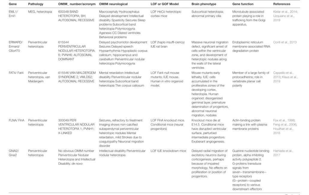
Protein Localization Implication in RG scaffold Phenotype References
Afadin Apical endfeet Apical process arrangement AJ maintenance
Apical process irregularly arranged Loss of AJ markers
Yamamoto et al., 2015
aPKCλ (Atypical protein kinase Cλ)
Apical endfeet RG polarity
Apical process maintenance
Apical process retraction RG detachment
Fumiyasu et al., 2006
APC (adenomatous poli C) RG tips Soma Maintenance and extension of RG processes
Scaffold polarity
Mis-oriented scaffold (basal process not directed at pial surface) Shorter processes
Yokota et al., 2009
Arp2/3 (Actin Related Protein 2/3)
Basal/apical endfeet Soma Nucleus
Formation and maintenance of AJs Shorter RG processes and misoriented Lower speed of basal process formation
Ventricular surface is altered
Wang P.S. et al., 2016
Bone morphogenic proteins/SMADs
eCSF Meninges Hem Control of neurogenesis Premature differentiation Thinner cortex/microcephaly
Najas et al., 2020
Brain Derived Neurotrophic Factor
eCSF RG Cajal-Retzius cells
RG self-renewal Decrease of RG proliferation Bartkowska et al., 2007
Cdc42 (Cell division control protein 42)
Leading process (basal fiber)
Basal process growth Inter−radial fiber interactions
Shorter basal process
Decreased contacts between RG fibers
Yokota et al., 2010
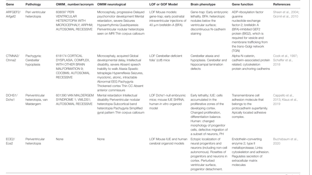
Ece2 (endothelin converting enzyme-2)
RG apical compartment Cortical plate
Apical process maintenance RG morphology
Ventricular surface integrity
Loss of apical process Ventricular surface alteration Loss of radial morphology
Buchsbaum et al., 2020
ECM components and receptors
Pial surface VZ SVZ Apical process integrity Basal process integrity RG morphology
Milev et al., 1996;Li et al., 2008;Loulier et al., 2009;Sittaramane et al., 2009;
Okamoto et al., 2013;Buchsbaum et al., 2020
Epidermal growth factor eCSF Maintenance of RG identity and self-renewal
Burrows et al., 1997;Lillien and Raphael, 2000;Sun et al., 2005
Fibroblast growth factor eCSF Production and maintenance of RG Decrease in cortical size Dono et al., 1998;Raballo et al., 2000
FSTL1 (Follistatin like-1) Pial basement membrane RG basal process orientation Basal endfeet branching
RG basal process not parallel Less endfeet branched
Liu et al., 2015
Glial growth factor Neuronal secretion Basal process elongation Loss of endfeet formation and disrupted morphology
Anton et al., 1997
GSK3 (Glycogen synthase kinase 3)
Leading process (basal fiber)
Basal process growth and orientation Whole scaffold morphology
Shorter basal process Basal process mis-oriented Scaffold morphology altered
Yokota et al., 2010
Insulin-like Growth factors eCSF RG proliferation Neurogenesis decrease Lehtinen et al., 2011
N-cadherin AJs AJ maintenance
Apical process maintenance
RG detachment Apical process retraction Premature differentiation
Rousso et al., 2012;Wong et al., 2012;
Das and Storey, 2014
(Continued) Fr ontiers in Cell and Developmental Biology | www .fr ontiersin.org 8 October 2020 | V olume 8 | Article 578341
fcell-08-578341 October 12, 2020 T ime: 15:46 # 9 Fer ent et al. Extracellular Cues and RG Scaf folding TABLE 1 | Continued
Protein Localization Implication in RG scaffold Phenotype References
Neuregulins RG Maintenance of RG proliferation and radial morphology
Reduced number of RG Schmid et al., 2003;Nakagawa et al., 2019
Notch RG RG identity Promotion of radial
morphology
Increase expression of adhesion proteins
Premature differentiation Overexpression: Radial morphology increased
Adhesion protein expression increased
Li et al., 2008;Yoon et al., 2008
Numb/Numbl Apical endfeet Radial polarity
Apical process maintenance AJ maintenance
Altered ventricular surface Loss of radial polarity Loss of apical process
Rasin et al., 2007
Plekha7 Apical endfeet Apical process maintenance Apical contact integrity
Loss of apical contact Apical process retraction
Tavano et al., 2018
Reelin Cajal-Retzius cells Maintenance of RG morphology RG process branching defects Hartfuss et al., 2003;Schaefer et al., 2008;Chai et al., 2015
Sonic Hedgehog eCSF and interneurons Radial glia proliferation Reduction in RG number Komada et al., 2008;Dave et al., 2011;
Wang L. et al., 2016
TAG-1 (Transient axonal glycoprotein-1)
Basal region Basal process maintenance Basal process loss Basal process retractation
Okamoto et al., 2013
Transforming growth factorβ 1
eCSF Control of RG morphology and processes
ND Stipursky et al., 2014
Wnt eCSF RG self-renewal RG radial morphology Basal process disruption Premature differentiation
Woodhead et al., 2006;Nakagawa et al., 2017
The column phenotype takes into account results of experiments done when the protein is lacking (cKO, KO, pharmacological inhibition etc.) Proteins are presented in alphabetical order.
Fr ontiers in Cell and Developmental Biology | www .fr ontiersin.org 9 October 2020 | V olume 8 | Article 578341
fcell-08-578341 October 12, 2020 Time: 15:46 # 10
Ferent et al. Extracellular Cues and RG Scaffolding
Thus, different growth factors play apparently critical roles
influencing the formation and maintenance of RG scaffolds.
Morphogens
Contrary to growth factors which are known classically to act at
the proliferation level, morphogens are instead also associated
with an action at the differentiation level and to control cell fate
decisions (
Briscoe and Small, 2015
). We review here the known
roles of morphogens in the maintenance of RGs and therefore the
RG scaffold (Figures 3, 4 and Table 1).
Certain members of the TGF family, e.g., the bone
morphogenic proteins
(BMP), have important roles in the
maintenance of RG scaffolding.
In vitro experiments on cultures
of RGs indicated that BMP signaling is involved in the control
of neurogenesis (
Li et al., 1998
;
Mabie et al., 1999
). Bmp7 has
been detected in the meninges, hem and also in the eCSF (
Segklia
et al., 2012
). When Bmp7 is removed from the mouse brain, this
leads to reduced cortical thickness and number of neurons at
E14.5. On the other hand, when Bmp signaling is activated by
expressing a constitutively active form of its receptors (Bmpr1a
or Bmpr1b), over proliferation and defects in global morphology
are observed in the developing cortex (
Panchision et al., 2001
).
In particular, folds can be seen at the brain surface, suggesting
differences in RG scaffolding. More recently, the implication
of Smad1/5 (canonical BMP transcription factors) was revealed
by loss of function experiments in both mouse and chick
(
Najas et al., 2020
). In these models, RG maintenance was
disrupted, and premature differentiation occurred, which leads
to a microcephaly phenotype. The consequences of KO were
assessed on neurogenesis but not the RG scaffold
per se. However,
SMADs are likely to regulate neurogenesis by modulating
YAP (Yes-associated protein) activity (
Najas et al., 2020
), since
decreasing SMAD1/5 leads to a decrease in YAP translocation
into RG nuclei. This is crucial for cortical development as Hippo
signaling has been linked to apical RG surface integrity and
adhesion (
Roy et al., 2019
). Moreover, KO of YAP and TAZ, a
transcriptional coactivator with PDZ-binding motif, can rescue
genetically driven (via a
Pard3 deletion in the mouse) cortical
heterotopia associated with detached RGs and higher YAP levels
(
Liu et al., 2018
, see also section “Further Factors Identified
via Human Pathology”
Human pathology). Overall, these data
indicate an important role of BMPs via their activation of SMADs
in the control of RG behavior during cortical development.
The Wnt morphogen is implicated at many levels of neural
system development and in particular in the cortex (
Harrison-Uy and Pleasure, 2012
). Wnt proteins are present and active
in the eCSF where they are transported by lipoprotein particles
(
Johansson et al., 2013
;
Kaiser et al., 2019
). Many different studies
point to the role of Wnt as an essential factor in maintaining
RG identity and self-renewal. A scaffolding disruption phenotype
as well as proliferative defects are described in the developing
hippocampus in the
Lrp6 gene mouse KO, one of the most
important Wnt co-receptors (
Pinson et al., 2000
;
Zhou et al.,
2004
;
Wang Y. et al., 2016
). Concerning the intracellular
signaling triggered by Wnt, the canonical pathway relying on
β-catenin inhibits neurogenesis by keeping RG undifferentiated
(
Woodhead et al., 2006
;
Wrobel et al., 2007
;
Mutch et al.,
2010
;
Munji et al., 2011
).
β-catenin can be involved in different
cellular processes such as cell-cell adhesion in addition to
its transcriptional role. In one study, the authors specifically
abrogated
β-catenin’s transcriptional role by expression of a
truncated form of this molecule in the telencephalon (
Draganova
et al., 2015
). This study showed that Wnt/
β-catenin signaling
regulates a network of transcription factors involved in specific
stages of cortical development including Dach1, Eya2, Etv5,
and also Nfix (
Draganova et al., 2015
). In the Wnt/
β-catenin
pathway, Adenomatous polyposis coli (APC) is a regulator of
β-catenin driving its degradation in the absence of Wnt binding
at the membrane (
Nelson et al., 2015
). In an APC conditional
KO in mouse RGs, the scaffold of basal processes is disturbed
(
Nakagawa et al., 2017
). It is also interesting to note that
Wnt signaling has been implicated in the maintenance of basal
progenitors via the regulation of N-myc (
Kuwahara et al., 2010
).
Therefore, since several Wnt molecules are expressed at different
levels of the developing cortex (i.e., Wnt7a at the apical surface
and Wnt7b in the basal parenchyma), it is possible that this
morphogen can regulate the RG scaffold throughout the cortex
and even in superficial regions.
The presence of the Sonic Hedgehog morphogen (Shh) ligand
in the developing cortex has been known for several years
(
Komada et al., 2008
). Shh is a well-known morphogen which
can control a lot of different aspects of neurodevelopment at
different locations of the nervous system (for review see
Ferent
and Traiffort, 2015
). Shh is present in the eCSF, providing a
source for the VZ, as identified by the ELISA method (
Huang
et al., 2010
;
Chau et al., 2015
;
Lun et al., 2015
). Shh production
occurs in cells of the choroid plexus of the fourth ventricle of
the hindbrain (
Huang et al., 2010
) but not from the choroid
plexus from the telencephalon (
Lun et al., 2015
). This would
suggest that ventricular derived-Shh derived from the hChP
would have to travel long distances to reach the ventricular wall
of the developing cortex. Very recently, Shh secretion in the eCSF
was linked to the ESCRT-III system (Endosomal sorting complex
required for transport). Indeed, the
Chmp1a (a gene coding for
the charged multivesicular body protein
1a, a subunit of the
ESCRT complex) null mice present a decrease in the amount of
Shh in the eCSF, correlated with a reduction in RG proliferation
and the development of microcephaly (
Coulter et al., 2018
).
This phenotype can be rescued when Shh signaling is genetically
activated, showing that the ESCRT system is indeed upstream
of Shh secretion. Migrating interneurons and Cajal-Retzius cells
also produce Shh locally within the cortex (
Dahmane et al., 2001
;
Flandin et al., 2011
).
Several studies focused on the role of receptors or downstream
signaling components of the Shh pathway during cortical
development. Loss of function of the Smoothened Shh signaling
activator in RG using GFAP-Cre or Nestin-Cre mice showed
a decrease in proliferation, whereas activating the pathway via
Patched1 receptor KO showed an increase (
Dave et al., 2011
;
Wang L. et al., 2016
). Overexpression of a constitutive form of
Smo (SmoM2) increases the proportion of bRG in the developing
cortex, suggesting a potential role for Shh signaling in the
formation of bRGs (
Wang L. et al., 2016
). The role of the Patched1
co-receptor
Cdon in cortical development has been highlighted
fcell-08-578341 October 12, 2020 Time: 15:46 # 11
Ferent et al. Extracellular Cues and RG Scaffolding
by a loss-of-function study showing that deletion of Cdon leads to
cortical microcephaly and reduction in RG proliferation (
Zhang
et al., 2006
). At the molecular level, Shh controls the activity of
Gli transcription factors to favor Gli2 activating forms over Gli3
repressor forms (Figure 4). Therefore,
Gli2 mutant mice present
a decrease in RG proliferation (
Palma et al., 2004
) whereas
Gli3 repressor form invalidation leads to an increase in cell
cycle speed (
Wilson et al., 2012
). Suppressor of Fused (Sufu) is
an important inhibitor of Shh signaling activity. Some ectopic
progenitor clusters are detected in the cortical wall, showing
over proliferation, when Sufu is conditionally knocked-out in the
murine cortex (using Emx1-Cre) (
Yabut et al., 2015
). Ultimately,
this leads to major defects such as a thinner cortex and strong
differentiation disruption. Very recently, Yabut et al. showed that
Sufu regulation of the Shh pathway controls the expression of
Fgf15 which is responsible for lineage progression of RGs (
Yabut
et al., 2020
). This is a good example of how different extracellular
cues can influence one another to modulate RG behavior.
Thus morphogens can have multiple effects but they
are notable in their impact on RG structure, maintenance
sand behavior.
SECRETED FACTORS FROM CLOSE
RANGE CELLS
The eCSF is not the only source of secreted factors controlling the
RG scaffold. Extracellular cues can be also sent from neighboring
cells throughout the tissue. For example, the formation and
maintenance of the basal process is dynamic (
Yokota et al.,
2010
), with important information received from the meninges
(
Radakovits et al., 2009
;
Siegenthaler et al., 2009
). This basal
communication is not well known, including the mechanisms
by which the meninges provide information to basal processes
for their maintenance. Here, we provide examples of proteins
involved in RG scaffold maintenance in response to extracellular
cues produced locally within the developing cortex or from the
meninges (Figure 3).
Neuregulins
(NRG) play a major role in neuronal migration
and RG integrity (
Anton et al., 1997
;
Lopez-Bendito et al., 2006
).
In particular, mouse KO of
Nrg-1 leads to reduced cell numbers in
primary cultures of embryonic progenitors (
Schmid et al., 2003
).
NRG activates the v-Erb-a erythroblastic leukemia viral oncogene
homolog (ErbB) family of tyrosine kinase receptors. ErbB2, 3,
and 4 are expressed by RGs and are present along their basal
processes (
Schmid et al., 2003
). Importantly, ErbB2 expression is
specific to RGs and its loss of function in the mouse unbalances
the astrocyte/RG population ratio by reducing the number of
elongated RGs in the developing cortex (
Schmid et al., 2003
).
ErbB2 interacts specifically with a redox active protein, Memo1
(
Newkirk et al., 2018
). Although Memo1 has been known for
some time to be important for cell migration (
Marone et al.,
2004
), its role in the branching and the maintenance of the
RG scaffold was identified relatively recently (
Nakagawa et al.,
2019
). A link has also been established between Nrg signaling
and mGluR5 receptors. Indeed mGluR5 is coupled to the
non-selective cation channel, canonical transient receptor potential 3
(Trpc3) (
Louhivuori et al., 2015
) and its loss of function in the
mouse disrupts the formation of RG processes. This RG growth
defect mediated by the mGluR5/Trpc3 signaling blockade can
be rescued by Nrg/ErbB4 signaling showing that Nrg/ErbB4 is
downstream of mGluR5/Trpc3 (
Louhivuori et al., 2018
).
Retinoic acid (RA)
is a very well-known neurogenesis
modulator. The particularity of this factor is that it is produced by
different sources which could each impact cortical development.
Although RA is secreted in the eCSF as described in chick
(
Alonso et al., 2011
) and in zebrafish (
Chang et al., 2016
), its
role on RG behavior has mainly been attributed to the meninges
source (
Siegenthaler et al., 2009
). Indeed, when meninges are
disrupted, limiting the supply of RA, or when a hypomorphic
allele for the RA synthesizing enzyme Rdh10 is generated in
the mouse, production of IPs is decreased (
Siegenthaler et al.,
2009
). Nevertheless, this phenotype was not observed in
Rdh10
-/- mouse embryos (
Chatzi et al., 2013
;
Haushalter et al., 2017
),
nor in conditional KO embryos for the other enzyme responsible
for RA synthesis, Raldh2 (
Haushalter et al., 2017
). Therefore, it
seems that although meninges-derived RA is important, its role
with respect to RGs stills needs clarifying. The role of eCSF RA
has also not yet been clearly identified.
Cajal-Retzius cells, present in basal regions in the MZ of
the developing cortex, secrete, amongst other factors, Reelin,
a glycoprotein which interacts extracellularly with receptors on
migrating neurons (
Sekine et al., 2014
, see also section “Further
Factors Identified via Human Pathology”
Human pathology).
When RG basal processes reach the MZ, they branch, however
this branching is impaired in the
reeler mutant mouse (deficient
for Reelin,
Chai et al., 2015
). This indicates that besides its
classical role influencing migrating neurons, Reelin may also
control some aspects of RG morphology and influence the
scaffolding (see also
Hartfuss et al., 2003
;
Schaefer et al., 2008
).
Also, Reelin was linked to maintaining hippocampal RG integrity,
since
reeler tissue also showed precocious conversion of RGs to
astrocytes, rescued by exogenous sources of Reelin (
Zhao et al.,
2004
). Amongst the signals secreted by the meninges, CXCL12
(chemokine (C-X-C motif) ligand 12) also called SDF1 (stromal
cell-derived factor 1) can directly act on Cajal-Retzius cells and
therefore indirectly modify the formation of RG scaffolding
(
Borrell and Marin, 2006
). Briefly, CXCL12 controls tangential
migration of Cajal-Retzius cells and disruption of its receptor
CXCR4 leads to their displacement in deeper layers of the cortex,
resulting in a dysplastic cortex (
Paredes et al., 2006
). Similarly
in the hippocampus, CXCR4 invalidation also leads to severe
phenotypes, including dentate gyrus granule neuron migration
defects but also reduced proliferation of RG-like progenitors (
Lu
et al., 2002
;
Berger et al., 2007
).
Bidirectional interactions between migrating neurons and
RGs are essential for RG fiber growth. The glial growth factor
(GGF), a soluble form of neuregulin, is expressed by migrating
neurons along RG fibers and influences positively the growth of
the RG fiber. In a pioneering study,
Anton et al. (1997)
provided
evidence suggesting that the effect of GGF signaling on fiber
elongation via ErbB2 is mediated through BLBP (brain lipid
binding protein), an RG-expressed molecule (see also
Hartfuss
et al., 2003
;
Poluch and Juliano, 2010
). It is thought that the rate of
fcell-08-578341 October 12, 2020 Time: 15:46 # 12
Ferent et al. Extracellular Cues and RG Scaffolding
migratory neurons influences the lengthening of RG fiber, which
also influences the rate of migratory neurons (
Anton et al., 1997
).
In the pial basement membrane (BM), a novel role for the
secreted glycoprotein, Follistatin like-1 (FSTL1) was identified
in RG scaffolding (
Liu et al., 2015
). Indeed, authors showed
that in embryonic mouse cortices, RG basal processes were not
parallel and their endfeet less branched. Thus, they provide data
suggesting that this protein is important for the basal but not the
apical process and plays its role through a unique mechanism
that does not include Cdc42 and GSk3
β (
Liu et al., 2015
). This
emphasizes the fact that multiple mechanisms are involved in the
formation of the RG scaffold.
ROLE OF CELL TO CELL AND CELL TO
ECM CONTACTS IN THE FORMATION
AND MAINTENANCE OF THE RG
SCAFFOLD AND PROLIFERATION
Because RGs extend across the whole cortical wall, they make
numerous and various contacts. This section will focus on
the impact of different contacts on their scaffold and their
proliferative capacity.
Adherens Junctions (AJs)
At the onset of neurogenesis, neuroepithelial cells become RGs
and lose tight junctions, but AJs are maintained (
Aaku-Saraste
et al., 1996
). These are composed of junctional complexes
including N-cadherin,
β catenin, α catenin and the cytoskeleton,
which connect the apical regions of RGs to each other at the apical
ventricular surface facing the eCSF (Figure 5).
The extracellular domain of N-cadherin enables the
anchoring of the cells to each other, while the intracellular
domain is connected to
β and α catenins to link the AJ to the
cytoskeleton. Therefore, this complex links the actin cytoskeleton
to the plasma membrane to form cadherin mediated cell-cell
adhesion sites (
Drees et al., 2005
;
Nelson, 2008
;
Pokutta et al.,
2008
;
Benjamin et al., 2010
;
Maiden and Hardin, 2011
). Many
studies (
Miyamoto et al., 2015
;
Veeraval et al., 2020
) emphasize
the fact that N-cadherin based AJs are key elements for the
development of cortical architecture. Several proteins involved in
the maintenance of AJs, including afadin, as well as N-cadherin,
αE catenin, β catenin, are essential for the formation and
maintenance of the RG scaffold (Table 1).
First, RG N-cadherin based AJs are Numb-dependent
(
Kadowaki et al., 2007
;
Rasin et al., 2007
). Numb acts as
an inhibitor of Notch signaling and is localized in apical
endfeet of RGs. Numb/Numbl (homolog of Numb) interact with
cadherin-catenin complexes during cortical development and
are essential to maintain N-cadherin AJs. When Numb and
Numbl are lacking in mouse cortices, the ventricular surface
is altered, and RGs lose their radial polarity and their apical
process suggesting an essential role of Numb/Numbl in apical
process maintenance (
Rasin et al., 2007
). Moreover, N-cadherin
has been shown to be required to prevent RG delamination,
apical process retraction and premature differentiation (
Rousso
et al., 2012
;
Das and Storey, 2014
;
Wang P.S. et al., 2016
).
The maintenance of cadherin based AJs enables the activation
of the
β catenin phosphodegradation complex (Gsk3β, APC,
Axin) and so reduces its level in the cytoplasm (
Maher
et al., 2009
). As mentioned in section “Secreted Factors
From Close Range Cells,”
β catenin is an effector of Wnt
signaling, and is stabilized in the cell, influenced by the
N-cadherin and Akt pathways. Interestingly, the presence of
N-cadherin also allows Akt activity, and the phosphorylation
of
β catenin by Akt increases its translocation to the nucleus
(
Zhang et al., 2010
;
Zhang et al., 2013
). These data further
suggest a role of N-cadherin in the regulation of
β catenin
level and distribution. N-cadherin function is also impacted
by the conditional KO of afadin in the cortex, and mice
exhibit a double cortex (a normotopic cortex as well as a
heterotopic cortex) due to detached RGs. In these mutants
Yamamoto et al. (2015)
showed that major proteins (including
N-cadherin) of RG AJs are not maintained at E14.5, suggesting
that afadin plays an essential role in the maintenance of the
AJs in the apical processes. In this case, ectopic detached
Sox2 + progenitors result. RG basal processes were not
altered but the apical processes were irregularly arranged
in the deficient embryonic mouse cortices. Interestingly, no
defects in RG proliferation and differentiation were found
in these mutants (
Yamamoto et al., 2015
), further suggesting
a crucial role of basal attachment on RG proliferation
(
Uzquiano et al., 2018
).
It is important to mention the Arp2/3 complex that is
involved in the formation and maintenance of AJs. When Arp2/3
is conditionally deleted in the mouse, RG processes are shorter
and mis-oriented (
Wang P.S. et al., 2016
). More precisely,
the ventricular surface is altered with the presence of ectopic
progenitors and the speed of formation of the basal process is
also reduced. The Arp2/3 complex is an effector of
β catenin,
and establishes a link between the formation of the RG scaffold
and the cytoskeleton (
Wang P.S. et al., 2016
). Cdc42 and RhoA
are known to be upstream regulators of the Arp2/3 complex, and
control both basal process extension and apical process adhesion
(
Cappello et al., 2006
;
Yokota et al., 2010
).
Also, Atypical protein kinase C
λ (aPKC λ) is a protein kinase
present at the level of AJs during mammalian corticogenesis,
and this protein forms complexes with polarity proteins Par6
and Par3. When aPKC is conditionally deleted in mouse
cortex, apical processes of RGs retract more often and RGs
detach from the ventricular surface. aPKC is indispensable for
neuroepithelial cells to form AJs and maintain cell polarity in
the neuroepithelium (
Imai et al., 2006
). Also, classical polarity
proteins (Par3, Llg1) are phosphorylated by aPKC during
RG polarity establishment (leading to the specific bipolar RG
morphology) (
Yamanaka et al., 2003
) emphasizing a role of
classical polarity proteins in this scaffolding. As mentioned above,
Pard3 conditional KO leads to detached RGs (
Liu et al., 2018
).
Plekha7
is another protein associated with apical AJs,
and is involved in the maintenance of the RG scaffold,
preventing RG delamination.
Tavano et al. (2018)
showed
the importance of this AJ-associated protein by forcing the
expression of
Insulinoma-associated 1 (Insm1) in RGs. Insm1 is
fcell-08-578341 October 12, 2020 Time: 15:46 # 13
Ferent et al. Extracellular Cues and RG Scaffolding
FIGURE 5 | Close range contacts controling the scaffolding of RGs. RGs directly receive signals from neighboring cells such as other RGs or migrating neurons. On the top panel are depicted the cell–cell interactions occurring at the apical side of aRGs. Adherens junctions between aRGs are crucial for the maintenance of the scaffold. In the enlarged box is represented the binding of N-cadherins which can link extracellular contacts with the cytoskeleton (via Plekha7 orβ-catenin) or with polarity proteins such as Par3, Par6, and PKC. On the bottom panel is illustrated basal cell–cell interactions. Basal processes of RGs can interact with each other inducing a Cdc42 response intracellularly. Neurons can also directly act on the glial scaffold by secreting factors such as GGF which controls growth and maintenance of basal processes. Finally, basal processes receive information from the extracellular matrix, especially via the interaction between intergins and laminins.
a transcription factor that represses
Plekha7 transcription. By
forced expression of Insm1, there was an increased proportion
of bIPs (multipolar cells) and bRGs, suggesting an alteration
of the RG scaffold across the brain. Thus, when the level
of Plekha7 is reduced by Insm1 forced expression, RGs lose
their apical contact and their apical processe retracts (
Tavano
et al., 2018
). This study confirms that AJ components are
crucial for the RG scaffold and more specifically for apical
process integrity.
Thus, perturbing or changing AJs influences RG attachment
and this is one method by which apically detached RGs can
arise (
Penisson et al., 2019
;
Kalebic and Huttner, 2020
). The
fate of the detached cell can be variable (e.g., aberrant RG,
bRG, IP or neuron) depending often on mutant conditions.
fcell-08-578341 October 12, 2020 Time: 15:46 # 14
Ferent et al. Extracellular Cues and RG Scaffolding