Carnosol Inhibits Pro-Inflammatory and
Catabolic Mediators of Cartilage Breakdown
in Human Osteoarthritic Chondrocytes and
Mediates Cross-Talk between Subchondral
Bone Osteoblasts and Chondrocytes
Christelle Sanchez1, Marie-Noëlle Horcajada2, Fanny Membrez Scalfo2, Laurent Ameye2,
Elizabeth Offord2, Yves Henrotin1,3*
1 Bone and Cartilage Research Unit, Arthropôle Liège, University of Liège, Liège, Belgium, 2 Nutrition and Health, Nestle Research Center, Vers- chez- les- Blanc, Lausanne, Switzerland, 3 Physical Therapy and Rehabilitation Department, Princess Paola Hospital, Marche-en-Famenne, Belgium
*yhenrotin@ulg.ac.be
Abstract
Aim
The aim of this work was to evaluate the effects of carnosol, a rosemary polyphenol, on pro-inflammatory and catabolic mediators of cartilage breakdown in chondrocytes and via bone-cartilage crosstalk.
Materials and Methods
Osteoarthritic (OA) human chondrocytes were cultured in alginate beads for 4 days in pres-ence or abspres-ence of carnosol (6 nM to 9μM). The production of aggrecan, matrix metallopro-teinase (MMP)-3, tissue inhibitor of metalloprometallopro-teinase (TIMP)-1, interleukin (IL)-6 and nitric oxide (NO) and the expression of type II collagen and ADAMTS-4 and -5 were analyzed. Human osteoblasts from sclerotic (SC) or non-sclerotic (NSC) subchondral bone were cul-tured for 3 days in presence or absence of carnosol before co-culture with chondrocytes. Chondrocyte gene expression was analyzed after 4 days of co-culture.
Results
In chondrocytes, type II collagen expression was significantly enhanced in the presence of 3μM carnosol (p = 0.008). MMP-3, IL-6, NO production and ADAMTS-4 expression were down-regulated in a concentration-dependent manner by carnosol (p<0.01). TIMP-1 produc-tion was slightly increased at 3μM (p = 0.02) and ADAMTS-5 expression was decreased from 0.2 to 9μM carnosol (p<0.05). IL-6 and PGE2production was reduced in the presence
of carnosol in both SC and NSC osteoblasts while alkaline phosphatase activity was not changed. In co-culture experiments preincubation of NSC and SC osteoblasts wih carnosol
a11111
OPEN ACCESS
Citation: Sanchez C, Horcajada M-N, Membrez Scalfo F, Ameye L, Offord E, Henrotin Y (2015) Carnosol Inhibits Pro-Inflammatory and Catabolic Mediators of Cartilage Breakdown in Human Osteoarthritic Chondrocytes and Mediates Cross-Talk between Subchondral Bone Osteoblasts and Chondrocytes. PLoS ONE 10(8): e0136118. doi:10.1371/journal.pone.0136118
Editor: Oreste Gualillo, SERGAS (Servizo Galego de Saude) and IDIS (Instituto de Investigación Sanitaria de Santiago), the NEIRID Lab, Research Laboratory 9, Santiago University Clinical Hospital. Santiago de Compostela, SPAIN, SPAIN
Received: May 13, 2015 Accepted: July 29, 2015 Published: August 20, 2015
Copyright: © 2015 Sanchez et al. This is an open access article distributed under the terms of the
Creative Commons Attribution License, which permits unrestricted use, distribution, and reproduction in any medium, provided the original author and source are credited.
Data Availability Statement: All relevant data are within the paper.
Funding: This project received a grant from Nestec. Nestec chose the compounds and doses evaluated in this study. Yves Henrotin who is the head of BCRU, received the funding. The funders also participated in study design, decision to publish and preparation of
resulted in similar effects to incubation with anti-IL-6 antibody, namely a significant increase in aggrecan and decrease in MMP-3, ADAMTS-4 and -5 gene expression by chondrocytes.
Conclusions
Carnosol showed potent inhibition of pro-inflammatory and catabolic mediators of cartilage breakdown in chondrocytes. Inhibition of matrix degradation and enhancement of formation was observed in chondrocytes cocultured with subchondral osteoblasts preincubated with carnosol indicating a cross-talk between these two cellular compartments, potentially medi-ated via inhibition of IL-6 in osteoblasts as similar results were obtained with anti-IL-6 antibody.
Introduction
The main feature of osteoarthritis (OA) is the progressive degradation and loss of the articular cartilage accompanied by other critical structural changes, such as synovial membrane inflam-mation, subchondral bone sclerosis, osteophytes formation at the joint margin, ligament laxity and muscle atrophy [1]. All these structural changes contribute to symptoms of OA (severe pain, stiffness, loss of joint mobility and disability).
Certain cytokines, such as IL-1β and TNF-α produced by activated synoviocytes, chondro-cytes or monochondro-cytes, play a major role in the onset and the progression of OA [2]. These cyto-kines stimulate their own production in a paracrine or autocrine manner and induce the production of a wide range of other pro- inflammatory mediators, such as 6, 8, 17, IL-18 and oncostatin M as well as reactive oxygen species such as nitric oxide, superoxide, hydro-gen peroxide and peroxynitrite by joint cells [3]. These factors, together with inflammatory prostaglandins (such as PGE2), leukotrienes and some adipokines, promote cartilage
destruc-tion by increasing the producdestruc-tion and secredestruc-tion of proteinases such as matrix metalloprotei-nases (MMP) and aggrecametalloprotei-nases [A Disintegrin and Metalloproteinase with Thrombospondin Motifs (ADAMTS)-4 and -5)] [2].
Due to the presence of microcracks, vascular channels and neovascularization linking the subchondral bone tissue and cartilage, it is hypothesized that cross-talk may occur between the two tissues. Thus expression of proinflammatory mediators such as IL-6, TGF-β1 and probably some other unexplored factors produced by subchondral osteoblasts could also contribute to the abnormal remodelling of OA cartilage [4,5].
We have previously demonstrated that osteoblasts isolated from subchondral OA bone expressed an altered phenotype. More precisely, we demonstrated that osteoblasts coming from the thickening (called sclerotic area, SC) of subchondral bone located just below a carti-lage lesion exhibits an elevated alkaline phosphatase activity and express higher levels of IL-6, IL-8, PGE2, TGF-β1 and type I collagen than osteoblasts coming from the non-thickening
neighbouring area (called non-sclerotic area, NSC) [5]. To investigate osteoblasts/chondrocytes crosstalk, we developed an original co-culture model, in which human OA subchondral osteo-blasts in monolayer are cultured with human OA chondrocytes in alginate beads. Using this co-culture model, we have previously shown that SC osteoblasts, but not NSC osteoblasts, induced a decrease of aggrecan and type II collagen mRNA levels and an increase of MMP-3 mRNA in chondrocytes [6,7].
The aim of this study was to assess the chondroprotective effects of carnosol and related mechanisms of action on cartilage breakdown in this co-culture model of chondrocytes and
the manuscript preparation. Nestle provided support in the form of salaries for authors MNH, FMS, LA and EO, but did not have role in the data collection and analysis.
Competing Interests: This project received a grant from Nestec. Nestec chose the compounds and doses evaluated in this study. Marie-Noëlle Horcajada, Fanny Membrez Scalfo, Laurent Ameye and Elizabeth Offord are employed by Nestle. Yves Henrotin receives honoraria from Artialis, Bioiberica, Danone, Expanscience, Ibsa, Merck, Pierre Fabre, Synolyne Pharma, Tilman, and is the founder and President of Artialis SA, a biomarker manufacturer and Synolyne Pharma, a manufacturer of chitosan hydrogel. Artialis SA and Synolyne Pharma are two spin-off companies of the University of Liège. There are no patents, products in development or marketed products to declare. This does not alter the authors' adherence to all the PLOS ONE policies on sharing data and materials, as detailed online in the guide for authors.
osteoblasts. Carnosol is an anti-inflammatory and anti-oxidant compound from rosemary. Rosemary polyphenols, in particular carnosol and carnosic acid, are among the most potent natural antioxidants, used in food preservation but also with health benefits such as anti-inflammation and detoxification [8]. Carnosol was largely investigated for tumoral, anti-inflammatory and anti-oxidant activities in several cell line and animal models [9–12], but no study was yet dedicated to its potential effect on joint cells and bone. Carnosol suppresses inducible nitric oxide synthase down-regulating nuclear factor-B in mouse macrophages [13]. It suppresses cyclooxygenase (COX)-2 transcription in human mammary epithelial cells [14]. Carnosol also induces Nrf-2, an important transcriptional regulator of antioxidant, anti-inflammatory and detoxification processes [15]. Furthermore, using neutralizing monoclonal antibodies, we have highlighted the role played by mediators modified by carnosol in this oste-oblast/chondrocyte crosstalk.
Materials and Methods
Origin of cells and ethics statement
OA human articular cartilage and subchondral bone was removed from knee joints patients undergoing total knee-replacement surgery. All subjects provided written informed consent, and ethical approval (ethics Committee of the University of Liège (reference number 2005/8) was granted for this study.
Chondrocytes culture in alginate beads
OA cartilage was obtained from the knees of 7 men and 2 women aged from 52 to 76 years old, being excised from the superficial and medium layers of cartilage and avoiding the calcified layer, enzymatically digested and primary chondrocytes were cultured in alginate bead at a density of 4 x 10⁶cells/ml in 1.2% low viscosity alginate (Sigma-Aldrich, Belgium) as previously described [16]. Ten alginate beads containing OA chondrocytes were placed in porous inserts (with 1μm pore size; Falcon, BD Biosciences, Erembodegem, Belgium). The chondrocytes in alginate beads were maintained two days in DMEM supplemented with 2% Ultroser G, 10 mM HEPES, penicillin (100 U/ml) and streptomycin (100μg/ml), to avoid contamination with drugs that donors might have taken before death. After this wash out period, culture medium was changed and the chondrocytes were either co-cultured with NSC or SC subchondral osteo-blasts for 4 days (see next paragraph), or cultured alone (mono-culture) with carnosol (Cay-man Chemical) from 6 nM to 9μM for 4 days. The experiment on chondrocytes mono-cultures were repeated three times, each time using cells coming from a different donor. Carno-sol was first disCarno-solved in DMSO and then diluted in incubation medium to the required final concentration. The final concentration of DMSO was 0.02% in all culture conditions. The nutrient media used was DMEM supplemented with 1% ITS+ (Lonza, Belgium), 10 mM HEPES, 100 U/ml penicillin, 100μg/ml streptomycin, 2mM glutamine (Lonza, Belgium), 50μg/ml ascorbic acid (Sigma-Aldrich, Belgium), 20 μg/ml proline (Invitrogen, Belgium). ITS + is a premixed cell growth system containing in one ml: 0.625 mg insulin, 0.625 mg transfer-rin, 0.625μg selenious acid, 0.125 g bovine serum albumin and 0.535 mg linoleic acid.
Subchondral osteoarthritic osteoblasts in monolayer culture
Tibial subchondral bone plates were obtained from 9 OA men (age ranged from 40 to 79 years). After careful elimination of trabecular bone and articular cartilage, OA subchondral bone was dissected to separate non sclerotic (NSC) from SC zones [5]. We have considered as SC bone only the subchondral bone zones with a thickness greater than 2 mm and either denuded or
overlaid by fibrillated cartilage. Also, we have considered as NSC bone only the subchondral bone zones with a maximal thickness of 1 mm. Osteoblasts from SC or NSC subchondral bone were then obtained by outgrowth from explants as previously described [5]. At confluence, pri-mary cells were collected by trypsinization, seeded (50,000 cells/cm2) in 12-well plates (12-well companion plates, Falcon, BD Biosciences, Erembodegem, Belgium) and grown for 3 days in DMEM containing 10% FBS, 100 U/ml penicillin, 100μg/ml streptomycin, 10 mM HEPES. After washings, osteoblasts were maintained for 12 days in a differentiation media, composed of DMEM containing 100 U/ml penicillin, 100μg/ml streptomycin, 10 mM HEPES, 2% Ultroser G, a serum substitute, 10−8M 1,25(OH)2vitaminD3(Sigma-Aldrich, Belgium), 2 mM glutamine,
50μg/ml ascorbic acid and 20 μg/ml proline. At the end of this differentiation period, cells expressed an osteoblastic phenotype characterized by the production of osteocalcin and alkaline phosphatase [5]. After washings, cells were then cultured in ITS+ medium for 72 hours in the absence or in the presence of carnosol ranging from 6 nM to 9μM. The experiment was repeated three times, each time using subchondral osteoblasts coming from a different donor.
OA osteoblasts/chondrocytes co-culture
The inserts (with a pore size of 1μm; Falcon, BD Biosciences, Belgium) containing ten alginate beads were co-cultured for 4 days with osteoblasts in monolayer, as previously described [17]. Before co-culture, osteoblasts were pre-incubated or not with carnosol for 72h. Before co-cul-ture, osteoblasts were extensively washed with PBS. Co-culture medium was DMEM supple-mented with 1% ITS+, 10 mM HEPES, 100 U/ml penicillin, 100μg/ml streptomycin, 2 mM glutamine, 50μg/ml ascorbic acid, 20 μg/ml proline. To identify the osteoblast-secreted media-tors involved in chondrocyte responses, we have also tested piroxicam (7μg/ml) or 5μg/ml of Mab neutralizing the activity of human IL-6, or TGF-β1 and β3 (TGFβ1/β3) or HGF (R&D sys-tems, UK) in co-culture and monoculture. Six wells of a 12-well plate were used for each cul-ture conditions. The experiment was repeated three times, each time using subchondral osteoblasts coming from a different donor. Conversely, the origin of OA chondrocytes was from a distinct donor than osteoblasts. As controls, OA chondrocytes in alginate beads were cultured alone (mono-culture). The cell pellets of chondrocytes were homogenized in 1 ml PBS by ultrasonic dissociation for DNA and aggrecan quantification or in 600μl of cell lysis buffer (Qiagen) for RNA isolation. Cell extracts were kept at -80°C until analysis.
DNA assay
The DNA content of the cell cultures was measured according to the fluorimetric method of Labarca and Paigen [18].
LDH release assay
The cell viability was estimated by the ratio of lactate deshydrogenase (LDH) released in the culture supernatant, as previously described [16].
Immunoassays for aggrecan, IL-6, MMP-3, TIMP-1 and PGE2
IL-6 and MMP-3 were directly measured in chondrocytes or osteoblasts-conditioned culture supernatant by specific EASIA (Enzyme Amplified Sensitivity ImmunoAssays, Invitrogen, Bel-gium). Aggrecan was measured in the different compartments of alginate bead and in culture supernatants by a specific EASIA (Invitrogen, Belgium). In the case of MMP-3, the antibodies were directed against pro-MMP-3, activated-MMP-3 and MMP-3 bound to TIMP-1 and TIMP-2. PGE2was assayed according to a previously described radioimmunoassay [19].
Alkaline phosphatase assay
Alkaline phosphatase activity was quantified in the cellular fraction of the osteoblasts culture, using p-nitrophenylphosphate as substrate, as previously described [17]. Results were expressed in nmoles of p-nitrophenol released per min and perμg of DNA.
Nitrite assay
Nitric oxide (NO) production was determined by quantifying its derived product, nitrite, in the culture supernatant using a spectrophotometric method based upon the Griess reaction, as pre-viously described [20]. Briefly, a sample of the supernatant or sodium nitrite (NaNO2) stan-dard dilutions was mixed with Griess reagent (0.5% sulphanilamide, 0.05% naphtyl ethylene diamine dihydrochloride, 2.5% H3PO4). The absorption was measured at 540 nm.
Quantitative Real-time RT PCR
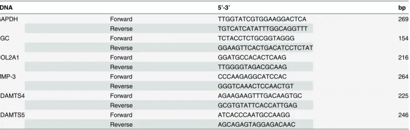
RNA from 1.106cells was isolated by RNeasy mini kit (Qiagen), reverse transcription made with superscript III (Invitrogen) and polymerase chain reaction (PCR) was performed by using the Light Cycler-FastStart DNA Master Sybr Green I (LC480, Roche Diagnostics, Brussels, Bel-gium). The PCR template source was either 3 ng first-strand cDNA or purified DNA standard. Primer sequences used to amplify the desired cDNA were as shown inTable 1. Glyceralde-hyde-3-phosphate dehydrogenase (GAPDH), a house keeping gene, was used as internal con-trol and gene expressions were normalised by calculating the expression of aggrecan, type II collagen (COL2A1), MMP-3, ADAMTS-4, -5 and that of GAPDH.
Statistical analysis
The results (mean ± SD) were expressed as GAPDH-normalized gene expression or as the con-centration perμg of DNA. A One-way Analysis of Variance (ANOVA) test followed by a Tukey-Kramer Multiple Comparisons Post-test was performed on means of all the experi-ments, and Pearson correlations were performed to compare concentration-dependence (GraphPad Prism 6.0). Log(agonist) or log(inhibitor) vs response non linear fit was used to estimate the IC50 (GraphPad Prism 6.0).
Results
Effect of carnosol on proliferation and viability of chondrocytes and
osteoblasts in culture
At concentrations ranging from 6 nM to 9μM, no significant variation of chondrocyte or oste-oblast viability, as evaluated by the LDH release, was observed in our cultures. The DNA con-tent remained stable in our culture conditions.
Effect of carnosol on OA chondrocyte metabolism
After 4-days of culture, carnosol did not significantly modify the aggrecan production or gene expression by chondrocytes, but significantly enhanced type II collagen gene expression at 3μM (1.61-fold, p = 0.0008,Fig 1A). Carnosol also significantly reduced in a concentration-dependent manner the ADAMTS-4 (IC50 = 8.5μM, r = -0.9463, p = 0.0012,Fig 1B) and ADAMTS-5 gene expression at 0.2μM and higher concentrations (1.5-fold, p<0.05,Fig 1C).
Carnosol significantly decreased MMP-3 protein production in a concentration-dependent manner (IC50 = ~9.6μM, r = -0.9684, p = 0.0003) and enhanced TIMP-1 protein production at 3μM (1.24-fold, p = 0.02) by OA chondrocytes (Fig 2A and 2B). These effects were associated
with a concentration-dependent decrease of the MMP3/TIMP-1 ratio (IC50 = 3μM, r = -0.9770, p = 0.0002).
Finally, carnosol significantly down-regulated chondrocyte production of IL-6 (IC50 = 2.3μM, r = -0.9067, p = 0.0049Fig 2C) and NO (IC50 = 5.3μM, r = -0.9942,
p<0.0001,Fig 2D) in a concentration-dependent manner. The inhibitory effect of carnosol on these parameters was statistically significant at concentration of 1μM and above.
Effect of carnosol on OA subchondral osteoblasts
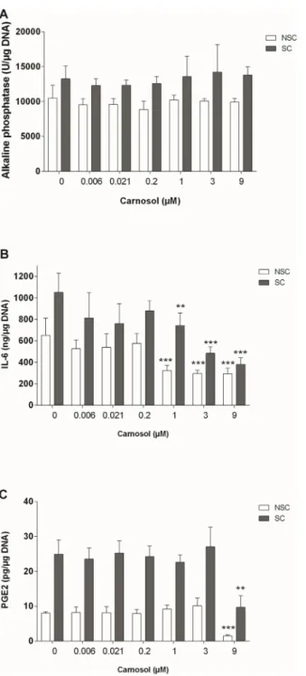
Alkaline phosphatase activity was higher in SC osteoblasts than in NSC osteoblasts (10494 +/-1833 vs 13254 +/- 1652 U/μg DNA, p = 0.006). Carnosol did not significantly affect the alkaline phosphatase activity of NSC or SC osteoblasts (Fig 3A).
SC osteoblasts produced higher levels of IL-6 and PGE2than NSC osteoblasts (1.6 and
3.1-fold, p<0.001), and NO production was below detection levels in both NSC and SC osteo-blasts. Carnosol reduced IL-6 production in a concentration-dependent manner
(IC50 = 0.6μM, r = -0.7674, p = 0.0440 in NSC and IC50 = 2.7μM, r = -0.7861, p = 0.0361 in SC osteoblasts, 0.45-fold at 9μM in NSC and 0.36-fold in SC,Fig 3B). At 9μM, carnosol inhib-ited PGE2production in both NSC and SC cultures (0.19-fold in NSC and 0.39 fold in SC
oste-oblasts, p<0.01,Fig 3C).
Effect of carnosol on OA chondrocytes co-cultured with subchondral
osteoblasts
When chondrocytes were co-cultured with osteoblasts, NSC osteoblasts did not significantly modify aggrecan protein production by chondrocytes, while SC osteoblasts significantly reduced total aggrecan production by 15+/-4% (p = 0.043). Aggrecan gene expression by chon-drocytes was decreased by 25+/-8% in co-culture with NSC osteoblasts (p = 0.032) and by 58 +/-7% with SC osteoblasts co-culture (p = 0.0053,Fig 4A). The pre-treatment of SC osteoblasts with carnosol at 1 or 5μM fully prevented the inhibition of aggrecan gene expression by chon-drocytes and even enhanced this gene expression by 3.7 and 4.2-fold (p<0.05 and p <0.01,Fig 4A). Type II collagen gene expression was not significantly modified in presence of NSC osteo-blasts, but was reduced by SC osteoblasts (0.55-fold, p = 0.0006,Fig 4B). Carnosol did not affect the inhibitory effect of SC osteoblasts on COL2A1 gene expression (Fig 4B).
Table 1. Primers Sequences .
cDNA 5’-3’ bp
GAPDH Forward TTGGTATCGTGGAAGGACTCA 269
Reverse TGTCATCATATTTGGCAGGTTT
AGC Forward TCTACCTCTGCGGTAGGG 154
Reverse GGAAGTTCACTGACATCCTCTAT
COL2A1 Forward GGATGCCACACTCAAG 216
Reverse TTGGGGTAGACGCAAG
MMP-3 Forward CCCAAGAGGCATCCAC 264
Reverse GGGTCAAACTCCAACTGT
ADAMTS4 Forward AGAAGAAGTTTGACAAGTGC 225
Reverse GCGTGTATTCACCATTGAG
ADAMTS5 Forward ATCACCCAATGCCAAGG 246
Reverse AGCAGAGTAGGAGACAAC
Fig 1. Effect of Carnosol on Chondrocyte Gene Expression. (A) COL2A1, (B) 4, (C) ADAMTS-5. Gene expression was normalized to GAPDH. The results are expressed as the mean± SD of three experiments performed with cells coming from three different donors. ANOVA with Tukey posttest performed between treated and untreated cells,* = p<0.05, ** = p<0.01 and *** = p<0.001.
Fig 2. Effect of Carnosol on Chondrocyte Protein Production. (A) MMP-3, (B) TIMP-1, (C) IL-6 and (D) NO. Production was normalized to DNA content. The results are expressed as the mean± SD of three experiments performed with cells coming from three different donors. ANOVA with Tukey posttest performed between treated and non-treated cells,* = p<0.05 and *** = p<0.001.
When the chondrocytes were co-cultured with NSC osteoblasts, ADAMTS-4 and -5 sion by chondrocytes were 3-fold or 1.8-fold upregulated (p<0.001), but MMP-3 gene expres-sion was unchanged. In co-culture with SC osteoblasts, ADAMTS-4 / -5 and MMP-3, mRNA levels were 4.6-fold, 4.4-fold (p<0.001) and 1.3-fold (p<0.05), respectively, increased
Fig 3. Effect of Carnosol on Activity of Alkaline Phosphatase (A), Production of IL-6 (B) and PGE2(C)
in NSC and SC Subchondral Osteoblasts. The results were normalized to the DNA content and are expressed as the mean± SD of three experiments performed with cells coming from three different donors. ANOVA with Tukey posttest performed between treated and non-treated cells,** = p<0.01 and *** = p<0.001.
Fig 4. Effect of Carnosol Pre-treatment of Subchondral Osteoblasts on Chondrocyte Gene Expression after 4 Days of Co-culture with Osteoblasts. (A) AGC, (B) COL2A1, (C) MMP-3, (D) ADAMTS-4, (E) ADAMTS-5. Gene expression was normalized to GAPDH. The results are expressed as the mean± SD of three experiments performed with cells coming from three different donors, and presented as ratio of chondrocytes monoculture experiments run in parallel. ANOVA with Tukey posttest statistical significances: Chondrocytes/osteoblasts co-culture compared to mono-culture* = p<0.05, ** = p<0.01, *** = p < 0.001; SC osteoblasts compared to NSC osteoblasts # = p<0.05, ## = p<0.01, ### = p<0.001; SC osteoblasts in co-culture with inhibitors
compared to SC osteoblasts without inhibitors: ° = p<0.05, °° = p<0.01 and °°° = p<0.001. doi:10.1371/journal.pone.0136118.g004
compared to chondrocytes in mono-culture. Pre-incubation of SC or NSC osteoblasts with car-nosol at 1 or 5μg/ml induced a significant reduction of ADAMTS-4 / -5 and MMP-3 gene expression by chondrocytes (SC: 0.3-fold, p<0.001, NSC: 0.5-fold, p<0.05,Fig 4C–4E).
Effects of subchondral sclerotic osteoblast mediators on chondrocyte
To identify the osteoblastic mediators triggering the chondrocytes responses, we incubated subchondral osteoblast/chondrocyte co-cultures in the presence of monoclonal antibodies (Mabs) neutralizing either IL-6 or TGF-β1 or HGF activity, or with piroxicam as PGE2
inhibi-tor (Fig 5). The addition of the Mabs or piroxicam completely neutralized the expression of these proinflammatory mediators as of IL-6, TGF-β1 or PGE2by were non detectable in the
culture supernatants by specific ELISA tests. In comparison to chondrocyte mono-culture with the same neutralizing agents, only anti-IL-6 Mab significantly inhibited the effect of SC osteo-blasts on AGC (1.45-fold, p = 0.0009), COL2A1 (1.8-fold, p = 0.0312), MMP-3 (0.5-fold, p = 0.0008), ADAMTS-4 (0.29-fold, p<0.0001) and -5 (0.29-fold, p<0.0001) gene expression (Fig 5). The same range of effect was observed also on NSC osteoblasts (Fig 5). Piroxicam also significantly decreased the MMP-3 expression (p = 0.03) in NSC osteoblasts co-culture.
Discussion
In this study, we have shown thatin vitro, at similar ranges of concentrations to those bioavail-able in tissues after rosemary extracts administration [21], carnosol is a good candidate as anti-OA agent. On chondrocytes, carnosol has an interesting stimulating effect on type II collagen gene expression and an inhibitory effect on MMP-3 production, and ADAMTS-4 and -5 gene expression. Further, TIMP-1 synthesis was increased by carnosol suggesting that this product can also down-regulate MMP activity. Carnosol showed also potent anti-inflammatory activi-ties by decreasing NO and IL-6 synthesis.
In addition to these direct effects on chondrocyte metabolism, carnosol also exerts a protec-tive effect on the deleterious activities of OA subchondral osteoblasts on chondrocytes, observed in our co-culture model. Indeed, pre-treatment of OA sclerotic subchondral osteo-blasts with carnosol mediated an increase in cartilage matrix formation components (aggrecan, type II collagen) and decrease in gene expression of degradation enzymes (MMP-3, ADAMTS4 and -5) by chondrocytes.
As previously shown by ourselves [17,22] and others [23,24], OA subchondral osteoblasts, in particular coming from sclerotic subchondral bone, can modify the gene expression of chon-drocytes in co-culture, leading to a phenotype suitable for matrix degradation We therefore tested the effect of carnosol on certain factors in OA subchondral osteoblasts which could be responsible for mediating the protective effects of carnosol on matrix formation and degrada-tion in cocultures. Carnosol did not modify alkaline phosphatase activity of the osteoblasts, but did it reduce IL-6 production at 1μM, and at 9 μM also PGE2production. These effects of
car-nosol were observed in the same way either on NSC and SC osteoblasts.
Finally, we investigated the effects of cytokines and growth factors involved in bone remod-elling for their potency to trigger the deleterious effects of SC osteoblasts on the overlying carti-lage in OA. TGF-β1/3, IL-6 and PGE2are regulators of bone remodelling and are involved in
subchondral bone sclerosis associated with OA. Moreover, they are overexpressed in OA scle-rotic subchondral bone [5,25,26]. Further, HGF, a factor produced by subchondral osteo-blasts, but not by chondrocytes, was found in the deep layer of the cartilage indicating that factors produced by bone cells can cross over the osteochondral bone plate [27]. In our co-cul-ture model, we tested the effects of neutralizing factors to each of these mediators, and among them only 6 seems to be involved. To the best of our knowledge, this is the first time that
IL-Fig 5. Effect of IL-6 Neutralizing Mab on Gene Expression by Chondrocytes. (A) AGC, (B) COL2A1, (C) MMP-3, (D) ADAMTS-4 and (E) -5 gene expressions by chondrocytes after four days of culture in the absence (mono-culture) or in the presence (co-culture) of osteoblasts isolated from sclerotic (SC) or non-sclerotic (NSC) zones of subchondral bone. The culture was performed in absence or in the presence of 5μg/ml of anti-IL-6, TGF-β1/3 or HGF Mab or 7μg/ml of piroxicam (Pirox). GAPDH-normalized gene expressions are reported to mono-culture expression level, and represented as the mean ± SD of three experiments performed with cells coming from three different donors. Comparison of mean values of gene expression was performed by
Mann-6 is identified as a key mediator of the cross talk between subchondral bone and cartilage in OA.
We observed that the inhibitory effect of SC osteoblasts on the production of aggrecan by chondrocytes was abolished by neutralizing the biological activity of IL-6. In addition, type II collagen gene expression inhibition by osteoblasts was reversed by about 50% in the presence of anti-IL-6. Neutralization of IL-6 also abolished the effect of SC osteoblasts on the expression of MMP-3, ADAMTS-4 and -5 by chondrocytes. Previously, Sanchez et al. [28] demonstrated that IL-6 in the presence of its soluble receptor, stimulated the production of MMP-3 and inhibited the synthesis of aggrecan by chondrocytes in alginate beads. Other authors have shown that IL-6 inhibited the expression of type II collagen and the aggrecan link protein by bovine chondrocytes cultured in monolayer, in parallel with an inhibition of the expression of the transcription factor sox9 [29]. IL-6 acts in synergy with IL-1 in vitro to degrade collagens and proteoglycans in cartilage explants [30]. These results emphasize the key role played by IL-6 in cartilage degradation. They also emphasize the important role of this cytokine in the cross-talk between subchondral bone and cartilage. Furthermore, these results are consistant with previous results showing that pre-treatment of NSC osteoblasts with IL-6 led to the same dele-terious effect than untreated SC osteoblasts on chondrocytes in co-culture [17]. Finally, these data indicate that the protective effects of carnosol against osteoblast-induced deregulation of chondrocyte metabolism is probably mediated by IL-6.
While we cannot exclude the involvement of other factors, our study clearly demonstrates that PGE2TGF-β1–3 and HGF are not involved in the cross-talk between osteoblasts and
chondrocytes, and identifies IL-6 as a key factor of the osteoblasts/chondrocytes crosstalk in OA.
Conclusion
Carnosol showed potent anti-inflammatory and anti-catabolic effects on chondrocytes. Fur-thermore, it was able to reduce cartilage matrix breakdown and enhance its formation in pres-ence of subchondral osteoblasts, probably by inhibition of IL-6 production by osteoblasts. Its effects would deserve to be further confirmed and investigated, in an appropriate animal model and then in clinical studies.
Acknowledgments
The authors want to thanks Elizabeth Kaut for this technical help, and Dr Delcour for the biopsies.
Author Contributions
Conceived and designed the experiments: CS MNH LA FMS EO YH. Performed the experi-ments: CS. Analyzed the data: CS MNH LA FMS YH. Contributed reagents/materials/analysis tools: MNH LA FMS. Wrote the paper: CS MNH LA EO YH.
References
1. Loeser RF, Goldring SR, Scanzello CR, Goldring MB. Osteoarthritis: a disease of the joint as an organ. Arthritis Rheum. 2012; 64(6):1697–707. doi:10.1002/art.34453PMID:22392533; PubMed Central PMCID: PMC3366018.
Whitney tests. Statistical significances: Chondrocytes/osteoblasts co-culture compared to mono-culture* = p<0.05, ** = p<0.01, *** = p < 0.001; SC osteoblasts compared to NSC osteoblasts # = p<0.05, ## = p<0.01, ### = p<0.001; SC osteoblasts in co-culture with inhibitors compared to SC osteoblasts without inhibitors: ° = p<0.05 °° = p<0.01 and °°° = p<0.001.
2. Liu-Bryan R, Terkeltaub R. Emerging regulators of the inflammatory process in osteoarthritis. Nat Rev Rheumatol. 2015; 11(1):35–44. doi:10.1038/nrrheum.2014.162PMID:25266449.
3. Martel-Pelletier J, Di Battista J, Lajeunesse D. Biochemical factors in joint articular tissue degradation in osteoarthritis. In: Reginster JY, Pelletier JP, Martel-Pelletier J, Henrotin Y, editors. Osteoarthritis, clin-ical and experimental aspects. Berlin: Springer; 1999. p. 156–87.
4. Pesesse L, Sanchez C, Henrotin Y. Osteochondral plate angiogenesis: A new treatment target in oste-oarthritis. Joint Bone Spine. 2011; 78(2):144–9. Epub 2010/09/21. doi: S1297-319X(10)00192-2 [pii] doi:10.1016/j.jbspin.2010.07.001PMID:20851653.
5. Sanchez C, Deberg MA, Bellahcene A, Castronovo V, Msika P, Delcour JP, et al. Phenotypic character-ization of osteoblasts from the sclerotic zones of osteoarthritic subchondral bone. Arthritis Rheum. 2008; 58(2):442–55. Epub 2008/02/02. doi:10.1002/art.23159PMID:18240211.
6. Sanchez C, Deberg MA, Piccardi N, Msika P, Reginster JY, Henrotin YE. Osteoblasts from the sclerotic subchondral bone downregulate aggrecan but upregulate metalloproteinases expression by chondro-cytes. This effect is mimicked by interleukin-6, -1beta and oncostatin M pre-treated non-sclerotic osteo-blasts. Osteoarthritis Cartilage. 2005; 13(11):979–87. Epub 2005/10/26. doi: S1063-4584(05)00094-4 [pii] doi:10.1016/j.joca.2005.03.008PMID:16243232.
7. Sanchez C, Deberg MA, Piccardi N, Msika P, Reginster JY, Henrotin YE. Subchondral bone osteo-blasts induce phenotypic changes in human osteoarthritic chondrocytes. Osteoarthritis Cartilage. 2005; 13(11):988–97. Epub 2005/09/20. doi: S1063-4584(05)00196-2 [pii] doi:10.1016/j.joca.2005.07.012
PMID:16168681.
8. Offord EA. In: Packer L, Ong CN, Halliwell BB, Wachtel-Galor S, Benzie IFF, editors. Herbal Medecine and molecular basis of health and disease management: Marcel Dekker 2004. p. 457.
9. Huang SC, Ho CT, Lin-Shiau SY, Lin JK. Carnosol inhibits the invasion of B16/F10 mouse melanoma cells by suppressing metalloproteinase-9 through down-regulating nuclear factor-kappa B and c-Jun. Biochem Pharmacol. 2005; 69(2):221–32. Epub 2005/01/04. doi: S0006-2952(04)00651-3 [pii] doi:10. 1016/j.bcp.2004.09.019PMID:15627474.
10. Johnson JJ. Carnosol: A promising anti-cancer and anti-inflammatory agent. Cancer Lett. 2011. Epub 2011/03/09. doi: S0304-3835(11)00067-X [pii] doi:10.1016/j.canlet.2011.02.005PMID:21382660. 11. Lopez-Jimenez A, Garcia-Caballero M, Medina MA, Quesada AR. Anti-angiogenic properties of
carno-sol and carnosic acid, two major dietary compounds from rosemary. European journal of nutrition. 2013; 52(1):85–95. doi:10.1007/s00394-011-0289-xPMID:22173778.
12. Takaki I, Bersani-Amado LE, Vendruscolo A, Sartoretto SM, Diniz SP, Bersani-Amado CA, et al. Anti-inflammatory and antinociceptive effects of Rosmarinus officinalis L. essential oil in experimental ani-mal models. J Med Food. 2008; 11(4):741–6. Epub 2008/12/05. doi:10.1089/jmf.2007.0524PMID:
19053868.
13. Lo AH, Liang YC, Lin-Shiau SY, Ho CT, Lin JK. Carnosol, an antioxidant in rosemary, suppresses inducible nitric oxide synthase through down-regulating nuclear factor-kappaB in mouse macrophages. Carcinogenesis. 2002; 23(6):983–91. Epub 2002/06/26. PMID:12082020.
14. Subbaramaiah K, Cole PA, Dannenberg AJ. Retinoids and carnosol suppress cyclooxygenase-2 tran-scription by CREB-binding protein/p300-dependent and-independent mechanisms. Cancer Res. 2002; 62(9):2522–30. Epub 2002/05/01. PMID:11980644.
15. Martin D, Rojo AI, Salinas M, Diaz R, Gallardo G, Alam J, et al. Regulation of heme oxygenase-1 expression through the phosphatidylinositol 3-kinase/Akt pathway and the Nrf2 transcription factor in response to the antioxidant phytochemical carnosol. J Biol Chem. 2004; 279(10):8919–29. doi:10. 1074/jbc.M309660200PMID:14688281.
16. Sanchez C, Mathy-Hartert M, Deberg MA, Ficheux H, Reginster JY, Henrotin YE. Effects of rhein on human articular chondrocytes in alginate beads. Biochem Pharmacol. 2003; 65(3):377–88. PMID:
12527330.
17. Sanchez C, Deberg MA, Piccardi N, Msika P, Reginster JY, Henrotin Y. Osteoblasts from the sclerotic subchondral bone downregulate aggrecan but upregulate metalloproteinases expression by chondro-cytes. This effect is mimicked by Interleukin-6, -1b and Oncostatin M pre-treated non sclerotic osteo-blasts. Osteoarthritis Cartilage. 2005; 13(11):979–87. PMID:16243232.
18. Labarca C, Paigen K. A simple, rapid, and sensitive DNA assay procedure. Anal Biochem. 1980; 102 (2):344–52. PMID:6158890.
19. Serteyn D, Deby-Dupont G, Pincemail J, Mottart E, Philippart C, Lamy M. Equine postanaesthetic myo-sitis: thromboxanes, prostacyclin and prostaglandin E2 production. Vet Res Commun. 1988; 12(2– 3):219–26. PMID:3188388.
20. Green LC, Wagner DA, Glogowski J, Skipper PL, Wishnok JS, Tannenbaum SR. Analysis of nitrate, nitrite, and [15N]nitrate in biological fluids. Anal Biochem. 1982; 126(1):131–8. PMID:7181105.
21. Romo Vaquero M, Garcia Villalba R, Larrosa M, Yanez-Gascon MJ, Fromentin E, Flanagan J, et al. Bio-availability of the major bioactive diterpenoids in a rosemary extract: metabolic profile in the intestine, liver, plasma, and brain of Zucker rats. Mol Nutr Food Res. 2013; 57(10):1834–46. doi:10.1002/mnfr. 201300052PMID:23625681.
22. Sanchez C, Deberg MA, Piccardi N, Msika P, Reginster JY, Henrotin Y. Subchondral bone osteoblasts induce phenotypic changes in human osteoarthritic chondrocytes. Osteoarthritis Cartilage. 2005; 13 (11):988–97. PMID:16168681.
23. Jiang J, Nicoll SB, Lu HH. Co-culture of osteoblasts and chondrocytes modulates cellular differentiation in vitro. Biochem Biophys Res Commun. 2005; 338(2):762–70. PMID:16259947.
24. Prasadam I, Crawford R, Xiao Y. Aggravation of ADAMTS and matrix metalloproteinase production and role of ERK1/2 pathway in the interaction of osteoarthritic subchondral bone osteoblasts and articu-lar cartilage chondrocytes—possible pathogenic role in osteoarthritis. J Rheumatol. 2012; 39(3):621– 34. doi:10.3899/jrheum.110777PMID:22247346.
25. Hilal G, Martel-Pelletier J, Pelletier JP, Ranger P, Lajeunesse D. Osteoblast-like cells from human sub-chondral osteoarthritic bone demonstrate an altered phenotype in vitro: possible role in subsub-chondral bone sclerosis. Arthritis Rheum. 1998; 41(5):891–9. PMID:9588742.
26. Massicotte F, Lajeunesse D, Benderdour M, Pelletier JP, Hilal G, Duval N, et al. Can altered production of interleukin-1beta, interleukin-6, transforming growth factor-beta and prostaglandin E(2) by isolated human subchondral osteoblasts identify two subgroups of osteoarthritic patients. Osteoarthritis Carti-lage. 2002; 10(6):491–500. PMID:12056853.
27. Guevremont M, Martel-Pelletier J, Massicotte F, Tardif G, Pelletier JP, Ranger P, et al. Human adult chondrocytes express hepatocyte growth factor (HGF) isoforms but not HgF: potential implication of osteoblasts on the presence of HGF in cartilage. J Bone Miner Res. 2003; 18(6):1073–81. PMID:
12817761.
28. Sanchez C, Deberg MA, Burton S, Devel P, Reginster JY, Henrotin YE. Differential regulation of chon-drocyte metabolism by oncostatin M and interleukin-6. Osteoarthritis Cartilage. 2004; 12(10):801–10. Epub 2004/09/29. doi:10.1016/j.joca.2004.06.011S1063-4584(04)00119-0 [pii]. PMID:15450530. 29. Legendre F, Dudhia J, Pujol JP, Bogdanowicz P. JAK/STAT but not ERK1/ERK2 pathway mediates
interleukin (IL)-6/soluble IL-6R down-regulation of Type II collagen, aggrecan core, and link protein transcription in articular chondrocytes. Association with a down-regulation of SOX9 expression. J Biol Chem. 2003; 278(5):2903–12. PMID:12419823.
30. Nietfeld JJ, Wilbrink B, Helle M, van Roy JL, den Otter W, Swaak AJ, et al. Interleukin-1-induced inter-leukin-6 is required for the inhibition of proteoglycan synthesis by interleukin-1 in human articular carti-lage. Arthritis Rheum. 1990; 33(11):1695–701. PMID:2242066.