Somatic MEN1 gene mutation does not contribute significantly
to sporadic pituitary tumorigenesis
Jacques Poncin1, Achille Stevenaert2and Albert Beckers3 1
Departments of Medical Genetics,3Endocrinology, and2Surgery, University Hospital, University of Lie`ge, 4000 Lie`ge, Belgium (Correspondence should be addressed to Jacques Poncin, Laboratoire de Ge´ne´tique Mole´culaire, Centre Hospital Universitaire de Lie`ge, Tour de Pathologie Sart-Tilman, 4000 Liege, Belgique; Email: [email protected])
Abstract
Pituitary adenomas are a common manifestation of multiple endocrine neoplasia type 1 (MEN1) but most of them occur sporadically. There are only a few well defined genetic abnormalities known to occur in these sporadic tumours. The MEN1 gene located on 11q13 has recently been cloned and allelic deletion and mutation analysis studies have implicated the MEN1 gene in a significant fraction of the sporadic counterparts of typical MEN1 neoplasms (parathyroid tumours, insulinomas and gastrinomas).
To determine if MEN1 gene inactivation is also involved in the development of sporadic pituitary adenomas, allelic deletions of chromosome 11q13 and MEN1 gene mutations and polymorphisms were assessed in 35 sporadic tumours of the anterior pituitary (9 prolactin-secreting, 8 GH-secreting, 3 TSH-secreting, 2 TSH/GH-secreting, 4 Cushing, 9 silent). Thirty-one tumours were found to be heterozygous for at least one MEN1 intragenic polymorphism (25 cases) or for a flanking gene polymorphism (6 cases). The remaining tumours were not informative.
No mutations were found in any tumour except in one prolactinoma which was homozygous or hemizygous for a mutation (1–117 C→T) in a region close to the promoter. Unfortunately, blood or normal tissue was not available in this case.
Our data show that somatic MEN1 mutations do not contribute significantly to tumorigenesis of sporadic pituitary adenomas and suggest that mutation of other genes are likely to contribute to the pathogenesis of these tumours.
European Journal of Endocrinology 140 573–576
Introduction
Pituitary adenomas are a common manifestation of multiple endocrine neoplasia type 1 (MEN1) but most of them occur sporadically. To date, only a few genetic abnormalities have been described in these sporadic tumours.
For example, a mutation in the GSP gene occurs in about 1/3 of somatotrophic adenomas, but is rarely found in other pituitary tumour types (1). Other candidate oncogenes and tumour suppressor genes may play a role in the formation of pituitary tumours, but to date the pathogenesis of most tumours remains unexplained (for a review, see (2)).
The MEN1 gene located on chromosome 11q13 has recently been cloned (3, 4), and allelic deletion and mutation analysis studies have implicated the MEN1 gene in a significant fraction of the sporadic counter-parts of typical MEN1 neoplasms (parathyroid tumours, carcinoid tumours of the lung, gastrinomas and insu-linomas) (5–7). To investigate whether MEN1 gene inactivation is involved in the development of sporadic pituitary adenomas, loss of heterozygosity (LOH) for
11q13 and MEN1 gene mutations was assessed in 35 sporadic tumours of the anterior pituitary.
Materials and methods
Tissue collection and DNA preparation
Thirty-five cryopreserved anterior pituitary tumours from the files of the Department of Endocrinology were included in this study. A MEN1 syndrome was excluded by familial anamnesis and by measurements in each patient of parathyroid hormone, Ca++, phosphate, gastrin, pancreatic polypeptide and adrenal hormones serum levels.
The sample comprised 9 prolactin (PRL)-secreting adenomas, 8 growth hormone (GH)-secreting adeno-mas, 3 thyrotrophin (TSH)-secreting adenoadeno-mas, 2 TSH/ GH-secreting adenomas, 9 silent adenomas and 4 ACTH-secreting adenomas.
Tissue was frozen in liquid nitrogen immediately after surgical excision and stored at ¹708C. Genomic DNA was extracted by standard methods.
European Journal of Endocrinology (1999) 140 573–576 ISSN 0804-4643
Mutation and intragenic polymorphism
analysis
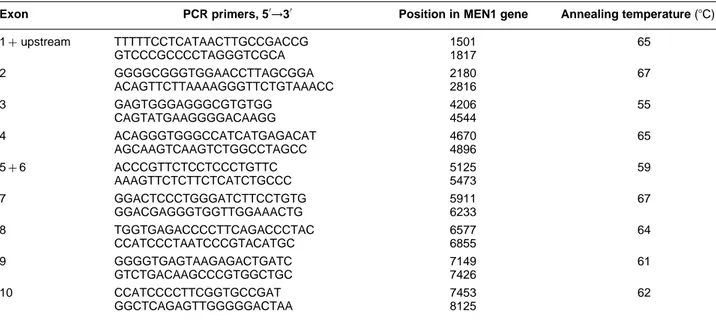
Exons 1–10 of the MEN1 gene were amplified in a volume of 50 ml containing 10 mmol/l Tris–HC1 (pH 8.3), 50 mmol/l KC1, 1.5 mmol/l MgCl2, 200 mmol/l dNTPs, 0.5 mmol/l of each of the primers, 2.5 units Taq polymerase (Perkin-Elmer Cetus, Foster City, CA, USA) and 200 ng genomic DNA. Samples were heated at 948C for 7 min. Forty PCR cycles (1 min at 94 8C, 1 min at the relevant annealing temperature (Table 1), and 2 min at 728C) were then performed. The DNA synthesis step of the final cycle at 728C was extended to 7 min. To secure detection of most splice site mutations, the PCR primers (Table 1) were designed so that each exon and at least 40 bases of its flanking sequences were contained in the amplified fragment.
The PCR products were controlled by electrophoresis in 1% agarose, purified using the QIAquick PCR purification kit (Qiagen GmbH, Hilden, Germany) and sequenced using Dye Terminator Cycle Sequencing chemistry with AmpliTaq DNA Polymerase FS (ABI PRISM Dye Terminator Cycle Sequencing kit, Perkin-Elmer, Foster City, CA, USA) and one of the primers described above. The sequencing reaction mixture was subjected to 25 repeated rounds of denaturation for 30 s at 968C, annealing for 15 s at 50 8C, and synthesis steps for 4 min at 608C. The extension products were purified by ethanol precipitation. The pellets were briefly dried and resuspended in 4 ml 5 mmol/l EDTA (pH 8.0), 80% formamide. The samples were heated at 958C for two min, loaded on 6% polyacrylamide denaturing gels and electrophoresed for 7 h at 1600 V in a ABI PRISM 377 DNA Sequencer (Perkin-Elmer). Both strands of the PCR
products were sequenced in each case. Sequence comparisons were performed by use of the Sequence Navigator software (Perkin-Elmer).
LOH testing with MEN1 flanking markers
Samples were screened for LOH with the microsatellite polymorphic markers D11S449, D11S1883, and D11S1889 flanking the MEN1 gene (8). The target DNA sequences were amplified by PCR in a volume of 50 ml containing 10 mmol/l Tris–HCl (pH 8.3), 50 mmol/l KCl, 1.5 mmol/l MgCl2, 200 mmol/l dNTPs, 2 mmol/l fluorescent-dUTP (TAMRA-, R110-, or R6G-dUTP, Perkin-Elmer), 1 mmol/l of each of the primers, 2.5 units Taq polymerase (Perkin-Elmer Cetus) and 200 ng genomic DNA. Samples were heated at 948C for 3 min. Thirty-five PCR cycles (1 min at 948C, 1 min at the relevant annealing temperature and 2 min at 728C) were then performed. The DNA synthesis step of the final cycle at 728C was extended to 7 min. The products were purified by centrifugation on a MicroCon-30 spin column (Amicon Inc., Beverly, MA, USA) and were analysed on 6% polyacrylamide denaturing gels in an ABI PRISM 377 DNA Sequencer (Perkin-Elmer). Purified PCR product (0.5–1.5 ml) was combined with 2.5 ml forma-mide and 0.5 ml of a fluorescent size marker (ROX 500, Perkin-Elmer). After denaturation at 908C, 2 ml of the mixture were loaded on the gel and electrophoresed for 2.5 h at 3000 V. The fluorescent gel data collected during the run were automatically analysed by the Genescan analysis program (Perkin-Elmer) at the end of the run. Each fluorescent peak was quantitated in terms of size (in base pairs), peak height and peak area.574 J Poncin and others EUROPEAN JOURNAL OF ENDOCRINOLOGY (1999) 140
Table 1 Oligonucleotides used for PCR amplification and PCR parameters of MEN1 gene exons.
Exon PCR primers, 50→30 Position in MEN1 gene Annealing temperature (8C)
1þupstream TTTTTCCTCATAACTTGCCGACCG 1501 65 GTCCCGCCCCTAGGGTCGCA 1817 2 GGGGCGGGTGGAACCTTAGCGGA 2180 67 ACAGTTCTTAAAAGGGTTCTGTAAACC 2816 3 GAGTGGGAGGGCGTGTGG 4206 55 CAGTATGAAGGGGACAAGG 4544 4 ACAGGGTGGGCCATCATGAGACAT 4670 65 AGCAAGTCAAGTCTGGCCTAGCC 4896 5þ6 ACCCGTTCTCCTCCCTGTTC 5125 59 AAAGTTCTCTTCTCATCTGCCC 5473 7 GGACTCCCTGGGATCTTCCTGTG 5911 67 GGACGAGGGTGGTTGGAAACTG 6233 8 TGGTGAGACCCCTTCAGACCCTAC 6577 64 CCATCCCTAATCCCGTACATGC 6855 9 GGGGTGAGTAAGAGACTGATC 7149 61 GTCTGACAAGCCCGTGGCTGC 7426 10 CCATCCCCTTCGGTGCCGAT 7453 62 GGCTCAGAGTTGGGGGACTAA 8125
Results
Mutation screening in pituitary tumours
The MEN1 transcripted sequence was screened for mutations by automatic sequence analysis in 35 sporadic anterior pituitary tumours including 26 hormone-secreting and 9 non-hormone-secreting tumours (Table 2). No mutations were found in any tumour except in one prolactinoma which was homozygous or hemizygous for a mutation (1–117 C→T) in a region close to the transcription start site.Polymorphisms of the MEN1 gene and
intragenic screening for LOH
Six relatively common polymorphisms were identified and their frequencies were evaluated in 120 normal
chromosomes. These were T-A at nucleotide 76 of exon 1 (45% T, 55% A), C-G in intron 116 nucleotides upstream of exon 2 (80% C, 20% G), S145S (AGC/AGT) (6%), R171Q (CGG/CAG) (3.7%), D418D (GAC/GAT) (42%), A541T (GCA/ACA) (2.5%). The first two of these polymorphisms (76: T-A and 88–16: C-G) were not described previously. They are highly informative and were therefore used as intragenic markers for LOH screening. Most of the tumors (25/35) were heterozygous for at least one of these intragenic polymorphisms.
LOH analysis with flanking markers
The 10 tumours which were uninformative for the MEN1 intragenic polymorphisms were screened for LOH using the flanking markers D11S1889, D11S449, and D11S1883. Six of the tumours were heterozygous for at least one marker.
Pituitary adenomas and MEN1 mutations 575 EUROPEAN JOURNAL OF ENDOCRINOLOGY (1999) 140
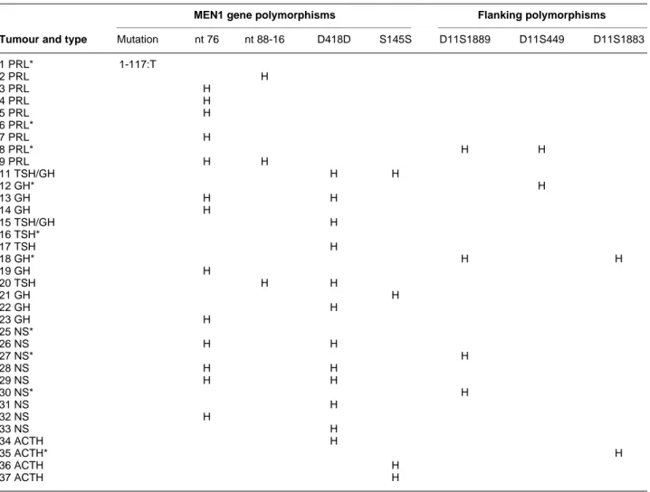
Table 2 Results of the polymorphisms analysis for 35 pituitary adenomas.
MEN1 gene polymorphisms Flanking polymorphisms
Tumour and type Mutation nt 76 nt 88-16 D418D S145S D11S1889 D11S449 D11S1883 1 PRL* 1-117:T 2 PRL H 3 PRL H 4 PRL H 5 PRL H 6 PRL* 7 PRL H 8 PRL* H H 9 PRL H H 11 TSH/GH H H 12 GH* H 13 GH H H 14 GH H 15 TSH/GH H 16 TSH* 17 TSH H 18 GH* H H 19 GH H 20 TSH H H 21 GH H 22 GH H 23 GH H 25 NS* 26 NS H H 27 NS* H 28 NS H H 29 NS H H 30 NS* H 31 NS H 32 NS H 33 NS H 34 ACTH H 35 ACTH* H 36 ACTH H 37 ACTH H
PRL, prolactinoma; TSH, TSH-secreting adenoma; GH, GH-secreting adenoma; NS, non-secreting adenoma; H, heterozygosity for the considered marker.
* The ten tumours which were uninformative for the MEN1 intragenic polymorphisms and which were screened for LOH with flanking markers.
Discussion
The pathogenesis of pituitary tumours remains unex-plained in most cases. The MEN1 gene has been cloned recently. As pituitary tumours are classically associated with MEN1, we tested the presence of MEN1 gene mutations in sporadic tumours.
We studied 35 secreting and non-secreting sporadic adenomas. Among them 31 were found to be hetero-zygous for at least one MEN1 intragenic polymorphism (25 cases) or for a flanking gene polymorphism (6 cases). In only 4 cases there remained a possibility for homo- or hemizygosity. In one case only we identified a possible point mutation in the promoter region. As the patient originated in South America, we cannot exclude a polymorphism. Unfortunately, peripheral blood was not available to discriminate between these two possibilities.
Our results are consistent with previous reports that suggested that MEN1 gene mutation is a rare event in pituitary adenomas. Indeed, Zhuang et al. using single strand conformational polymorphism analysis found only 2 missense mutations among 4 pituitary adenomas with LOH for MEN1 in a sample of 39 sporadic tumours (9). Prezant et al. using dideoxy fingerprinting analysis (which is more sensitive) studied 45 sporadic tumours but failed to reveal any mutations in the coding sequence of MEN1 (10). Recently, in a sample of 31 sporadic tumours, Tanaka et al. detected only 1 nonsense mutation in a GH/PRL adenoma with LOH in 11q13 (11).
These results are disturbing taking into account that Boggild et al. found LOH on chromosome 11 in as many as about 20% of 88 sporadic adenomas (12). This discrepancy could have been explained by mutation(s) affecting the transcription level of the MEN1 gene. Therefore, we decided to sequence exon 1 and promoter but this experiment failed to reveal any mutation. While this work was in progress Prezant et al. also addressed this question by studying MEN1 mRNA expression which was found to be normal (10), and Asa et al. demonstrated by competitive reverse transcription-PCR that there was a lack of menin down-regulation in the majority of tumours with LOH at 11q13 (13).
To summarise, only 1 nonsense and 2 missense mutations (with unknown functional effects) were found in a total of 150 sporadic pituitary adenomas screened (including our series). Therefore, contrary to the other sporadic counterparts of typical MEN1 neoplasms, MEN1 gene mutation does not appear to be a common event explaining the formation of pituitary tumours. The discrepancy between LOH and mutation remains to be explained.
Acknowledgements
This work was supported by the Fonds National de la Recherche Scientifique (grants FRSM no. 3.4566.89 and 3.4628.93) and the Fonds de Recherche de la Faculte´ de Me´decine de l’Universite´ de Lie`ge. We thank G Albert-Theate and J Jamin for expert technical assistance.
References
1 Spada A, Vallar L & Faglia G. Cellular alterations in pituitary tumors. European Journal of Endocrinology 1994 130 43–52. 2 Shimon I & Melmed S. Genetic basis of endocrine disease: pituitary
tumor pathogenesis. Journal of Clinical Endocrinology and Metabo-lism 1997 82 1675–1681.
3 Chandrasekharappa SC, Guru SC, Manickam P, Olufemi S-E, Collins FS, Emmert-Buck MR et al. Positional cloning of the gene for multiple endocrine neoplasia type 1. Science 1997 276 404– 407.
4 Lemmens I, Van de Ven WJ, Kas K, Zhang CX, Giraud S, Wautot V et al. Identification of the multiple endocrine neoplasia type 1 (MEN1) gene. Human Molecular Genetics 1997 6 1177–1183. 5 Heppner C, Kester MB, Agarwal SK, Debelenko LV, Emmert-Buck
MR, Guru SC et al. Somatic mutation of the MEN1 gene in parathyroid tumors. Nature Genetics 1997 16 375–378. 6 Debelenko LV, Brambilla E, Agarwal SK, Swalwell JI, Kester MB,
Lubensky IA et al. Identification of MEN1 gene mutations in sporadic carcinoid tumors of the lung. Human Molecular Genetics 1997 6 2285–2290.
7 Zhuang Z, Vortmeyer AO, Pack S, Huang S, Pham TA, Wang C et al. Somatic mutations of the MEN1 tumor suppressor gene in sporadic gastrinomas and insulinomas. Cancer Research 1997 57 4682–4686.
8 Courseaux A, Grosgeorge J, Gaudray P, Pannet AAJ, Forbes SA, Williamson C et al. Definition of the minimal MEN1 candidate area based on a 5-Mb integrated map of proximal 11q13. Genomics 1996 37 354–365.
9 Zhuang Z, Ezzat SZ, Vortmeyer AO, Weil R, Oldfield EH, Park W-S et al. Mutations of the MEN1 tumor suppressor gene in pituitary tumors. Cancer Research 1997 57 5446–5451.
10 Prezant TR, Levine J & Melmed S. Molecular characterization of the MEN1 tumor suppressor gene in sporadic pituitary tumors. Journal of Clinical Endocrinology and Metabolism 1998 83 1388– 1391.
11 Tanaka C, Kimura T, Yang P, Moritani M, Yamaoka T, Yamada S et al. Analysis of loss of heterozygosity on chromosome 11 and infrequent inactivation of the MEN1 gene in sporadic pituitary adenomas. Journal of Clinical Endocrinology and Metabolism 1998 832631–2634.
12 Boggild MD, Jenkinson S, Pistorello M, Boscaro M, Scanarini M, McTernan P et al. Molecular genetics studies of sporadic pituitary tumors. Journal of Clinical Endocrinology and Metabolism 1994 78387–392.
13 Asa SL, Somers K & Ezzat S. The MEN1 gene is rarely down-regulated in pituitary adenomas. Journal of Clinical Endocrinology and Metabolism 1998 83 3210–3212.
Received 13 November 1998 Accepted 12 February 1999