Université de Montréal
EXPRESSION OF STEROIDOGENIC PROTEINS AND GENES IN BOVINE PLACENTA FROM CONVENTIONAL AND SOMATIC CELL NUCLEAR TRANSFER (SCNT) GESTATIONS
par
ADRIANA REBECA VERDUZCO GÓMEZ
Département de biomédecine vétérinaire Faculté de médecine vétérinaire
Thèse présentée à la Faculté de médecine vétérinaire en vue de l’obtention du grade de
philosophiae doctor (Ph.D.) en sciences vétérinaires
option reproduction
Décembre, 2010
Université de Montréal Faculté de médecine vétérinaire
Cette thèse intitulée
EXPRESSION OF STEROIDOGENIC PROTEINS AND GENES IN BOVINE PLACENTA FROM CONVENTIONAL AND SOMATIC CELL NUCLEAR TRANSFER (SCNT) GESTATIONS
présentée par
ADRIANA REBECA VERDUZCO GÓMEZ
a été évaluée par un jury composé des personnes suivantes
Paul D. Carrière, président-rapporteur
Bruce D. Murphy, directeur de recherche
Lawrence C. Smith, membre du jury
Éric Asselin, examinateur externe
RÉSUMÉ
Pendant la grossesse, les hormones stéroïdes jouent un rôle indispensable dans la régulation des principales manifestations physiologiques telles que la reconnaissance maternelle de la gestation, la réceptivité de l'endomètre, le début du développement embryonnaire ainsi que le maintien de la gestation. Cependant, on sait très peu sur la production de ces hormones et les principaux facteurs des voies intracellulaires impliqués dans le processus de stéroïdogenèse dans le placenta bovin pendant les stades initiaux et plus avancés de la gestation. Par ailleurs, certaines anomalies du placenta chez les bovins suite à une mauvaise production de stéroïdes n'ont pas encore été démontrées. Les objectifs de cette thèse étaient donc de : 1) déterminer la présence et la localisation des principales protéines stéroïdiennes dans le placenta de bovins provenant de gestations de 50 à 120 jours, 2) comparer l'expression placentaire d'une série de gènes et de protéines stéroïdiennes entre une gestation impliquant un transfert de noyaux de cellules somatiques (SCNT) et une gestation non-clonale; 3) étudier l'impact des hormones trophiques et des seconds messagers sur la stéroïdogenèse dans le placenta bovin à 140 +10 jours de gestation.
L’utilisation de techniques d’immunohistochimie, d’immunobuvardage et de PCR quantitatif nous a permis d’évaluer la présence d'un large éventail de gènes stéroïdiens (STAR, CYP11A1, HSD3B1, CYP17A1 et SCARB1) qui participent au transport du cholestérol et dans la production de différents types de stéroïdes. Dans cette thèse, nous avons démontré la capacité du placenta bovin d’initier la stéroïdogenèse au début de la gestation et nous avons également déterminé les principales cellules impliquées dans ce processus. Nous avons constaté que les tissus maternels expriment les principaux marqueurs de stéroïdogenèse suggérant une plus grande capacité stéroïdogénique que les tissus fœtaux. En outre, un modèle d'expression des protéines complémentaires stéroïdogéniques entre la caroncule et le cotylédon a été observé, indiquant que la stéroïdogenèse placentaire exige une communication cellule à cellule entre les cellules de la mère et du fœtus.
Après avoir démontré les principales cellules impliquées dans la synthèse des hormones stéroïdiennes dans le placenta bovin en début de gestation, nous avons ensuite étudié les modifications possibles de la stéroïdogenèse dans les tissus SCNT cotylédonaires à 40 jours de gestation. Nous avons identifié d'importantes modifications dans l'expression
des gènes STAR, CYP11A1, HSD3B1, CYP17A1, et SULT1E1. Conséquemment, nous postulons que l'expression réduite des gènes stéroïdiens peut provoquer une insuffisance de la biosynthèse des hormones stéroïdiennes, ce qui pourrait contribuer à un développement anormal du placenta et du fœtus dans les gestations SCNT à court ou long terme.
Finalement, nous avons développé un modèle efficace de culture d’explants de placentome qui nous a permis d'explorer les mécanismes sous-jacents spécifiques à la stéroïdogenèse placentaire. Nous avons exploré l'effet stimulant des hormones trophiques et différents messagers secondaires sur l'expression de différentes protéines stéroïdogéniques ainsi que le taux de progestérone (P4) dans les explants de placentome. En utilisant les techniques de RIA et de PCR quantitatif, nous avons constaté que même si les analogues de l'hormone lutéinisante (hCG) ont un effet stimulant sur plusieurs gènes stéroïdiens, le calcium ionophore est le principal modulateur dans la synthèse de la P4. Ces résultats suggèrent que dans le placenta bovin, la synthèse de la P4 est modulée principalement par l'afflux de calcium intracellulaire, et apparemment les nucléotides cycliques ne semblent pas contrôler ce processus.
En conclusion, cette étude contribue de manière significative à une meilleure compréhension des mécanismes d'entraînement de la synthèse des stéroïdes placentaires au début de la gestation et permet aussi d’apporter de nouveaux éclairages sur l'importance des stéroïdes placentaires dans la régulation du développement du placenta et du fœtus.
ABSTRACT
During pregnancy, steroid hormones have essential roles in regulating key physiological events such as maternal recognition, endometrial receptivity, early embryonic development, and maintenance of pregnancy. However, very little is known about the production of these hormones nor about the principal factors and intracellular pathways implicated in the steroidogenic process in bovine placenta, during early and advanced pregnancy. In addition, placental abnormalities in cattle following an improper steroid production in bovine placenta have not been yet demonstrated. The aims of this thesis were to: 1) determine the occurrence and localization of the principal steroidogenic proteins in bovine placenta from day 50 to day 120 of pregnancy; 2) compare the placental expression of a series of steroidogenic genes and proteins between somatic cell nuclear transfer (SCNT) pregnancies and non-SCNT gestations; 3) investigate the impact of trophic hormone, and second messengers on steroidogenesis in bovine placenta at 140 +10 days of gestation.
Using immunohistochemistry, western blot and qPCR techniques, we evaluated the presence of a wide range of steroidogenic genes (STAR, CYP11A1, HSD3B1, CYP17A1 and SCARB1), that participate in the cholesterol transport and in the production of different types of steroids. In this thesis, we demonstrated the capability of the early bovine placenta to initiate steroidogenesis, and we also determined the principal cells implicated in this process. We found that maternal tissue expresses the principal steroidogenic markers suggesting it has a greater steroidogenic capacity compared to fetal tissue. Moreover, a complementary pattern of steroidogenic protein expression between the caruncle and the cotyledon were found, indicating that placental steroidogenesis requires cell to cell communication between the maternal and fetal cells.
Having shown the principal cells involved in the synthesis of steroid hormones in bovine placenta during early pregnancies, we then studied possible
alterations in steroidogenesis in cotyledonary tissue in SCNT at 40 days of pregnancy. We identified significant alterations in the expression of STAR, CYP11A1, HSD3B1, CYP17A1 and SULT1E1 transcripts. Therefore, we postulate that reduced expression of steroidogenic genes may cause an insufficient local biosynthesis of steroid hormones, which might contribute to the abnormal placental and fetal development in SCNT gestations at short or long term.
Finally, we developed an efficient placentome explants culture model that allowed us to explore the specific mechanisms underlying placental steroidogenesis. We explored the stimulatory effect of trophic hormones and different second messengers on the expression of various steroidogenic proteins and the progesterone levels in placentome explants. By RIA and qPCR techniques, we found that although LH-like hormones (hCG), had a stimulatory effect on multiple steroidogenic genes, the calcium ionophore was the principal modulator in the synthesis of progesterone. These results suggest that in bovine placenta, the synthesis of progesterone is modulated principally by intracellular calcium influx, and cyclic nucleotides do not seem to be controlling this process.
In conclusion, these studies significantly contribute to a better understanding of the driving mechanisms of placental steroid synthesis in early gestations and also provide new insights into the importance of placental steroids in the regulation of placental and fetal development.
TABLE OF CONTENTS
RÉSUMÉ ... iii
ABSTRACT ... v
TABLE OF CONTENTS ... vii
LIST OF TABLES ... xi
LIST OF FIGURES ... xii
LIST OF ABBREVIATIONS ... xv
ACKNOWLEDGEMENTS ... xxiv
CHAPTER 1. Introduction ... 1
CHAPTER 2. Literature Review ... 5
2.1. Pregnancy ... 6
2.1.1. Definition and general aspects of pregnancy ... 6
2.1.2. Pregnancy recognition ... 6
2.1.3. Animal models of implantation ... 9
2.2. Bovine pregnancy ... 12
2.2.1. Implantation ... 12
2.2.2. Type of placentation ... 12
2.2.3. Morphometry and histology of bovine placenta ... 13
2.2.3.1. Bovine Trophoblast Giant Cells ... 17
2.2.4. Pregnancy recognition in cattle ... 19
2.2.4.1. Ruminant endometrial receptivity ... 19
2.3. Steroid Biosynthesis ... 23
2.3.1. General aspects ... 23
2.3.2.1. Importing cholesterol into the mitochondria ... 27
2.3.2. Steroidogenic enzymes ... 30
2.3.3. Estrogen sulfoconjugation ... 36
2.3.4. Regulation of steroidogenesis and principal steroidogenic pathways ... 37
2.4. Steroids synthesis during pregnancy ... 39
2.4.1. General aspects ... 39
2.4.2. Cholestrol uptake in placenta ... 40
2.5. Steroids synthesis in bovine placenta ... 41
2.5.1. Placental production of progesterone and estrogens in cattle ... 41
2.5.2. Bovine steroids and parturition ... 43
2.5.3. Estrogen sulfoconjugation in bovine placenta ... 45
2.6. Calcium ... 46
2.6.1. General aspects ... 46
2.6.2. Calcium and bovine placenta ... 47
2.6.2.1. Calcium and placental steroidogenesis ... 48
2.7. SCNT pregnancies ... 49
2.7.1. General aspects ... 49
2.7.2. Bovine SCNT pregnancies ... 49
2.7.2.1. Poor efficiency and alterations ... 49
2.7.2.2. Abnormal placental and fetal development ... 50
2.7.2.3. Possible alterations in hormones and factors ... 51
2.7.2.4. Abnormal expression of genes and transcription factors ... 52
2.8. Problem, hypothesis and objectives ... 53
MATERIALS, METHODS AND RESULTS ... 56
3.1. Abstract ... 59
3.2. Introduction ... 60
3.3. Materials and Methods ... 63
3.4. Results ... 69
3.5. Discussion ... 72
3.6. References ... 78
3.7. Tables and Figures ... 87
CHAPTER 4. SECOND ARTICLE ... 102
4.1. Abstract ... 104
4.2. Introduction ... 106
4.3. Results ... 107
4.4. Discussion ... 109
4.5. Materials and methods ... 113
4.6. References ... 120
4.7. Tables and figures ... 128
CHAPTER 5. THIRD ARTICLE ... 139
5.1. Abstract ... 140
5.2. Introduction ... 141
5.3. Material and methods ... 144
5.4. Results ... 149
5.5. Discussion ... 151
5.6. References ... 158
5.7. Tables and figures ... 166
CHAPTER 6. GENERAL DISCUSSION ... 176
CHAPTER 8. REFERENCES ... 192 CHAPTER 9. ANNEXES ... XXVIII ANNEX 1. Separation of subpopulation of bovine placental cells by size, using fluorescence activated cell sorting method (FACS) ... xxix ANNEX 2. Preliminary studies to determine the viability and sampling time of bovine
LIST OF TABLES
CHAPTER 2. LITERATURE REVIEW
Table 1. Pregnancy recognition factors. 8
Table 2. Different types of placenta, implantation and time of attachment. 10 Table 3. Principal steroidogenic proteins and genes expressed in a
different number of bovine tissues and cells. 34
MATERIALS, METHODS AND RESULTS CHAPTER 3. FIRST ARTICLE
Expression of steroidogenic proteins in bovine placenta during the first third of the gestation.
Table1. First antibodies and concentrations employed for western blotting (WB) and immunohistochemical staining procedures (IMH) in bovine placenta.
87
Table 2. Primer pairs used for mRNA detection in bovine placenta. 88 CHAPTER 4. SECOND ARTICLE
Atypical expression of steroidogenic genes in somatic cell nuclear transfer-derived bovine placenta at day 40 of pregnancy
Table 1. Primer pairs used for RNA detection 128
CHAPTER 5. THIRD ARTICLE
Regulation of placental steroid synthesis in bovine placentome explants during the first half of gestation
LIST OF FIGURES
CHAPTER 2. LITERATURE REVIEW
Figure 1. Cartoon representing the initial phases of murine implantation. 11 Figure 2. Diagrammatic representation of the bovine placentome. 14 Figure 3. Cross section of a bovine placentome at approximately 90 days of
gestation.
16
Figure 4. Schematic representation of binucleate giant cells (BNGC) or trophoblast giant cells (TGCs) migration from the chorion to the endometrial epithelium in ruminants.
18
Figure 5. Pleiotropic effects of P4 and IFNT affect conceptus development, pregnancy and maternal well-being
22
Figure 6. Overview of steroidogenesis and the principal steroid hormones. 24
Figure 7. Uptake and transport of cholesterol to the mitochondria for steroidogenesis.
29
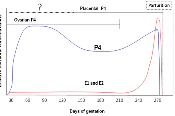
Figure 8. Conventional pathways and associated enzymes of steroid biosynthesis. 32 Figure 9. 17beta estradiol (E2) and progesterone (P4) profiles during gestation in
the cow.
44
MATERIALS, METHODS AND RESULTS CHAPTER 3. FIRST ARTICLE
Expression of steroidogenic proteins in bovine placenta during the first third of the gestation.
Figure 1. Positive and negative controls to validate the western blot system in bovine placenta.
89
Figure 2. Diagram representing the bovine placentome. 90 Figure 3. Characterization of steroidogenic proteins in bovine placental tissues by
western blot.
92
Figure 4. Characterization of mRNA in bovine placental tissues. 94 Figure 5. Characterization of STAR expression in bovine placentomes. 96 Figure 6. Characterization of CYP11A1 expression in bovine placentomes. 98 Figure 7.Characterization of HSD3B1 expression in bovine placentomes. 100
CHAPTER 4. SECOND ARTICLE
Atypical expression of steroidogenic genes in somatic cell nuclear transfer-derived bovine placentae at day 40 of pregnancy.
Figure 1. Characterization of levels of mRNA for STAR, CYP11A1, HSD3B1, CYP17A1,
SCARB1 ,STS and SULT1E1 in cotyledonary tissue of artificial insemination (AI) and
somatic cell nuclear transfer (SCNT) gestations at day 40.
129
Figure 2. Immunoblotting analysis for STAR, CYP11A1, HSD3B1 and ACTB in cotyledonary tissues at day 40 of gestation.
131
Figure 3. Immunofluorescence staining of SCARB1 in bovine cotyledons at day 40 of AI pregnancy.
133
Figure 4. Immunofluorescence staining of CYP17A1 in bovine cotyledons at day 40 of AI pregnancy.
135
Figure 5. Immunofluorescence staining of CYP11A1 in bovine placentome at day 40 of AI pregnancy.
137
CHAPTER 5. THIRD ARTICLE
Regulation of placental steroid synthesis in bovine placentome explants during the first half of gestation.
Figure 1. Pilot studies to determine the influence of different concentrations of dbcAMP, PMA, hCG and A2387 on P4 concentrations in bovine placental explants after 1 and 6 hours post-treatment.
167
Figure 2. Quantification of LHR gene expression. 169
Figure 3. Expression of CYP11A1 and HSD3B1 mRNA in bovine placental explants around day 140 +10 of gestation.
170
Figure 4. Expression of SULT1E1 and STS mRNA in bovine placental explants around day 140 +10 of gestation.
172
Figure 5. STAR mRNA abundance and P4 concentrations after 6, 12 and 18 h treatment.
174
ANNEXES ANNEX 1
Figure 1. Isolation of different bovine placental cells by FACS in caruncular and cotyledonary tissues.
ANNEX 2
Figure 1. Progesterone (P4) concentrations after 1, 2, 4, 6, 8, 10, 12, 14, 16, 18, 20, 22 and 24 h of incubation to determine the viability and sampling time of placenta explants.
LIST OF ABBREVIATIONS
25-OHD3 25-hydroxyvitamin D3
A2387 Calcium ionophore
ACTB Actin beta
ACTH Adrenocorticotropic hormone
AI Artificial insemination
ANGPT1 Angiopoietin 1
BTGC or BNGC Binucleate trophoblast giant cells
Ca2 +BPs Binding proteins known as the calbindins
Ca2+ Calcium
Ca2+BP9k Calbindin-D9k
calcitriol 1,25-dihydroxyvitamin D3
cAMP Cyclic-AMP
CEC Caruncular epithelial cells
CL Corpus luteum
CP Chorionic plate
CRL Crown-rump length
CSC Caruncular stromal cells
CSH Chorionic somatomammotropin hormones
(placental lactogen)
CST3 Cystatin C
CTSL Cathepsin L, cysteine proteinase
CV Chorionic villi
Cy3 Cyanine 3
CYP11A1 Cytochrome P450, family 11 subfamily, A
polypeptide 1
CYP17A1 Cytochrome P450, family 17, subfamily A,
polypeptide 1
CYP19A1 Cytochrome P450, family 19 subfamily, A
polypeptide 1
DAPI 4',6-diamidino-2-phenylindole
DEDTC Diethyldithiocarbama
DHEA Dehydroepiandrosterone
DHEA-S DHEA sulfate
DHT Dihydrotestosterone
DKK1 Dickkopf homolog 1 (Xenopus laevis)
DOC Deoxycorticosterone
E1 Estrone
E1S Estrone sulfate
E2 Estradiol
EGF Epidermal growth factor
ERK Extracellular-signal-regulated kinases
ESR1 Estrogen receptor 1
FACS Fluorescence activated cell sorting method
FGF Fibroblast growth factor
FGF2 Fibroblast growth factor 2 (basic)
FGFR1 Fibroblast growth factor receptor 1
FITC Fluorescein isothiocyanate
FSH Follicle-stimulating hormone
GAPDH Glyceraldehyde-3-phosphate dehydrogenase
GE Glandular epithelium
GH Growth hormone
GnRH Gonadotrophin-releasing hormone
GNRH1 Gonadotrophin-releasing hormone 1
HAND1 Heart and neural crest derivatives expressed 1
hCG Human chorionic gonadotrophin
HCGR Human chorionic gonadotrophin
HDL High density lipoproteins
HIF1A Hypoxia inducible factor 1, alpha subunit (basic
helix-loop-helix transcription factor)
HOXA10 Homeobox A10
HSD17B Hydroxysteroid (17-beta) dehydrogenase
HSD17B1 Hydroxysteroid (17-beta) dehydrogenase 1
HSD17B3 Hydroxysteroid (17-beta) dehydrogenase 3
HSD17B7 Hydroxysteroid (17-beta) dehydrogenase 7
HSD3B Hydroxy-delta-5-steroid dehydrogenase, 3 beta-
and steroid delta-isomerases
HSD3B1 Hydroxy-delta-5-steroid dehydrogenase, 3 beta-
and steroid delta-isomerase 1
HSD3B2 Hydroxy-delta-5-steroid dehydrogenase, 3 beta-
and steroid delta-isomerase 2
HSL Hormone-sensitive lipase
IFIH1 Interferon induced with helicase C domain 1
IFNT Interferon-tau
IGF1 Insulin-like growth factor 1 (somatomedin C)
IGF2 Insulin-like growth factor 2 (somatomedin A)
IGF2R Insulin-like growth factor 2 receptor
IGFBP1 Insulin-like growth factor binding protein 1
IGFBP2 Insulin-like growth factor binding protein 2
IgG Immunoglobulin G
IL-1 Interleukin-1
IMH Immunohistochemistry
ISG15 IFN-stimulated gene 15 or ISG15 ubiquitin-like modifier
KDR or VEGFR-2 Kinase insert domain receptor (a type III receptor tyrosine kinase)
LDL Low density lipoproteins
LDLr LDL family receptors
LE Uterine luminal epithelium
LGALS15 Galectin 15; lectin
LH Luteinizing hormone
LHR Luteinizing hormone receptor
LIF Leukemia inhibitory factor (cholinergic
differentiation factor)
MAPK Mitogen activated protein kinase
Mash 2 Achaete-scute complex-like protein 2
MDF Macrophage-derived factor
MLN64 or STARD3 StAR-related lipid transfer (START) domain containing 3
MMP Matrix-metalloproteinases
MMP10 Matrix metallopeptidase 10 (stromelysin 2)
MMP26 Matrix metallopeptidase 26
MMP8 Matrix metallopeptidase 8 (neutrophil
collagenase)
MS Maternal septum
MTGC Mononucleate trophoblast giant cells
MUC4 Mucin 4, cell surface associated
MUC5B Mucin 5B, oligomeric mucus/gel-forming
NG Non-clone gestations
NPC Niemann-Pick type C proteins
OXTR Oxytocin receptor (OXTR)
P13K Phosphoinositide 3-kinases
P4 Progesterone
PAF Paraformaldehyde
PAG Pregnancy-associated glycoproteins
PAP7 or ACBD3 acyl-CoA binding domain containing
PGE (1) Prostaglandin E(1)
PGE (2) Prostaglandin E(2)
PGF Placental growth factor
PGF(2α) Prostaglandin F2α
PGR Progesterone receptor
PHLDA2 Pleckstrin homology-like domain, family A,
member 2
PKA Protein kinase A
PKC Protein kinase C
PRL Prolactin
PRLR Prolactin receptor
qPCR Quantitative real time PCR
RSAD2 Radical S-adenosyl methionine domain
containing 2
SAP Steroidogenic activator polypeptide
SC Stromal cells
SCARB1 or SRB1 Scavenger receptor class B member I
SCNT Somatic cell nuclear transfer
SCP2 Sterol carrier protein 2
sGE Superficial glandular epithelum
SNRPN Small nuclear ribonucleoprotein polypeptide N
SSP1 Secreted phosphoprotein 1
STAR Steroidogenic acute regulatory protein
STARD4 StAR-related lipid transfer (START) domain
containing 4
STARD5 StAR-related lipid transfer (START) domain
containing 5
STARD6 StAR-related lipid transfer (START) domain
containing 6
SULT1E1 Sulfotransferase family 1E, estrogen-preferring member 1
TBS Tris Buffered Saline
TGC Trophoblast giant cells
TGFB1 Transforming growth factor beta 1
TNF Tumor necrosis factor
TSPO Translocator protein (18 kDa)
TTGC Trinucleate trophoblast giant cells
UTC Uninucleate trophoblast cells
VDCA Voltage-dependent anion channel
VEGFA Vascular endothelial growth factor A
VEGFR-2 or KDR Kinase insert domain receptor (a type III receptor tyrosine kinase)
WB Western blotting
WNT7A Wingless-type MMTV integration site family,
member 7A
ZF Zona fasciculata
ZG Zona glomerulus
ZP Zona pellucida
“Sueño con que mi México despierte del profundo letargo en
el que se encuentra sumido; y sobre todo sueño con un
México que vuelva a vivir sus años de esplendor como la
gran Tenochtitlán que fue algún día”
ACKNOWLEDGEMENTS
I would like to begin by thanking my advisor, Bruce D. Murphy. I am deeply grateful to him for his valuable supervision and financial support throughout the course of my PhD. I am also grateful to him for giving me the opportunity to attend several national and international meetings. Gracias por haber confiado en mí plenamente para la realización de este proyecto; y sobre todo por haberme dado plena libertad intelectual a lo largo de todos mis estudios.
Also, I would like to acknowledge the CONACYT, the funding source that allowed me to pursue a PhD program outside my country.
I would like to thank my committee members, for all your time and invaluable advice.
Heartfelt thanks to Mira Dobias not only for the technical assistance and training in laboratory techniques, but also for the generous, loving human being that you are.
Thank you to the past and present members of the Dr. Murphy’s lab, which offered me their unconditional support at some point during my studies: Adrian Quero, Leonor Miranda, Danila Campos, Reza Kohan-Ghadr, Pavine Lefèvre, Catherine Dolbec, Kalyne Bertolin, Evelyn Llerena, Deborah Salmeron, Vickie Roussel, Cong Zhang, Anne-Marie Bellefleur, Mario Binelli, Joëlle Desmarais and trainee students. Whether it was by helping with an experiment or method, providing thoughtful discussion, you have made the lab feel like home for the past five years.
Merci à tous les membres du CRRA pour avoir contribué directement ou indirectement à ma formation doctorale. Je voudrais remercier en particulier Micheline Sicotte pour son aide administrative et sa qualité humaine.
I want to thank the members of Dr. Smith’s lab and Dr. Price’s lab, for sharing their lab equipment and their technical assistance, especially Patrick Vincent for his valuable help with the confocal microscopy and the FACS machine.
Je voudrais remercier profondément à tous les membres du secrétariat, en particulier Micheline St-Germain pour être toujours à l’écoute pour toutes mes questions concernant mon Ph.D; et aussi pour m’avoir orienté et m’avoir aidé à trouver des solutions raisonnables pour les diverses difficultés que j'ai eu à traverser pendant mes études.
Thanks to all my friends in Saint Hya Hya la jolie, Richard Graveline, Johanna Kaartinen, Aron Cory, Brïte Pauchet, Audrey Simon, Joëlle Desmarais, Simon Demers, Lan Tran and Erika Guerreiro. Thank you guys for the good times we had spent together during the last 5 years.
Special thanks to Pavine Lefèvre, merci infiniment pour ta compagnie pendant nos études, et surtout merci d’être là pour moi pendant mes moments de faiblesse comme une vraie hermana.
De igual forma quiero agradecer a mis amigos hispanoamericanos en Canadá, Stephane Meder, Luisa, Gabriela Miguez, Norma Ybarra, Maricruz Domínguez, Karla Agurto, Eduardo, Jackie, Clara y Germán. Gracias no solo por compartir el mismo idioma sino también por dejarme formar parte de esta pequeña familia hispana que hemos formado, y que no olvida sus raíces.
Continuando, quisiera agradecer de forma particular a la ninia Maricruz, gracias amiga por estar conmigo en las buenas y en las malas. Gracias por todos tus consejos, y sobre todo por ayudarme a crecer como ser humano, no sabes cómo te admiro.
De igual forma quiero agradecer a Norma por todos los buenos momentos que vivimos juntas, y por su apoyo incondicional.
J’aimerais remercier extraordinairement mes partenaires-amis d’escalade avec lesquels j’ avais vécu plein d’aventures pendant mon séjour au Canada, Maxime Labelle, Brigitte Teasdale, Jean-Paul Aubry, Pier Hamel, Veronique Champagne, Simon Grenier, Jean Philippe Malenfant et Valerie Garcia. Merci les amis pour partager la même passion que moi, merci pour votre amitié, et surtout merci de m’avoir aidé à décrocher du monde de la science pendant nos sorties d’escalade.
Merci aux petits anges que j’avais rencontré pendant mes moments d’obscurité, et qui m’ont guidé à retrouver la lumière dans ma vie. Merci Micheline Goulet, Sujey Alemán, Johanne Gourde, Isabelle Turcotte, Charo Segura et Katia Laflamme. De la même manière je voudrais remercier mes sœurs d’âme Yolanda L, Isabelle, Jessica, Lysette, Nicole, Lyne, Elange et Sylvie, merci les filles pour votre compagnie et votre support pendant notre cheminement intérieur vers la santé et la liberté de nos âmes.
Quiero agradecer a mis viejos amigos de battalla Karla Rodriguez , Arlette Barrera, Nadia Ramírez y Martín Maquivar, quienes siempre están conmigo a pesar de la distancia.
Una mención especial a mis mentores, el Dr. Carlos Galina, Ivette Rubio, Manuel Corro y el Dr. Alejandro Villa-Godoy quienes me iniciaron en el mundo de la investigación.
Porque uno no puede ser agradecido con la vida y con los demás, si uno no lo es consigo mismo: Gracias Adriana Verduzco porque sin ti y tu gran fortaleza para afrontar nuevos retos, gran parte de este trabajo no hubiera podido llevarse a término.
No olvidando mis orígenes, gracias padre por las cosas buenas que sembraste en mi vida.
Al gran pilar de mi existencia Rebeca Gómez. Gracias mamá por apoyarme a lo largo de todos estos años, te quiero.
A mi compañero y hermano Salvador Verduzco, gracias por demostrarme que con determinación todo se puede lograr, me siento muy orgullosa de ti.
Gracias en particular a mi abuela, mis tíos Pepé y Male, mi primo Arturo, y a mis preciosas sobrinas Renata y Vic, por tenerme siempre presente en sus vidas a pesar de la distancia y el tiempo.
Deliberately last but first in priority, merci to my partner and best friend Claude Lachance for walking alongside me depuis novembre 2009, and for being my strongest supporter in Canada. Nunca dejaré de agradecer a la vida el que te haya puesto en mi camino.
In the last decade, the manipulation of gametes and embryos in farm animals has improved significantly. New reproductive biotechnologies have had a great impact in the development of the productive and reproductive fields, and are powerful tools that may provide a solution to the coming world food demands. SCNT technology has many potential applications in basic research, medicine and agriculture. Some examples of the potential benefits and research applications of this technology, including conservation of animal genetic resources, study of genetic and epigenetic mechanisms underlying developmental biology, aging and carcinogenesis and preservation of endangered species (Tong, et al., 2002; Mastromonaco & King, 2007; Murphey, et al., 2009; Rodriguez-Osorio, et al., 2009; Liu, et al., 2010). Although bovine cloning by SCNT has improved significantly in the last decade, the cloning process remains largely inefficient because of losses during gestation. In cattle, most SCNT pregnancies are lost in the first trimester and resulting from abnormalities of the embryo or its placenta or alterations in maternal uterine environment or fetomaternal interactions (Wilmut, et al., 1986; Wilmut, et al., 1997; Cibelli, et al., 1998; Wells, et al., 1999; Koo, et al., 2002; Pace, et al., 2002). A variety of placental dysfunctions have been reported for the SCNT pregnancies, including fewer placentomes, rudimentary and reduced number of cotyledons, increased caruncular weight, failures in development of the allantois, and abnormalities of placental vasculature (Hill, et al., 2000; Hashizume, et al., 2002; Lee, et al., 2004; Farin, et al., 2006). Faulty gene expression during implantation and placentogenesis has been suggested to contribute to the pathologies observed in cloned offspring.
Variations in the pattern of expression found in some genes, proteins and factors which control placental growth (IGF1, IGF2, IGF2R, IGFBP1, IGFBP2, VEGFA, VEGFR-2, DKK1 and PHLDA2), have been associated with placental dysfunction, restraining placental growth, delayed parturition and a generalized hypoxic condition in early placental tissues in SCNT pregnancies (Ravelich, et al., 2004) (Ledgard, et al., 2009; Perecin, et al., 2009; Campos, et al., 2010).
Among the key functions of the placenta is the production of steroid hormones, such as progesterone, estrogens and sulfoconjugated estrogens. Placental steroid hormones are required for pregnancy recognition, maternal support of conceptus, regulation of proliferation and/or differentiation of glandular epithelium, conceptus-uterine interactions, placental vascular development and process of parturition (Pepe & Albrecht, 1995; Spencer, et al., 2004; Mesiano & Welsh, 2007; Johnson, et al., 2010). In addition, excess of estrogen sulfoconjugation is the possible cause for a poor sign of parturition in SCNT pregnancies (Hirayama, et al., 2008). Nonetheless, in SCNT gestations, little is known about sub-optimal steroid synthesis, and its possible impact in the placental and fetal development.
Previous research in bovine gestation has demonstrated that ruminant placenta produces estrogens and progesterone, and, as mentioned before, these hormones are important factors controlling caruncular growth, differentiation and function, and also inducing histotrophic and hematotrophic support (Hoffmann & Schuler, 2002; Dunlap, et al., 2008; Schuler, et al., 2008). Furthermore, estrogen and progesterone receptors are present in bovine placenta, suggesting that placental
steroids act as local regulators of placental growth (Hoffmann & Schuler, 2002). Nevertheless, further research is needed to identify the underlying processes of steroidogenesis in bovine placenta on a molecular level.
Given the importance of steroid hormones in placental and fetal development, the objectives of this PhD project were to understand the mechanisms involved in the regulation of steroid synthesis by bovine placenta, and to explore if abnormalities in bovine cloned pregnancies may originate from disturbed placental steroidogenesis.
2.1. PREGNANCY
2.1.1. Definition and general aspects of pregnancy
Pregnancy is defined as a condition where there is carrying of an embryo or fetus inside a female viviparous animal. Establishment and maintenance of pregnancy is an intricate and complex interaction between the conceptus and the mother that comprises pregnancy recognition, endometrial receptivity, implantation, placental and fetal development and parturition. In most species, a complex dialogue between the embryo and the uterus is initiated immediately after fertilization with remarkable and visible micro-remodeling of the endometrium, followed somewhat later by embryo implantation (Hashizume, 2007; Lefevre, et al., 2007).
2.1.2. Pregnancy recognition
In ruminants, pregnancy recognition is the physiological process whereby the conceptus signals its presence to the maternal system and prolongs lifespan of the corpus luteum (CL), allowing it to continue to secrete progesterone (Spencer, et al., 2004). Progesterone is the hormone of pregnancy and unequivocally required in all mammals to stimulate and maintain the uterine functions that will facilitate the early embryonic development, implantation, placentation, and successful fetal and placental development to term (Spencer & Bazer, 2002; Spencer & Bazer, 2004). The role of progesterone and other steroid hormones during gestation will be discussed in more detail in later sections of this review. As mentioned before,
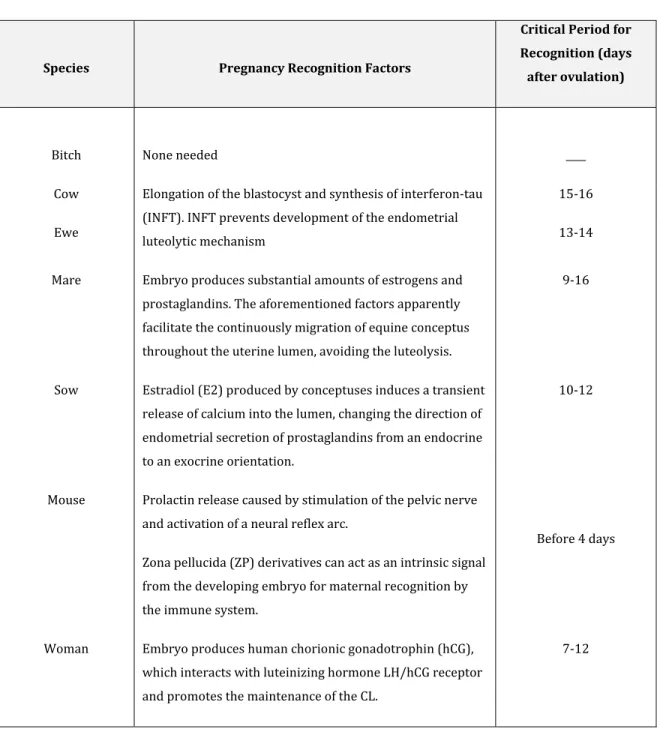
recognition of ruminant pregnancy requires conceptus signals, which alter endometrial physiology, resulting in establishment of the pregnancy. Precisely timed embryo recognition in the genital tract is essential for establishment of an optimal maternal environment for embryo implantation (Spencer, et al., 2007). A deficient signal leads to luteolysis, progesterone level decline and finally, pregnancy loss. The pregnancy recognition factors, critical days of pregnancy recognition and time of conceptus attachment in different mammal species are shown in the following table:
Table 1. Pregnancy recognition factors.
Species Pregnancy Recognition Factors
Critical Period for Recognition (days after ovulation)
Bitch None needed ____
Cow Elongation of the blastocyst and synthesis of interferon-tau
(INFT). INFT prevents development of the endometrial luteolytic mechanism
15-16
Ewe 13-14
Mare Embryo produces substantial amounts of estrogens and
prostaglandins. The aforementioned factors apparently facilitate the continuously migration of equine conceptus throughout the uterine lumen, avoiding the luteolysis.
9-16
Sow Estradiol (E2) produced by conceptuses induces a transient
release of calcium into the lumen, changing the direction of endometrial secretion of prostaglandins from an endocrine to an exocrine orientation.
10-12
Mouse Prolactin release caused by stimulation of the pelvic nerve
and activation of a neural reflex arc.
Zona pellucida (ZP) derivatives can act as an intrinsic signal from the developing embryo for maternal recognition by the immune system.
Before 4 days
Woman Embryo produces human chorionic gonadotrophin (hCG),
which interacts with luteinizing hormone LH/hCG receptor and promotes the maintenance of the CL.
7-12
(Bazer & Roberts, 1983; Gandolfi, et al., 1992; Roberts, et al., 1996; Duc-Goiran, et al., 1999; Spencer, et al., 2004; Spencer, et al., 2004; Senger, 2005; Bazer, et al., 2008; Fujiwara, et al., 2009; Walker, et al., 2009; Farin, et al., 2010; Klein, et al., 2010).
2.1.3. Animal models of implantation
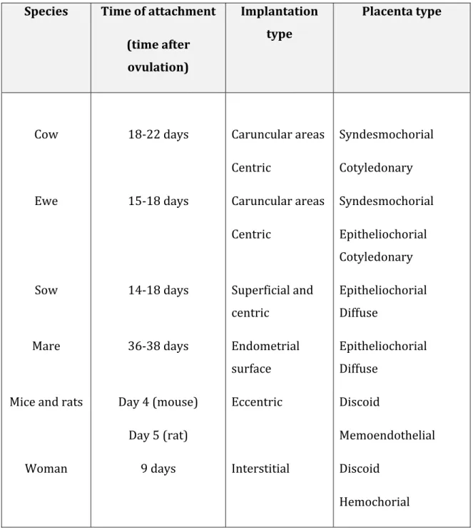
The embryo implantation is a complex process where local paracrine signaling between embryo and the endometrium occurs in order to obtain a strong attachment between them. In species with invasive placenta, the free floating blastocyst attaches to the maternal endometrium, invades into the stroma and establishes the placenta. This requires a synchronization of a viable blastocyst to implant and an endometrium able to respond to embryonic signals. There is diversity in types and time of implantation among species (see _Table. 2).
Table 2. Different types of placenta, implantation and time of attachment.
Species Time of attachment
(time after ovulation)
Implantation type
Placenta type
Cow 18-22 days Caruncular areas
Centric
Syndesmochorial Cotyledonary
Ewe 15-18 days Caruncular areas
Centric
Syndesmochorial Epitheliochorial Cotyledonary
Sow 14-18 days Superficial and
centric
Epitheliochorial Diffuse
Mare 36-38 days Endometrial
surface
Epitheliochorial Diffuse
Mice and rats Day 4 (mouse)
Day 5 (rat)
Eccentric Discoid
Memoendothelial
Woman 9 days Interstitial Discoid
Hemochorial (Dey, et al.; Lee & DeMayo, 2004; Senger, 2005)
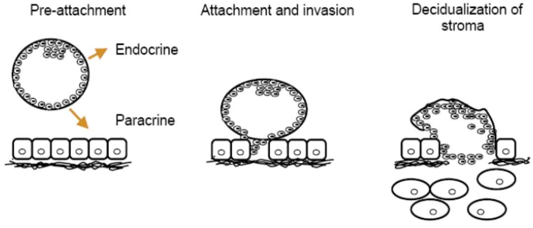
In general, the first step in implantation after the preattachement is when cells of the trophectoderm from the blastocyst migrate between the epithelial cells, displacing them and penetrating as far as the basement membrane. The second step is when the trophoblast cells invade through the basal membrane coming in contact with the maternal stroma. The final step in this process is the decidualization, which consists of transformation of the endometrial stromal cells in highly secretory cells to prepare the endometrium for implantation (Sharkey & Smith, 2003) (see figure. 1).
Figure 1. Cartoon representing the initial phases of murine implantation. During the pre-attachment phase, the embryo uses endocrine and paracrine mediators to signal its presence to the mother. Following apposition and attachment, the embryo invades and promotes decidualization of the maternal stroma. Taken from Sharkey and Smith, 2003.
After implantation, the conceptus undergoes massive growth, due largely to the development of extraembryonic membranes. The formation of these membranes is the vital importance in the ability of the embryo to attach in some species. In fact the chorioallantoic membrane is the foetal contribution to the placenta and will provide the surface for attachments to the endometrium (Senger, 2005). Regardless of the placental type, the purpose of attachment is to bring embryonic tissue and conceptus blood vessels into contact with the maternal blood supply to allow nutrient uptake, waste elimination, and gas exchange between the mother and the conceptus (Lee & DeMayo, 2004).
2.2. Bovine pregnancy
2.2.1. Implantation
In ruminants implantation takes place when the spherical blastocysts elongates to a tubular form, after which the embryo and associated membranes begin to adhere strongly to the maternal endometrium (Spencer, et al., 2004; Song, et al., 2010)
2.2.2. Type of placentation
The bovine placenta is classified as synepitheliochorial (Wooding, 1992). In contrast to haemochorial placenta, placentation in the ruminants is characterized by multiple discrete areas of attachment, called placentomes, and formed by interactions of patches of the chorioallantois with the endometrium (Igwebuike, 2006).
2.2.3. Morphometry and histology of bovine placenta
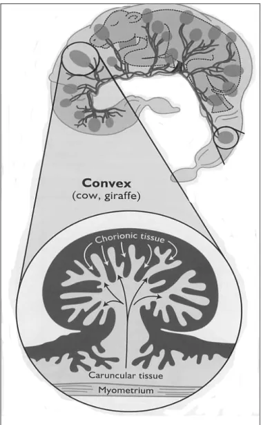
The placentome consists of a fetal portion (the cotyledon) contributed by the chorion, while the maternal contact sites are the caruncles, originated from uterus (Figure 2).
Figure 2. Diagrammatic representation of the bovine placentome. The cotyledonary tissue is characterized by numerous “button-like” structures distributed across the surface of the chorion. The cotyledons (black) fuse with the maternal caruncles (gray), and they form a placentome. Source: (Senger, 2005).
The formation of placentomes is a ongoing process that begins immediately after pregnancy recognition, and the chorion becomes strongly attached to the caruncles of the uterus approximately at day 40 of gestation (Leiser & Kaufmann, 1994; Senger, 2005). In cows there are between 70 to 120 cotyledons distributed across the surface of the chorion, and apparently there is no change in the total number of placentomes during gestation (Laven & Peters, 2001). Moreover, the cotyledons display a villous interdigitation of fetal villi with maternal crypts of the complementary caruncles (Leiser, et al., 1997). This adhesion of the trophectoderm and the endometrial luminal epithelium occurs in caruncular and intercaruncular areas of the endometrium. In the trophectoderm, two morphological and functional cell population have been identified, these are the mononucletae trophoblast cells and the binucleate trophoblast cells (Igwebuike, 2006). Mononucleate trophoblast cells are cuboidal to columnar cells that constitute four-fifths of the trophoblast population, and are primarily involved in nutrient exchange (Igwebuike, 2006) . On the other hand, mature binucleate cells or trophoblast giant cells (TGC) are large cells that contain two nuclei, but the presence of mononuclear and trinuclear forms have been also reported (Klisch, et al., 1999)(See Figure 3).
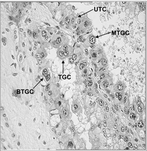
Figure 3. Cross section of a bovine cotyledon at approximately 90 days of gestation. The image shows the different types of trophoblast cells lying in the chorionic villi. UTC, uninucleate trophoblast cells, MTGC, mononucleate trophoblast giant cells, BTGC, binucleate trophoblast giant cells, TTGC, trinucleate trophoblast giant cells (A. Verduzco, unpublished).
UTC
MTGC
BTGC
2.2.3.1. Bovine Trophoblast Giant Cells
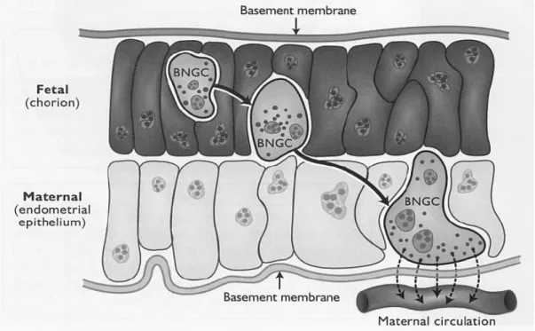
The trophoblast giant cells represent approximately 20% of the trophectoderm and it has been demonstrated that they originate from mononucleate trophoblast cells by acytokinetic mitoses (mitoses uncoupled from cell division) (Igwebuike, 2006). Moreover the occurrence of trinuclear cells is the result of the fusion of binuclear trophoblast cells with uterine epithelial cells (Klisch, et al., 1999). The TGCs are believed to be endocrine cells that produce factors, which are necessary to mediate gas and micronutrient exchange through the placenta and specially to guarantee fetal survival (Wooding, 1992). These cells are the only placental cells that have the capacity to migrate across the feto-maternal interface to fuse with columnar cells of the feto-maternal uterine epithelium. The TGCs have at least two functions: 1. to form a hybrid feto-maternal syncytium essential for successful implantation and subsequent placentomal growth; 2. to synthesize and secrete protein and steroid hormones as placental lactogen (CSH1) and progesterone (P4) (Spencer et al, 2004). Migration and fusion of TGCs with maternal endometrial cells are imperative for the delivery of the aforementioned factors into the maternal compartment and subsequently to the maternal circulation (Senger, 2005; Igwebuike, 2006)(Figure 4).
Figure 4. Schematic representation of how binucleate giant cells (BNGC) or Trophobast giant cells (TGCs) migrate from the chorion to the endometrial epithelium in ruminants. These cells are thought to secrete placental lactogens, pregnancy specific proteins and steroid hormones. Source (Senger, 2005).
2.2.4. Pregnancy recognition in cattle
In ruminants the columnar trophoblast cells synthesize and secrete IFNT, the pregnancy recognition signal from Days 8 to 24 of gestation. The effect of the IFNT is the maintenance of a functional CL, abrogation of the endometrial luteolytic mechanism, and also establishment of uterine receptivity to implantation (Kubisch, et al., 2001; Spencer & Bazer, 2004; Bazer, et al., 2009; Farin, et al., 2010). IFNT acts directly on the uterine endometrium blocking the expression of estrogen receptor 1 (ESR1), and therefore indirectly silences oxytocin receptor (OXTR) expression in the endometrial epithelium, preventing endometrial prostaglandin F2α (PGF(2α) synthesis and release (Walker, et al., 2009; Banu, et al., 2010; Farin, et al., 2010).
2.2.4.1. Ruminant endometrial receptivity
Endometrial receptivity is a self-limited period in which the endometrium acquires the ability to adhere the developing blastocyst (Dominguez, et al., 2003). In bovine, this process involves numerous physiological, molecular and cellular interactions between the conceptus, the receptive endometrium, and the corpus luteum; which play a pivotal role in establishment of the pregnancy and development of the placenta (Hashizume, 2007; Farin, et al., 2010).
Steroid hormones, cytokines, chemokines, growth factors, IFNT, pregnancy-associated glycoproteins (PAGs) and chorionic somatomammotropin hormones (CSH) secreted by the ovary, endometrium and developing embryo, have been
shown to be involved in the coordination of the endometrial receptivity (Adamson, 1993; Sharkey, 1998; Hashizume, 2007).
In ruminants the coordinate expression and sequential exposure of the pregnant endometrium to P4, IFNT, CSH1, prolactin (PRL) and growth hormone (GH), are necessary to activate and maintain endometrial remodeling, secretory function, and uterine growth during gestation (Spencer, et al., 1999). In this context, IFNT and P4 are essential factors for endometrial receptivity in early bovine gestation. Exposure of endometrium to steroid hormones, especially P4 and estrogens, prepare the endometrium for implantation and then maintain the gestation by promoting uterine growth, modulating the maternal immune system and suppressing myometrial contractions (Graham & Clarke, 1997; Sharkey & Smith, 2003; Goff, 2004).
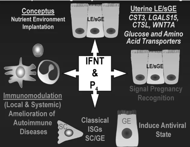
The primary action of IFNT is to abrogate the pulsatile secretion of PGF(2α), blocking the luteolytic mechanism (Bazer, et al., 2009). Moreover recent studies in ovine and bovine, indicate that conceptus IFNT also stimulated genes such as radical S-adenosyl methionine domain containing 2 (RSAD2), interferon induced with helicase C domain 1 (IFIH1), IFN-stimulated gene 15 (ISG15); which are important genes in the establishment of uterine receptivity to implantation, conceptus development and modulation of local immune cells in the endometrium (Song, et al., 2007; Yang, et al., 2010) .Figure 5 presents the principal pleiotropic (producing more than one effect) effects of P4 and IFNT in mammal’s pregnancy.
In addition to the molecules listed above, other factors and genes such as metalloproteinases (MMP8,MMP10,MMP26), integrins, mucins (MUC4,MUC5B),
secreted phosphoprotein 1 (SSP1), calcitonin, leukaemia inhibitory factor (LIF), homeobox genes (HOXA10 and HOXA11), have been reported essential for uterine receptivity in the human and mouse (Lim, et al., 1999; Sharkey & Smith, 2003; Altmae, et al., 2010; Casals, et al., 2010; Sendag, et al., 2010). Some of the aforementioned molecules have yet proven useful as biomarkers of endometrial receptivity (Casals, et al., 2010; Fogle, et al., 2010; Rackow, et al., 2011).
Although the implantation process is different in bovine compared to human and mice; a recent study points to the potential use of endometrial and embryo gene expression patterns as predictors of pregnancy success in cattle (Salilew-Wondim, et al., 2010). In this study cell division, kinesin, integrins, chemokines, insulin growth factors, solute carriers, WD repeats, adapter protein, leucine reach repeats, claudins, coiled-coil domain, inositol, ATPases, Zink finger, S100 calcium binding protein, collagen families, RNA binding motif, Rho GTPase activating protein, CD molecules, keratins, protein kinases and phosphatase gene clusters were differentially expressed in embryo biopsies resulting in calf delivery.
In addition, transcript levels of many new candidate genes involved in the regulation of transcription, cell adhesion, modulation of the maternal immune system and endometrial remodeling were found to be increased during the implantation window (Bauersachs, et al., 2006).However, the precise mechanism or mechanisms involved in the ruminant endometrial receptivity have not been fully investigated.
Figure 5. General pleiotropic effects of progesterone (P4) and interferon-tau (IFNT)on conceptus development, pregnancy and maternal well-being in mammal’s pregnancy. Uterine luminal epithelium (LE), stromal cells (SC), glandular epithelium (GE), superficial glandular epithelum (sGE), cystatin C (CST3), galectin 15; lectin (LGALS15), cathepsin L, cysteine proteinase (CTSL) and wingless-type MMTV integration site family, member 7A (WNT7A). Source: (Bazer, et al., 2008).
2.3. Steroid Biosynthesis
2.3.1. General aspects
The synthesis of steroids is a vital process for essential physiologic functions such as blood salt balance, carbohydrate metabolism and reproduction. Steroid hormones are synthesized predominantly in the adrenal cortex, testis, ovary, placenta and central nervous system (Simpson & Waterman, 1983; Hornsby, 1987; Payne & Youngblood, 1995; Arensburg, et al., 1999; Pasqualini, 2005; Rekawiecki, et al., 2008; Rone, et al., 2009). The steroids occur in a large diversity of biologically active forms; this variety is due partially by steroid-synthesizing tissues, depending upon the set of enzymes present in it. Steroids include principally: testosterone (androgens), estradiol (estrogen), progesterone
(progestin), cortisol/corticosterone (glucocorticoid), aldosterone
(mineralocorticoids), neurosteroids and a wide variety of intermediate metabolites (Bogan & Hennebold, 2010). In general, P4 and estradiol are classified as sex-steroids, and are produced primarily in ovary and placenta; whereas glucocorticoids and mineralocorticoids are produced in adrenal glands, androgens in testes, and neurosteroids in brain (Enyeart, 2005; Pasqualini, 2005; Ghayee & Auchus, 2007; Mellon, 2007; Rekawiecki, et al., 2008; Lavoie & King, 2009) (Figure 6) . Although steroid hormones are synthesized with different physiological actions in each tissue, their synthesis depends on a common precursor, namely cholesterol (Ghayee & Auchus, 2007).
Ch
ol
este
rol
Pr
egnenolone
Pro
ge
st
ero
ne
17 OH-Pre
gn
en
ol
on
e
DHE
A
DHE
A-S
An
dr
os
te
ne
di
on
e
Te
st
os
te
ro
ne
Estr
one
Es
tra
di
ol
Ad re na l Z R Go nads17
OH-Pr
ogeste
rone
Ovary placentaDOC
Cort
ic
ost
er
on
e
Deoxy
cor
tis
ol
Cor
tis
ol
Ad renal ZF /ZRAl
do
st
er
on
e
Ad re na l Z GFigure 6. Overview of steroidogenesis and the principal steroid hormones. Boxes indicate
the major tissues for each reaction (adrenal glands, gonads and placenta). Abbreviations include DOC (deoxycorticosterone), DHEA (dehydroepiandrosterone), DHEA-S (DHEA sulfate), ZF (zona fasciculata), ZR (zona reticularis) and ZG (zona glomerulus). From Lavoie and King, 2009.
2.3.2. Cholesterol transport
In all mammals, cholesterol is an important sterol found in the cell
membranes, which can be derived from the diet or be synthesized de novo (Bloch,
1952). It is well known that cholesterol is the starting point for biosynthesis of
steroids, oxysterols and bile acids in mitochondria (Russell, 2003; Steck & Lange, 2010). Cholesterol is transported in the blood plasma to the target tissue as lipoproteins (Plosch, et al., 2007). High density lipoproteins (HDL) and low density lipoproteins (LDL) are the principal carriers responsible for cholesterol transport. The relative contribution of HDL or LDL in cholesterol uptake varies among species and tissues. There are two known pathways for cholesterol absorption and import into the cell: a non-selective endocytic pathway and a selective absorption pathway. In the non-selective pathway, LDL are specifically bound and internalized
via the LDL family receptors (LDLr); and the receptor fuses with the endosomal
pathway for distribution of the lipoproteins (Murphy & Silavin, 1989; Argov, et al.,
2004). On the other hand, the selective pathway uses a specific receptor, the
scavenger receptor class B member I (SCARB1) situated at the plasma membrane to bind both LDL and HDL (Hoekstra, et al., 2009; Kolmakova, et al., 2010). The selective pathway takes the cholesterol present in the lipoproteins, and it is transferred directly to the cell membrane without absorption of the lipoprotein particles (Argov, et al., 2004; Steck & Lange, 2010). The mRNA expression of the
was determined in bovine ovarian cells at different stages of follicular development (Argov, et al., 2004).
Once inside the cell the cholesterol esters and triglycerides in the cell can be stored in lipid droplets, which are bound by a phospholipid membrane (Rone, et al., 2009). The stored cholesterol esters present in the lipid droplets are converted to
free cholesterol. In steroidogenic cells, upon hormonal stimulation there is
increased cholesterol absorption through the plasma membrane; and cholesterol
esters are hydrolyzed by lipases, principally hormone-sensitive lipase (HSL)(Kraemer & Shen, 2002). HSL interacts with various cholesterol-binding proteins to direct the cholesterol to the outer mitochondrial membrane, where the first step in the steroidogenic process takes place (Lavoie & King, 2009).
2.3.2.1. Importing cholesterol into the mitochondria
In the acute steroidogenic response, cells must deliver large amounts of cholesterol: from the cytoplasm to the outer mitochondrial membrane, and from there to the inner mitochondrial membrane. It is believed that two strategic pathways could be the responsible for the movement of cholesterol across aqueous cytoplasmic spaces (Soccio & Breslow, 2004; Rone, et al., 2009). In the first “the vesicular pathway”, the cholesterol is incorporated into vesicular membranes (e.g. lysosomes, endosomes, peroxisomes) which subsequently fuse with other membranes, delivering cholesterol into the inner mitochondrial membrane. However this route appears to be a minor pathway. On the other hand, in the
second route “the non-vesicular pathway”, cholesterol can be solubilized and transferred by cholesterol-binding proteins, through the cytosol to the mitochondria (Soccio & Breslow, 2004; Rone, et al., 2009).
Several candidate proteins, including SNARE complexes, vimentin-intermediate
filaments, nonspecific transporters like sterol carrier protein 2 (SCP2), voltage-dependent anion channel (VDCA) and a subfamily of lipid binding proteins referred to as STARD4, STARD5 and STARD6 have been proposed for the cytoplasmic cholesterol transfer to the outer membrane (Bogan & Hennebold, 2010). The transfer of cholesterol from the outer to the inner membrane is considered as a rate-limiting step in steroid formation; and until now the prototypical steroidogenic acute regulatory protein (STAR), is the principal molecule implicated in this process. Mutations in STAR protein cause a specific phenotype in intracellular cholesterol distribution (lipoid congenital adrenal hyperplasia)(Lin, et al., 1995; Sugawara, et al., 1995). Nevertheless studies suggest that STAR proteins presumably work in concert with the translocator protein (18 kDa, TSPO) and endozepine, which is the natural ligand for TSPO (Niswender, 2002; Papadopoulos, et al., 2006; Rone, et al., 2009). Figure 7 illustrates the trafficking of cholesterol to the mitochondria for steroidogenesis more precisely.
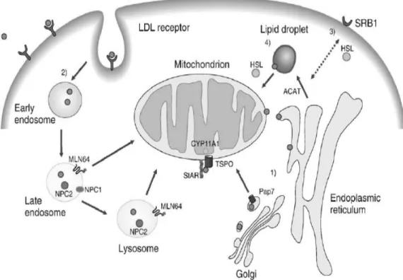
Figure 7. Uptake and transport of cholesterol to the mitochondria for steroidogenesis. Pathway 1 shows the passive diffusion of cholesterol from the endoplasmic reticulum to the mitochondria, via
the acyl-CoA binding domain containing (PAP7 or ACBD3) protein. In pathway 2, low density
lipoprotein (LDL),binds to the LDLr and cholesterol is trafficked through the endosomes to the mitochondria via the Niemann-Pick type C proteins (NPC) proteins and StAR-related lipid transfer
(START) domain containing 3 (MLN64) protein. In pathway 3 cholesterol is transported from high
density lipoprotein (HDL) to the plasma membrane by the SR-BI receptor. In pathway 3 and 4 hormone-sensitive lipase (HSL) converts esterified cholesterol from the plasma membrane or from the lipid droplets in free cholesterol. Finally free cholesterol interacts with lipid-binding proteins present in the cytosol for delivery to the mitochondria, where the enzyme cytochrome P450, family
11 subfamily, A polypeptide 1 (CYP11A1) transforms cholesterol into pregnenolone. Taken from
2.3.2. Steroidogenic enzymes
The biosynthesis of steroids involves several enzymes, however all the steroid hormones are synthesized through a common precursor steroid, pregnenolone (Hu, et al., 2010). In the inner mitochondrial membrane, the enzyme CYP11A1 converts the insoluble cholesterol into soluble pregnenolone, by the enzymatic cleavage of a 6-carbon side-chain of the 27-carbon cholesterol molecule (Ghayee & Auchus, 2007). Pregnenolone is next metabolized to progesterone by hydroxy-delta-5-steroid dehydrogenase, 3 beta- and steroid delta-isomerases (HSD3B). HSD3B isoforms are membrane-bound enzymes, which are found in both mitochondrial and microsomal membranes, depending on the type of cell. Their enzymatic actions are essential to catalyze the conversion of intermediary metabolites into active steroid hormones (see figure 8). The variant isoforms of HSD3B are highly homologous in their amino acid sequence, and are specific for each species (Lavoie & King, 2009). In humans, two isoforms have been identified: HSD3B1 and HSD3B2, and both function as steroid dehydrogenase/isomerases and are involved in the biosynthesis of all active steroid hormones (Payne & Youngblood, 1995). Moreover, the isoforms of HSD3B are expressed in a cell and tissue-specific manner. HSD3B1 is expressed in placenta, skin and breast tissue, whereas HSD3B2 is expressed in adrenal gland, ovary, and testis (Payne & Youngblood, 1995; Pasqualini, 2005). In cows, HSD3B1 is present in ovary and placenta (Takagi, et al., 2007; Vanselow, et al., 2010).
Like HSD3Bs, the hydroxysteroid (17-beta) dehydrogenase enzymes (HSD17B) play essential roles in steroidogenesis, catalyzing the final step in the biosynthesis of active gonadal steroid hormones, estradiol, and testosterone (see figure 8). They are found as either membrane-bound or soluble enzymes and, unlike HSD3B, there is very little homology among different HSD17B isozymes (Payne & Youngblood, 1995). Among the different forms of human HSD17B, only types 1, 3 and 7 participate in the final step of biosynthesis of active steroid hormones in gonads (Payne & Youngblood, 1995). In cattle, Cytochrome P450, family 17, subfamily A, polypeptide 1 (CYP17A1) is expressed in ovary and trophoblast cells (Schuler, et al., 2006; Luo, et al., 2010; Mossa, et al., 2010)
Further, pregnenolone can also be hydroxylated in certain tissues by CYP17A1 to 17-OH-pregnenolone to DHEA (Lavoie & King, 2009). The progesterone and 17-OH-pregnenolone are basic steroids, which can be used as intermediary metabolites for all other steroid hormones (see figures 6 and 8). Table 3 give a general view of the principal steroidogenic proteins and enzymes expressed in bovine.
Figure 8. Conventional pathways and associated enzymes of steroid biosynthesis. Abbreviations include: DHEA dehydroepiandrosterone and DHT Dihydrotestosterone. Taken from Ghayee and Auchus, 2007 with some modifications.
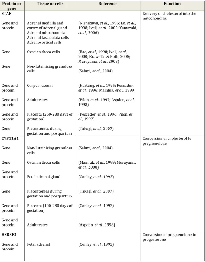
Table 3. Principal steroidogenic proteins and genes expressed in a different number of bovine tissues and cells.
Protein or
gene Tissue or cells Reference Function
STAR Gene and protein Gene Gene Gene and protein Gene and protein Gene and protein Gene
Adrenal medulla and cortex of adrenal gland Adrenal mitochondria Adrenal fasciculata cells Adrenocortical cells Ovarian theca cells Non-luteinizing granulosa cells Corpus luteum Adult testes Placenta (260-280 days of gestation) Placentomes during gestation and postpartum
(Nishikawa, et al., 1996; Lo, et al., 1998; Ivell, et al., 2000; Yamazaki, et al., 2006)
(Bao, et al., 1998; Ivell, et al., 2000; Braw-Tal & Roth, 2005; Murayama, et al., 2008) (Sahmi, et al., 2004)
(Hartung, et al., 1995; Pescador, et al., 1996; Mamluk, et al., 1999) (Pilon, et al., 1997; Aspden, et al., 1998)
(Pescador, et al., 1996; Pilon, et al., 1997)
(Takagi, et al., 2007)
Delivery of cholesterol into the mitochondria. CYP11A1 Gene Gene Gene and protein Gene Gene and protein Gene and protein Non-luteinizing granulosa cells
Ovarian theca cells Fetal adrenal gland Placentomes during gestation and postpartum Placenta (100-280 days of gestation)
Adult testes
(Sahmi, et al., 2004)
(Mamluk, et al., 1999; Murayama, et al., 2008) (Conley, et al., 1992) (Takagi, et al., 2007) (Conley, et al., 1992) (Aspden, et al., 1998) Conversion of cholesterol to pregnenolone HSD3B1 Gene and
protein Fetal adrenal (Conley, et al., 1992)
Conversion of pregnenolone to progesterone
Gene Gene Enzymatic activity by substrate metabolism Gene and protein Gene Gene and protein Non-luteinizing granulosa cells Theca cells
Caruncles and cotyledons (120-280 days of gestation)
Placenta (100-280 days of gestation)
Placentomes during gestation and postpartum Adult testes (Sahmi, et al., 2004) (Murayama, et al., 2008) (Tsumagari, et al., 1994) (Conley, et al., 1992) (Takagi, et al., 2007) (Aspden, et al., 1998) CYP17A1 Gene and protein Gene and protein Gene and protein Gene and protein Placentomes from pregnant (days 80-284), prepartal and parturient period.
Fetal adrenal Adult testes Adrenocortical cells
(Conley, et al., 1992; Schuler, et al., 2006)
(Conley, et al., 1992) (Aspden, et al., 1998)
(McCarthy, et al., 1983; Zuber, et al., 1985)
Catalysed androgen synthesis
CYP19A1 Gene Gene Gene and protein Granulosa cells Theca cells Placentomes from pregnant (days 80-284), prepartal and parturient period.
(Vanselow, et al., 2001) (Luo, et al., 2010) (Schuler, et al., 2006)
Conversion of androgens into oestrogens
SULT1E1
Gene Gene
Villi and caruncle (Day 25-250 of gestation) Caruncular and cotyledonary tissue at parturition (Ushizawa, et al., 2007) (Hirayama, et al., 2008) Sulfation of estrogens STS
Gene Caruncular and
cotyledonary tissue at parturition
(Hirayama, et al., 2008)
Conversion of sulfoconjugated estrogens into estrogens