Active-site-directed inactivators of the Zn2+-containing
D-alanyl-D-alanile-cleaving
carboxypeptidase
of
Streptomyces albus G
Paulette
CHARLIER,*t
OttoDIDEBERG,t Jean-ClaudeJAMOULLE,$
Jean-Marie FRERE,* Jean-Marie GHUYSEN,*GeorgesDIVE: and Josette LAMOTTE-BRASSEURt*Service deMicrobiologie,
Faculte
deMedecine,Universite
deLie'ge, InstitutdeChimie, B6,B-4000 Sart Tilman, Liege, Belgium, tServicede Cristallographie, Faculte' des Sciences, Universite de Liege, Institut de Physique,B5,B-4000 SartTilman, Liege, Belgium, and$Service de ChimieAnalytique, Faculte deMedecine,Universite deLiege, InstitutdePharmacie, Fl,RueFusch, 3-5, B-4000
Lie'ge,
Belgium
(Received6October 1983/Accepted 16 January 1984)
Several types ofactive-site-directed inactivators (inhibitors) of the Zn2+-containing
D-alanyl-D-alanine-cleaving carboxypeptidase were tested. (i) Among the
heavy-atom-containingcompounds examined, K2Pt(C204)2inactivates the enzyme with a second-order rate constant of about 6 x 10⁻²M-1. 1 andhas only one binding site
located close to the Zn2+ cofactor within the enzyme active site. (ii) Several
compounds possessingboth aC-terminalcarboxylate function and, at the other end of the molecule, a thiol, hydroxamate or carboxylate function were also examined.
3-Mercaptopropionate (racemic) and 3-mercaptoisobutyrate (L-isomer) inhibit the enzymecompetitivelywith a
Ki
value of 5 x10⁻⁹-10 x10-9M. (iii)ClassicalP-lactam
compoundshaveaveryweakinhibitory potency. Dependingon thestructure of thecompounds,enzymeinhibitionmay becompetitive(and binding occurs to the active
site)ornon-competitive (and binding causes disruption of the protein crystal lattice).
(iv) 6-,B-Iodopenicillanateinactivates the enzyme in a complex way. At high
P-lactam
concentrations,thepseudo-first-orderrateconstant of enzymeinactivation has a limit value of 7 x 10⁻⁴s- 1.6-p-Iodopenicillanate
binds to the active site just in front of theZn2+ cofactor and superimposes histidine-190, suggesting that permanent enzyme inactivation is by reaction with this latter residue.
The transpeptidation and carboxypeptidation reactions involved in the last stages of wall
peptidoglycan synthesis in bacteria are catalysed bymultiple
D-alanyl-D-alanine-cleaving
peptidases (in short DD-peptidases) (Ghuysen et'al.,
1981).The serine
DD-carboxypeptidases/transpeptidases
operate via acyl-enzyme formation and aresus-ceptible to inactivation by
P-lactam
antibiotics.One of them has been
crystallized
and thepenicillin-binding site has been located (Kelly et
al.,
1982).
A metallo DD-peptidase is also known thatrequiresaZn2+ cation boundtotheactive site forcatalysis(Didebergetal., 1980b). This enzyme functions solely as a carboxypeptidase and ishighly resistant to penicillins and cephalosporins (Ghuysenetal., 1981).Its
primary
structure(Joris etal.,1983)andcrystalstructure at0-25nm(2.5
A)
resolution (Dideberg et al.,1982)-
are known.Attempts were made
tofjind
effective inhibitors(inactivators)
of theZn2+
pn-carboxypeptidase.
The results obtained during this first exploratory
phase are described below. Materials and methods ,B-Lactam compounds
Cephalosporin C, cephalothin and
cephalo-glycinewerefrom EliLilly and Co., Indianapolis, IN, U.S.A. The
P-iodopenicillanate
was agift from Dr J. T. Hendersonand Dr. J. E. G. Kemp from Pfizer Central Research, Sandwich, Kent, U.K.; 7-[2
-(p-iodophenyl)acetamido]cephalosporanate
was that previously used (Dideberg et al., 1982)
and was synthesized by Dr. L. Christiaens, Department ofOrganic Chemistry, University of
Lege.
Heavy-metal-containingcompounds
K3UO2F5wasagiftfrom Dr. D.Stuart,Oxford, U.K.
K2PtCl4,
(NH4)2IrCl6
andNaAuCl4
were Vol. 219from Aldrich Chemical Co., Milwaukee, WI, U.S.A., NaUO2(CH3CO2)3 was from Merck,
Darmstadt,Germany, and K2Pt(C204)2 was
syn-thesized asdescribed in Pascal (1958).
Zn2+-directedinactivating reagents
Compounds(2), (3), (4)and(10)of Fig. 4(see the Results section)were generously given by Dr. M. Ondetti of the Squibb Institute for Medical Research, Princeton, N.J.,U.S.A.Compounds(1), (5), (6), (7), (8), (9) and (11) were from Aldrich
EuropeDivision, Janssen Pharmaceutica N.V., B-2340 Beerse, Belgium. Compounds(12), (13)and
(14)were synthesized during the present work.
Buffers
Buffer A was 10mM-Tris/HCl, pH 8.3,
contain-ing 5mM-MgCl2, and buffer B was 10mM-Hepes
[4-(2-hydroxyethyl)-1-piperazine-ethanesulphonic acid]/NaOH, pH8.0, containing 5mM-MgCl2.
Enzyme
The Zn2+ DD-carboxypeptidase was purified to
protein homogeneity as described in Duez et al.
(1978). The enzyme activity was determined by incubating the enzyme with 1.8 mM-N"Nt-diacetyl-L-lysyl-D-alanyl-D-alanineat37°C in a total volume of
30Ml
of either buffer A or buffer B and measuring enzymically the amount of C-terminal D-alanineliberated(Frereetal., 1976; DeCoen et al., 1981).Interaction between the Zn2+ DD-carboxypeptidase and
P-lactam
compounds (Frere et al., 1978)The
P-lactam
compounds react with the enzymeaccording to:
lactam compound by dialysis, A, is the activity
after incubationofthe enzyme- -lactamcomplex E-I* at 37°C, andA is the activity ofanenzyme
sample treated as above but without
P-lactam
compound).Determination ofthe apparent rate constants (ka values)for theformation of complexes E-I* andK
andk+2 values. The enzyme (3.4Mm) and the JJ-lactamcompound(at concentrations rangingfrom 1to 15mM)wereincubatedtogether at37°Cin60 1 of buffer B. Formation of complex E-I* was
determinedon 1
MI
samplesremovedafter increas-ing times ofincubation (andfurther incubated for 15min at 37°C with 1.8 mM-Ac2-L-Lys-D-Ala-D-Ala). Plots of ln(A,/Ao)
versus time yieldedstraight lines
(A,
is the residual enzymeactivityat time t andAo
is the initial enzyme activity), permitting calculationofka. Plots of ka versus[I]showeddeviation fromlinearityathigh[I]values. Individual values of K and
k+2were
obtainedfrom the reciprocalplotsl/ka
versus 1/[I].Effect
ofthe presenceofAc2-L-Lys-D-Ala-D-Ala.The enzyme(1.2Mm), the f-lactamcompound(at
concentrations ranging from 0.05 to 10mM) and
Ac2-L-Lys-D-Ala-D-Ala(at concentrations ranging
from 0.275 to 1.1mM)were incubated together for 2min at37°Cin100I of bufferA,andthe amount of released D-Ala was determined.
Lineweaver-Burkplots (1/vversus
1/[S]
for various[I] values)allowed determination of the type of enzyme inhibition (competitive ornon-competitive).
Interaction between the Zn2+ DD-carboxypeptidase andnon-fi-lactam inhibitors (inactivators)
All theexperimentswereperformed in buffer A.
E+I
K'
E.Ik-
2.E-I*-'
E +degradationproduct(s)whereE is enzyme, I is
fl-lactam
compound,E-I*isinactivecomplex,Kis thedissociationconstant and k+2 andk+
3are
first-orderrate constants. TheZn2+ DD-carboxypeptidase shows high intrinsic resistance to all
fl-lactam
compounds tested be-cause oflow k+2/Kvalues.Determination ofthe k+3 values. The enzyme
(33
gM)
and theP-lactam
compound (20mm)
wereincubatedtogetherfor 2hat37°Cin 25Mlofbuffer B. The reaction mixture was 100-fold diluted in bufferB,dialysed againstthesamebuffer for2hat
4°C and then incubated at 37°C. Samples were removed afterincreasingtimes ofincubation,and the extentofenzyme recovery (orbreakdown of
complexE-I*)wasdetermined afteranadditional incubation of 15min at 37°C with 1.8mM-Ac2-L-Lys-D-Ala-D-Ala.Plots of ln
[ 1-(A,
-AO)/(A
-AO)]
versus time gave rise to
straight
lines(Ao
is the residual activity after removal of the unboundf,-Asafirst approximation,theinhibiting
(inactivat-ing)potencyofacompound was expressed by the
concentration necessary to decrease the enzyme
activity by 50%
(ID50
value) when enzyme, substrate andinhibitorwereincubatedtogether at37°C. Pseudo-first-order rate constants (ka) of enzymeinactivationweredetermined as indicated above. Lineweaver-Burk plots and Dixon plots
(1/vversus
[I]
for various[S] values)were used to determine the type of enzyme inhibition and to measure theKi
values.Enzymecrystals
Crystallization of the Zn2+
DD-carboxy-peptidasewascarriedoutasdescribed inDideberg
et al. (1979).
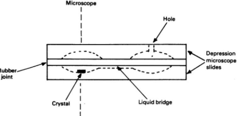
Preparationof enzyme crystalderivatives
A cellconsisting oftwomicroscope slides with
depressions
asshown in Fig. 1, and fixedtoeachMicroscope Hole _ 4 L__b- Depression microscope - / slides Liquidbridge
Fig. 1. Schematic representationofthe cell usedforthepreparationof complexes oftheZn2+ DD-carboxypeptidase crystal
For further detailssee thetext.
7.5 16 33 45
I,-Iodopenicillanatel(mM)
Fig.2. Relative variationofthe birefringence ofacrystal of the Zn2+ DD-carboxypeptidase asafunction ofincreasing
concentrationsof J-iodopenicillanate
AB/B is the relative variation of birefringence. Diffusion of ,-iodopenicillanate was carried out
within a total period of time of 24h. I-lodo-penicillanate wasadded stepwise after 30min (1),
90min (2),180min(3),270min (4), 18h (5),20h(6) and 24h(7). For further detailsseethetext.
other byarubberjoint, wasused. Before the cell
was mounted, an enzyme crystal (of thickness
0.05mm)anda30plsampleof motherliquorwere
deposited intheleft-hand well,and another
30pl
sampleof motherliquorwasdepositedinthe
right-hand well. Thetwodroplets wereconnected bya
thinliquidbridge bymeansofafine needle. After
the cell had beensealed,asolution of theselected
compound (heavy atomororganic inhibitor) was
injected in theright-hand well and thecompound concentration wasincreased stepwise, thesystem being allowed to equilibrate between each step. Formation of the enzyme derivative was
moni-toredby measuringthechangesinbirefringenceof
thecrystal withtime, with aBerekcompensator. Fig. 2 shows the results obtained with
fi-iodo-penicillanate. All the above operations were
performed at 200C. X-ray-diffraction studies
The experimentswereperformed witha
Hilger-Watts diffractometer fitted with an He-filled
tunnel and with the use of Ni-filtered Cu
K.
radiation (40kV, 26mA). Absorption corrections
were deduced from scans (North etal., 1981), and the decreased intensity due to radiation damage was corrected by a time function. The
maximum decay correction was 20%. The
posi-tions ofthe heavy atoms were determined from
FHLE Patterson functions, and parameters were
refined by a least-squares procedure. For the
complexes formed with Pt(C204)2-, U02F53
-and AuCl4-, the process was followed by three
cycles of phase refinement at0.25nm resolution. Thecrystal data for the nativeenzymeand for
theenzyme-,B-iodopenicillanate complexaregiven
in Table 1.Thecomplete0.28nmdataset(hkl and h1cl) was measured on one crystal. The structure
factor amplitudes of the enzyme-,B-lactam
com-plexwerescaledtothose ofthenativeenzyme.The
Rdif.
factorwas0.11.Adifference electron-density map was computed from the coefficients mIFcj-IFpj
and the multipleisomorphous-replacement phases(Didebergetal., 1982). Reflex-ions of resolution lower than 1nm were omitted from these summations.
Results
Inactivation of the Zn2+ DD-carboxypeptidase by heavy-metal-containing compounds
Pseudo-first-order kineticsofenzyme inactiva-tion were observed with(NH4)2IrCl6, NaAuCl4,
K2Pt(C204)2 and K2PtCl4. The corresponding
second-order rate constants were 2.4x 10⁻²,
5.7x10⁻², 6.2x 10⁻² and 18.6x 10⁻²M-1- 1
respectively. With the two uranyl compounds Vol. 219
Table 1. Crystal data for the native Zn2 + DD-carboxypeptidase and the complex formed withP-iodopenicillanate
Rsym=ZZIIi
-7h1I/1IIh
h h
where
Iid
is theintensity of anhklreflexion and7his the mean intensity.Rdif.
=EIIF
-IFplIl/ZlFpl
where
IFJI
is thestructure-factor amplitudeof thef-iodopenicillanate-Zn2+DD-carboxypeptidasecomplexandIFpI
that of the native protein.a (nm) 5.11 5.119 b (nm) 4.97 4.964 c (nm) 3.87 3.866 O
(0)
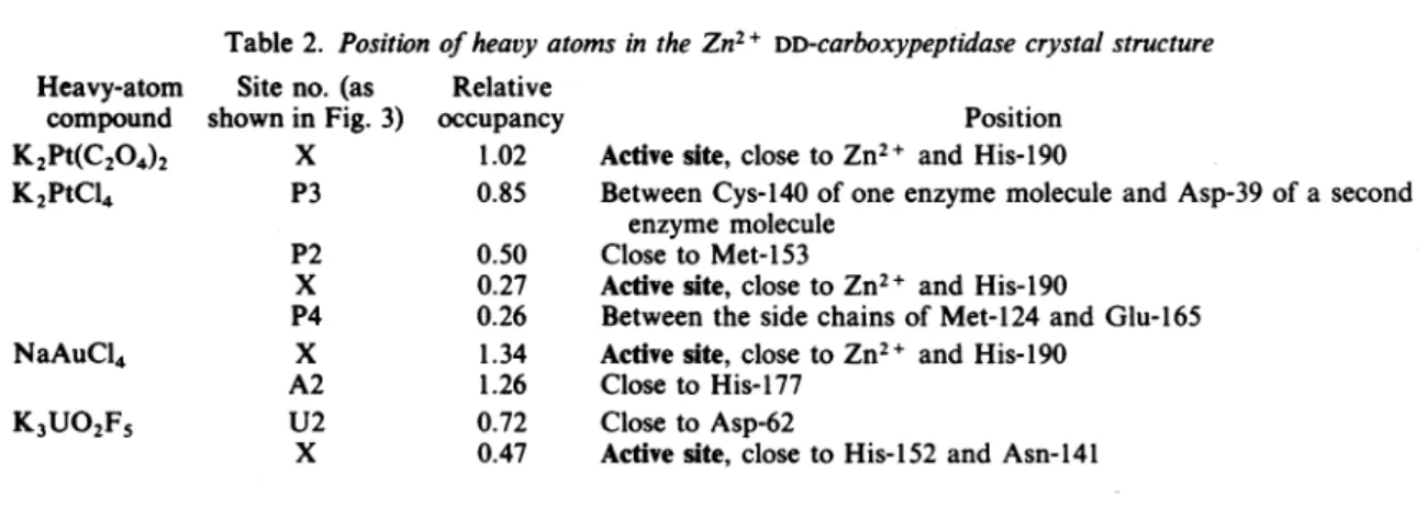
100.60 100.56 Rsym (%) 3.2 4.6Table 2. Positionofheavy atoms in theZn2+ DD-carboxypeptidase crystalstructure
Siteno. (as Relative
shown inFig. 3) occupancy
X 1.02 P3 0.85 P2 x P4 x A2 U2 x 0.50 0.27 0.26 1.34 1.26 0.72 0.47 Position Activesite, close toZn2+ and His-190
BetweenCys-140 ofoneenzymemoleculeand Asp-39ofasecond
enzymemolecule
Closeto Met-153
Activesite, closetoZn2+ and His-190
Between the side chains of Met-124 and Glu-165 Activesite, closetoZn2+ and His-190
Closeto His-177 Closeto Asp-62
Activesite, closeto His-152 and Asn-141
[20mM-NaUO2(CH3CO2)3and
30mM-K3UO2F5],
rapid but partial enzyme inactivation (35% and 25% respectively) occurred during the first 5-10min of incubation. During subsequent incuba-tion, enzyme inactivation by NaUO2(CH3CO2)3 continued to proceed, but at a very low rate(O.93xlO-2M-1'-s1). K3UO2F5 had no further
detectable effect, at least within 60min of
treatment.
The positions of the aforementioned heavy
atoms[except(NH4)2IrCl6 and NaUO2Ac3] in the Zn2+ DD-carboxypeptidase and the relative
occu-pancies of the binding sites were determined (Table 2 and Fig. 3). Compound K2Pt(C204)2
gave rise to one single binding site, which was
locatedwithin theenzymecavity.The other heavy-atom-containing compoundsgave risetomultiple (two tofour) binding sites, but in eachcase only one of these was also located within the enzyme
active site.
CompoundK2PtCi4wasthemostpotent
inacti-vator (as expressed by the second-order rate
constant ofenzyme inactivation), although bind-ingtothe active sitewasratherweak(as expressed by the relative occupancy value). However,
K2PtCl4 was the only heavy-atom-containing compound that occupied four distinct binding
sites,andtheenzyme-K2PtCl4 derivativewasthe
onlyone that showedlack of isomorphism.
Attempts to construct 'bi-product' analogue
inactivators
A general application of the concept of 'bi-product' analogues (Byers & Wolfenden, 1972) makes use of functional groups able to interact regio- and stereo-specifically with the different
enzymebinding areasandtoanchorthe molecule firmly within the enzyme active site. This
ap-proach ledtothesynthesis of specificinhibitors of the Zn2+-containing carboxypeptidases A and B, thermolysin and angiotensin-converting enzyme
(Cushman etal., 1977).
TheZn2+ DD-carboxypeptidase active site prob-ably possesses at least three subsites (Fig. 4). SubsitesS2,
SI
and,withalessstrict requirement,subsite S' must be suitably complemented by the substrate side chains, and Arg-136 is probably involved in charge-pairing. Several bifunctional compoundsweretested(Fig. 4).Most oftheactive
compounds possessed a C-terminal carboxylate
function capable ofbindingtothecationiccentre
Arg-136, and either a thiol, hydroxamate or
carboxylate function capable of binding to the Zn2+cofactor.Exceptincompound(10),the back-Native Complex Heavy-atom compound K2Pt(C204)2 K2PtC14 NaAuC14 K3UO2F5
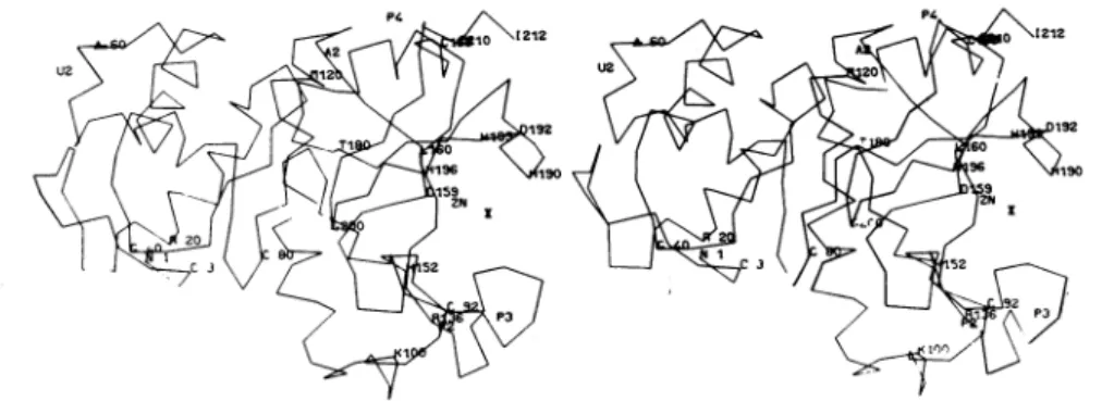
Fig.3. Stereoscopic view of thethree-dimensional structure of the Zn2+ DD-carboxypeptidase
Thea-carbon-positions were derived from a Kendrew model. For thelabelling of the heavy-atom-binding sites, see Table 2. Theamino acid residues were designated by the one-letter code (IUPAC-IUB Commission on Biochemical Nomenclature,1969). Numbering of the amino acid residues is that derived from X-ray data (Dideberg etal., 1982). Atpresent, there are still minordiscrepancies between this sequence and that derived from chemical and enzymic degradation (Joris etal., 1983). Strong experimental evidence suggests (i) that His-152, His-193 and His-196 serve as Zn2+ ligands, (ii) that charge-pairing may occur between the C-terminal carboxylate function of the bound substrate and Arg-136, and (iii) thatHis-190 may play the role of a proton donor in catalysis. The model was drawn with the
useof thePackgraph programme (Katz&Levinthal, 1972).
bones ofthecompoundsweretoo short topermit interaction with subsites
S,
and S2. Unless otherwise specified, racemic compounds wereused. From this preliminary exploration, the following are apparent.
1. Inactivating potency requires a C-terminal anionic group
[compare
compounds (13)and(14)]
andgreatlydependsonthenature of the functionthat is directed towards the Zn2+ cofactor. The
comparable pairs compounds (1) and (12) and
compounds (2) and (11) showed that replacing a
thiol by a hydroxamate or a carboxylate at this position caused a 1000-fold and 100000-fold decreased affinity
respectively.
2. As shown by Lineweaver-Burk plots of l/v
versus 1
/[S]
forvarious concentrationsof I(wherevis theinitialvelocity of substrateconsumption,Sis the substrate Ac2-L-Lys-D-Ala-D-Ala and I is the
inactivator), compounds
(1)-(5)
were potentcom-petitive
inhibitors. Contrary totheexpectations,
comparable effectiveness was achieved whether the lateralchainassumed tocomplementsubsite
S;
was aproton,amethyl groupin theLconfiguration
or a methyl group in the Dconfiguration,
andwhether one single or two carbon atoms were
disposedbetween theterminalthiol and
carboxy-latefunctions. Moreover,themostpotentinactiva-tors were
3-mercaptopropionate
(Ki
=5x10- 9M)
and the L-isomer of
3-mercaptoisobutyrate
(Ki
= 10x10-9M),
whereas the D-isomer of3-mercaptoisobutyrate
wassomewhatbutdistinctly
lesspotent(Ki
=60x10-9M).
Suchsmall truncat-edmolecules did notofferthepossibility
ofcom-plementing
subsitesSI
andS2,
and,
astheabove observations stronglysuggested,
subsiteS;
aloneseemed not to play any significant role in the binding process. Considering the flexibility of these molecules, enzyme inactivation may be causedby bidentate interaction with formation of
apenta-orhexa-co-ordinate Zn2+ ion (Fig. 5). 3. As models showed, folding the above mole-culestoformapenta-orhexa-co-ordinateZn2+ ion orientedthe lateralchain towards His-190, which mayplay the roleofprotondonorin catalysis. A
methyl group, especially inan Lconfiguration, had
noprominentstericeffect,but morebulky
substi-tuentsonthea-carbonatom[compounds
(6)-(10)]
were much less favourable, thus considerably decreasing the binding efficacy.4. Alternative mechanisms for the loss of enzymeactivityontreatment with 3-mercaptopro-pionatewere envisaged. One of them might have beenby
opening
of thedisulphide bridges[which
occurinthreeplaces in theprotein
(Dideberg
etal., 1982; Joris et al.,1983)].
This possibility waseliminated by the following experiment. The enzyme
(1.1 M)
and the inhibitor(100
tM)
wereincubatedtogether for
5min
at37°C in buffer A,which treatment caused complete enzyme inhi-bition. The reaction mixture was then supple-mented with 1mm-dithiobis-2-nitrobenzoate, which causedenzyme recovery with a first-order
rateconstantof about6.4 x 10-4s 1
(90%
recoverywas observed after
60min).
Another alternativewasthat loss of enzymeactivity might have been
by
removalof theZn2+
cofactor from the active site by the inhibitor. Inturn,
thispossibility waseliminated by the following experiment. The enzyme
(5nM)
and the inhibitor(100nM)
wereincubatedtogetherat37°CinbufferAuntil
80%
of Vol. 219Enzyme cavity S2 Si \ Zn Arg-136 CH3 CO NH
,CH
0 0 H214 CCH3R CH3 0 CH3-CO-NH-CH-C-NH-CH-C-NH-CH-C=0 L D Inactivators (1) .3-Mercaptopropionate 3-Mercaptoisobutyrate racemic D-isomer L-isomer (5) 2-Mercaptopropionate (6) Acetylcysteine (7) D-Cysteine (8) Penicillamine (9) Formylpenicillamine (10) Captotril (1 1) Methylsuccinate(12) Succinicacid monohydroxyamate
13) 2-Hydroxylpminopropionate (14) 2-HydrgxylaminoprQpionitrilp -S -CH2-( -S CHM2-k, 0-IH2-C=O iH3 0 :H-C-0 CH 0--S-CH-C=0 CH3 C=O NH 0-II -S CH2-lH-t;=0 H+0--9 C -2-H-C,= CH3CH3 NH3+ -S-C CH-C=u H CH3CH3NCH 0 \/ L &- CH3O S CH CH-CI0 HO 0 0-NH-C-CH2- H2--=0 HO CH3 0 NH-CH-C=0H0 CH3 0
H?
-H NH-CH-eC_-N ID50 (M) 15x1 0-K, (M) 5 x 10⁻⁹ 35 x10-9 45 x10-9 60x10-9 lOx10-9 SOx10-9 40 x10-6 90x10-6 70x10-6 105x10-6 25x10-6 65x10-6 3x10⁻³ 15x10-6 1x 10⁻³ No activityFig. 4. Positioning ofthe tripeptide Ac2-L-Lys-D-Ala-i-Alaintheac#tv siteoftheZn2+DV-carboxypeptWdaseand inhibittn
potencyofbifunctional compounds
ID50is defined in theMaterialsand methods section. With compounds (1)-(5) theenzyme(Sum)andtheinhibitor
(10-200nM)wereincubated for60min. With compounds (6)-(10) and(12)theenzyme(0.8yM)and theinhibitor
(33 pm-1 mM)wereincubated for5min. With compounds (11), (13) and (14) theenzyme
(0.8.pm)
and the inhibitor(1-10mM)wereincubated for5min. All the experimentswereperformedat37°C and in buffer A. For further details
seethetext.
(2) (3)
(4)
His-i 93
His-152 His-196
\/
Zn2+
-s
Fig. 5. Possiblebidentate interaction between theZn2+
DD-carboxypeptidase and a bifunctional (thiol/carboxylate)
inhibitor
the initial activity had disappeared (i.e. after 5min). The reaction mixture was then supple-mentedwith120nM-ZnSO4, i.e. at aconcentration much higher than that of 3-mercaptopropionate, which treatment failed to regenerate an active
carboxypeptidase.
Inactivation by bicyclic ring-fused azetidinone structures
(a)Classical penams and 3-cephems. On the basis of reaction (1) (see the Materials and methods
section), thelower the K value and thehigher the
k+2andk+
3values,
the more potent aP-lactam
as an enzyme inactivator. Although, once formed,complexesE-I* are very stable(low k+3 values),
the 6-f-substituted penicillins were found to be
extremely poor inactivators of the Zn2+
DD-carboxypeptidase because of very high K values and very lowk+2 values(150mMand 8 x 10⁻⁴s-1 respectivelyfor phenoxymethylpenicillin, which is the most potent penam inactivator so far tested) (Frere et al., 1978). The
7-p-substituted
cephalo-sporinsweresomewhatbetterinactivators,mainly because of more favourable K values (1-10mM).
Lineweaver-Burk plots showed that
7-fl-substituted A3-cephems fall into two groups.
Cephalothin (K=9.5mM; k+2=5 x
10-4S-1;
k+3=5x10⁻⁶s-; in buffer B) and
cephalo-sporin C (K=2mM; k+2=1 x10⁻⁴s-I;
k+3=2x10⁻⁶s- ; in buffer B) acted
non-com-petitively. Inparallel to this, cephalothin(10mM) and cephalosporin C (1.66mM) destroyed the crystal lattice, thus preventing any study from
beingcarried outby X-raydiffraction. Incontrast, cephaloglycine (K=4.5mM; k+2=3x 10⁻⁴s-I; k+3 =9x 10⁻⁶S-1; in bufferB)and
7-[2-(p-iodo-phenyl)acetamido]cephalosporanate
(K=3.5mM; k+2=6x 10⁻⁴s-l; k+3=1+10-6s-1; in bufferA) acted competitively. The enzyme complex
formed with thislattercompoundshowed
satisfac-toryisomorphismandindicated that the
f-lactam
wasbound to the activesite(Didebergetal., 1980a,1982).
(b)
P-Iodopenicillanate. 6-p-Halogenopenams
aref-lactamase
inactivators,and,in thisrespect, 6-p-iodopenicillanate has been much studied (Frereet al., 1982, and other references included in that paper). Kinetically, the interaction between
the Zn2+ DD-carboxypeptidase and
6-p-iodo-penicillanatewasfound toresemblethat observed with the ,B-lactamases, in that complete and permanentinactivationwasobservedonly at high
(>12500) inactivator/enzyme ratio. When this ratio was decreased, enzyme inactivation was incomplete, and, after subsequentprolonged incu-bation, partial and spontaneous recovery of en-zymeactivity was observed. Althoughmorework remainsto be donein order toelucidatethe under-lying mechanism, these observations suggested a branched pathway including (i)normal turnover through anintermediateE-I*, (ii)re-arrangement
of this intermediate into a transitorily inactive complex E-I**, and (iii) re-arrangement of the same intermediate into a permanently inactive complex E-I*** (see Fig. 6 legend). The first phases of enzyme inactivation by increasing concentrations of
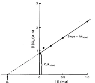
P-iodopenicillanate
proceededaccording to pseudo-first-orderkinetics.Fromthe
ka
valuesthusobtained,aHanesplot,[I]/ka
versus[I],gave riseto astraightline(Fig. 6) intersecting
theordinateaxis at
KIlka
(limit)and the abscissaaxisat -
Ki.
Fromthe slope(l/ka(limit)),
aka(limit)
value of 7x10⁻⁴s-1 was computed. TheKi
value was0.66mM.
An enzyme crystal was treated with ,B-iodo-penicillanate as described in the Materials and methodssection. The[I]/[E] ratiowasestimatedto be about 8300, and extensive inactivation oc-curred.(Indeed, whenthemeasurementsdescribed
below were completed, the treated crystal was
dissolved,andDD-carboxypeptidaseassayscarried outon asampleofthis solutionshowedthat85%of the enzyme has been permanently
inactivated.)
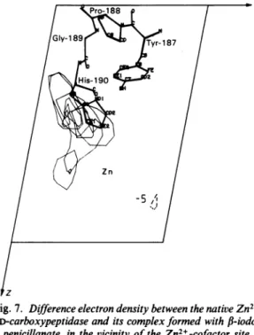
The enzymederivativewasperfectlyisomorphous,and the difference Fourier synthesis at 0.28nm
resolution gave one main peak that was much higher than any other features ofthe map. The
shape and the height of the peak did not
correspond to an iodine atom with a full occu-pancy, strongly suggesting that iodine had been eliminated.Thesiteofinteractionclearly extended
from His-190just in front of the Zn2+ cofactor (Fig. 7) to an open cavity formed by the two segmentsHis-190-Gly-189-Pro-188 and
Asn-141-Ser-Asn-Val-144-Gly-Gly-Ala-147.
Theseobser-vations strongly suggest that, in the process of permanent inactivation ofthe Zn2+ DD-carboxy-peptidase by
6-f-iodopenicillanate,
thecatalytic-ally activeHis-190 was
alkylated
with lossofI-.Such a mechanism would drastically differ from
the inactivation oftheserine ,B-lactamases, whose serineresidueisirreversibly acylated
by
P-bromo-(iodo)penicillanate
(Cohen &Pratt,1980).
Aminor peakwasalso foundin thedifferencemapclosetoArg-136. The shape and the size of the
density
suggestedthat this
peak
wasattributabletoanion. Vol. 2190.5 1.0
[II (mm)
Fig.6. Hanesplot forthe interaction between theZn2+ DD-carboxypeptidaseandf-iodopenicillanate (1) ka(s-1) is thepseudo-first-order rate constant of enzyme inactivation. Assumingthat the interactionobeysthe
scheme:
k E+P
K kk2 +
where P and P E E-I* dg ai prE-I**dc thE+Pe \E-I***
where P and P'=degradationproducts, then:
I k+2+k+3
and
ka(Iim)
k+2(k+4+k+5)
Discussion
Non-f,-lactam bifunctional reagents may be
potent competitive inhibitors of the Zn2+
DD-carboxypeptidase. Enzyme inactivation by these
agents(see Fig.4)cannotbe attributedtocleavage of the disulphide bridges and subsequent dis-organization of the protein three-dimensional
structure, norto the detachment of the Zn2+ ion
from the active site. Mostprobablyit is duetothe immobilization of the Zn2+ cation by co-ordina-tion,thuspreventing the cofactor from performing itscatalytic function. The bifunctional compounds
so far examined complement only part of the bindingsites oftheenzymeactivesite. Specificity and perhaps increasing inactivating potency
re-quires more elaborate structures that not only wouldfirmly anchor theinhibitor molecules in the activesite butwould beapttointeractspecifically with the
SI
and S2 subsite targets.Classical
P-lactams
areextremelypoor inactiva-tors(aswellassubstrates)of the Zn2+ DD-carboxy-peptidase.Some of them(cephaloglycine and7-[2-(p-iodophenyl)acetamido]cephalosporanate) are
weak competitive inhibitors. X-ray-diffraction studies have provided direct evidence that this
lattercompound binds tothe enzyme active site.
Other
f,-lactams
(cephalosporin C and cepha-lothin) are weak non-competitive inhibitors. Inparallel to this, these compounds destroy the protein crystal lattice and profoundly alter the scattering behaviour of the protein by inducing large conformational changes and/or aggregation (Labischinskietal., 1984).6-,B-Iodopenicillanate is
an unusualpenam in that it has a veryshortside chain. Its interaction with the Zn2+ D-carboxy-peptidaseiscomplex,butathigh ,B-lactam
concen-trations it inactivates the enzymewith a
pseudo-first-order rate constant of 7x10⁻⁴s-i (limit
value). X-ray analysis shows that this
P-lactam
compoundis located in theenzymeactive sitejustinfrontof Zn2+ and superimposes His-190 (which isthoughtto actasprotondonorduring catalysis of
a bound peptide substrate), suggesting that
His-190actsasanucleophile byitsloneelectron pair of
thenitrogenatomandundergoesalkylation by 6-f-iodopenicillanate with loss of iodine.
The abovestudiesstronglysuggestthat both the high intrinsic resistance of the Zn2+ DD-carboxy-peptidaseto,B-lactam compoundsand the
extreme-ly low substrate activity of these ,B-lactam
com-poundstowards the Zn2+DD-carboxypeptidaseare
k+3K
Ki=
Pro188X XGIy-189 Tyr187 Hs-190 2
14R
Zn -5A lzFig.7. Differenceelectron density between the nativeZn2+ DD-carboxypeptidaseand its complexformedwith
P-iodo-penicillanate, inthe vicinity of theZn2+-cofactor site
The contourlevels,shown on anarbitraryscalewith maximal value of10,are -5, 5and 7. The contour of negative value(-5)is shown as a broken line. The unit cell was divided into 48 x 48 x 48points. The
Figureis a composite ofsectionsof Y= 19 to24,and
theareashown extendsfromX=38 to 48 and from Z=14to34.The sequences Tyr-187-Pro-188-Gly-189-His-190 (as positionedin the Kendrewmodel)
areshownasboldlines.
caused,mostprobably,by a particular geometry of the enzyme active site. Production of enzyme-ligand associations of highproductiveness occurs only with carbonyl donor peptides (thus, for example, the standardtripeptide substrate
AC2-L-Lys-D-Ala-D-Ala is hydrolysed with a turnover number of 2.5s-1 at 37°C). In contrast, the
fi-lactam compounds are little predisposed to aligncorrectly with regard to the active functional groups. In fact, models show that with these compounds it is difficult to align simultaneously
boththe
P-lactam
amide bond with the Zn2+ ion and the C-terminal carboxylate with Arg-136 because of collisions with other side chains of the enzyme active site (P. Charlier & 0. Dideberg,unpublished work).
One way to resolve the crystal structure of a protein by X-ray-diffraction methods rests upon the preparation ofheavy-atomcomplexes with this protein. In this procedure,attachment of the heavy atomto theprotein should not perturb the atomic arrangement of the crystal and should involve as fewaminoacidside chainsaspossible(Blundell&
Johnson, 1976). Among the heavy-atom-contain-ing compounds tested, K2Pt(C204)2 is remarkable inthat it hasonly onebinding site, located within theenzymecavity, closetothe Zn2+ cofactorand the putative proton donor His-190. Inactivation proceeds with a second-order rate constant of about 6x1-2M-1-S-1.
The work was supported in part by the Fonds de la Recherche Scientifique Medicale,Brussels (contract no.
3.4501.79),the FondsNational de la Recherche Scienti-fique, Brussels (contract no.1.5.212.82F),and anAction
Concerteefinanced by theBelgianState(conventionno.
79/84-Il).Wethank Dr. M. A.Ondetti from the Squibb
Institute for Medical Research, Princeton, NJ, U.S.A., fordiscussionand advice, and forprovidingcompounds
(2), (3)and(4)shown in Fig. 4, and Dr. J. T. Henderson and Dr. J. E. G. Kemp from Pfizer Central Research for
providing6-p-iodopenicillanate.
References
Blundell, T. L. & Johnson, L. N.(1976) Protein Crystallo-graphy, pp. 183-239, Academic Press, London Byers, L. D. & Wolfenden, R.(1972) J. Biol. Chem.247,
606-608
Cohen,S. A.& Pratt, R. F. (1980) Biochemistry 19, 3996-4003
Cushman,D.W.,Cheung, H. S., Sado, E. F. &Ondetti,
M. A.(1977) Biochemistry 16, 5484-5491
DeCoen, J. L., Lamotte-Brasseur, J., Ghuysen, J. M., Frere, J. M. &Perkins, H. R.(1981) Eur. J. Biochem. 121, 221-232
Dideberg,O.,Frere, J. M. & Ghuysen, J. M. (1979)J. Mol. Biol. 129, 677-679
Dideberg, O., Charlier, P., Dupont, L., Vermeire, M., Frere, J. M. &Ghuysen, J. M.(1980a)FEBSLett.117, 212-214
Dideberg, O., Joris, B., Frere, J. M., Ghuysen, J. M., Weber, G., Robaye, R., Delbrouck, J. M. & Roelandts, I. (1980b) FEBS Lett. 117, 215-218
Dideberg, O., Charlier, P., Dive, G., Joris, B., Frere,
J. M. &Ghuysen,J. M. (1982)Nature(London) 299,
469-470
Duez, C., Frere, J. M., Geurts, F., Ghuysen, J. M.,
Dierickx,L. &Delcambe, L. (1978)Biochem. -J. 175,
793-800
Frere, J. M., Leyh-Bouille, M.,Ghuysen, J. M., Nieto,
M.&Perkins, H. R.(1976)Methods Enzymol. 45, 610-636
Frere, J. M., Geurts, F. & Ghuysen, J. M. (1978)
Bio-chem. J. 175, 801-805
Frere, J. M., Dormans, Ch., Duyckaerts, C. & De
Graeve,J. (1982) Biochem. J.207,437-444
Ghuysen, J. M., Frere, J. M., Leyh-Bouille, M., Dideberg,O.,Lamotte-Brasseur, J., Perkins,H. R.& De Coen, J. L. (1981) in Topics in Molecular Pharmacology(Burgen,A.S. V. &Roberts,G. C.K., eds.),pp. 63-97, Elsevier/North-Holland Biomedical Press, Amsterdam
IUPAC-IUB Commission on Biochemical
Nomencla-ture(1969)Biochem. J. 113, 1-4
Joris, B., Van Beeumen, J., Casagrande, F., Gerday, Ch.,Frere, J. M. &Ghuysen,J. M.(1983)Eur.J. Bio-chem. 130, 53-69
Katz, L. & Levinthal, C. (1972) Annu. Rev. Biophys. Bioeng. 1, 465-504
Kelly, J. A., Moews, P. C., Knox, J. R., Frere, J. M. & Ghuysen, J. M. (1982) Science 218, 479-481
Labischinski,H., Giesbrecht,P., Fischer, E.,Barnikel, G., Bradaczek, H., Frere, J. M., Houssier, Cl., Charlier, P., Dideberg, 0. & Ghuysen,J. M. (1984) Eur.J. Biochem. 138, 83-87
North, A. C.T., Philips, D. C. & Matthews, F. S.(1981)
ActaCrystallogr. Sect. A 24, 351-359
Pascal, P.(1958)NouveauTraitedeChimieMinerale,vol. 19, pp. 774-775, Masson, Paris