Keywords: Immunotherapy; Immune Molecules; Rituximab; Burkitt’s Lymphoma
Objective: Burkitt’s lymphoma is an aggressive B cell malignancy that is associated with Epstein-Barr virus (EBV) infection. The
monoclonal antibody rituximab is used to treat many B cell malignancies, including Burkitt’s lymphoma. Studying immune mol-ecule modulation in Burkitt’s lymphoma allows insight on developing new or refining existing immunotherapy agents for refractory or chemotherapy-resistant patients. The main purpose of this study was to explore rituximab’s impact on the expression of immune molecules associated with immune activation or immune inhibition by comparing the expression of rituximab-treated cells to IgG-treated cells using flow cytometry.
Methods: Burkitt’s lymphoma cell lines Raji, Ramos, Bjab, and an EBV-transformed B cell line, COX, were cultured and treated with
an optimally-determined concentration of rituximab or human IgG for 24 hours. Immune modulation was determined by flow cy-tometric analysis of the immune molecules HLA-I, HLA-DR, PD-L1, and CD40.
Results: Treatment of cells with rituximab, 10 µg/ml, completely downregulated CD20 expression and modulated expression of
immune molecules. Compared to human IgG control, rituximab treatment decreased HLA-1, HLA-DR and CD40 expression on all cell lines, but significantly only for HLA-I on Bjab. Interestingly, the immune inhibitor PD-L1 was decreased on EBV-positive COX and Raji but increased on EBV-negative Ramos and Bjab.
Conclusion: Since HLA-I expression is critical for CD8 T-cell mediated tumor cell destruction, the downregulation of HLA-I could
contribute to an immune escape mechanism. As this was a small study, there is limited transferability of the results to the clinical setting and further experiments comprising larger cell panels are needed.
Rebecca Quilty
1, Kendra Smith
2, David Jones
1, Sheila Drover
21Faculty of Medicine,Memorial University of Newfoundland, Newfoundland & Labrador, Canada
2Department of Biomedical Sciences, Memorial University of Newfoundland, Newfoundland & Labrador, Canada
ABSTRACT
Objective: Le lymphome de Burkitt est une malignité agressive de cellules B qui est associée avec l’infection par le virus
d’Ebstein-Barr (EBV). L’anticorps monoclonal rituximab est utilisé pour traiter plusieurs malignités de cellules B, y inclut le lymphome de Burkitt. Étudiant la modulation de molécules immunitaires dans le lymphome de Burkitt donne un aperçu sur le développement de nouveaux agents d’immunothérapie et le raffinement d’agents existants pour les patients réfractaires ou chimiorésistants. Le but principal de cette étude était d’explorer l’impact de rituximab sur l’expression de molécules immunitaires associées à l’activation ou l’inhibition immunitaire, en comparant l’expression de cellules traitées par rituximab aux cellules traitées par IgG, en utilisant la cytométrie en flux.
Méthodes: Les lignées de cellules du lymphome de Burkitt : Raji, Ramos, Bjab et une lignée de cellules B transformées par le EBV,
COX ; ont été cultivées et traitées avec une concentration optimale de rituximab ou d’IgG humain pendant 24 heures. La modulation immunitaire a été déterminée par analyse de cytométrie en flux des modulateurs immunitaires HLA-I, HLA-DR, PD-L1, et CD40.
Résultats: Le traitement de cellules avec le rituximab, 10 µg/ml, a complètement réprimé l’expression de CD20 et l’expression
modulée des molécules immunitaires. Comparé au contrôle d’IgG humain, le traitement par rituximab a diminué l’expression de HLA-1, HLA-DR et CD40, mais seulement de façon significative pour l’HLA-I de la lignée Biab. Curieusement, l’inhibiteur immunitaire PD-L1 a diminué pour les lignées EBV-positive COX et Raji, mais a augmenté pour les lignées EBV-négatives Ramos et Bjab.
Conclusion: Comme l’expression de HLA-I est critique pour la destruction de cellules tumeurs par les cellules T CD8, réprimer le
HLA-I peut contribuer à un mécanisme d’évasion immunitaire. Comme cette étude était très petite, la transférabilité de ces résultats est limitée dans le contexte clinique et donc, plus d’expériences comprenant plusieurs types de cellules sont nécessaires.
RÉSUMÉ
Rituximab and Immune Molecule Modulation in
Burkitt’s Lymphoma Cell Lines
B
urkitt’s lymphoma is a rare aggressive mature B cell malignancy (1). It accounts for 40% of all childhood non-Hodgkin lymphomas but only less than 5% of adult lymphomas (2). Burkitt’s lymphoma is found in three epidemiologically distinct forms that are prevalent in different populations: the endemic form, the sporadic form, and the immunodeficiency-associated form (3). The endemic form is most prevalent in Africa and the Middle East and is the form that is associated with EBV. It is diagnosed at a median age of 4-7 years. The sporadic form can be detected all over the world and at any age and represents less than 3% of all non-Hodgkin lymphomas (3). The final form is associated with the human immunodeficiency virus (HIV), is not linked with EBV, and its development does not correlate with CD4 T cell levels. All three subtypes have the same morphology and involve the overexpression of the MYC oncogene expressed on chromosome 8 (4). The activation of the MYC oncogene and resultant uncontrolled cell proliferation is triggered by a translocation with one of three immunoglobulin genes on chromosomes 2, 14, or 22. The translocation between chromosomes 8 and 14 is the most common, triggering 80% of cases of Burkitt’s lymphoma.Over the past few decades, therapeutic techniques boosting the immune system’s ability to fight cancer, or immunotherapy, have become a mainstay in cancer treatment (5). For example, “checkpoint” molecules involved in immune activation that subdue the immune response and prevent autoimmunity have been identified and have been engineered as an additional avenue to prevent cancer growth (6). T cell activation occurs with two main stimulatory steps. The first step is triggered by the binding of the T cell receptor on naïve CD4+ or CD8+ T-cells to the human leukocyte antigen (HLA) on an antigen presenting cell (APC). The HLA is antigen-bound and this first step allows specificity of the T cell’s effector function. The second signal entails the co-stimulatory signals that are also required for appropriate immune activation. The main co-stimulatory signal includes the interaction between CD28 on the T cell and B7 on the APC, but the binding of CD40L on the T cell and CD40 on the APC is also important in certain T cell functioning (7). The absence of these co-stimulatory signals would result in anergy of the T cell (6). This intricate activation process is further sophisticated by its regulation, both centrally and peripherally. Occurring in the lymph nodes, central regulation arises through the binding of cytotoxic T-lymphocyte-associated antigen 4 (CTLA-4) on the surface of T cells to B7 on the APC. As this interaction induces anergy of the T cell, CTLA-4 is typically only upregulated when there is
strong antigen stimulation, and its upregulation out-competes the CD28:B7 interaction. Peripheral regulation occurs at a later stage of the immune response and involves the interaction between programmed cell death protein 1 (PD-1) on T cells and its ligand programmed cell death ligand 1 (PD-L1) on its target cells, including cancer cells. This interaction also results in decreased T cell proliferation and signalling, thus preventing immune-mediated damage towards targeted tissues.
As cancer develops, there is a dynamic interplay between the immune system and cancer called immunoediting, whereby tumours can escape immune system defenses. Although there are many mechanisms by which tumour cells can escape the host immune system, interfering with antigen presentation is an important theme. Tumour cells can down-regulate the expression of HLA Class I on the surface to prevent recognition and attack by CD8+ cytotoxic T cells (8). Tumour cells can also interfere with CD4+ T cell-mediated signalling through the down-regulation of HLA Class II (9). Checkpoint pathways are also not exempt from tumour cell interference, and significant evidence exists of the manipulation of PD-L1 and CTLA-4 pathways by cancer cells to evade immune defenses (6). Rituximab, a monoclonal antibody targeting the CD20 receptor, has been shown to be an effective biologic agent against other types of non-Hodgkin lymphomas such as diffuse large B cell lymphoma (DLBCL) (10). The use of rituximab has also been extended to other B cell lymphomas and there have been several studies that have shown its safety and effectiveness in treating Burkitt’s lymphoma (11,12). Most importantly, a recent randomized control clinical trial was conducted to see if the addition of rituximab to a short chemotherapy protocol improves event-free survival in Burkitt’s lymphoma patients (2). This study included 260 HIV-negative Burkitt’s lymphoma adult patients. These patients were first stratified into two groups based on the presence or absence of central nervous system or bone marrow involvement, and then randomly assigned to the rituximab or no rituximab treatment groups. The rituximab group achieved a better prognosis with an event-free survival of 75% compared to 62% in the no rituximab group. This study solidified the evidence to support rituximab as a treatment for Burkitt’s lymphoma in adult patients.
The implementation of rituximab in chemotherapy protocols has also been studied in pediatric Burkitt’s lymphoma populations and a recent randomized controlled trial showed favourable results (13). This international study randomized 600 patients with high-risk B cell non-Hodgkin’s lymphoma to
RESEARCH
standard Lymphome Malins de Burkitt (LMB) chemotherapy with or without the addition of rituximab. A planned interim analysis including 310 randomized patients, of which 85% had Burkitt’s lymphoma, showed a 1-year event-free survival rate of 94% in the rituximab group, compared to 81% in the standard chemotherapy group. This finding led to an early closure of the study as the rituximab arm had significantly improved outcomes, confirming that this drug’s efficacy also extends to the pediatric population.
Despite the improved outcome with chemotherapy in Burkitt’s lymphoma, poor outcomes continue to be observed in patients with relapsed disease or who are chemotherapy-resistant (3). A retrospective study conducted by the European Group for Blood and Marrow Transplantation described a 3-year overall survival rate of only 7% in chemo-resistant adult patients (14). For pediatric patients with relapsed or refractory mature B cell non-Hodgkin lymphoma, the 5-year overall survival rate was noted to be less than 30% (15). As well, even with the addition of rituximab as salvage therapy in relapsed mature B cell lymphoma pediatric patients, poor survival rates were observed (16). The survival rates of the 37 pediatric and adolescent patients at 1 and 3 years were 40.5% and 37.6%, respectively.
The purpose of this study is to gain further insight into the mechanistic actions of rituximab in the treatment of Burkitt’s lymphoma and to delineate an explanation for the poor outcomes in refractory or relapsed diseased states. The study comprised investigating the cell surface expression of antigen-presenting molecules (HLA-1 and HLA-DR), and two paradoxical immune molecules: PD-L1, an immunosuppressive checkpoint regulator, and CD40, a costimulatory molecule involved in T cell activation. Rituximab treatment of the cell lines may uncover an altered modulation of immune molecules playing an important role in tumor immune escape mechanisms. These changes could help explain potential resistance to rituximab treatment.
This is a pre-clinical study to determine whether rituximab treatment modulates HLA Class I, HLA DR (HLA Class II), CD40 or PD-L1 expression in a panel of Burkitt’s lymphoma cell lines. The first objective of this study was to determine the optimal concentration of rituximab to deplete CD20 on the surface of lymphoma cells. The second objective was to determine if rituximab treatment of Burkitt’s cells alters the expression of HLA Class I, HLA-DR, CD40, or PD-L1.
MATERIALS AND METHODS Cells
Cell lines included three well-characterized Burkitt’s lymphoma cell lines Bjab (Dr. Jacques Thibodeau, Université de Montréal), Ramos (Dr. Mani Larijani, Memorial University), and Raji (Dr. Gerald T Nepom, Benaroya Research Institute), and one EBV-transformed B cell line COX (11th International Histocompatibility Workshop). Cells were grown in complete medium (CM) consisting of RPMI-1640 (Invitrogen) containing 10% complement-inactivated fetal bovine serum (Invitrogen), 2mM L-glutamine (Invitrogen), 2mM antibiotic antimycotic (Invitrogen), and 1mM sodium pyruvate (Invitrogen) at 37 °C in a CO2 incubator. The medium was replenished when the cells reached a density of 5-8 x 10⁵/ml and all experiments were completed on healthy and viable cells as determined by Trypan blue exclusion.
Rituximab Treatment
Rituximab (Health Sciences Centre Pharmacy, St. John’s, NL), a humanized anti-CD20 monoclonal antibody, was tested on COX to determine the optimal concentration of rituximab. Cells were harvested, counted, and cultured at 2.5 x 10⁵ cells/ ml in CM. Cell cultures were treated with rituximab or human IgG (hIgG) (Sigma), ranging from 5 µg/ml to 100 µg/ml, for 24 hours, after which CD20 expression was determined by flow cytometry. The assay was conducted to determine the rituximab concentration that most effectively down-regulated CD20 expression.
To determine if rituximab treatment altered the expression of HLA Class I, HLA DR, CD40 or PD-L1 in Burkitt’s cells, cell lines were treated at time 0 with the appropriate concentration of rituximab or the control antibody, hIgG, at the same concentration. The cells were incubated for the same time period as the previous experiment (24 hours), followed by flow cytometry to determine expression of immune molecules.
Antibodies and Flow Cytometry
Flow cytometry was conducted to measure surface expression of CD20, irrelevant antibody IgG1/2a (ebioscience/local source), HLA Class I (antibody W6/32, in house), HLA-DR (antibody L243, in house), CD40 (antibody B-B20, Abcam), and PD-L1 (antibody MIH2, Abcam). Briefly, 2.5 x 10⁵ cells/assay tube was removed and centrifuged at 1400 rpm for 7 mins. The cells were washed twice with FACS (Fluorescence Activated Cell Sorting) buffer and after resuspending the cells, 100 µL was added to each 5 mL tube. 25 µL of primary mouse antibody
(IgG1/2a, W6/32, L243, B-B20, or MIH2) was then added to each tube and the tubes incubated on ice for 30 minutes. After incubation, they were washed twice with FACS buffer and then 25 µL of secondary antibody (phycoerythrin (PE)-labelled goat anti-mouse IgG, Jackson ImmunoResearch) was added to each tube. The cells were then incubated in the dark on ice for 30 minutes. After incubation, the cells were washed twice and resuspended in 150 µL of paraformaldehyde to fix the cells. Samples were then analyzed on a BD FACSCalibur flow cytometer. Each cell line was tested three times, aside from COX for which only two experiments were conducted. Only two experiments were conducted for COX due to technical difficulties in the experimental protocol.
Interpretation and Statistics
The expression of the immune molecules was determined by the mean fluorescence intensities (MFI) quantified by Kaluza software (Figure 1). The MFI for each marker was adjusted by
subtracting the background MFI, accounting for the irrelevant IgG1/2a binding. The MFIs from the three experiments were averaged for use in statistical calculations. Statistical analysis was carried out using Microsoft Excel software. The significance of differences between hIgG and rituximab treatments was calculated using a two-tailed, paired students t-test. A p-value of less than 0.05 was considered to be significant.
RESULTS
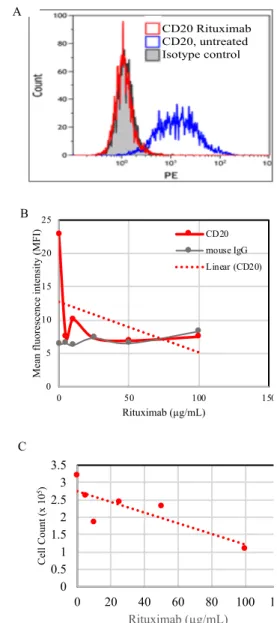
The first objective was to establish a concentration of rituximab that downregulated cell surface CD20 expression but did not have a huge impact on cell viability. COX cells, which have high levels of CD20, were treated with various concentrations for 24 hours, after which flow cytometry and viability assays were performed. As shown in Figure 1 (A, B), all concentrations of
rituximab blocked CD20 expression. Compared to untreated cells, the number of viable cells was reduced in a dose-dependent manner (Figure 1C), as compared to untreated
with only a small impact at the lower concentrations. Based on these results and literature reports (17, 18), 10 µg/mL was selected as the concentration of rituximab for subsequent experiments.
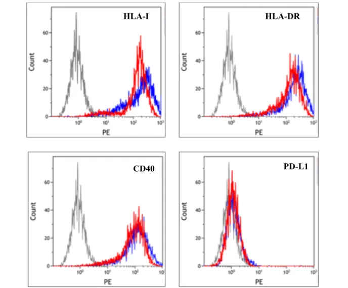
Three sets of experiments were performed on rituximab or hIgG-treated Burkitt’s lymphoma cells, Raji, Ramos and Bjab, and two sets on EBV-transformed COX cell line, followed by flow cytometric analysis. Representative histograms comparing expression of HLA-I, HLA-DR, CD40, and PD-L1 on rituximab and hIgG treated Bjab cells is shown in Figure 2. Compared to
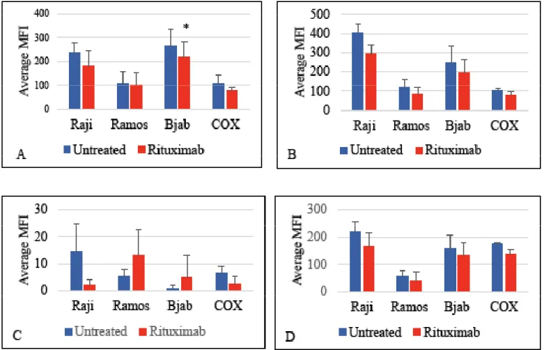
the control hIgG-treated cells, rituximab treatment somewhat reduced expression of HLA-I, HLA-DR, and CD40 on all cells whereas the effect on PD-L1 was variable and cell-dependent (Figure 3).
Figure 1. Determination of the optimal concentration of rituximab concentration to downregulate cell surface expression of CD20 on COX cells. Cells were treated or
not with indicated concentrations of rituximab for 24 hours and then assayed for CD20 expression by flow cytometry and viable cells. A: Representative flow histogram illustrating loss of CD20 using rituximab (10μg/ml) to treat COX cells. B: CD20 downregulation by rituximab is dose dependent, as indicated by decreased mean fluorescence intensities for all concentrations of rituximab compared to untreated cells.
C: The number of viable cells was inversely correlated with
increasing concentrations of rituximab.
CD20 Rituximab CD20, untreated Isotype control A B C
Figure 1
0 5 10 15 20 25 0 50 100 150Mean fluorescence intensity (MFI)
Rituximab (µg/mL) CD20 mouse IgG Linear (CD20) 0 0.51 1.5 2 2.53 3.5 0 20 40 60 80 100 120 Cell Count (x 10 5) Rituximab (µg/mL)
RESEARCH
HLA-I
HLA-DR
CD40
PD-L1
Figure 2. Flow histograms of a representative experiment on cell line Bjab showing expression of HLA-I, HLA-DR, PD-L1 and CD40
following rituximab treatment (red lines) or hIgG treatment (blue lines).correlated with increasing concentrations of rituximab. HLA-class I was significantly (p = 0.013) reduced on rituximab
treated Bjab cells and although reduced on other cells, the effect was not statistically significant (Figure 3A). Similarly, the downward trend for expression of HLA DR, a class II MHC molecule, and CD40 on all treated cells is apparent (Figure
3B and 3B), but not significant. None of the cell lines
expressed much constitutive PD-L1 and rituximab treatment had variable effects (Figure 3C). As shown, Raji and COX cells displayed decreased expression after rituximab treatment, whereas Ramos and Bjab showed increased and variable PD-L1 expression after rituximab. None of the rituximab-mediated changes in PD-L1 were statistically significant.
DISCUSSION
While rituximab treatment of Burkitt’s lymphoma cell lines did show a trend to downregulate surface expression of the
immune molecules HLA DR, CD40 and PD-L1, in the majority of cases this was not statistically significant. There was a significant downregulation of HLA Class I expression in the Bjab cell line.
The downregulation in HLA Class I expression could potentially have negative implications in the use of rituximab to treat Burkitt’s lymphoma. As mentioned earlier, a mechanism through which tumour cells evade immune surveillance is by downregulating HLA Class I on their surface to limit the attack of cytotoxic T cells (8). These CD8+ T cells typically recognize tumor-associated antigens on cancer cells through binding to the antigen on HLA Class I on the cancer cell, an interaction that is limited when this HLA molecule is downregulated (19). This result suggests that rituximab treatment could potentially contribute to an immune escape mechanism in Burkitt’s
RESEARCH
lymphoma, which could negatively impact prognosis.
Despite the immune mechanism illustrated above, the repercussions of the loss of HLA Class I expression in the setting of B cell lymphomas are not well-defined. Interestingly, our results showed a downward trend for HLA-DR expression, which is supported by previous reports showing downregulation of HLA Class II in patients receiving standard chemotherapy with rituximab. This was found to be a negative prognostic factor in B cell lymphomas, but the same has not been shown for HLA Class I expression (19,20). In a retrospective analysis of 144 patients with diffuse large B cell lymphoma and that were treated with standard chemotherapy, the loss of HLA Class I expression did not correlate with a worsened prognosis (19). The authors suggest the work of natural killer cells and rituximab in conducting antibody-dependent cellular cytotoxicity or direct cell killing has compensated for
the decreased activity of cytotoxic T cells through HLA Class I interactions.
CONCLUSION
Based on these results, rituximab treatment tends to result in a decreased cell surface expression of the immune molecules HLA Class I, HLA Class II, PD-L1, and CD40 on Burkitt’s lymphoma cell lines. However, aside from HLA Class I expression on the Bjab cell line, the results were not statistically significant. With these limited results, it is difficult to extrapolate them to the clinical setting to determine whether rituximab therapy will improve treatment outcomes in refractory or relapsing cases of Burkitt’s lymphoma. More data should be collected through further experiments including a wider variety of Burkitt’s lymphoma and B cell lines. If subsequent experiments confirmed these results, exploring the mechanism by which rituximab downregulated HLA-I expression is the next avenue to pursue.
Figure 3. The expression of immune molecules HLA Class I (3A), HLA-DR (3B), PD-L1 (3C), and CD40 (3D), measured by mean
fluorescence intensity (MFI), on the cell surface of Burkitt’s lymphoma cell lines and an EBV-positive B cell line after rituximab treatment. The figures show the averages of three independent experiments, with the exception of two independent experiments for COX line. The error bars represent standard error with * signifying p < 0.05.
This could be significant as one of the central anti-tumour immune mechanisms is cytolytic CD8+ T cell destruction of tumour cells, which require ample HLA expression.
REFERENCES
1. Wasterlid T, Brown PN, Hagberg O, Hagberg H, Pedersen LM, D’Amore F et al. Impact of chemotherapy regimen and rituximab in adult burkitt lymphoma: A retrospective population-based study from the nordic lymphoma group. Ann Oncol. 2013; 24(7):1879-1886.
2. Ribrag V, Koscielny S, Bosq J, Leguay T, Casasnovas O, Fornecker L et al. Rituximab and dose-dense chemotherapy for adults with burkitt’s lymphoma: A randomised, controlled, open-label, phase 3 trial. Lancet. 2016; 387(10036): 2402-2411.
3. Casulo C & Friedberg J. Treating burkitt’s lymphoma in adults. Curr Hematol Malig R. 2015; 10(3): 266-271.
4. Casulo C & Friedberg J. Burkitt lymphoma – a rare but challenging lymphoma. Best Pract Res Clin Heamatol. 2018; 31(3): 279-284.
5. Shore ND. Advances in the understanding of cancer immunotherapy. BJU Int. 2015; 116(3): 321-329.
6. Goodman A, Patel S, Kurzrock R. PD-1-PD-L1 immune-checkpoint blockade in B-cell lymphomas. Nat Rev Clin Oncol. 2017; 14: 203-220. 7. Howland K, Ausubel L, London C, Abbas A. The roles of CD28 and CD40
ligand in T cell activation and tolerance. J Immunol. 2000; 164: 4465-4470. 8. Kubo T, Hirohashi Y, Matsuo K, Sonoda T, Sakamodo H, Furumura K
et al. Mismatch repair protein deficiency is a risk factor for aberrant expression of HLA class I molecules: a putative “adaptive immune escape” phenomenon. Anticancer Res. 2017; 37: 1289-1295.
9. Thibodeau J, Bourgeois-Daigneault M, Lapointe R. Targetting the MHC class II antigen presentation pathway in cancer immunotherapy. Oncoimmunology. 2012; 1: 908-916.
10. Coiffier B, Pfreundschuh M, Stahel R, Vose J, Zinzani PL. Aggressive lymphoma: Improving treatment outcome with rituximab. Anticancer Drugs. 2002; 13 Suppl 2: S43-50.
11. Rizzieri D, Johnson J, Byrd J, Lozanski G, Blum K, Powell B et al. Improved efficacy using rituximab and brief duration, high intensity chemotherapy with filgrastim support for Burkitt or aggressive lymphomas: cancer and leukemia group B study 10 002. Br J Haematol. 2014;. 165: 102-111. 12. Hoelzer D, Walewski J, Dohner H, Viardot A, Hiddemann W, Spiekermann
K et al. Improved outcome of adult Burkitt lymphoma/leukemia with rituximab and chemotherapy: report of a large multicenter trial. Blood. 2014; 124: 3870-9.
13. Minard-Colin V, Auperin A, Pillon M, Burke A, Anderson JR, Barkauskas DA et al. Results of the randomized Intergroup trial Inter-B-NHL Ritux 2010 for children and adolescents with high-risk B-cell non-Hodgkin lymphoma (B-NHL) and mature acute leukemia (BAL): evaluation of rituximab (R) efficacy in addition to standard LMB chemotherapy (CT) regimen. J Clin Oncol. 2016; 34 (15- suppl): 10507–10507.
14. Sweetenham J, Pearce R, Taghipour G, Blaise D, Gisselbrecht C, Goldstone A. Adult Burkitt’s and Burkitt-like non-Hodgkin’s lymphoma – outcome for patients treated with high-dose therapy and autologous stem-cell transplantation in first remission or at relapse: results from the European Group for Blood and Marrow Transplantation. J Clin Oncol. 1996; 14: 2465-72.
15. Jourdain A, Auperin A, Minard-Colin V, Aladjidi N, Zsiros J, Coze C et al. Outcome of and prognostic factors for relapse in children and adolescents with mature B-cell lymphoma and leukemia treated in three consecutive prospective “Lymphomes Malins B” protocols. A Societe Francaise des Cancers de l’Enfant study. Haematologica. 2015; 100: 810–817.
16. Minard-Colin V, Auperin A, Jourdain A, Schmitt C, Thomas C, Aladjidi N et al. Outcome of relapse in children and adolescents with mature B-cell lymphoma and leukemia treated in the rituximab era: a study of the Société Française des Cancers de l’Enfant [abstract]. In: Childhood, adolescent and young adult NHL in low and middle income countries workshop. Fifth International Symposium on Childhood, Adolescent
and Young Adult Non–Hodgkin Lymphoma; 2015 Oct 21-24; Varese, Italy. Hoboken, New Jersey: Blackwell Publishing Ltd; 2015. p 6.
17. Takei K, Yamazaki T, Sawada U, Ishikuka H, Aizawa S. Analysis of changes in CD20, CD55, and CD59 expression on established rituximab-resistant B-lymphoma cell lines. Leuk Res. 2016; 30: 625-631.
18. Gopal A, Press O, Wilbur S, Maloney D, Pagel J. Rituximab blocks binding of radiolabeled anti-CD20 antibodies (Ab) but not radiolabeled anti-CD45 Ab. Blood. 2008; 112: 830-835.
19. Tada K, Maeshima A, Hiraoka M, Yamauchi N, Maruyama D, Kim S et al. Prognostic significance of HLA class I and II expression in patients with diffuse large B cell lymphoma treated with standard chemotherapy. Cancer Immunol Immunother. 2016; 65: 1213-1222.
20. Yamamoto W, Nakamura N, Tomita N, Takeushi K, Ishii Y, Takayashi H et al. Human leukocyte antigen-DR expression on flow cytometry and tumor-associated macrophages in diffuse large B-cell lymphoma treated by rituximab, cyclophosphamide, doxorubicin, vincristine and prednisone therapy: retrospective cohort study. Leuk Lymphoma. 2014; 55: 2721-2727.